HAL Id: hal-02191553
https://hal.archives-ouvertes.fr/hal-02191553
Submitted on 27 May 2021
HAL is a multi-disciplinary open access
archive for the deposit and dissemination of
sci-entific research documents, whether they are
pub-lished or not. The documents may come from
teaching and research institutions in France or
abroad, or from public or private research centers.
L’archive ouverte pluridisciplinaire HAL, est
destinée au dépôt et à la diffusion de documents
scientifiques de niveau recherche, publiés ou non,
émanant des établissements d’enseignement et de
recherche français ou étrangers, des laboratoires
publics ou privés.
Distributed under a Creative Commons Attribution| 4.0 International License
II-associated protein 3 (RPAP3) and recruits the heat
shock proteins 70 and 90 (Hsp70 and Hsp90) during the
assembly of cellular machineries
H. Benbahouche Nel, I. Iliopoulos, I. Torok, J. Marhold, J. Henri, A. V.
Kajava, R. Farkas, T. Kempf, M. Schnolzer, P. Meyer, et al.
To cite this version:
H. Benbahouche Nel, I. Iliopoulos, I. Torok, J. Marhold, J. Henri, et al.. Drosophila Spag is the
homolog of RNA polymerase II-associated protein 3 (RPAP3) and recruits the heat shock proteins
70 and 90 (Hsp70 and Hsp90) during the assembly of cellular machineries. Journal of Biological
Chemistry, American Society for Biochemistry and Molecular Biology, 2014, 289 (9), pp.6236–47.
�10.1074/jbc.M113.499608�. �hal-02191553�
Drosophila Spag Is the Homolog of RNA Polymerase
II-associated Protein 3 (RPAP3) and Recruits the Heat Shock
Proteins 70 and 90 (Hsp70 and Hsp90) during the Assembly
of Cellular Machineries
*
⽧Received for publication, July 22, 2013, and in revised form, December 24, 2013 Published, JBC Papers in Press, January 6, 2014, DOI 10.1074/jbc.M113.499608 Nour El Houda Benbahouche,a,b,c1,2Ioannis Iliopoulos,d1,3István Török,d1Joachim Marhold,d1Julien Henri,e,f Andrey V. Kajava,g,hRobert Farkasˇ,iTore Kempf,jMartina Schnölzer,jPhilippe Meyer,e,fIstván Kiss,d4
Edouard Bertrand,a,b,c1,5Bernard M. Mechler,d,k,l1,6and Bérengère Pradet-Baladea,b,c1,7
From theaEquipe Labellisée Ligue Contre le Cancer, Institut de Génétique Moléculaire de Montpellier, UMR 5535 CNRS, 1919 Route de Mende, 34293 Montpellier Cedex 5, France, thebUniversité Montpellier 2, Place Eugène Bataillon, 34095 Montpellier Cedex 5,
France, thecUniversité Montpellier 1, 5 Bd. Henry IV, F-34967 Montpellier Cedex 2, France, thedDepartment of Developmental Genetics, DKFZ-ZMBH Alliance, German Cancer Research Center, D-69120 Heidelberg, Germany, theeCNRS, FRE 3354, Laboratoire
de Biologie Moléculaire et Cellulaire des Eucaryotes, Institut de Biologie Physico-Chimique, 13 Rue Pierre and Marie Curie, F-75005, Paris, France, thefSorbonne Universités, UPMC Université Paris 6, FRE 3354, Laboratoire de Biologie Moléculaire et Cellulaire des
Eucaryotes, Institut de Biologie Physico-Chimique, 13 Rue Pierre et Marie Curie, F-75005 Paris, France, thegCentre de Recherches de Biochimie Macromoléculaire, UMR5237 CNRS, Montpellier 1 and 2, 1919, Route de Mende, 34293 Montpellier Cedex 5, France, the
hInstitut de Biologie Computationnelle, 95 Rue de la Galéra, 34095 Montpellier Cedex, France, theiLaboratory of Developmental
Genetics, Institute of Experimental Endocrinology, Slovak Academy of Sciences, Vlárska 3, 83306 Bratislava, Slovakia, the
jFunctional Proteome Analysis, German Cancer Research Center, Im Neuenheimer Feld 580, D-69120 Heidelberg, Germany, the kInstitute of Cellular Biology and Pathology, First Faculty of Medicine, Charles University in Prague, Prague CZ 128 01, Czech
Republic, and thelVIT-University, Vellore, 632 014 Tamil Nadu, India
Background:Mammalian RNA polymerase II-associated protein 3 (RPAP3) recruits heat shock protein 90 (Hsp90) to assemble cellular machineries such as RNA polymerases.
Results:Spaghettiencodes the Drosophila homolog of RPAP3. Spaghetti is essential for development. Spag protein binds and stimulates Hsp90 and Hsp70.
Conclusion:RPAP3 function is conserved among metazoans.
Significance:Our data suggest that Hsp70 assists RPAP3 in complex assembly.
The R2TP is a recently identified Hsp90 co-chaperone, com-posed of four proteins as follows: Pih1D1, RPAP3, and the AAAⴙ-ATPases RUVBL1 and RUVBL2. In mammals, the R2TP is involved in the biogenesis of cellular machineries such as RNA polymerases, small nucleolar ribonucleoparticles and phos-phatidylinositol 3-kinase-related kinases. Here, we characterize the spaghetti (spag) gene of Drosophila, the homolog of human
RPAP3. This gene plays an essential function during Drosophila
development. We show that Spag protein binds Drosophila orthologs of R2TP components and Hsp90, like its yeast coun-terpart. Unexpectedly, Spag also interacts and stimulates the chaperone activity of Hsp70. Using null mutants and flies with inducible RNAi, we show that spaghetti is necessary for the sta-bilization of snoRNP core proteins and target of rapamycin activity and likely the assembly of RNA polymerase II. This work highlights the strong conservation of both the HSP90/R2TP sys-tem and its clients and further shows that Spag, unlike
Saccha-romyces cerevisiae Tah1, performs essential functions in
meta-zoans. Interaction of Spag with both Hsp70 and Hsp90 suggests a model whereby R2TP would accompany clients from Hsp70 to Hsp90 to facilitate their assembly into macromolecular complexes.
Hsp90 is an essential chaperone. It is involved in the folding of a great range of substrates, called clients proteins, that are selected through dedicated co-chaperones. Recently, a multim-eric Hsp90 co-chaperone called R2TP was discovered in
Sac-charomyces cerevisiae, which is composed of RVB1 and RVB2 (also called Pontin/Reptin or TIP49a and TIP49b), Tah1, and Pih1 (1). RVB1 and -2 are two highly conserved AAA⫹ -*This work was supported in part by La Ligue Nationale Contre le Cancer and
ANR Grant Hsp90 Assembly.com (to E. B.); work in the laboratory of B. M. M. was supported by the Czech Grant Foundation Grant P302/11/1640, the Charles University Center UNCE 204022, and the First Faculty of Medicine Prvouk/ILF/1 (to B. M. M.); and work in the laboratory of P. M. was sup-ported by Agence National de la Recherche Grant ANR-11-BSV8-015-03, the LABEX DYNAMO Grant ANR-11-LABX-0011, and the Fondation Agence National de la Recherche pour la Recherche sur le Cancer Grant SFI20101201793.
⽧This article was selected as a Paper of the Week. 1These authors contributed equally to this work.
2Recipient of the Algerian “Programme National d’Excellence” scholarship
programme.
3Present address: Division of Basic Sciences, University of Crete Medical
School, Heraklion 71110, Voutes, Greece.
4Present address: Institute of Genetics, Biological Research Center,
Hungar-ian Academy of Sciences, Temesvári krt. 62, H-6726 Szeged, Hungary.
5To whom correspondence may be addressed. E-mail: edouard.bertrand@
igmm.cnrs.fr.
6To whom correspondence may be addressed. E-mail: mechler@bluewin.ch. 7To whom correspondence may be addressed. E-mail: pradet@igmm.cnrs.fr.
ATPases that associate in hexameric rings and are implicated in various cellular functions such as chromatin remodeling and snoRNP8biogenesis (2). Because of sequence similarity with the bacterial RuvB proteins, they were initially thought to be DNA helicases. However, biochemical studies have shown a very modest or no unwinding activity at all, and their molecular functions remain poorly understood. RVB1/2 associate with a number of protein complexes (2), and it is currently unclear in which case this relates to the R2TP function. In contrast, Pih1 and Tah1 activity seems to be restricted to the R2TP. In mam-malian cells, R2TP contains similar proteins, namely RUVBL1, RUVBL2, RPAP3 (RNA polymerase interacting protein 3, also called Spagh, homologous to Tah1), and Pih1D1 (Pih1 domain containing 1) (3). In addition, R2TP co-purifies with a set of seven prefoldin and prefoldin-like proteins, which together form the R2TP/prefoldin-like complex (4). The role of these additional proteins remains elusive.
One of the first described role of R2TP concerns the biogen-esis of the L7Ae ribonucleoparticles (RNPs). This family of structurally related RNPs includes the snoRNPs, necessary for ribosomal RNA maturation, the spliceosomal snRNA U4, and mRNP encoding selenoproteins. This function is conserved from yeast to mammals and requires a dedicated adaptor, called Rsa1 in S. cerevisiae and Nufip in mammals (3, 5). Later on, we showed that the R2TP is also involved in the early cytoplas-mic steps of RNA polymerase II biogenesis (6). Finally, mam-malian R2TP also stabilizes proteins from the PI3 kinase-like kinase family (PIKKs), including mammalian TOR and SMG-1, two regulators of protein synthesis (7). This func-tion in PIKK stabilizafunc-tion is dependent on an adaptor called Tel2 (7). In all these processes, R2TP appears to stabilize newly synthesized proteins by recruiting Hsp90 and to assemble them into macromolecular complexes by yet poorly understood mechanisms (8).
These studies reveal that mammalian R2TP plays a role in the formation of cellular machineries that are necessary for cell growth and proliferation (8). Yet RPAP3 can be knocked down in cell lines without any gross effect on cell viability (6). In S.
cerevisiae, R2TP function has been mostly documented for snoRNP biogenesis (5). Surprisingly, although snoRNPs are essential, the deletion of TAH1 is viable, with no clear effect on cell growth, although that of PIH1 results in thermo-sensitivity (5). Whether R2TP plays an essential or accessory role in meta-zoans and whether its clients would be conserved, besides snoRNP, remain open questions.
To address the role of the R2TP in a multicellular organism, we used Drosophila melanogaster as a model system to investi-gate the spaghetti gene (or spag) as it encodes a protein similar to the mammalian RPAP3. Homozygosity of a P-element inser-tion in the spag gene produces larval lethality. In mosaic flies, it gives rise to the formation of narrow strips of mutant cells in the
wings, hence the spaghetti designation for the gene (9). There-fore, it is of particular interest to determine whether the func-tion of Spag could be similar to that of the mammalian RPAP3 and, if so, whether Spag would be part of a multimeric Hsp90 co-chaperone R2TP complex.
EXPERIMENTAL PROCEDURES
Animals—All fly stocks were maintained on a standard
Dro-sophilamedium at room temperature, and the crosses were done at 25 °C. The w1118 stock was used as a control. The
l(2)k12101 mutant line derives from a large P-element screen described previously (10). All second chromosomal mutations were further balanced with [CyO] or [CyO, Dfd GFP⫹] (kindly provided by M. Crozatier). Deficiency Px4was obtained from the Bloomington Drosophila Stock Center. Isolation of viable and lethal revertants was carried out as described previously (11).
We generated three different transgenes in the CaSpeR4
P-element vector (9, 12) carrying different DNA fragments from the spag locus. The transgenic P[DnaJ60] is made of a 2.7-kb segment containing the DnaJ60 gene (12); the transgenic fragment P[spag⫹] consists of a 4.3-kb DNA segment covering
spagtranscription unit located upstream from the DnaJ60 gene, and the transgenic fragment P[DnaJ60/spag⫹] contains a 6.5-kb DNA segment encompassing both genes (Fig. 2A). Sev-eral transgenic lines were established for each construct. Trans-genic flies expressing RNAi by the Gal4 upstream-activating sequence system were obtained from Vienna Drosophila RNAi Center and maintained at 25 °C: fly strains 23896 and 103353 were used to induce RNAi against spag, and strains 21784 and 106393 were used against Nufip (13).
Protein Extracts, Immunoprecipitations, Western Blots, and Antibodies—For protein extracts, 10 snap-frozen animals (lar-vae or pupae) were crushed, lysed in Laemmli buffer, boiled, and centrifuged to discard cell debris and lipids. For immuno-precipitations, Schneider’s S2 cells were extracted in HNTG buffer (6). Following incubation at 4 °C for 10 min, extracts were centrifuged at 15,000⫻ g at 4 °C to sediment cell debris. Supernatants were collected and incubated for 1 h with agarose beads previously bound with serum or mouse monoclonal anti-Rpb1 antibody PB-7C2 (Euromedex, Souffelweyersheim, France). Bound complexes were then analyzed by Western blot. Proteins separated by SDS-PAGE were transferred onto nylon or PVDF (small proteins) membranes, according to the size of proteins to be detected. Polyclonal antibodies against a 22-mer synthetic peptide corresponding to the C-end of Spag (CKNWPSKNPAVLDNLFKEYGVA) were raised in rabbits. Polyclonal anti-dHsp90 antibody was kindly given by Renato Paro. Proteins were detected as follows: Rpb1 detected with mouse monoclonal PB7-C2 antibody; Rpb2 with goat S20 from Santa Cruz Biotechnology; Nop58 with polyclonal antibodies generated from rabbits immunized with an KKLQEVD-SLWKEFETPEK peptide (14); p70 S6K with monoclonal anti-body SC-9027 from Santa Cruz Biotechnology; phospho-Thr-398 p70 S6K with monoclonal antibody provided by Cell Signaling Technology (reference 9209); fibrillarin with mono-clonal antibody 5821 from Abcam; tubulin with monomono-clonal 12G10 (Developmental Studies Hybridoma Bank, Iowa City,
8The abbreviations used are: snoRNP, small nucleolar ribonucleoparticle;
RNP, ribonucleoparticle; R2TP, complex of RVB1, RVB2, Tah1, and Pih1; RPAP3, RNA polymerase-associated protein 3; Pih1D1, protein interacting with Hsp90 domain containing 1; TOR, target of rapamycin; Hsp, heat shock protein; Hsc, heat shock constitutive; L7Ae, ribosomal protein L7 from archaea; PIKK, phosphatidylinositol 3-kinase-related kinase; Rpb1, RNA polymerase II subunit 1; aa, amino acid; TPR, tetratricopeptide repeat.
IA); polyclonal anti-15.5K antibodies from Santa Cruz Biotech-nology (SC-86760); polyclonal anti-P40 antibodies have been described previously (15), and FLAG M2 monoclonal anti-body was from Sigma.
Tissue Analysis—Larvae were dissected in PBS, and tissues were fixed with 4% paraformaldehyde and mounted in Vectashield mounting medium (Vector Laboratories).
Northern Blots—RNA from animals at the indicated stage was extracted, separated on agarose gel, and transferred to nylon membrane as described (12). The Northern blots were hybridized with32P-labeled spag or DnaJ60 cDNA probes.32 P-Labeled-tubulin and actin cDNA probes were used as loading controls.
Isolation of Spaghetti Interactants—S2 cells were stably transformed with constructs in pMT/V5-His vector as follows: FLAG-Spag (aa 1–534), FLAG-Spag⌬TPR (lacking aa 96–197), or full size Spag without the FLAG epitope as a control. Cells were collected by centrifugation and washed with 20 mMTris
(pH 7.5), 50 mMNaCl and lysed in a buffer containing 20 mM
HEPES (pH 7.2), 50 mMKCl, 2 mMMgCl2, and 0.2% Nonidet P-40 supplemented with 1⫻ protease inhibitor mixture (com-plete, Roche Applied Science) on ice. Insoluble components were separated by sedimentation for 15 min at 10,000⫻ g, and the supernatant was centrifuged at 4 °C for 30 min at 100,000⫻
g. The FLAG-Spag or FLAG-Spag⌬TPR complexes were then separated by affinity chromatography on FLAG M2 anti-body-agarose column. After washing in lysis buffer, the com-plexes were released with 200g/ml FLAG peptide in low salt lysis buffer, and the proteins were separated by PAGE. The most abundant protein bands were then analyzed by high mass accuracy matrix-assisted laser desorption/ionization (MALDI) peptide mapping.
Transient transfections in S2 cells were performed using Effectene (Qiagen), according to the Drosophila RNAi Screen-ing Center (16) experimental procedures. 3⫻FLAG-Pih1D1 was obtained by recombination of the pDON clone LD15349 in the pAFW vector (both from the Drosophila Genomics Resource Center, Bloomington, IN), using Gateway technology (Invitrogen).
Yeast Two-hybrid Screen—A yeast two-hybrid screen was carried out as described previously (15) using full-length ORF of a spag-cDNA fused in-frame into pBD(GAL4) vector (Strat-agene) and transformed into YRG-2 yeast cells. The pBD-(GAL4)-spag transformed yeast cells were retransformed with embryonic (0 –5 h) or ovarian cDNA libraries, in Hibry-Zap II phagemid AD(GAL4) vector (15). Clones were selected for growth on⫺Leu, ⫺Trp, and ⫺His SD plates and -galactosid-ase filter assay.
In Vitro Assays—For GST pulldown assays, ORF encoding
DrosophilaHsp90 (aa 535–717), Spag, Spag⌬TPR (lacking aa 96 –197), or Spag⌬2Ct (lacking aa 231–490) were cloned into pGEX 4T-1 and transformed into BL21-CodonPlus-RIL com-petent cells (Stratagene). The induction was performed with 400 M isopropyl 1-thio--D-galactopyranoside for 5 h, and
GST fusion proteins were purified as described earlier (15). GST fusion proteins (⬃1–2g) bound to ⬃20 l of glutathi-one-Sepharose 4B (GE Healthcare) were incubated with35 S-labeled Spag, Spag⌬TPR, or Hsc70-3 previously synthesized by
TNT coupled reticulocyte lysate in vitro translation system
(Promega) or with 2 g of unlabeled recombinant bovine Hsc70-4 protein (StressGen) in 20 mMTris-HCl (pH 7.5), 100 mMNaCl, 1 mMEDTA, 1 mMDTT, 0.5% Nonidet P-40, and
protease inhibitor mixture (complete, Roche Applied Science) buffer for 3 h at 4 °C by rotation of the tubes. Following three 20-min washes with the same buffer, the resin was boiled in SDS-PAGE loading buffer. Two SDS-polyacrylamide gels were run in parallel and transferred to membranes. The first mem-brane was exposed to detect radioactive proteins and then incu-bated with anti-Hsp70/Hsc70 monoclonal antibody (Stress-Gen, Enzo Life Sciences) followed by a second incubation with chemiluminescent antibodies. The second membrane was stained with Coomassie R-250.
The Hsp70 ATPase measurements were performed as described (17, 18) using His-Spag and recombinant bovine Hsc70 and human Hsp40 available from StressGen/Enzo Life Sciences. 1g of the indicated recombinant proteins was mixed in 10l of reaction containing ATPase buffer. 2.5 l of ATP mix, including 50 M ATP and 2.5 Ci [␣-32P]ATP (3000
Ci/mmol, 10 mCi/ml; PerkinElmer Life Sciences), was added and incubated for 30 min at 30 °C. At the end, the reaction was loaded on a Micro-Spin G-50 column (GE Healthcare) and cen-trifuged for 2 min. 2-l aliquots of the G-50 spin column flow-through (containing the proteins and the bound nucleotides) were chromatographed on Polygram Cel 300 PEI/UV thin layer plate (Macherey-Nagel) using 1MLiCl, 1MHCOOH buffer. Unlabeled ATP and ADP were simultaneously run as standards and identified by UV light. The dried plate was exposed to X-ray film.
The Hsp90 ATPase measurements were performed with recombinant His-tagged Drosophila Hsp90 and Spag proteins using a previously described coupled enzymatic assay (19), with modified conditions (50 mMHEPES buffer (pH 7.5), KCl 50 mM,
2 mMATP and 5 mMMgCl2). ATPase activity was evaluated using 5MHsp90. 20Mof Spag co-chaperone was added to
the reaction, and background ATPase activity was determined by the addition of 15Mof geldanamycin. All experiments were
carried out at 37 °C on a UVIKON spectrophotometer. Reagents were purchased from Sigma.
The aggregation of denatured rhodanese was determined according to Ref. 20. Bovine liver rhodanese (50M, Sigma) was
denatured in 6Mguanidine HCl, 30 mMMOPS-KOH (pH 7.2), and 2 mMDTT buffer for 30 min at room temperature. 2l of
the denatured rhodanese was added to 198l of 20 mM
MOPS-KOH (pH 7.2), 50 mMKCl, 2 mMMgCl2, 2 mMDTT, 2.5 mM
ATP buffer containing the proteins (His-Spag, bovine Hsc70, human Hsp40, and bovine serum albumin; 15g of each) in the described combinations. Aggregation of the denatured rhoda-nese was determined at the indicated time points by measuring the light scattering (absorbance) at 320 nm. The measured time point values were subtracted with the corresponding to 0-min value.
RESULTS
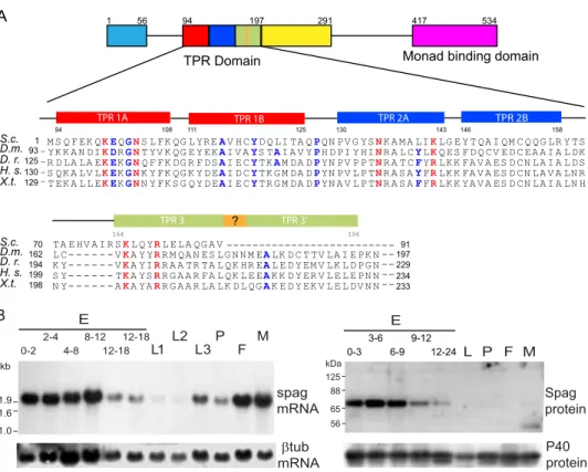
Characterization of the Spaghetti Gene in D. melanogaster— The spag gene is referred to in FlyBase as Dmel/CG13570 and encodes a protein of 534 residues (NCBI accession number
NP_524664.1). Spag protein displays one complete set of three adjacent tetratricopeptide repeats (TPR). TPR domains are responsible for Hsp binding and typically contain a triple repe-tition of 34 amino acids as follows: X5(WLF)X2
(LIM)(GAS)-X2(YLF)X8(ASE)X3(FYL)X2(ASL)X4(PKE). S. cerevisiae Tah1 possess a single TPR domain, and residues responsible for its interaction with Hsp90 are conserved in Spag (21, 22). Addi-tional residues important for the binding to the C-terminal EEVD motif in most Hsp70 and Hsp90 are also present (Fig. 1A) (23). Spag TPR domain is preceded by a structured domain at its N terminus, and followed by a second, incomplete TPR domain and a “potential Monad-binding” domain in the C terminus (InterPro 025986). Human RPAP3 (AAH5615.1) harbors two domains of triple TPR motifs and the potential binding domain at its C terminus (24). Such a potential Monad-binding domain is absent in Tah1 (Fig. 1A) (21).
In vitrotranslation of spag ORF produced a single peptide with an apparent molecular mass of⬃68 kDa corresponding to the predicted molecular mass of Spag (data not shown). Poly-clonal antibodies raised against a 22-mer synthetic peptide cor-responding to the C-terminal end of Spag reacted on a Western blot with a protein of⬃70 kDa (see below for the specificity of the antibodies). In embryo extracts, Spag migrated with a slightly higher mass than expected. Treatment with potato
acidic phosphatase (PAP) indicated that Spag is phosphory-lated in embryos but not in larvae nor in S2 Schneider’s cells (data not shown). Spag expression is at its highest level dur-ing early embryogenesis and then drops to a low level durdur-ing the second half of embryogenesis to become barely detecta-ble in successive developmental stages and in adult flies. This developmental profile partly reflects mRNA expression and suggests that spag may play a critical role during develop-ment (Fig. 1B).
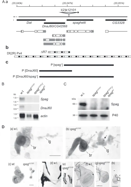
The spagI(2)k12101mutation (hereafter termed spagk12101) was
previously identified in a screen for P-element insertion causing tissue overgrowth during larval development (10). In this line, the P-lacW-transposon is inserted 52 nucleotides downstream from the spag ORF initiation codon (Fig. 2A-a). As a result, expression of both spag and its upstream gene DnaJ60 mRNAs is reduced, whereas the expression of the neighboring genes
Dat and CG3328 is not (Fig. 2B, and data not shown) (12). Accordingly, we did not detect Spag mRNA nor protein in
spagk12101 animals, also demonstrating the specificity of the
antibodies (Fig. 2, B and C).
Homozygous spagk12101larvae die 6 –7 days after egg laying
with overgrowth of the hematopoietic organs and occasional formation of melanotic pseudo-tumors (Fig. 2D, compare panel
awith panel b) (25). Overgrowth of the hematopoietic organs A L P F M 56 65 88 125 kDa P40 protein Spag protein B S.c. D.m. D. r. H. s. X.t. S.c. D.m. D. r. H. s. X.t. TPR 1A TPR 1B TPR 2A TPR 2B TPR 3 130 129 93 125 1 _ _ _ _ - --94 108 111 125 130 143 164 194 234 233 197 229 91 146 158 199 198 162 194 70 TPR 3’ M S Q F EK QKEQGNS L FK QG L YREAV H CYD Q LI TA QPQN PV G YSNK A M A LIKLG E Y T QA I QM C Q QG LR Y TS TA E HV A I R SKL Q YRL E LA QG A V YK K A N DIKDRGNT YV K QG E Y EKAI V AYS TAI A V YPH D PI Y HINR A L CYLKQ ES F D Q CV E D C EA A I A L DK L C- - - VKA Y YRR M QA N ES LG N N M EALK D C T TV LA I E PK N R D LA LA EKEKGNQ F FK DG R F DSAI E CYTKAM DA DPYN PV P P TNR A T CFYRLK K FA V A ES D CN LA I A L DS K Y- - - VKA YIRR A A TR TA L QK HR EAL E D Y EM V LK L D PG N S QK A LV LKEKGNK Y FK QG K Y D EAI D CYTK G M DA DPYN PV L P TNR A S AYFRLK K FA V A ES D CN LA V A LN R S Y- - - TKA YSRR G A A R FA L QK L E EAK K D Y ER V L E L E PN N T EK A L L EKEKGNN Y FK S G Q Y D EAI E CYTR G M DA DPYN A V L P TNR A S AFFRLK K YA V A ES D CN LA I A LN H N Y- - - AKA YARR G A A R LA LK D L QGAK E D Y EK V L E L DV N N ---0-3 3-6 6-9 9-12 12-24 E ? 534 1 TPR Domain 94 197 291 417 56
Monad binding domain
E L1 L2 L3 P F M βtub mRNA 1.9 1.6 1.0 kb 0-2 2-4 4-8 8-12 12-18 12-18 spag mRNA
FIGURE 1. Spag protein structure and patterns of gene expression and protein accumulation during Drosophila development. A, schematic structure of the Spag protein and evolutionary conservation of the TPR region in Spag orthologs from yeast to human. Filled boxes represent predicted structural domains and lines unstructured domains. The three TPR motifs forming the TPR region are indicated as red, blue, and green boxes. TPR 3⬘ is a putative helical domain absent in Tah1. Orange box represents a potential␣-helical junction between TPR 3 and TPR 3⬘. The potential monad binding motif (interPro 025986) overlaps with a predicted structured domain (magenta). Below are aligned the amino acid residues characterizing the three TPR motifs in S. cerevisiae (S.c.), D.
melano-gaster (D.m.), Danio rero (D.r.), Homo sapiens (H.s.), and Xenopus tropicalis (X.t.). Conserved residues identified in Tah1 for Hsp90 binding are indicated in bold red
(21). Blue indicates additional residues conserved in TPR domains and important for Hsp70/Hsp90 binding, as defined previously (23). B, expression pattern of
spag mRNA (left panel) and pattern of Spag protein accumulation (right panel) during Drosophila development. RNAs and proteins were extracted from (E)
embryos with age in hours after egg laying indicated above the lanes (L1, L2, and L3) first, second, and third instar larvae, (P) pupae, (F) female, and (M) male
3-days old imagos. The Northern blot was hybridized with either32P-labeled spag or-tubulin cDNA. The Western blot was probed with anti-Spag or
concerns more particularly the distal and the secondary lobes, which in wild-type larvae are usually the smallest. All other tissues such as imaginal discs (Fig. 2D, panels c and f– h) and salivary glands (Fig. 2D, panel d versus panel e) are reduced in size.
Following P-element excision, we recovered numerous
whiteflies producing homozygous viable progeny, as well as
a series of 20 nonviable w⫺revertants with larval lethality. Molecular studies using Southern blotting and PCR analysis revealed that the majority of these revertants contained a partially excised P-element, whereas the lethal revertant line
R7 displayed an interstitial chromosomal deficiency (Fig. 2A-b). These findings indicate that the P-insert is responsi-ble of the lethality.
w t spag k12101 spag k12101 / spag R7 1.9 1.9 1.0 1.6 1.6 0.6 kb Spag DnaJ60 actin D I(2)k12101 spaghetti CG3328 DnaJ60 Dat P [DnaJ60/spag+] P [DnaJ60]
b
c
B ∆R7 66 Spag P40 38 kDa w.t. spag k12101 spag k12101 / C spag R7lg
(a) w.t.lg
br
br
(b) spagk12101 spagk12101 (c) wt (d) w.t. (e) (f) w.t. spagk12101 (g) spagk12101 (h) P [spag+] /CG42568 (20,043k) (20,047k) (20,051k) Df(2R) Px4FIGURE 2. A, sketch of genomic map for the spaghetti locus. A-a, filled black boxes represent the genes, with the corresponding transcripts below. P-element insertion in the beginning of the spag ORF prevents mRNA accumulation of spag and its neighboring gene, DnaJ60. A-b, reversion of the P-element produced the lethal mutations R7, which is a deletion encompassing the spaghetti locus. Px4 is a wider deletion that encompasses the locus. A-c, three fragments encompassing either the spaghetti locus (P[spag⫹]), DnaJ60 locus (P[DnaJ60]), or both loci (P[DnaJ60/spag⫹]) were used to generate transgenic flies for rescue of the null mutants. B, no mRNA for spag nor DnaJ60 are detected by Northern blot in null mutants spagk12101nor in animals carrying this null mutation with the respective chromosomal deficiency spagk12101/spagR7, as compared with wild-type (Oregon-R). The Northern blots were hybridized with32P-labeled spag or
DnaJ60 cDNA probes. The32P-labeled-tubulin mRNA was used as loading control. C, Western blot analysis shows a signal of ⬃70 kDa in extracts from wild
type but not in homozygous null mutants spagk12101nor in spagk12101/spaghR7animals. D, lymph gland (lg), but not the brain (br), is overgrown in the spaghk12101 larvae (panel b) as compared with the wild-type w1118(panel a). Arrow points toward a melanocytic tumor. In contrast, there is atrophy of the imaginal discs for
the legs and halteres (panel c), the salivary glands (panels d and e), and the imaginal discs of the wings (panels f– h) in the spagk12101null mutants (panels e, g, and
In addition, transgenes encompassing the spag locus (trans-formation fragments P(spag⫹] and P(DnaJ60/spag⫹] from Fig. 2A-c) rescued the lethality of animals with either P-element insertion (homozygous spagk12101) or deletion in the spag locus
(heterozygous spagk12101/spagR7, spagk12101/Px4, or spagR7/Px4 flies; see Fig. 2A-b for a representation of these deletions). In contrast, a DnaJ60 transgene alone (P(DnaJ60] from Fig. 2A-c) was unable to rescue the development of the mutant animals. These data show that DnaJ60 is dispensable, although spag is essential for the survival of the fly (12).
We then took advantage of two different strains carrying transgenes with inducible RNAi against spag (P(GD8058) and P(KK100112), hereafter referred to as spagRNAi1and spagRNAi2, respectively) (13). Induction of gene silencing was performed with the upstream activating sequence Gal4 system. RNAi expression driven by Gal4 under either an act5c or a tubulin promoter resulted in pupal lethality shortly after metamorpho-sis. These animals die later than homozygous spagk12101,
prob-ably because gene silencing is a lengthy process, as compared with a genetic null mutation. This result further demonstrates that spag is necessary for fly development.
Identification of Spag Partners—The yeast Tah1 and mam-malian RPAP3 act in the context of a multimolecular co-chap-erone to assist client stabilization by Hsp90. To investigate the partners of Drosophila Spag, we isolated soluble, cytoplasmic complexes from Schneider’s S2 cells expressing either FLAG-Spag (full length) or FLAG-FLAG-Spag⌬TPR, in which the three TPR motifs were excised from amino acid residues 96 –197 (Fig. 1A). Following binding to an anti-FLAG M2 antibody-agarose col-umn, complexes were eluted with the FLAG peptide, to reduce contaminants. High mass accuracy matrix-assisted laser des-orption/ionization (MALDI) peptide mapping identified numerous heat shock proteins, including Hsp90 and Hsp70 iso-forms (Table 1). In the case of Hsc70-3 and Hsc70-5 (both devoid of a C-terminal EEVD), in contrast to the other Hsps, more peptides were detected with Spag⌬TPR than with
full-length Spag. In addition, we detected peptides for Pontin and Reptin in full-length Spag immunopurified complexes (Table 1).
We also performed a yeast two-hybrid screen using Spag pro-tein fused to the DNA-binding domain of Gal4 as bait. We identified interacting clones corresponding to the genes
CG1242(Hsp90), CG5792, in accordance with another two-hybrid screen (26), and CG13849 (Nop56) (Table 1). Alterna-tive splicing of CG5792 gives rise to two transcripts, one of which (CG5796-RA) produces a polypeptide of 334 amino acids, which contains a Pih1 domain (CG5792-PA or NP-609590.1). It is thereafter described as D.mel/Pih1D1, as its likely ortholog. Interaction of Spag with Hsp90 parallels the proteomic data described above, as in both cases it required the presence of the TPR domain. This was also the case for Nop56, a core protein from the box C/D snoRNPs. S. cerevisiae Nop56 also interact with Tah1 in two-hybrid assays (3). In contrast, Spag interaction with dPih1D1 was independent from the TPR domain.
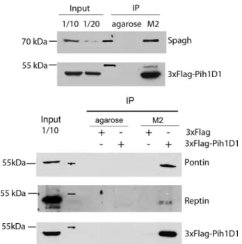
Co-immunoprecipitations in S2 cells, using a 3 ⫻FLAG-tagged dPih1D1 confirmed that it binds to endogenous Spag, Pontin, and Reptin (Fig. 3). Altogether, these data indicate that the R2TP core complex is conserved in Drosophila.
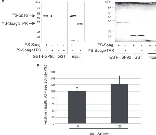
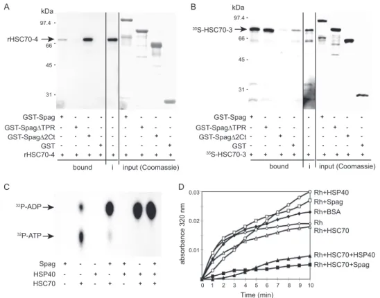
Spag Is a Co-chaperone of Hsp90—To test whether the inter-action between Spag and Hsps was direct, we used GST pull-down assays. Full-length in vitro translated Spag protein, albeit not Spag⌬TPR (lacking aa 96–197), could bind to immobilized C-terminal domain of Hsp90 (aa 535–717, encompassing the EEVD-binding motif for co-chaperones) (Fig. 4A). This finding is in accordance with the results from the proteomic and yeast
TABLE 1
Identification of full-length Spag or Spag⌬TPR (lacking aa 96–197) interactants by proteomic analysis or yeast two-hybrid
The number of prototypic peptides in the proteomic is indicated. ND⫽ not
detected. The presence of an EEVD motif at the C terminus of the Hsps is indicated.
Annotation symbol Gene
FL Spag Spag⌬TPR C-terminal EEVD Proteomic data CG13570 Spaghetti 116 29 CG4003-PA Pontin 8 ND CG9750-PA Reptin 3 ND CG14207 Hsp20-like protein 4 ND ⫺ CG4460-PB Hsp22 5 ND ⫺ CG4463-PA Hsp23 10 ND ⫺ CG4183-PA Hsp26 14 ND ⫺ CG4147-PA HSC70-3 12 18 ⫺ CG4264-PA HSC70-4 19 7 ⫹ CG31366 Hsp70Aa 6 5 ⫹ CG8542-PA HSC70-5 2 7 ⫺ CG1242-PA Hsp90/Hsp83 21 ND ⫹ CG5520-PA Hsp90/GP93 19 ND ⫺ Yeast two-hybrid CG1242 Hsp90/Hsp83 ⫹ ⫺ ⫹ CG13849 Nop56 ⫹ ⫺ CG5792 Pih1D1 ⫹ ⫹
FIGURE 3. Spag, dPih1D1, Reptin, and Pontin associate in Drosophila cells
to form R2TP. Drosophila S2 cells were transiently transfected to express
3⫻FLAG-dPih1D1 or 3⫻FLAG alone. Cell lysates were incubated with agarose or M2-agarose beads, and complexes were eluted with 3⫻FLAG peptide. Western blot demonstrates an interaction of 3⫻FLAG-Pih1D1 with endoge-nous Spagh, Pontin, and Reptin. As controls, cell lysates (transfected or not) were incubated with agarose beads. 3⫻FLAG-tagged dPih1D1 was detected using an anti-FLAG antibody. IP, immunoprecipitation.
two-hybrid assays. In Tah1, the TPR domain is also responsible for binding to Hsp90 (21, 27).
To monitor the effect of Spag on Hsp90 function, we fol-lowed its effect on Hsp90 ATPase activity. We observed a weak stimulation in the presence of a 4-fold excess of Spag (Fig. 4B). The level of stimulation of Hsp90 ATPase by Spag appears sim-ilar to the effect reported for Tah1 on yeast Hsp82 (22, 27, 28).
Spag Is a Co-chaperone of Hsp70—In vitro GST pulldown assay showed that binding to Hsc70-4 was also direct and depended on the TPR domain (Fig. 5A). This is consistent with the fact that Hsc70-4 harbors a C-terminal EEVD motif, known to bind to TPR domains of co-chaperones (5, 23, 27). Yet, in S.
cerevisiae, Tah1 does not bind directly to Ssa-1, a cytoplasmic Hsp70 isoform (28). Conversely, direct binding of Spag to Hsc70-3, which lacks a C-terminal EEVD motif, was not dependent on the Spag TPR domain, further supporting the fact that deletion of the TPR did not induce a gross disorgani-zation of the protein (Fig. 5B). Indeed, Hsc70-3 was unable to bind Spag⌬2Ct, which lacks aa 231–490. Altogether, our results indicated that Spag contains distinct domains able to bind to distinct chaperones.
To investigate the functional consequences of Spag binding to Hsp70, we first measured the in vitro ATPase activity of Spag in the presence of commercially available Hsc70 (HspA8, homolog of Hsc70-4). Neither Spag nor Hsp40 was able to hydrolyze ATP to ADP (Fig. 5C). In contrast, Spag could signif-icantly enhance the intrinsic ATPase activity of Hsc70, to a level
comparable with that of the Hsc70-Hsp40 complex (Fig. 5C). Finally, we investigated the effect of Spag on rhodanese disag-gregation. In this assay, rhodanese was first denatured to induce aggregate formation. Addition of Spag, Hsp40, or the control protein BSA alone exerted no effect on those aggregates, which were moderately dissociated by Hsc70. In contrast, combina-tion of Hsc70 with either Hsp40 or Spag strongly enhanced rhodanese disaggregation by as much as ⬃75% (Fig. 5D). Hence, similarly to Hsp40, Spag can act as an Hsp70 co-chap-erone. All these data show that Spag is also a bona fide Hsp70 co-chaperone.
Conservation of the R2TP Clients—Several substrates have been identified for R2TP. These include the box C/D snoRNPs (3, 5). Experiments in mammalian cells showed that accumula-tion of newly synthesized 15.5K and Nop58 depend on Hsp90 activity (3). We assessed the stability of Nop58 (also called Nop5 in Drosophila) (29) and 15.5K (Hoip encoded by the
hoi-polloigene or hoip (9)) in spag null mutant larvae (spagk12101),
as well as in pupae in which spag was knocked down by the induction of RNAi under the control of Gal4act5cdriver. West-ern blot analysis showed a decrease in Nop58 and 15.5K con-tent, in spagk12101larvae and act5C⬎spagRNAipupae. Different
protein turnovers in larvae and pupae could explain the differ-ences observed for Nop58 and 15.5K destabilization in
spagk12101or act5C⬎spagRNAianimals. In contrast, fibrillarin,
another core protein of the C/D snoRNPs, was not affected (Fig. 6, A and B). This is coherent with results from mammals. 125 88 65 56 38 31 kDa 35S-Spag 35S-Spag∆TPR 35S-Spag 35S-Spag∆TPR + + + -- + -- + -- + 125 88 65 56 38 31 kDa 35S-Spag 35S-Spag∆TPR + + + -- + -- + -- + input GST-HSP90 GST GST-HSP90 GST input A B 0 20 40 60 80 100 120 140 160 0 20 Relative Hsp90 A TPase activity (%) μM Spagh
FIGURE 4. Spag is a genuine Hsp90 co-chaperone. A, Spag binds Hsp90 directly through its TPR domain. The C-terminal domain of D. melanogaster Hsp90 (aa 535–717) was fused to GST in pulldown assays with rabbit reticulocyte35S-labeled proteins. Hsp90 retains Spag but not the truncated Spag⌬TPR, which lacks
the tripartite TPR domain (amino acids 96 –197). Ponceau-stained filter shows that identical levels of the GST and GST-Hsp90 C terminus were retained on the column (right panel). B, ATPase activation of Drosophila Hsp90 by Spag. Relative levels of ATPase activity of 5MHsp90 in the absence or presence of 20Mof purified His-tagged Spag protein are comparable with that obtained for HSP90 with S. cerevisiae Tah1.
Indeed, fibrillarin is incorporated at a latter step during snoRNP assembly, and it does not seem to be a client of R2TP (3, 30).
Interaction between R2TP and 15.5K requires Nufip as an adaptor (3). CG4076 encodes a protein with 18% identity to mammalian Nufip. Induction of two different transgenic lines expressing RNAi against CG4076/Nufip driven by Gal4act5c
produced early pupal lethality. In these animals, as in the case of
spag, there is a decrease in endogenous Nop58 and 15.5K, as compared with controls (Fig. 6C). In conclusion, Spag and Nufip are required for the stabilization of the C/D snoRNP core proteins.
A second class of clients described for mammalian R2TP are the PIKKs. Mammalian R2TP is recruited to these kinases via the adaptor Tel2, with a predominant effect on mammalian TOR and SMG1 stability (7). In Drosophila, proteomic data indicate that TOR similarly interacts with Pontin, Reptin, and Lqfr, a distant ortholog of Tel2 (31). To test whether these interactions are functionally relevant, we measured TOR activ-ity in wild-type and Spag-deficient flies (as null mutant or trans-genic RNAi). Phosphorylation of S6K is classically used as a surrogate marker of TOR activity, to circumvent the lack of antibodies to detect TOR in Drosophila. In animals where
spagwas inactive (as null mutant or transgenic RNAi), we detected a down-regulation of S6K phosphorylation (Fig. 7,
A and B). In contrast, phosphorylation of S6K was not affected upon Nufip inactivation (Fig. 7C). These data are consistent with a role for spag, but not Nufip, in the activity of TOR and are in accordance with a recent report in the
DrosophilaS2 cell line (4, 31).
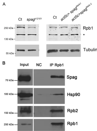
Finally, in mammals, RNA polymerase II is also a substrate of R2TP (4, 6, 32). Indeed, human Spag binds to neo-synthesized Rpb1, the largest subunit of RNA polymerase II, to stabilize it before its association with the other polymerase II subunits. In flies devoid of Spag (spagk12101
), we found a moderate decrease in total Rpb1 (RPII215). However no effect was seen in spagRNAi
flies (Fig. 8A). Immunoprecipitation of soluble Rpb1 in S2 cells shows a tight interaction with Spag and Hsp90 (Fig. 8B). This suggests a role of Spag in RNA polymerase II assembly rather than Rpb1 stabilization.
DISCUSSION
Conservation of the R2TP Complex—A multimeric Hsp90 co-chaperone known as R2TP was first identified in S. cerevisiae (1) and later in human cells (3, 4). In R2TP, Tah1/RPAP3 plays a central role, binding directly to Pih1/Pih1D1 and Hsp90 (3, 5, 27). In this study, we characterize spaghetti, a gene essential for
Drosophiladevelopment. Spag interacts with Pontin, Reptin, and CG5792-PA, the Drosophila orthologs of the mammalian
97.4 66 45 31 kDa GST-Spag GST-Spag∆TPR + + + + -GST-Spag∆2Ct + + -GST- - - + - - - - + GST-Spag GST-Spag∆TPR + + + + -GST-Spag∆2Ct + + -GST - - - + - - - - + bound 35S-HSC70-3+ + + + + + + + + + + + + + + + + + rHSC70-4 rHSC70-4 35S-HSC70-3 97.4 66 45 31 kDa Spag HSP40 + - - + + - + - - + - + + + HSC70 - + - + - + + 32P-ADP 32P-ATP 0.03 0.01 0.02 0 1 2 3 4 5 6 7 8 9 10 absorbance 320 nm Time (min) Rh+HSP40 Rh Rh+BSA Rh+Spag Rh+HSC70 Rh+HSC70+HSP40 Rh+HSC70+Spag C D
i input (Coomassie) bound i input (Coomassie)
FIGURE 5. Spag is a genuine Hsp70 co-chaperone. Spag interacts differentially with different Hsp70 isoforms. Full-length or truncated Spag missing either the TPR domain (Spag⌬TPR lacking aa 96–197) or the C-terminal domain (Spag⌬2Ct lacking aa 231–490) was fused to GST and immobilized on beads for pulldown assays. Prey input is detected in the i lane. Coomassie stainings of the gels (right parts of A and B) show identical amounts of baits on the columns. A, Hsc70-4 is retained by GST-Spag and GST-Spag⌬2Ct, but not GST-Spag⌬TPR. B, in contrast,35S-labeled Hsc70 –3 is specifically retained on GST-Spag or GST-Spag⌬TPR
but not GST-Spag⌬2Ct. C, Spag stimulates ATP hydrolysis by Hsc70. Chromatography on PEI-cellulose thin layer plate shows that [32P]ATP hydrolysis into ADP
by Hsc70 alone is weak and undetectable in the presence of Spag or Hsp40 alone. ATP hydrolysis by Hsc70 is strongly enhanced by the addition of recombinant Spag, Hsp40, or both. D, combination of Hsc70 with either Hsp40 or Spag strongly reduces rhodanese aggregation by⬃75%, as measured by light scattering at 320 nm. In addition, addition of Spag, Hsp40, or the control protein BSA alone exerts no effect on rhodanese aggregation, which is moderately suppressed when Hsc70 is alone. This experiment is representative of three.
Pih1D1, in a complex very similar to the mammalian and S.
cerevisiaeR2TP.
In mammals, R2TP associates with seven prefoldin-like pro-teins (4, 8). So far, only the association of Spag, Pontin, and Reptin with the product of the CG14353 gene has been docu-mented (31). The CG14353-encoded protein contains a domain with 56% sequence identity with the human prefoldin Monad, associated with the R2TP (24). Besides, an ortholog of Uri, also part of R2TP/prefoldin-like system (4), was iden-tified as a specific interactor of protein phosphatase 1␣ (33). dUri is cytoplasmic and is strongly expressed in embryo, pupae, and adult gonads. Interestingly, dUri co-localizes with RNA polymerase II on polytene chromosomes, yet its link with R2TP remains elusive. Conservation of the prefol-din-like association with R2TP in D. melanogaster thus remains open to question.
Conservation of R2TP Clients across Metazoans—In mam-mals, three types of clients have been found for the Hsp90/ R2TP system as follows: PIKKs, snoRNPs, and RNA poly-merases. In Drosophila, we detect a decreased activity for the
PIKK TOR when spag is inactivated. Proteomic experiments in Schneider S2 cells showed that dTOR interacts with Pontin, Reptin, Spag, and liquid facet-related, a distant homolog of Tel2 (31, 34). In mammalian cells, recruitment of the whole R2TP by Tel2 is necessary to stabilize mammalian TOR (7). Taken together, these data support the conservation of R2TP in the stabilization and assembly of PIKK complexes.
We found that the amounts of two box C/D snoRNP core proteins are diminished in animals devoid of Spag or Nufip. This is consistent with the role described for R2TP and Rsa1/ Nufip in the biogenesis of snoRNPs in S. cerevisiae and mam-mals (3, 5). In Arabidopsis thaliana, atNufip hypomorph mutants are viable but show severe developmental defects, with a subclass of snoRNPs being affected (35). Yet, in this organism, no gene encoding products similar to Pih1D1 has been so far identified (36).
Mammalian RPAP3 stabilizes nascent Rpb1 prior to its association with other RNA polymerase II subunits (6). In
Dro-sophila, Spag depletion only showed a mild destabilization of Rpb1 in the null mutant. Yet the strong binding of Spag to Rpb1
FIGURE 6. Spag and Nufip are required for box C/D sno core protein stabilization. A, Western blot analysis of protein extracts from third instar larvae
spagk12101, as compared with wild-type w1118(Ct), showed a significant diminution in the content of Nop58 (Nop5) and 15.5K (Hoip) but not fibrillarin. Tubulin
was used as a loading control. B, this phenomenon was also observed in pupae extracts from animals in which RNAi was induced by Gal4act5Cto knock down
spag (Gal4act5c/spagRNAias compared with Gal4act5c/⫹). C, similar results were observed in pupae extracts from animals in which RNAi was induced against Nufip (C, Gal4act5c/nufipRNAicompared with w1118).
supports a role in RNA polymerase II assembly. Quantitative analysis of Hsp90 interaction with its clients demonstrates that some, but not all, are degraded upon Hsp90 inhibition (37). Hsp90 could act as a molecular tweezers, by maintaining Rpb1 via R2TP in a state amenable for interaction with Rpb2, as it maintains glucocorticoid receptors in a state compatible with ligand binding (38).
The phenotype observed in spag null mutant larvae, in which most organs are atrophied, could thus be explained by altera-tions in the funcaltera-tions of TOR, snoRNP, and/or RNA polymer-ase II, as all these machineries are required for cell growth (39, 40).
Spaghetti Is a Co-chaperone for Both Hsp70 and Hsp90—D.
melanogaster Spag is able to bind directly through its TPR domain, with Hsp90 and Hsp70 isoforms containing a C-termi-nal EEVD. In addition, the conserved C-termiC-termi-nal domain is implicated in recruiting Hsc70 –3, an Hsp70 isoform devoid of EEVD motif. Finally, Spag is able to stimulate Hsc70 activity, suggesting a role for Hsp70 in R2TP function.
Some co-chaperones such as CHIP, display a single TPR domain, which binds alternatively Hsp70 or Hsp90 (15, 17). In contrast, HOP harbors three TPR domains, which enable it to transfer clients from Hsp70 to Hsp90 (23). In invertebrates genomes, genes encoding proteins displaying both TPR and Monad-binding motifs can be identified, which are likely
Spaghomologs (41). Most insect Spag, including D.
melano-gaster, show a single TPR domain, although other insect Spag contain two such regions, as in vertebrates. Spag homologs with two TPR domains could bind simultaneously to Hsp70 and Hsp90 and transfer the client between these chaperones. In species with a single TPR domain (such as D.
melanogaster), another co-chaperone or Hsp (such as Hsp22, -23, or -26) could assist the transfer of R2TP/clients from Hsp70 to Hsp90.
Intriguingly, the Ssa-1 and Ssb-1 proteins of S. cerevisiae, which are orthologs of cytoplasmic Hsp70, interact with Pih1 as well as the box C/D sno core proteins (42). This supports a role for Hsp70 in box C/D snoRNP biogenesis, and R2TPs function in general. Based on all these data, we propose a model whereby nascent R2TP substrates would first be pre-folded by Hsp70 and then transferred to Hsp90 to maintain them in a conformation amenable for integration into molecular complexes (Fig. 9).
Conclusion—In S. cerevisiae, single deletions of some R2TP components (namely Tah1 and Pih1) or adaptors (Rsa1) are not lethal unless cells are subjected to heat or nutrient stress (5). In particular, deletion of TAH1, the subunit that contacts Hsp90, generates only very mild phenotypes, thus suggesting that the chaperone plays an auxiliary role in the assembly of R2TP cli-ents. In contrast, spaghetti is essential during fly development. This suggests that within the R2TP context, the role of Hsp90 might have evolved from a conditional machinery operating under stress conditions in yeast to a constitutive assembly
fac-FIGURE 7. Phosphorylation of p70 S6K, a readout of TOR activity, is
decreased upon spag inactivation but not Nufip. Western blot analysis
showed a diminution of phospho-p70-S6K (Ph-S6K) but not total p70-S6K, as compared with tubulin. This was observed upon spag inactivation, either in spagk12101null mutants as compared with w1118(A) or in pupae
with spag knockdown (B, Gal4act5c/spagRNAi). This was not the case, how-ever, when Nufip was silenced (C, Gal4act5c/NufipRNAias compared with
w1118) (C).
FIGURE 8. Spag interacts with Rpb1. A, largest subunit in RNA polymerase II, Rpb1 (RpII215), showed a moderate decrease in extracts from spagk12101 L3 and no sensible variation in Gal4act5C/spagRNAipupae, as compared with matching controls. B, immunoprecipitation of Rpb1 in D. melanogaster S2 cells showed a robust interaction with Spag and Hsp90, as well as Rpb2.
tor in metazoans. In addition, our data support a role for Hsp70 in R2TP-assisted complex assembly.
Acknowledgments—We are indebted to Dorothee Albrecht, as well as the organizers of the EMBO workshop on Drosophila and François Juge, Céline Fernando, and Charlotte Grimaud for their constant help and support in this project. We thank Janice Fischer, Tao Wang, Yves Henri, Carl Wu, Séverine Chambeyron, Renato Paro, Michèle Croza-tier, Krzysztof Rogowski, Bruno Beaumelle, and Martine Simonelig for generous gifts of reagents, and Jonathan Bizarro for critical review of the manuscript. We thank the Bloomington Stock Center, the Vienna Drosophila RNAi Center, the DrosophilaGenomics Resource Center at Bloomington, and Developmental Studies Hybridoma Bank at the University of Iowa for providing materials.
REFERENCES
1. Zhao, R., Davey, M., Hsu, Y.-C., Kaplanek, P., Tong, A., Parsons, A. B., Krogan, N., Cagney, G., Mai, D., Greenblatt, J., Boone, C., Emili, A., and Houry, W. A. (2005) Navigating the chaperone network: an integrative map of physical and genetic interactions mediated by the hsp90 chaper-one. Cell 120, 715–727
2. Jha, S., and Dutta, A. (2009) RVB1/RVB2: running rings around molecular biology. Mol. Cell 34, 521–533
3. Boulon, S., Marmier-Gourrier, N., Pradet-Balade, B., Wurth, L., Verheg-gen, C., Jády, B. E., Rothé, B., Pescia, C., Robert, M.-C., Kiss, T., Bardoni, B., Krol, A., Branlant, C., Allmang, C., Bertrand, E., and Charpentier, B. (2008) The Hsp90 chaperone controls the biogenesis of L7Ae RNPs through conserved machinery. J. Cell Biol. 180, 579 –595
4. Cloutier, P., Al-Khoury, R., Lavallée-Adam, M., Faubert, D., Jiang, H., Poi-tras, C., Bouchard, A., Forget, D., Blanchette, M., and Coulombe, B. (2009) High-resolution mapping of the protein interaction network for the hu-man transcription machinery and affinity purification of RNA polymerase II-associated complexes. Methods 48, 381–386
5. Zhao, R., Kakihara, Y., Gribun, A., Huen, J., Yang, G., Khanna, M., Costanzo, M., Brost, R. L., Boone, C., Hughes, T. R., Yip, C. M., and Houry, W. A. (2008) Molecular chaperone Hsp90 stabilizes Pih1/Nop17 to main-tain R2TP complex activity that regulates snoRNA accumulation. J. Cell
Biol. 180,563–578
6. Boulon, S., Pradet-Balade, B., Verheggen, C., Molle, D., Boireau, S., Geor-gieva, M., Azzag, K., Robert, M.-C., Ahmad, Y., Neel, H., Lamond, A. I., and Bertrand, E. (2010) HSP90 and its R2TP/Prefoldin-like cochaperone are involved in the cytoplasmic assembly of RNA polymerase II. Mol. Cell
39,912–924
7. Horejsí, Z., Takai, H., Adelman, C. A., Collis, S. J., Flynn, H., Maslen, S., Skehel, J. M., de Lange, T., and Boulton, S. J. (2010) CK2 phospho-depen-dent binding of R2TP complex to TEL2 is essential for mTOR and SMG1 stability. Mol. Cell 39, 839 – 850
8. Boulon, S., Bertrand, E., and Pradet-Balade, B. (2012) HSP90 and the R2TP co-chaperone complex: building multiprotein machineries essential for cell growth and gene expression. RNA Biol. 9, 148 –154
9. Roch, F., Serras, F., Cifuentes, F. J., Corominas, M., Alsina, B., Amorós, M., López-Varea, A., Hernández, R., Guerra, D., Cavicchi, S., Baguñá, J., and García-Bellido, A. (1998) Screening of larval/pupal P-element induced lethals on the second chromosome in Drosophila melanogaster: clonal analysis and morphology of imaginal discs. Mol. Gen. Genet. 257, 103–112 10. Török, T., Tick, G., Alvarado, M., and Kiss, I. (1993) P-lacW insertional mutagenesis on the second chromosome of Drosophila melanogaster: iso-lation of lethals with different overgrowth phenotypes. Genetics 135, 71– 80
11. Török, I., Strand, D., Schmitt, R., Tick, G., Török, T., Kiss, I., and Mechler, B. M. (1995) The overgrown hematopoietic organs-31 tumor suppressor gene of Drosophila encodes an Importin-like protein accumulating in the nucleus at the onset of mitosis. J. Cell Biol. 129, 1473–1489
12. Iliopoulos, I., Török, I., and Mechler, B. M. (1997) The DnaJ60 gene of
Drosophila melanogasterencodes a new member of the DnaJ family of proteins. Biol. Chem. 378, 1177–1181
13. Dietzl, G., Chen, D., Schnorrer, F., Su, K.-C., Barinova, Y., Fellner, M., Gasser, B., Kinsey, K., Oppel, S., Scheiblauer, S., Couto, A., Marra, V., Keleman, K., and Dickson, B. J. (2007) A genome-wide transgenic RNAi library for conditional gene inactivation in Drosophila. Nature 448, 151–156
14. Pradet-Balade, B., Girard, C., Boulon, S., Paul, C., Azzag, K., Bordonné, R., Bertrand, E., and Verheggen, C. (2011) CRM1 controls the composition of nucleoplasmic pre-snoRNA complexes to license them for nucleolar transport. EMBO J. 30, 2205–2218
15. Török, I., Herrmann-Horle, D., Kiss, I., Tick, G., Speer, G., Schmitt, R., and Mechler, B. M. (1999) Down-regulation of RpS21, a putative translation initiation factor interacting with P40, produces viable minute imagos and larval lethality with overgrown hematopoietic organs and imaginal discs.
Mol. Cell. Biol. 19,2308 –2321
16. Santos, M. G., Jorge, S. A., Brillet, K., and Pereira, C. A. (2007) Improving heterologous protein expression in transfected Drosophila S2 cells as as-sessed by EGFP expression. Cytotechnology 54, 15–24
17. Ballinger, C. A., Connell, P., Wu, Y., Hu, Z., Thompson, L. J., Yin, L. Y., and Patterson, C. (1999) Identification of CHIP, a novel tetratricopeptide re-peat-containing protein that interacts with heat shock proteins and neg-atively regulates chaperone functions. Mol. Cell. Biol. 19, 4535– 4545 18. Minami, Y., Höhfeld, J., Ohtsuka, K., and Hartl, F. U. (1996) Regulation of
the heat-shock protein 70 reaction cycle by the mammalian DnaJ homo-log, Hsp40. J. Biol. Chem. 271, 19617–19624
19. Meyer, P., Prodromou, C., Hu, B., Vaughan, C., Roe, S. M., Panaretou, B., Piper, P. W., and Pearl, L. H. (2003) Structural and functional analysis of the middle segment of hsp90: implications for ATP hydrolysis and client protein and cochaperone interactions. Mol. Cell 11, 647– 658
20. Höhfeld, J., Minami, Y., and Hartl, F. U. (1995) Hip, a novel cochaperone involved in the eukaryotic Hsc70/Hsp40 reaction cycle. Cell 83, 589 –598 21. Jiménez, B., Ugwu, F., Zhao, R., Ortí, L., Makhnevych, T., Pineda-Lucena, A., and Houry, W. A. (2012) Structure of minimal tetratricopeptide repeat domain protein Tah1 reveals mechanism of its interaction with Pih1 and Hsp90. J. Biol. Chem. 287, 5698 –5709
22. Back, R., Dominguez, C., Rothé, B., Bobo, C., Beaufils, C., Moréra, S., Meyer, P., Charpentier, B., Branlant, C., Allain, F. H., and Manival, X. (2013) High-resolution structural analysis shows how Tah1 tethers Hsp90 to the R2TP complex. Structure 21, 1834 –1847
23. Muller, P., Ruckova, E., Halada, P., Coates, P. J., Hrstka, R., Lane, D. P., and Vojtesek, B. (2013) C-terminal phosphorylation of Hsp70 and Hsp90 reg-FIGURE 9. Hypothetical model for R2TP function in complex assembly,
such as RNA polymerase II. Nascent Rpb1 could be tethered to Hsp70 via
R2TP to undergo early folding steps, upon which it would be transferred to Hsp90, to maintain it in a conformation compatible with Rpb2 binding. Data from Ref. 31 suggest that an ortholog of the prefoldin-like Monad (light green) is associated to R2TP.
ulates alternate binding to co-chaperones CHIP and HOP to determine cellular protein folding/degradation balances. Oncogene 32, 3101–3110 24. Itsuki, Y., Saeki, M., Nakahara, H., Egusa, H., Irie, Y., Terao, Y., Kawabata,
S., Yatani, H., and Kamisaki, Y. (2008) Molecular cloning of novel Monad binding protein containing tetratricopeptide repeat domains. FEBS Lett.
582,2365–2370
25. Minakhina, S., and Steward, R. (2006) Melanotic mutants in Drosophila: pathways and phenotypes. Genetics 174, 253–263
26. Giot, L., Bader, J. S., Brouwer, C., Chaudhuri, A., Kuang, B., Li, Y., Hao, Y. L., Ooi, C. E., Godwin, B., Vitols, E., Vijayadamodar, G., Pochart, P., Machineni, H., Welsh, M., Kong, Y., Zerhusen, B., Malcolm, R., Varrone, Z., Collis, A., Minto, M., Burgess, S., McDaniel, L., Stimpson, E., Spriggs, F., Williams, J., Neurath, K., Ioime, N., Agee, M., Voss, E., Furtak, K., Renzulli, R., Aanensen, N., Carrolla, S., Bickelhaupt, E., Lazovatsky, Y., DaSilva, A., Zhong, J., Stanyon, C. A., Finley, R. L., Jr., White, K. P., Braver-man, M., Jarvie, T., Gold, S., Leach, M., Knight, J., Shimkets, R. A., Mc-Kenna, M. P., Chant, J., and Rothberg, J. M. (2003) A protein interaction map of Drosophila melanogaster. Science 302, 1727–1736
27. Eckert, K., Saliou, J.-M., Monlezun, L., Vigouroux, A., Atmane, N., Caillat, C., Quevillon-Chéruel, S., Madiona, K., Nicaise, M., Lazereg, S., Van Dorsselaer, A., Sanglier-Cianférani, S., Meyer, P., and Moréra, S. (2010) The Pih1-Tah1 cochaperone complex inhibits Hsp90 molecular chaper-one ATPase activity. J. Biol. Chem. 285, 31304 –31312
28. Millson, S. H., Vaughan, C. K., Zhai, C., Ali, M. M., Panaretou, B., Piper, P. W., Pearl, L. H., and Prodromou, C. (2008) Chaperone ligand-discrim-ination by the TPR-domain protein Tah1. Biochem. J. 413, 261–268 29. Vorbrüggen, G., Onel, S., and Jäckle, H. (2000) Restricted expression and
subnuclear localization of the Drosophila gene Dnop5, a member of the Nop/Sik family of the conserved rRNA processing factors. Mech. Dev. 90, 305–308
30. Verheggen, C., Lafontaine, D. L., Samarsky, D., Mouaikel, J., Blanchard, J.-M., Bordonné, R., and Bertrand, E. (2002) Mammalian and yeast U3 snoRNPs are matured in specific and related nuclear compartments.
EMBO J. 21,2736 –2745
31. Glatter, T., Schittenhelm, R. B., Rinner, O., Roguska, K., Wepf, A., Jünger, M. A., Köhler, K., Jevtov, I., Choi, H., Schmidt, A., Nesvizhskii, A. I., Stocker, H., Hafen, E., Aebersold, R., and Gstaiger, M. (2011) Modularity and hormone sensitivity of the Drosophila melanogaster insulin receptor/ target of rapamycin interaction proteome. Mol. Syst. Biol. 7, 547
32. Forget, D., Lacombe, A.-A., Cloutier, P., Al-Khoury, R., Bouchard, A., Lavallée-Adam, M., Faubert, D., Jeronimo, C., Blanchette, M., and Cou-lombe, B. (2010) The protein interaction network of the human transcrip-tion machinery reveals a role for the conserved GTPase RPAP4/GPN1 and microtubule assembly in nuclear import and biogenesis of RNA polymer-ase II. Mol. Cell. Proteomics 9, 2827–2839
33. Kirchner, J., Vissi, E., Gross, S., Szoor, B., Rudenko, A., Alphey, L., and White-Cooper, H. (2008) Drosophila Uri, a PP1␣ binding protein, is es-sential for viability, maintenance of DNA integrity and normal transcrip-tional activity. BMC Mol. Biol. 9, 36
34. Lee, J.-H., and Fischer, J. A. (2012) Drosophila Tel2 is expressed as a trans-lational fusion with EpsinR and is a regulator of wingless signaling. PLoS
ONE 7,e46357
35. Rodor, J., Jobet, E., Bizarro, J., Vignols, F., Carles, C., Suzuki, T., Nakamura, K., and Echeverría, M. (2011) AtNUFIP, an essential protein for plant development, reveals the impact of snoRNA gene organisation on the assembly of snoRNPs and rRNA methylation in Arabidopsis thaliana.
Plant J. 65,807– 819
36. Rodor, J., Letelier, I., Holuigue, L., and Echeverria, M. (2010) Nucleolar RNPs: from genes to functional snoRNAs in plants. Biochem. Soc. Trans.
38,672– 676
37. Taipale, M., Krykbaeva, I., Koeva, M., Kayatekin, C., Westover, K. D., Karras, G. I., and Lindquist, S. (2012) Quantitative analysis of HSP90-client interactions reveals principles of substrate recognition. Cell 150, 987–1001
38. Zhao, R., and Houry, W. A. (2005) Hsp90: a chaperone for protein folding and gene regulation. Biochem. Cell Biol. 83, 703–710
39. Oldham, S., Montagne, J., Radimerski, T., Thomas, G., and Hafen, E. (2000) Genetic and biochemical characterization of dTOR, the Drosophila homolog of the target of rapamycin. Genes Dev. 14, 2689 –2694 40. Zhang, H., Stallock, J. P., Ng, J. C., Reinhard, C., and Neufeld, T. P. (2000)
Regulation of cellular growth by the Drosophila target of rapamycin dTOR. Genes Dev. 14, 2712–2724
41. Haslbeck, V., Eckl, J. M., Kaiser, C. J., Papsdorf, K., Hessling, M., and Richter, K. (2013) Chaperone-Interacting TPR Proteins in Caenorhabditis
elegans. J. Mol. Biol. 425, 2922–2939
42. Makhnevych, T., Wong, P., Pogoutse, O., Vizeacoumar, F. J., Greenblatt, J. F., Emili, A., and Houry, W. A. (2012) Hsp110 is required for spindle length control. J. Cell Biol. 198, 623– 636