Cell-type specific cholinergic modulation of the cortex
by Naiyan Chen
B.Eng., Biomedical Engineering, Imperial College London (2006)
ASSACHUSE TjiT TUTEf OFTECHNOLOGY
SEP
2
5
2013
SUBMITTED TO THE COMPUTATIONAL AND SYSTEMS BIOLOGY PROGRAM IN PARTIAL FULFILLMENT OF THE REQUIREMENTS FOR THE DEGREE OF DOCTORATE OF PHILOSOPHY IN COMPUTATIONAL AND SYSTEMS BIOLOGY
AT THE MASSACHUSETTS INSTITUTE OF TECHNOLOGY SEPTEMBER 2013
C Massachusetts Institute of Technology. All rights reserved
Author ...
Naiyan Chen Computational and Systems Biology Program July 29 2013
Certified by ... ... ...g
Mriganka Sur, PhD Paul E. Newton Professor of Neuroscience
Thesis Supervisor
A ccepted by ... . ... ... Christopher B. Burge, PhD Professor of Biology and Biological Engineering Director, Computational and Systems Biology Graduate Program
Cell-type specific cholinergic modulation of the cortex
by Naiyan Chen
SUBMITTED TO THE COMPUTATIONAL AND SYSTEMS BIOLOGY PROGRAM IN PARTIAL FULFILLMENT OF THE REQUIREMENTS FOR THE DEGREE OF DOCTORATE OF PHILOSOPHY IN COMPUTATIONAL AND SYSTEMS BIOLOGY
AT THE MASSACHUSETTS INSTITUTE OF TECHNOLOGY SEPTEMBER 2013
0 Massachusetts Institute of Technology. All rights reserved
Abstract
The cholinergic innervation of the neocortex by afferent fibers originating in the nucleus basalis (NB) of the basal forebrain is implicated in modulating diverse neocortical functions including information processing, cortical plasticity, arousal and attention. To understand the physiological basis of these brain functions during cholinergic modulation, it is critical to identify the cortical circuit elements involved and define how their interactions contribute to cortical network dynamics. In this thesis, I present evidence showing how specific neuronal and glial cell types can be differentially modulated by acetylcholine (Ach), resulting in dynamic functional interactions during ACh-modulated information processing and cortical plasticity. Chapter 2 identifies somatostatin-expressing neurons as a dominant player in driving decorrelation and information processing through its intimate interactions with parvalbumin-expressing and pyramidal neurons. Chapter 3 highlights astrocytes and their interactions with pyramidal neurons as important drives for stimulus-specific cortical plasticity during cholinergic modulation. This is one of the earliest works that has mapped the functional role of distinct cell-types and their interactions to specific brain functions modulated by ACh, thereby setting the foundation for future studies to manipulate these specific functional interactions in both normal and diseased brains.
Thesis Supervisor: Mriganka Sur, PhD
Title: Paul E. Newton Professor of Neuroscience Thesis Committee:
Acknowledgements
Mama, for her loving support, encouragement, understanding and unwavering belief that kept me going during the darkest days. This would not have been possible without her. Papa, for being the first to teach me math and science and for planting the seed of knowledge in me. My brother, Naizhen for engaging me intellectually. Dr. Martyn Boutelle, Dr. Mauricio Barahona and especially Prof John Lever, my dearest teachers at Imperial College for going out of their way to support my graduate school dream. My friends, Shaoying, Cathelline, Agnes, Beijia, Lumine+Oliver, Lek, Yifeng, Prasit, Shireen, Yoong Keok, Mornin+Bingfie, Raga, Manzhi,
Claus, Geoffrey, Juanjuan, Liz, Ikue for all the lovely adventures. CSBi 2007 classmates, Yuanyuan, Michelle, Albert, Tracy and Jonathan for walking the grad school journey together. Dr. Bruce Tidor and Dr. Chris Burge, Darlene Ray, Bonnie Whang and Jacqueline Carota for the great CSBi vision and administrative support. Members of Sur lab, especially Sami El Boustani, Caroline Runyan, Michael Goard, Travis Emery, Chuong Le, Jorge Castro, Eleana Ricci, Jonathan Woodson and Vardhan Dani. I will also like to specially thank Jitendra Sharma for his kind encouragement and for the insightful discussion on planning my scientific career.
My thesis committee, Dr. Guoping Feng for sharing his invaluable advice and insights on the
scientific career, Dr. Peter So and Dr. Fatih Yanik for making themselves available for scientific discussions. Dr. Philip Haydon for his invaluable presence at my thesis defense. Dr. Ed Boyden, Dr. Ken McCarthy, Dr. Aimei Yang, Holly Robertson, Dr. Abhishek Banerjee, Dr. Jeremy Petravicz for sharing resources for my thesis work. The Singapore A *STAR National Science
Graduate Fellowship for their generous funding.
There are two scientists who have been instrumental in my scientific growth. My immense gratitude to Hiroki Sugihara, whose intellect, patience, positivity, curious mind, tireless persistence and camaraderie made the acetylcholine journey an unforgettable experience. My neuroscience efforts will not have been possible without my advisor, Mriganka Sur whose open-mindedness, intelligence, creativity, unwaning enthusiasm, optimism and support have been a great source of motivation for my graduate studies.
Table of Contents
Chapter 1: Introduction ... 10
1.1. Organization of the basal forebrain cholinergic system ... 12
1.2. Properties of acetylcholine and its receptors... 13
1.3. Cholinergic m odulation of information processing ... 14
1.4. Cholinergic m odulation of brain states ... 15
1.5. Cholinergic m odulation of plasticity... 16
1.6. Cholinergic m odulation of distinct cell types ... 16
1.7. M odel system ... 17
1.8. Figures... 18
1.9. References... 19
Chapter 2: Differential cholinergic modulation of a specific inhibitory microcircuit drives acetylcholine-induced changes in tem poral dynam ics of cortical activity ... 25
2.1. Introduction...26
2.2. Results...27
2.2.1. In vivo optogenetic stimulation of ChAT-ChR2 expressing axons induces desynchronization of local field potentials and drives neuronal decorrelation in V I ... 27
2.2.2. In vivo optogenetic stimulation of ChAT-ChR2 expressing axons evokes specific response signatures in SOM , PV and PYR neurons ... 28
2.2.3. ACh application in slices evokes specific responses in SOM, PV and PYR neurons ... 29
2.2.4. ACh-induced facilitation of SOM responses leads to ACh-induced inhibition in other cell types ... 31
2.2.5. Desynchronization of local field potentials and neuronal decorrelation in VI in vivo is m ediated by SOM neurons... 33
2.2.6. Computational analyses show that both SOM-induced direct inhibition and indirect PV disinhibition are necessary for rate-independent neuronal decorrelation... 35
2.3. Discussion ... 37
2.4. A cknowledgm ents... 41
2.5. Figures...42
2.6. M ethods... 49
2.7.1. Supplem entary M ethods ... 50
2.7.2. Supplem entary Figures ... 60
2.8. References...69
Chapter 3: Cholinergic activation of astrocytes mediates nucleus basalis-enabled stimulus-specific plasticity in the visual cortex ... 73
3.1. Introduction...74
3.2. Results...76
3.2.1. Pairing NB and visual stimulation potentiates visual responses in excitatory neurons in vivo. 76 3.2.2. In vivo NB stimulation evoke robust calcium responses in visual cortical astrocytes via m uscarinic receptors ... 77
3.2.3. ACh stimulation in VI slices evokes calcium responses in astrocytes via muscarinic receptors ... 78
3.2.4. Cholinergic activation of astrocytes contributes to prolonged depolarizing responses in excitatory neurons via calcium-m ediated processes ... 79
3.2.5. Cholinergic activation of astrocytes evokes slow NMDAR-mediated currents in neurons...80
3.2.6. Astrocytic IP3R2-mediated calcium mediates NB stimulation-evoked potentiation of visual responses in excitatory neurons ... 82
3.2.7. NB-induced cholinergic activation of astrocytes contributes to potentiation of stimulus-specific responses...84
3.3. Discussion...86
3.4. Acknowledgm ents...88
3.5. Figures...89
3.6. M ethods...97
3.7. Supporting Inform ation... 98
3.7.1. Supplem entary M ethods ... 98
3.7.2. Supplem entary Figures ... 108
3.8. References...118 Chapter 4: Discussion ... 121 4.1. Sum m ary ... 121 4.2. Im plications... 122 4.3. Future work... 123 4.4. References... 125 8
Chapter 1: Introduction
Our conscious experience is one of the most familiar yet also most mysterious aspect of our lives (1). Philosophers and theologists have attempted to comprehend the nature of consciousness since the 1500s. Their understanding is further extended in the 1970s by psychologists and neuroscientists who have characterized consciousness as arousal, vigilance, mental experience and selective attention. More recently, it has been further elucidated that these behavioral correlates of our conscious experience can be driven and modulated by the cholinergic system in our brains (2). Specifically, the cholinergic innervation of the neocortex by afferent fibers originating in the nucleus basalis (NB) of the basal forebrain (3) was found to be implicated in the modulation of diverse functions spanning consciousness and learning, including arousal (4), attention (5), information processing (6) and cortical plasticity (7, 8).
It is well known that the NB neurons can alter their activity (9) in a task- and stimulus-dependent manner to release acetylcholine (ACh) to the cortex across multiple timescales (10). It is however unclear how the released ACh can modulate rapid and dynamic changes in task demands and inputs across different brain functions. Despite increasing ex vivo immunohistochemistry, pharmacological and electrophysiological evidence of multiple cell types being activated by ACh in distinct species and brain regions, several questions still remain unresolved: What are the cortical cell-types involved and how do they interact during cholinergic
modulation in vivo? Are the roles of ACh-responsive cell-types and their functional interactions preserved across different brain functions? If not, how do their roles and functional dynamics change? This thesis attempts to address these questions by focusing on two functions found implicated in cholinergic modulation: information processing and cortical plasticity. The experiments in this thesis employ the use of transgenic mice that allow targeted recording of genetically defined cell classes both in vivo and ex vivo using both calcium imaging and electrophysiology. A combination of optogenetics, pharmacology and knockout mice approaches was also used to dissect the functional interactions among the distinct cell classes.
Chapter one provides an overview on what has been known about the organization of the basal forebrain cholinergic system, properties of ACh and its receptors as well as that of cholinergic modulation of information processing, plasticity and of different cell types. A description of the visual pathway, used as a model system in my thesis, is also included. Chapter two lays out evidence on how a specific inhibitory microcircuit that is differentially modulated by ACh can enhance information processing and alter brain states in the visual cortex. Chapter three describes how cholinergic activation of a special subtype of glial cell known as astrocytes and their interactions with excitatory neurons can surprisingly play a dominant role in driving stimulus-specific plasticity in the visual cortex. Chapter four offers concluding thoughts on the implications of a functional circuit that dynamically changes across different cholinergic modulated brain functions.
1.1. Organization of the basal forebrain cholinergic system
Two major groups of cholinergic neurons exist and they lie in the basal forebrain and brainstem. The basal forebrain cholinergic group is located within the medial septal nucleus, vertical and horizontal limb nuclei of the diagonal band of Broca as well as the nucleus basalis (NB) magnocellularies (also known as nucleus basalis of Meynert). On the other hand, the brain stem cholinergic group is found in the pedunculopontine tegmental nucleus and the laterodorsal pontine tegmentum. The basal cholinergic neurons innervate the neocortex, cingulate cortex, hippocampus, basolateral amygdala and olfactory bulb while those in the brainstem innervate mainly the thalamus (11).
In this thesis, we focus on the visual cortex as a model system where majority of the cholinergic projections it receives originate from the NB (Fig. 1). The distribution of the cholinergic axons in the neocortex has been known to be diffuse (12, 13) and vary in density across the cortical layers (14-17). Cholinergic neurotransmission is traditionally believed to be volumetric where ACh spills into the extracellular space to stimulate distant extrasynaptic cholinergic receptors. Recent evidence however contradicts the volume transmission hypothesis by suggesting that wired transmission through classical synapses can also exist (18). In addition, contrary to the traditional belief that ACh release is tonic and slow-changing, there has been increasing evidence that seem to support phasic ACh signaling where transient release of ACh at the time scale of seconds can occur in attentional tasks (10, 18-20).
1.2. Properties of acetylcholine and its receptors
ACh binds to two classes of receptors: metabotropic muscarinic receptors (mAChRs) and ionotropic nicotinic receptors (nAChRs) (21), named based on their differential sensitivity to muscarine and nicotine.
The mAChRs can be further subdivided into five different subtypes (22) where M1, M3, M5
activation is characterized by phosphatidyl inositol hydrolysis through G-proteins-mediated activation of phospholipase while M2 and M4 activation leads to inhibition of adenylyl cyclase
activity via G protein Gi or G. Distributions of Mi- M5 are inhomogeneous across species, brain
regions, layers (23-27) and in subcellular structures (28).
The nAChRs are composed of ligand binding subunits (a2 - a8) and structural subunits (p2 -
p4)
(29). Multiple combinations can create a diversity of receptor subtypes with different physiological functions. The subtypes can also be classified based on their sensitivity to the toxin a-bungarotoxin where high and low affinity nAChRs are a-bungarotoxin-insensitive and sensitive respectively (30). High and low affinity nAChRs are dense in middle cortical layers and cortical layers I and VI respectively (31). nAChRs have been found to occur in pyramidal, spiny stellate cells and inhibitory neurons, sometimes with co-expression of mAChRs (32).
1.3. Cholinergic modulation of information processing
A wealth of studies using NB lesion, NB electrical stimulation and pharmacology have presented rich evidence in support of cholinergic modulation in the detection, selection and processing of
stimuli, particularly during attention.
Some of the earliest revealing evidence on the role of ACh in information processing came from NB lesion studies. In these studies, selective excitotoxic lesions of the NB neurons were performed by injecting excitatory amino acid agonists or immunotoxin into the NB (33). Animals with their NB neurons impaired by this method showed reduced attentional functions (34-38) and stimulus processing abilities (39) as reflected by lower accuracy and longer latencies in their response to attentional tasks. Pharmacological studies using cholinergic receptor agonists and antagonists as well as with cholinesterase inhibitors to enhance or suppress cholinergic action have provided further understanding at the biochemistry level. Particularly, researchers have demonstrated that both nicotinic and muscarinic receptors can mediate the change in performance during cholinergic modulation of sensory processing (40-43) and attentional (5,
44-46) tasks.
The advent of single/multi- unit recording allows unprecedented understanding of cholinergic modulation of information processing at a single neuronal level. These studies typically involve recording of the spike responses of a population of single units before, during and after local iontophoretic application of ACh or electrical stimulation of the NB. These studies have shown
that cholinergic modulation can affect contrast gain (47), orientation and direction selectivity (5,
48-51), spatial integration (52), receptive field size (53, 54) and attention (5).
More recently, similar studies performed with more elegant analysis has also revealed that ACh can enhance information processing through interneuronal decorrelation (6, 55), particularly during execution of attentional tasks (56, 57).
1.4. Cholinergic modulation of brain states
Several studies have shown that the cholinergic system can control cortical states. Cholinergic output in the cortex have been observed to co-vary with brain state changes (58) while increased NB neuronal firing have been found to occur during cortical desynchronization (59, 60). The causal relationship between the cholinergic system and cortical states are further established in the following studies: Lesions of the NB leads to increased low-frequency local field potential power (61) while electrical and optogenetic stimulation of the basal forebrain or cholinergic projections to the cortex induces mAChR-mediated cortical desynchronization (9, 62). This ACh-induced desynchronization is similar to that observed during normal physiological changes such as that observed during tail pinch of animals (62).
1.5. Cholinergic modulation of plasticity
One of the earliest studies that show that ACh can induce long-lasting changes in the excitability of neurons is performed by pairing ACh iontophoresis with intracellular depolarization of cortical neurons in awake cats (63). A subsequent study shows that co-application of ACh or muscarinic agonists with glutamate can induce a prolonged increase in response to glutamate in somatosensory cortical neurons (64, 65).
When the application of ACh or NB electrical stimulation is paired with sensory stimulation in the somatosensory (64, 66-71) and auditory cortex (53, 54, 72-75), prolonged changes in responses are also observed. It is shown in these studies that cholinergic antagonists cannot reverse the prolonged changes, thereby confirming that the induction but not maintenance of prolonged changes require ACh (67). It is worth mentioning that the cortical plasticity can occur at both single cell and cortical map levels (8, 53, 54, 76, 77).
1.6. Cholinergic modulation of distinct cell types
Multiple cortical cell types have been found to be responsive to ACh. These cells have complex functional interactions as supported by observations of both hyperpolarizing and depolarizing responses in excitatory (78-81) and different subtypes of inhibitory neurons (81-83) across distinct neocortical layers in multiple species. A recent study also reveals that ACh can elicit facilitatory and suppressive responses in non fast-spiking (FS) and FS cortical intemeurons
respectively(81). In the hippocampus, the circuitry of cholinergic modulation also include astrocytes (84, 85), which have been discovered to be ACh-responsive (86-88).
The functional interactions between these ACh-responsive cortical cell types and how they contribute to the ACh-modulated brain functions remain to be investigated.
1.7. Model system
The visual pathway in mice is used as a model system in my thesis work (Fig. 2). Visual responses in the primary visual cortex are evoked by presentation of visual stimuli to the eyes of anesthetized mice. The cholinergic projections from the nucleus basalis are stimulated to induce acetylcholine release to the visual cortex, where visual responses are recorded before and after cholinergic stimulation.
1.8. Figures
Visual cortex
Nucleus Basalis
Fig. 1. Schematic showing the
(11).
afferent cholinergic projection (red line) from the nucleus basalis to visual cortex
Retina stimull
LGN
Visual Cortex
Fig. 2. Schematic showing the visual pathway in mice
1.9. References
1. M. Velmans, S. Schneider, The Blackwell companion to consciousness. (Wiley-Blackwell,
2008).
2. E. Perry, M. Walker, J. Grace, R. Perry, Acetylcholine in mind: a neurotransmitter correlate of consciousness? Trends in Neurosciences 22, 273 (1999).
3. C. A. Kitt, C. Hohmann, J. T. Coyle, D. L. Price, Cholinergic innervation of mouse forebrain structures. The Journal of Comparative Neurology 341, 117 (1994).
4. J. W. Phillis, Acetylcholine release from the cerebral cortex: Its role in cortical arousal. Brain
Research 7, 378 (1968).
5. J. L. Herrero et al., Acetylcholine contributes through muscarinic receptors to attentional
modulation in V1. Nature 454, 1110 (2008).
6. M. Goard, Y. Dan, Basal forebrain activation enhances cortical coding of natural scenes. Nat
Neurosci 12, 1444 (2009).
7. M. F. Bear, W. Singer, Modulation of visual cortical plasticity by acetylcholine and
noradrenaline. Nature 320, 172 (1986).
8. R. C. Froemke, M. M. Merzenich, C. E. Schreiner, A synaptic memory trace for cortical
receptive field plasticity. Nature 450, 425 (2007).
9. R. Metherate, C. Cox, J. Ashe, Cellular bases of neocortical activation: modulation of neural
oscillations by the nucleus basalis and endogenous acetylcholine. The Journal of Neuroscience 12, 4701 (1992).
10. V. Parikh, R. Kozak, V. Martinez, M. Sarter, Prefrontal acetylcholine release controls cue
detection on multiple timescales. Neuron 56, 141 (2007).
11. B. J. Everitt, T. W. Robbins, Central cholinergic systems and cognition. Annual review of
psychology 48, 649 (1997).
12. D. Rye, B. Wainer, M. Mesulam, E. Mufson, C. Saper, Cortical projections arising from the basal forebrain: a study of cholinergic and noncholinergic components employing combined retrograde tracing and immunohistochemical localization of choline acetyltransferase.
Neuroscience 13, 627 (1984).
13. F. Eckenstein, R. Baughman, J. Quinn, An anatomical study of cholinergic innervation in rat
cerebral cortex. Neuroscience 25, 457 (1988).
14. A. Lysakowski, B. Wainer, G. Bruce, L. Hersh, An atlas of the regional and laminar distribution of choline acetyltransferase immunoreactivity in rat cerebral cortex. Neuroscience 28, 291 (1989).
15. K. Satoh, D. Armstrong, H. Fibiger, A comparison of the distribution of central cholinergic
neurons as demonstrated by acetylcholinesterase pharmacohistochemistry and choline acetyltransferase immunohistochemistry. Brain research bulletin 11, 693 (1983).
16. M. Mesulam, L. B. Hersh, D. C. Mash, C. Geula, Differential cholinergic innervation within
functional subdivisions of the human cerebral cortex: a choline acetyltransferase study.
Journal of Comparative Neurology 318, 316 (1992).
17. M. A. Raghanti et al., Cholinergic innervation of the frontal cortex: differences among humans,
chimpanzees, and macaque monkeys. Journal of Comparative Neurology 506, 409 (2008).
18. M. Sarter, V. Parikh, W. M. Howe, Phasic acetylcholine release and the volume transmission
hypothesis: time to move on. Nature Reviews Neuroscience 10, 383 (2009).
19. V. Parikh, M. Sarter, Cholinergic Mediation of Attention. Annals of the New York Academy of
20. V. Parikh, K. Man, M. W. Decker, M. Sarter, Glutamatergic contributions to nicotinic acetylcholine receptor agonist-evoked cholinergic transients in the prefrontal cortex. The
Journal of Neuroscience 28, 3769 (2008).
21. J. R. Cooper, F. E. Bloom, R. H. Roth, The biochemical basis of neuropharmacology. (Oxford University Press, USA, 2003).
22. T. I. Bonner, N. Buckley, A. Young, M. Brann, Identification of a family of muscarinic
acetylcholine receptor genes. Science 237, 527 (1987).
23. N. Buckley, T. Bonner, M. Brann, Localization of a family of muscarinic receptor mRNAs in rat
brain. The Journal of Neuroscience 8, 4646 (December 1, 1988, 1988).
24. M. S. Lidow, D. W. Gallager, P. Rakic, P. S. Goldman-Rakic, Regional differences in the
distribution of muscarinic cholinergic receptors in the macaque cerebral cortex. Journal of
Comparative Neurology 289, 247 (1989).
25. R. Schliebs, C. Walch, M. Stewart, Laminar pattern of cholinergic and adrenergic receptors in
rat visual cortex using quantitative receptor autoradiography. Journalfur Hirnforschung 30, 303 (1989).
26. K. Zilles et al., in Central Cholinergic Synaptic Transmission. (Springer, 1989), pp. 212-228.
27. G. Prusky, M. Cynader, The distribution of M< sub> 1</sub> and M< sub> 2</sub> muscarinic acetylcholine receptor subtypes in the developing cat visual cortex. Developmental Brain
Research 56, 1 (1990).
28. L. Mrzljak, A. I. Levey, P. S. Goldman-Rakic, Association of ml and m2 muscarinic receptor
proteins with asymmetric synapses in the primate cerebral cortex: morphological evidence for cholinergic modulation of excitatory neurotransmission. Proceedings of the National Academy
of Sciences 90, 5194 (1993).
29. P. B. Sargent, The diversity of neuronal nicotinic acetylcholine receptors. Annual review of
neuroscience 16,403 (1993).
30. Q. Gu, Neuromodulatory transmitter systems in the cortex and their role in cortical plasticity.
Neuroscience 111, 815 (2002).
31. P. Clarke, R. D. Schwartz, S. M. Paul, C. B. Pert, A. Pert, Nicotinic binding in rat brain: autoradiographic comparison of [3H] acetylcholine,[3H] nicotine, and [1251]-alpha-bungarotoxin. The Journal of Neuroscience 5, 1307 (1985).
32. K. Zilles et al., Distribution of cholinergic receptors in the rat and human neocortex. EXS 57,
212 (1989).
33. G. L. Wenk, The Nucleus Basalis Magnocellularis Cholinergic System: One Hundred Years of
Progress. Neurobiology of Learning and Memory 67, 85 (1997).
34. J. L. Muir, K. J. Page, D. Sirinathsinghji, T. W. Robbins, B. J. Everitt, Excitotoxic lesions of basal forebrain cholinergic neurons: effects on learning, memory and attention. Behavioural brain
research 57, 123 (1993).
35. M. L. Voytko et al., Basal forebrain lesions in monkeys disrupt attention but not learning and
memory [published erratum appears in J Neurosci 1995 Mar; 15 (3): following table of contents]. The Journal of Neuroscience 14, 167 (1994).
36. T. W. Robbins et al., Comparative effects of ibotenic acid- and quisqualic acid-induced lesions
of the substantia innominata on attentional function in the rat: further implications for the role of the cholinergic neurons of the nucleus basalis in cognitive processes. Behavioural brain
research 35, 221 (1989).
37. J. Turchi, M. Sarter, Cortical acetylcholine and processing capacity: effects of cortical
cholinergic deafferentation on crossmodal divided attention in rats. Cognitive Brain Research 6, 147 (1997).
38. J. McGaughy, J. W. Dailey, C. H. Morrison, B. J. Everitt, T. W. Robbins, Selective Behavioral and
Neurochemical Effects of Cholinergic Lesions Produced by Intrabasalis Infusions of 192
IgG-Saporin on Attentional Performance in a Five-Choice Serial Reaction Time Task. The Journal of
Neuroscience 22, 1905 (March 1, 2002, 2002).
39. A. Chiba, D. Bucci, P. Holland, M. Gallagher, Basal forebrain cholinergic lesions disrupt increments but not decrements in conditioned stimulus processing. The Journal of
Neuroscience 15, 7315 (November 1, 1995, 1995).
40. C. Hutchison, P. Nathan, L. Mrazek, C. Stough, Cholinergic modulation of speed of early information processing: the effect of donepezil on inspection time. Psychopharmacology 155, 440 (2001/06/01, 2001).
41. C. Stough et al., Effects of nicotine on perceptual speed. Psychopharmacology 119, 305 (1995).
42. C. Stough, Nicotine and information processing: Recent studies. Brain topography today.
Amsterdam: Elsevier, 799 (1998).
43. J. Thompson, C. Stough, D. Ames, C. Ritchie, P. Nathan, Effects of the nicotinic antagonist mecamylamine on inspection time. Psychopharmacology 150, 117 (2000).
44. M. Bauer et al., Cholinergic Enhancement of Visual Attention and Neural Oscillations in the
Human Brain. Current Biology 22, 397 (2012).
45. R. Thienel et al., Muscarinic antagonist effects on executive control of attention. The
International Journal of Neuropsychopharmacology 12, 1307 (2009).
46. M. L. Furey, P. Pietrini, J. V. Haxby, W. C. Drevets, Selective effects of cholinergic modulation
on task performance during selective attention. Neuropsychopharmacology 33, 913 (2007).
47. A. A. Disney, C. Aoki, M. J. Hawken, Gain Modulation by Nicotine in Macaque V1. Neuron 56,
701 (2007).
48. A. M. Sillito, J. A. Kemp, Cholinergic modulation of the functional organization of the cat visual cortex. Brain research 289, 143 (1983).
49. W. Singer, Acetylcholine-induced inhibition in the cat visual cortex is mediated by a GABAergic
mechanism. Brain research 487, 335 (1989).
50. H. Sato, Y. Hata, H. Masui, T. Tsumoto, A functional role of cholinergic innervation to neurons
in the cat visual cortex. Journal of Neurophysiology 58, 765 (1987).
51. A. Thiele, J. L. Herrero, C. Distler, K.-P. Hoffmann, Contribution of Cholinergic and GABAergic Mechanisms to Direction Tuning, Discrim inability, Response Reliability, and Neuronal Rate Correlations in Macaque Middle Temporal Area. The Journal of Neuroscience 32, 16602 (November 21, 2012, 2012).
52. M. J. Roberts et al., Acetylcholine Dynamically Controls Spatial Integration in Marmoset
Primary Visual Cortex. Journal of Neurophysiology 93, 2062 (April 1, 2005, 2005).
53. M. P. Kilgard, M. M. Merzenich, Cortical Map Reorganization Enabled by Nucleus Basalis
Activity. Science 279, 1714 (March 13, 1998, 1998).
54. J. S. Bakin, N. M. Weinberger, Induction of a physiological memory in the cerebral cortex by
stimulation of the nucleus basalis. Proceedings of the National Academy of Sciences 93, 11219 (October 1, 1996, 1996).
55. A. Thiele, Optimizing brain processing. Nature neuroscience 12, 1359 (2009).
56. M. R. Cohen, J. H. R. Maunsell, Attention improves performance primarily by reducing
interneuronal correlations. Nat Neurosci 12, 1594 (2009).
57. J. F. Mitchell, K. A. Sundberg, J. H. Reynolds, Spatial Attention Decorrelates Intrinsic Activity Fluctuations in Macaque Area V4. Neuron 63, 879 (2009).
58. F. Marrosu et al., Microdialysis measurement of cortical and hippocampal acetylcholine
59. 1. D. Manns, A. Alonso, B. E. Jones, Discharge properties of juxtacellularly labeled and
immunohistochemically identified cholinergic basal forebrain neurons recorded in association with the electroencephalogram in anesthetized rats. The Journal of Neuroscience 20, 1505 (2000).
60. A. Duque, B. Balatoni, L. Detari, L. Zaborszky, EEG correlation of the discharge properties of identified neurons in the basal forebrain. Journal of Neurophysiology 84, 1627 (2000). 61. G. Buzsaki et at., Nucleus basalis and thalamic control of neocortical activity in the freely
moving rat. The Journal of Neuroscience 8, 4007 (1988).
62. A. Kalmbach, T. Hedrick, J. Waters, Selective optogenetic stimulation of cholinergic axons in
neocortex. Journal of Neurophysiology 107, 2008 (April 1, 2012, 2012).
63. C. D. Woody, B. E. Swartz, E. Gruen, Effects of acetylcholine and cyclic GMP on input resistance of cortical neurons in awake cats. Brain research 158, 373 (1978).
64. R. Metherate, N. Tremblay, R. W. Dykes, Acetylcholine permits long-term enhancement of
neuronal responsiveness in cat primary somatosensory cortex. Neuroscience 22, 75 (1987).
65. Y. Lin, J. W. Phillis, Muscarinic agonist-mediated induction of long-term potentiation in rat
cerebral cortex. Brain research 551, 342 (1991).
66. J. P. Donoghue, K. L. Carroll, Cholinergic modulation of sensory responses in rat primary somatic sensory cortex. Brain research 408, 367 (1987).
67. Y. Lamour, P. Dutar, A. Jobert, R. Dykes, An iontophoretic study of single somatosensory
neurons in rat granular cortex serving the limbs: a laminar analysis of glutamate and
acetylcholine effects on receptive-field properties. Journal of Neurophysiology 60, 725 (1988).
68. D. Rasmusson, R. Dykes, Long-term enhancement of evoked potentials in cat somatosensory
cortex produced by co-activation of the basal forebrain and cutaneous receptors.
Experimental Brain Research 70, 276 (1988).
69. N. Tremblay, R. A. Warren, R. W. Dykes, Electrophysiological studies of acetylcholine and the
role of the basal forebrain in the somatosensory cortex of the cat. 11. Cortical neurons excited
by somatic stimuli. Journal of Neurophysiology 64, 1212 (1990).
70. N. Tremblay, R. A. Warren, R. W. Dykes, Electrophysiological studies of acetylcholine and the
role of the basal forebrain in the somatosensory cortex of the cat. I. Cortical neurons excited by glutamate. Journal of Neurophysiology 64, 1199(1990).
71. M. A. Howard IlIl, D. J. Simons, Physiologic effects of nucleus basalis magnocellularis
stimulation on rat barrel cortex neurons. Experimental Brain Research 102, 21 (1994).
72. M. P. Kilgard, M. M. Merzenich, Plasticity of temporal information processing in the primary
auditory cortex. Nat Neurosci 1, 727 (1998).
73. M. P. Kilgard et al., Sensory Input Directs Spatial and Temporal Plasticity in Primary Auditory
Cortex. Journal of Neurophysiology 86, 326 (July 1, 2001, 2001).
74. J. M. Edeline, B. Hars, C. Maho, E. Hennevin, Transient and prolonged facilitation of tone-evoked responses induced by basal forebrain stimulations in the rat auditory cortex.
Experimental Brain Research 97, 373 (1994).
75. M. A. Dimyan, N. M. Weinberger, Basal forebrain stimulation induces discriminative receptive
field plasticity in the auditory cortex. Behavioral neuroscience 113, 691 (1999).
76. A. C. Puckett, P. K. Pandya, R. Moucha, W. Dai, M. P. Kilgard, Plasticity in the Rat Posterior Auditory Field Following Nucleus Basalis Stimulation. Journal of Neurophysiology 98, 253 (July 1, 2007, 2007).
77. S. Bao, E. F. Chang, J. D. Davis, K. T. Gobeske, M. M. Merzenich, Progressive Degradation and
Subsequent Refinement of Acoustic Representations in the Adult Auditory Cortex. The Journal
of Neuroscience 23, 10765 (November 26, 2003, 2003).
78. A. T. Gulledge, G. J. Stuart, Cholinergic Inhibition of Neocortical Pyramidal Neurons. J. Neurosci. 25, 10308 (2005).
79. D. A. McCormick, D. A. Prince, Mechanisms of action of acetylcholine in the guinea-pig
cerebral cortex in vitro. The Journal of Physiology 375, 169 (1986).
80. A. Bandrowski, S. Moore, J. Ashe, Cholinergic synaptic potentials in the supragranular layers of auditory cortex. Synapse 41, 118 (2001).
81. S. Arroyo, C. Bennett, D. Aziz, S. P. Brown, S. Hestrin, Prolonged Disynaptic Inhibition in the
Cortex Mediated by Slow, Non-z7 Nicotinic Excitation of a Specific Subset of Cortical Interneurons. The Journal of Neuroscience 32, 3859 (March 14, 2012, 2012).
82. Z. Xiang, J. R. Huguenard, D. A. Prince, Cholinergic Switching Within Neocortical Inhibitory
Networks. Science 281, 985 (1998).
83. Y. Kawaguchi, Selective Cholinergic Modulation of Cortical GABAergic Cell Subtypes. J
Neurophysiol 78, 1743 (1997).
84. P. G. Haydon, Glia: listening and talking to the synapse. Nat Rev Neurosci 2, 185 (2001).
85. M. Nedergaard, B. Ransom, S. A. Goldman, New roles for astrocytes: Redefining the functional
architecture of the brain. Trends in Neurosciences 26, 523 (2003).
86. M. K. Shelton, K. D. McCarthy, Hippocampal Astrocytes Exhibit Ca2+ -Elevating Muscarinic
Cholinergic and Histaminergic Receptors In Situ. Journal of Neurochemistry 74, 555 (2000). 87. A. Araque, E. D. Martin, G. Perea, J. 1. Arellano, W. Buno, Synaptically Released Acetylcholine
Evokes Ca2+ Elevations in Astrocytes in Hippocampal Slices. J. Neurosci. 22, 2443 (2002).
88. G. Perea, A. Araque, Properties of Synaptically Evoked Astrocyte Calcium Signal Reveal
Chapter 2: Differential cholinergic modulation of a specific
inhibitory microcircuit drives acetylcholine-induced changes in
temporal dynamics of cortical activity
Naiyan Chen*, Hiroki Sugihara*, Sami El Boustani, Mriganka Sur
t
* Equal contribution
This chapter is presented as the draft prepared for publication submission.
N.C., H.S. and M.S. designed research
N.C., H.S. conducted and analyzed in vivo experiments. N.C. conducted and analyzed ex vivo experiments. S.E.B., H.S., N.C. performed computational simulations.
N.C., M.S. and H.S. wrote the paper.
Chapter 2: Differential cholinergic modulation of a specific
inhibitory microcircuit drives acetylcholine-induced
changes in temporal dynamics of cortical activity
Cholinergic modulation of the cortex is a powerful system for enhancement of information processing and dynamic modulation of brain states. The cellular circuit that underlies such neuromodulation, however, remains unresolved. Here we show that stimulation of cholinergic inputs to the visual cortex specifically and differentially drives a unique microcircuit: cholinergic inputs facilitate somatostatin-expressing inhibitory neurons (SOM) and via these, inhibit parvalbumin-expressing inhibitory neurons (PV) and inhibit/disinhibit excitatory pyramidal neurons. This is accompanied by robust desynchronization of local field potentials and strong decorrelation of visual responses between neurons. Using optogenetics to selectively block the responses of SOM neurons, we show that direct cholinergic activation of SOM neurons is necessary to drive both desynchronization and decorrelation. Computational simulations show that SOM-induced direct inhibition and indirect PV disinhibition are both necessary for decorrelation between pyramidal neurons. Together, these findings demonstrate a mechanistic basis of temporal representations in cortical populations, and the powerful role of specific inhibitory-excitatory circuits in actively shaping the dynamics of neuronal activity.
2.1. Introduction
Cholinergic innervation of the neocortex by afferent axons originating in the nucleus basalis (NB) of the basal forebrain(1) is a fundamental mechanism for modulating cortical sensory processing at both the level of brain states(2) and the temporal dynamics of neurons(3). Specifically, acetylcholine (ACh) can induce a highly desynchronized state as measured by the field potential activity of neuronal populations(2), accompanied by prominent decorrelation between the spike activity of individual neurons(3, 4); these related phenomena(5) are proposed to enhance information processing in alert, active and attentive conditions(6-10). Although recent computational studies have suggested inhibitory neurons can drive decorrelation and sparse coding in the cortex(11, 12) and experimental evidence shows that inhibitory neuronal activity correlates with(13) and can induce(14, 15) specific neuronal activity patterns, the cellular and circuit mechanisms that underlie the desynchronization and decorrelation observed during cortical cholinergic modulation remain unresolved. Specifically, many key questions remain open: Is ACh-induced desynchronization and decorrelation driven by inhibitory neurons? If so, which subtypes of inhibitory neurons are responsible, and how do their functional interaction with each other and other cell types in the circuit lead to brain state and neuronal spike correlation changes?
2.2. Results
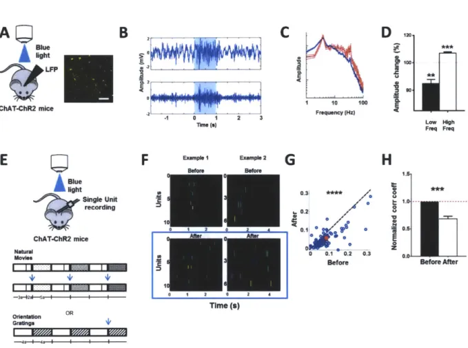
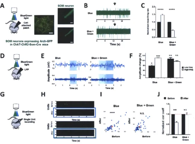
2.2.1. In vivo optogenetic stimulation of ChAT-ChR2 expressing axons induces desynchronization of local field potentials and drives neuronal decorrelation in V1
We addressed these questions in the superficial layers of primary visual cortex (VI) in adult mice. ACh release in vivo was induced by photostimulation (473 nm, Is, 20 Hz, 10 ms pulses, ~
10 mW/mm2) of channelrhodopsin-expressing cholinergic axons from the basal forebrain, in VI of ChAT-ChR2 transgenic mice(16) (Fig. 1A). The effect of cholinergic activity was tested by measuring the local field potential (LFP) in VI (17). Indeed, we observed that photostimulation of ChAT-ChR2 axons induced robust desynchronization of the LFP, similar to that induced by electrical stimulation of the nucleus basalis(18) (Fig. 1B-1C, 1D: post-stimulation decrease of low frequency events (<10 Hz, P < 0.005, paired t-test. Detailed numbers for all figures in Supplementary Table I and 2), increase of high frequency events (10 - 100 Hz, P < 0.0005, paired t-test).
One of the ways ACh(3) and attention(7) have been proposed to enhance the representation of information is through decorrelation between neurons, but the mechanisms underlying such decorrelation remain unresolved. We next examined whether activation of neocortical cholinergic axons can induce decorrelation by measuring the activity of single units with an array of multiple electrodes (Fig. 1E), in response to both natural movies and gratings of random orientation. Indeed, we observed a significant decorrelation between cortical neurons upon photostimulation (Fig. 1F) at the level of single units (Fig. 1G, P << 0.0001, paired t-test,
comparing the correlation coefficients before and after photostimulation; Supplementary Fig. 1A) and population of units (Fig. 1H: P = 0.0001, paired t-test, comparing population averaged correlation coefficients across animals before and after photostimulation; Supplementary Fig.
1B, see Supplementary Methods: In vivo single unit data analysis).
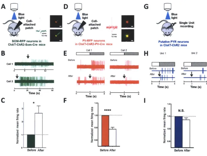
2.2.2. In vivo optogenetic stimulation of ChAT-ChR2 expressing axons evokes specific response signatures in SOM, PV and PYR neurons
To deconstruct the circuit that underlies ACh-induced LFP desynchronization and neuronal decorrelation, we performed in vivo cell-attached recordings from identified RFP-labeled SOM neurons (labeled with viral injection of a floxed RFP construct in VI of ChAT-ChR2-SOM-cre mice, Fig 2A-C) and PV neurons (similarly labeled in ChAT-ChR2-PV-cre mice, Fig 2D-F), as well as in vivo recording of putative pyramidal (PYR) neuron single units (in ChAT-ChR2 mice, Fig. 2G-I) within the superficial layers of V1. Neuron types were also distinguished by their spike shape (Supplementary Fig. 2A; Supplementary Methods). Interestingly, activation of ChAT-ChR2 axons modulated different cell types differently. Robust facilitatory responses were observed in SOM neurons (Fig. 2B, C: P < 0.05, paired t-test, comparing normalized firing rate before and after photostimulation; duration = 30.9 ± 7.49 s) while suppressive responses were observed in PV neurons (Fig. 2E, F: P <<< 0.0001, paired t-test comparing normalized visual responses before and after photostimulation). In putative PYR neurons however, there was no significant change in response rate (Fig. 2H, I: P > 0.2, paired t-test, comparing normalized visual response before and after photostimulation). Thus, endogenous ACh release via
photostimulation evokes distinct in vivo signatures from SOM, PV and PYR neurons: SOM neurons are facilitated, PV neurons are suppressed, and the mean spike rate of PYR neurons does not change but their visual responses are decorrelated.
2.2.3. ACh application in slices evokes specific responses in SOM, PV and PYR neurons
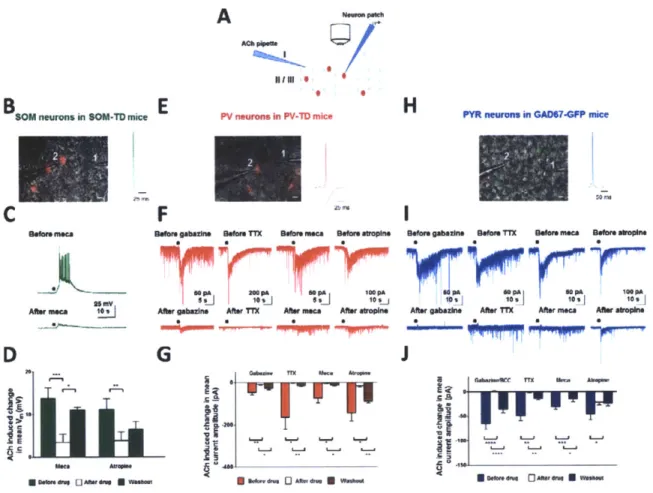
To further examine the functional interaction that underlies the in vivo responses described above, we performed ex vivo whole-cell patch clamp recordings (Fig. 3A) from SOM neurons (tdTomato positive neurons in SOM-TD mice; Fig. 3B-D), PV neurons (in PV-TD mice; Fig. 3E-G) and PYR neurons (GFP-negative neurons in GAD67-GFP mice; Fig. 3H-J). A total of 101 neurons (from 42 animals) were examined in slices under current and voltage clamp, after ACh application and with different antagonists applied singly or in combination. Consistent with the in vivo recordings (Fig. 2B), ACh application evoked a robust train of action potentials in non-fast spiking, SOM-expressing neurons (Fig. 3C top, Supplementary Fig. 3A). These responses were significantly reduced in the presence of the cholinergic antagonists mecamylamine (Fig. 3D: P < 0.0001, paired t-test, comparing ACh-induced depolarization before and after mecamylamine) and atropine (Fig. 3D: P = 0.005, paired t-test, comparing ACh-induced depolarization before and after atropine; see also Supplementary Figs 2B-C demonstrating that superficial layer inhibitory neurons express both muscarinic and nicotinic ACh receptors)._he facilitatory response in non-fast spiking inhibitory neurons persisted in the presence of glutamatergic and GABAergic antagonists (Supplementary Fig. 3B-C), indicating that it was due to direct action of ACh.
In PV neurons (patched in voltage clamp mode with Cl- based internal solution), ACh application increased the frequency of IPSCs (Fig. 3F top), which were sensitive to cholinergic antagonists (Fig. 3F bottom, Fig. 3G: P < 0.05, paired t-test, comparing ACh-induced current amplitudes before and after mecamylamine; P < 0.05, paired t-test, comparing amplitudes before and after atropine). Unlike SOM neurons however, the responses in PV neurons were observed to be indirect and of inhibitory origin as they were significantly reduced by GABA antagonist (Fig. 3F bottom, Fig. 3G: P < 0.01, paired t-test, comparing ACh-induced current amplitudes before and after gabazine). Additionally, the ACh-induced increase in frequency of IPSCs in PV neurons was also abolished in TTX (Fig. 3F bottom, Fig. 3G: P < 0.05, paired t-test, comparing ACh-induced current amplitudes before and after TTX). The cholinergic response in PYR neurons was similar to that in PV neurons, so that ACh application led to an increase in frequency of IPSCs (Fig. 31 top). This response was also significantly reduced in the presence of cholinergic antagonists (Fig. 31 bottom, Fig. 3J: P < 0.001, paired t-test, comparing ACh-induced current amplitudes before and after mecamylamine; P < 0.02, paired t-test, comparing amplitudes before and after atropine). Likewise, GABA antagonists and TTX also significantly reduced the response (Fig. 31 bottom, Fig. 3J: P < 0.0001, paired t-test, comparing ACh-induced current amplitudes before and after gabazine/bicuculline; P < 0.005, paired t-test, comparing ACh-induced current amplitudes before and after TTX). Thus, the ACh-induced responses in PV and PYR neurons appear to be indirectly mediated.
2.2.4. ACh-induced facilitation of SOM responses leads to ACh-induced inhibition in other cell types
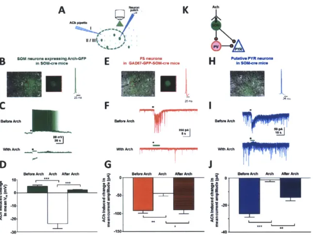
We hypothesized that the TTX and gabazine sensitive increase in frequency of IPSCs in PV and PYR neurons is due to activation of SOM neurons by ACh and subsequent inhibitory drive from SOM neurons to PV and PYR neurons. To test the hypothesis, we transiently blocked the activity of SOM neurons by selectively expressing archaerhodopsin-3 (Arch)(19) in them by viral injection of a flexed Arch construct into VI of SOM-cre mice; in addition, we crossed SOM-cre mice with GAD67-GFP mice for identifying inhibitory/fast-spiking neurons in conjunction with Arch blockade of SOM neurons (GAD67-GFP-SOM-cre mice). We carried out current and voltage clamp recordings in slices, with ACh application before and during green light stimulation of Arch in SOM neurons (Fig. 4A). Specific cell types were also identified by their electrophysiological characteristics (Supplementary Fig. 4A). Stimulation of Arch in SOM neurons (532 nm, 5-15s, continuous light, 0.174 mW/mm2) indeed abolished their cholinergic responses (Fig. 4B-D: P < 0.0005, paired t-test, comparing ACh-induced depolarization in SOM neurons before and after Arch activation). Control recordings from SOM neurons in SOM-TD mice without Arch expression showed that green light stimulation alone did not induce any changes in ACh-evoked responses (Supplementary Fig. 4B-C). To test the causal relationship between the ACh-induced facilitation of SOM responses and ACh-induced IPSCs in PV and PYR neurons, we carried out voltage clamp recordings from putative PV neurons (identified by their fast-spiking properties and presence of GFP in GAD67-GFP-SOM-cre mice; Fig. 4E-G) and from putative PYR neurons (identified by their pyramidal morphology and absence of GFP in SOM-cre mice; Fig. 4H-J). Photostimulation of Arch in SOM neurons completely blocked the
ACh-induced increase in frequency of IPSCs in both putative PV neurons (Fig. 4F, G P < 0.01, paired t-test, comparing ACh-induced current amplitudes before and after Arch) and putative PYR neurons (Fig. 41, J: P < 0.0001, comparing ACh-induced current amplitudes before and after Arch).
Thus, by a combination of optogenetics and pharmacology, these data reveal for the first time the circuit interactions between SOM, PV and PYR neurons in the context of ACh modulation. SOM neurons are directly activated by ACh, and release GABA to evoke IPSCs in PV and PYR neurons via GABA-A receptors (Fig. 4K). Because PV neurons widely inhibit PYR neurons (20-22), the SOM-mediated inhibition of PV neurons would subsequently disinhibit PYR neurons. Indeed, direct inhibition followed by disinhibition at sub-threshold levels is captured in ACh-induced responses of PYR neurons recorded in current clamp mode with a normal internal solution (Supplementary Fig. 4D-E). Such a circuit, comprising direct inhibition by SOM neurons and disinhibition via PV on PYR neurons, is also consistent with the in vivo observation of no net change in firing rate of PYR neurons during cholinergic modulation (Fig. 2H, I).
2.2.5. Desynchronization of local field potentials and neuronal decorrelation in V1 in vivo is mediated by SOM neurons
We next examined whether this specific inhibitory microcircuit can drive the ACh-induced changes in temporal dynamics of cortical activity, including LFP desynchronization and neuronal
decorrelation (Fig.1). To establish a causal relationship between these phenomena and SOM
neurons, we crossed ChAT-ChR2 mice with SOM-cre mice and expressed Arch in SOM neurons
by viral injection (ChAT-ChR2-Som-cre mice; Fig. 5A). Blue light induced ChAT-ChR2
stimulation facilitated SOM neurons, but green light activation of Arch simultaneous with blue
light stimulation blocked the facilitation and even reduced responses below background,
consistent with hyperpolarization of SOM neurons by Arch (Fig. 5A-C; activation by blue light:
P < 0.0001; suppression by green + blue light: P < 0.0001, paired t-test). We then measured the
LFP in the Arch-injected VI area (Fig. 5D). Activation of ChAT-ChR2 axons desynchronized the LFP (Fig. 5E, F: P <0.05 comparing decrease in low frequency amplitudes, P < 0.001
comparing increase in high frequency amplitudes, paired t-test); however, this modulation was
absent when Arch was activated during ChAT-ChR2 stimulation (Fig. 5E, F P > 0.3 comparing
low and high frequency amplitudes separately, paired t-test). We further confirmed in
ChAT-ChR2 control experiments that without Arch expression, green light presentation was not
sufficient to abolish the desynchronization (Supplementary Fig. 5A-C). These findings thus
directly demonstrate that SOM neurons play a critical role in cholinergic-induced
To examine the effect of SOM neurons on decorrelation of neuronal activity, we carried out single-unit recordings in ChAT-ChR2-SOM-cre mice within the region of VI where Arch was expressed in SOM neurons (Fig. 5G, Supplementary Fig. 6A). ChAT-ChR2 activation with blue light induced decorrelation between neurons (Fig. 5H, 51: p << 0.0001, paired t-test, 5J: P < 0.002, paired t-test), but the decorrelation was blocked during green light activation of Arch (Fig. 5H, 51: P > 0.4, paired t-test, 5J: P > 0.7, paired t-test). In control ChAT-ChR2 experiments without Arch expression, green light alone did not alter the extent of decorrelation (Supplementary Fig. 5D-F).
Decorrelation can improve visual coding by reducing redundancy between neurons(3, 23). We carried out a simple discrimination analysis(3) and found that decorrelation indeed improved discrimination performance; this improvement was also blocked by activation of Arch in SOM neurons (Supplementary Fig. 6B). Collectively, these data demonstrate that cholinergic activation of SOM neurons drives neuronal decorrelation in VI and thus potentially contributes to enhanced information processing.
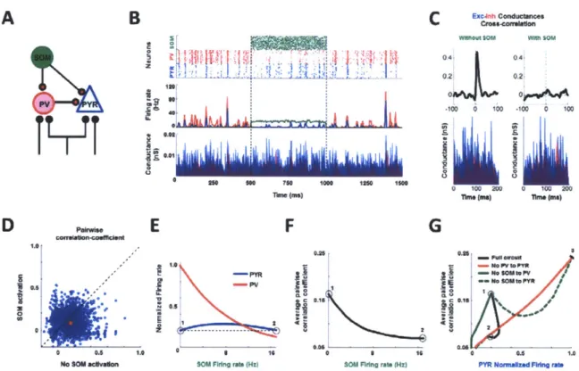
2.2.6. Computational analyses show that both SOM-induced direct inhibition and indirect PV disinhibition are necessary for rate-independent neuronal decorrelation
Previous computational studies have shown that correlations between pairs of neurons generated
by common inputs is a function of their spike rate(12). Our findings however showed no significant change in the spike rate of pyramidal neurons (Fig. 21) during ACh-induced
decorrelation (Fig. 1G-H). Although SOM neurons directly inhibit PYR neurons, they also disinhibit PYR neurons through suppression of PV neurons (Fig. 2E-F). It is thus be possible that
a different mechanism than inhibition-dependent spike rate modulation exists for this
rate-independent neuronal decorrelation. To investigate possible mechanisms, we simulated a
network model of 2000 SOM, 1000 PV and 1000 PYR neurons where varying SOM inputs could
alter the strengths of the direct inhibitory connection (SOM-PYR) and indirect disinhibitory
connection (SOM-PV-PYR). All neurons were modeled as conductance-based leaky
integrate-and-fire neurons (Fig.6A; Supplementary Methods: Computational model). We first
generated a synchronously firing network that resembled basal correlated states (Fig. 6B top and
middle panels) by providing common inputs with independent noise to PYR and PV neurons.
We then introduced a PV-PYR synaptic delay(24, 25) that filtered out independent noise
to favor signal propagation of common input. This led to the excitatory conductance being
tightly followed by the inhibitory conductance(26) (Fig. 6B bottom panel, Fig. 6C left). With
SOM activation, the temporal relationship between the inhibitory and excitatory conductance
was disrupted (Fig. 6B bottom; Fig. 6C right). This was accompanied by a robust decorrelation of the spike profile of PYR neurons (Fig. 6B top; Fig. 6D, 6F) with little change in the firing rate of PYR neurons (Fig.6E, Fig. 6G), similar to that observed experimentally (Fig. I G-H; Fig. 2F,
21). The model demonstrated that this relationship held for a wide range of SOM firing rates (Fig. 6F). As the SOM rate increased, the PV rate decreased while the PYR rate remained relatively unchanged and PYR correlation decreased (Fig. 6E - F), until the direct SOM-PYR inhibition became too strong for the indirect SOM-PV-PYR disinhibition to counter, thereby allowing rate-dependent decorrelation to dominate (12) (Fig. 6G, spike rates below point '2').
We further probed which connections were necessary for rate-independent decorrelation, by repeating the simulation in reduced circuits. In the three reduced circuits where each connection was selectively removed, we observed a rate-dependent correlation as reflected by the covariation of correlation with PYR rates (Fig. 6G, Supplementary Fig. 7). Thus, these analyses indicate that both the direct SOM-PYR inhibition and indirect SOM-PV-PYR disinhibition are necessary for rate-independent neuronal decorrelation of PYR neurons.
2.3. Discussion
Decorrelation between neurons can enhance(3) and even optimize(27) information processing. During execution of attentional tasks, decorrelation has been demonstrated to enhance population sensitivity to stimulus changes and the signal to noise ratio of neural signals (7, 8). Our study provides the first demonstration of a neural circuit that allows a mechanistic understanding of such temporal alterations of spike trains in the context of cholinergic modulation, a major neuromodulatory pathway implicated in attention and arousal(6). Specifically, we show experimentally that direct cholinergic facilitation of SOM neurons can activate both direct inhibitory and indirect PV disinhibitory pathways as well as decorrelate PYR neuronal spike profiles. Computational simulation further reveals that the two pathways together serve to destabilize intrinsic correlated network activity maintained by balanced excitation and PV inhibition(26), which are necessary for driving a and drive firing rate-independent decorrelation. These findings point to a crucial functional role of SOM neurons in intact cortical circuits, not only as players in spatial summation(28) and possibly in associative fear learning(29) but also in active shaping of the temporal structure of neural activity.
Our results indicate that direct SOM activation by ACh is also critical for the generation of network desynchronization (1, 5). This agrees with parallel observations of state-dependent SOM activation(28, 30, 31) and state-dependent variation in cholinergic output in the cortex(32) where more awake states favor both greater ACh release and higher SOM activity. Taken together, our data support the hypothesis that cortical circuits recruit unique inhibitory neuronal
cell types to modulate distinct brain states(14, 31, 33, 34) possibly for different brain functions, similar to that proposed in the hippocampus(35).
The co-existence of neuronal decorrelation and LFP desynchronization during ACh-induced SOM activation supports co-variation of temporal population activity with cortical states(5). Global cortical fluctuations have been proposed to induce neuronal correlations(5). During cholinergic modulation, these global fluctuations are suppressed, as reflected by a decrease in large amplitude, low frequency oscillations (Fig. 1D)(2) thereby leading to neuronal decorrelation(5). Small amplitude, high frequency oscillations then dominate(2), possibly due to sparsely distributed but simultaneously active neurons that fire synchronously(9). Indeed, previous findings have revealed the existence of such highly correlated neuronal pairs in decorrelated states(36). Future work will be necessary to characterize possible cell assemblies in the ACh-induced decorrelated states.
It is worth mentioning the link between high frequency events (gamma oscillations) and surround suppression through SOM activation. It has been recently shown that surround suppression in VI neurons is mediated by SOM neurons(28). Interestingly, the same visual stimulus that evokes surround suppression has also been shown to induce gamma oscillations(37). Likewise, ACh has also been shown to modulate surround suppression(38) and gamma oscillations(2). Our finding on SOM neurons mediating ACh-induced desynchronization provides the missing link between these various observations. Further investigation will be
important to understand the difference, if any, between SOM- and PV-mediated gamma oscillations.
Our findings help to reconcile seemingly contradictory effects of cholinergic modulation and nucleus basalis stimulation on visual responses of VI neurons(27). Cortical cholinergic activation has been linked to both fast GABA-mediated suppression(3, 39) and slow facilitation of visual responses of VI neurons(4, 27, 38-40). These findings can in principle be explained by our results where direct cholinergic activation of SOM neurons can drive both direct inhibition and indirect disinhibition(41) on PYR neurons to vary their firing rate according to the relative strengths of the two pathways. In addition, cholinergic activation of astrocytes(18) can provide excitatory influences at a slower timescale. Our results also extend the conclusions of earlier slice studies (42, 43) which have demonstrated the excitation of non fast-spiking inhibitory neurons and induction of an inhibitory barrage in fast-spiking and pyramidal neurons by ACh. The findings we describe do not rule out the possibility of other neuronal cell types (18, 44-46) being active during cholinergic modulation. These include facilitatory responses observed in vasoactive intestinal peptide (VIP)(44) and cholecystokinin-expressing interneurons(47) where the former has recently been shown to exclusively provide weak inhibition to SOM neurons(20). However, these cell types, unlike SOM neurons, have little connection to the two other principal cell types, PV and PYR neurons(20) that we have demonstrated to be critical players in driving the ACh-induced temporal changes in cortical dynamics. It is likely that these VIP neurons are involved in the modulation of energy supply during cholinergic modulation(47). Additionally, mAChR mediated activation of prolonged facilitatory responses in cortical astrocytes and hippocampal PYR neurons have also been proposed to drive ACh-induced plasticity (18, 45, 46).
Together with our data showing SOM neurons as a dominant driver of both decorrelation and desynchronization in cortical networks, we propose that these distinct ACh-responsive cell-types can reorganize to form specialized microcircuits for enabling multiple brain functions during cholinergic modulation.
2.4. Acknowledgments
We thank Guoping Feng, Holly Robertson (MIT) for providing the ChAT-ChR2 mice; Ed Boyden, Aimei Yang (MIT) for providing Arch virus; Chuong Le for technical assistance with viral injections and immunohistochemistry; Travis Emery for technical assistance with the optogenetics laser setup; Michael Goard and Jitendra Sharma for providing technical advice. This work was supported by an A*STAR (Singapore) Fellowship (NC), NIH (R01EY007023 and RO 1 EYO 18648), NSF and Simons Foundation grants (MS).