HAL Id: hal-00173388
https://hal.archives-ouvertes.fr/hal-00173388
Submitted on 19 Sep 2007HAL is a multi-disciplinary open access
archive for the deposit and dissemination of sci-entific research documents, whether they are pub-lished or not. The documents may come from teaching and research institutions in France or
L’archive ouverte pluridisciplinaire HAL, est destinée au dépôt et à la diffusion de documents scientifiques de niveau recherche, publiés ou non, émanant des établissements d’enseignement et de recherche français ou étrangers, des laboratoires
To cite this version:
Christophe Marecaux, Matthieu Chabanas, Yohan Payan, Franck Boutault. Computer aided plan-ning and navigation for orbito-zygomatic reconstruction.. Surgetica’2007: Computer-Aided Medical Interventions: tools and applications., 2007, France. pp.271-281. �hal-00173388�
3D Navigation Systems (3DNS) were introduced in the late 80s in neurosur-gery and no later described in maxillo-facial surgery. Concomitant develop-ment of computed imaging techniques offered exciting horizons for surgical planning and simulation and principles of Computer Aided Surgery (CAS) could be formulated for a consistent use in Cranio-maxillofacial surgery. To date it has been widely developed and increa-sing fields of indications were reported. However, no 3DNS specifically designed for maxillofacial surgery or one patholo-gy dedicated planning software plat-form are yet commercially available. We suggest a full protocol of CAS as previously recommended in literature addressing the challenging task of pri-mary or secondary reconstruction of OZMC dislocation. First, on a specifi-cally developed planning software, the best zygoma reduction and orbital boundaries reconstruction to achieve skeletal symmetry are determined. This treatment plan is then transferred to
the 3DNS within the operating room. After patient’s anatomy registration to his preoperative CT scan data, the navi-gation system allows zygomatic gui-ding to its planned reduced location and bone orbital volume restoration control. The feasibility of this tech-nique was checked in 3 patients with major OZMC deformities. Preliminary clinical results are presented.
MATERIALS AND
METHODS
Preoperative planning
Planning method for primary or secon-dary surgical treatment of OZMC was previously widely described [2]. Main steps are reminded.
3D models generation
The planning workstation provides from a regular CT scan dataset axial
COMPUTER AIDED PLANNING
AND NAVIGATION FOR
ORBITO-ZYGOMATIC RECONSTRUCTION
CH. MARECAUX*,**, M. CHABANAS*,
Y. PAYAN*, F. BOUTAULT***
* TIMC/GMCAO Laboratory, In3S - Université Joseph Fourier, Faculté de Médecine - 38706 La Tronche cedex. ** Clinic Cours Dillon – 1, rue Peyrolade - 31300 Toulouse.
*** Department of maxillofacial and facial plastic surgery, Hopital Purpan, TSA 40031 - 31059 Toulouse cedex 9.
and reconstructed coronal and sagittal views, as well as 3D reconstructed sur-face soft-tissue and skeletal. Practitioner can interactively browse through the different series, manipula-te the 3D models or select anatomical points or Regions Of Interest (ROI).
Target determination
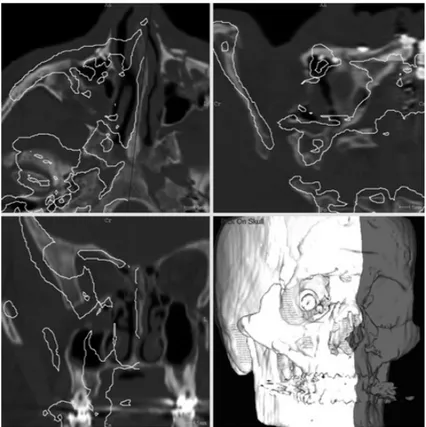
Whereas the target location for the fractured bone segment is defined sym-metrically to the healthy side, a theore-tical symmetric skull is computed around a midsagittal plane (fig. 1). This plane is defined by 3 included and manually selected anatomical land-marks.
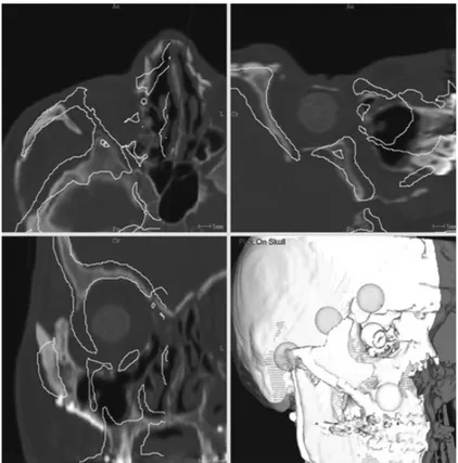
To take into account inaccuracy due to midsagittal plane manual definition, a double registration, first rigid then elastic, of the computed hemiskull to the bone structures surrounding the fractured zygoma is performed (fig. 2). Thanks to this process, an estimation of zygomatic location before injury has been obtained and will be able to be used as the target location for zygoma repositioning in the following steps of the planning.
Zygoma segmentation
Displaced bone is semi-automatically segmented using a spherical ROI which is defined by 4 points belonging
Fig. 1 : Native skull overimpressed with symmetrical skull computed from the controlateral healthy side around midsagittal plane.
to its limit and rather corresponding to the fractured buttresses. This sphere includes the body of zygomatic bone which is the most significant skeletal part for facial symmetry restoration.
Surgical planning for zygoma reduction
Finally, an automatic rigid registration of the previously segmented fractured bone to the target skull computed in step 2 is performed to determine the best zygomatic bone repositioning to achieve skeletal symmetry (fig. 3). Since matching provides all the better
result that initial position of both sur-faces is close, a prior point-to-point registration can be required in case of large displacement.
Non-injured orbit segmentation
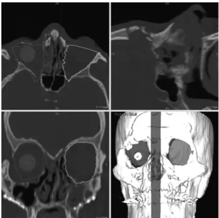
The same as for zygomatic reconstruc-tion planning, orbital boundaries resto-ration is planned symmetrically from the healthy side. Orbital volume is semi-automatically segmented with a cone-shaped growing ROI defined by its main axis corresponding to the neuro-optic axis and a closing anterior plane (fig. 4).
Fig. 2 : Result from the double registration of symmetrical skull to native skull according to selected surrounding ROI (spheres).
Surgical planning for orbital boun-daries reconstruction
A virtual orbit is then computed from the previously segmented orbit symme-trically around the midsagittal plane. Its boundaries are superimposed on the different axial, coronal and sagittal series and will be used as a guiding for accurate orbital walls reconstruction (fig. 5).
Intraoperative navigation and guiding
The Surgetics™ navigation system and the Craniologics™ software (Praxim-Medivision, La Tronche, France) were
fitted and used for intraoperative navi-gation. This navigation system uses a passive and infrared light based optical technology. Novelty of the Craniologics™ software is a double point-surface registration of patient’s anatomy to preoperatively acquired CT scan data set, first cutaneous, second skeletal after surgical exposure to improve navigation accuracy around the ROI. Then, location of calibrated surgical instruments in relation to its actual location in the surgical field can be presented in the different series or 3D models and in real time updated. Software adaptation consists of an additional zygomatic navigation step. First, a Rigid Body is rigidly fixated to
Fig. 4 : Segmentation of the healthy orbit with a cone-shaped growing ROI defined by the neuro-optic axis and an average closing anterior plane.
zygomatic body by a 2 pins transcuta-neous fixation (Digital Zygomatic Frame, DZF) (fig. 6). Next, a point-to-surface registration between zygoma and its model transferred with the planning data set is performed. Thus, zygoma can be guided to the planned best repositioning. Relative position between actual zygomatic location and target location is in real time updated and presented by a distance map bet-ween both surfaces and by boundaries of both elements superimposition on axial, sagittal and coronal series (fig. 7). Navigation interface also provides ove-rimpression of planned orbital recons-tructed boundaries on axial, coronal and sagittal series. Accuracy of orbital walls reconstruction or implant
place-ment according to planning is checked by scanning reconstructed orbital walls surface with the calibrated pointer.
Fig. 5 : Planning for orbital reconstruction defined by symmetrical orbit from the healthy one around midsagittal plane.
Fig. 6 : 3DNS setting. DRF : Digital Reference Frame for registering patient’s anatomy to its CT scan data set. DZF : Digital Zygomatic Frame for navigating bone fragment.
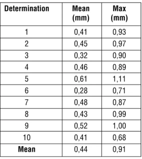
Problem raised by our method was to know if the point-to-surface zygomatic registration according to the small bone surface exposed through the limi-ted surgical approach is enough accu-rate for Zygoma repositioning. This has been evaluated on a one piece dry skull previously CT scan acquired. 3DNS was set up as previously presented. Mean and max distances of zygomatic surface in DZF referential to the same surface in DRF referential was compu-ted in 10 successive manipulations.
Patients
A pilot study of a non-comparative, non-randomized series of 3 patients
(table 1) was carried out to check method feasibility. Advisory Comity for People Protection Consenting to Clinical Research approval was obtai-ned through the Toulouse University Hospital, France. Informed consent forms were provided to patients, signed by both sides and collected by investi-gators.
2 patients had post traumatic malar asymmetry with associated enophthal-mos. One of them (patient 1) could not have received primary surgical treat-ment because of initial neurosurgical injury and the second (patient 2) was first operated 6 months before in ano-ther center. Patient 2 underwent bilate-ral OZMC fracture and left side was
Fig. 7 : 3DNS interface. Navigated zygoma location is in real time updated and tracked through its boundaries overimpression on axial, coronal and sagittal views in comparison to target loca-tion or a distance map between actual and target localoca-tions.
considered as the reference side for controlateral symmetrization. Patient 1 initially lost vision by optic nerve inju-ry and patient 2 by eyeball dilaceration which required prosthetic rehabilita-tion. The last patient (patient 3) had an acute largely displaced zygomatic frac-ture with severe comminutive fracfrac-ture of zygomatico-maxillary buttress and orbital rim. No ophthalmologic com-plication was associated.
Surgical approaches and number of miniplates for zygomatic fixation were individualized according to the patien-t’s fracture. Intraoral upper sulcus and transconjunctival incisions were used in all patients. In secondary treatment, a temporal or coronal approach pre-viously used in a neurosurgical treat-ment was associated to control zygo-matic arch.
RESULTS
Accuracy of the point-to-surface zygomatic registration
The computed mean distance between the same zygomatic registered in DRF referential and DZF referential was 0,44 mm in mean and 0,91mm in maxi-mum (Table 1).
Pilot study
Computer aided planning and naviga-tion procedures were successfully car-ried out for all 3 clinical cases. Surgical procedures and postoperative courses were uneventful.
Preoperative planning was completed in 7 to 10 minutes and was judged quite convenient by practitioners. Virtual repositioning alone was useful to conceptualize 3D displacement of zygomatic bone. Navigation and gui-ding device and specially DZF did not stop performing surgical treatment. Surgical time increase due to 3DNS installation was about 20 minutes. Whereas no anatomical landmarks allowed intraoperatively to check sym-metry achievement, zygoma repositio-ning was done as indicated by intrao-perative guiding whatever practitioner thought.
Surgical outcomes are presented (fig. 8, 9, 10). They have been conside-red as satisfying by both patient and surgeon in all 3 cases. Patient 2 who had initially bilateral OZMC fracture has not been back on his pretraumatic appearance but on a slightly enlarged facial width. However, he was totally satisfied with facial symmetrization. None of the 3 patients complained of the cheek scare.
Determination Mean Max (mm) (mm) 1 0,41 0,93 2 0,45 0,97 3 0,32 0,90 4 0,46 0,89 5 0,61 1,11 6 0,28 0,71 7 0,48 0,87 8 0,43 0,99 9 0,52 1,00 10 0,41 0,68 Mean 0,44 0,91
Table 1 : Distances computed of zygomatic surface registered in DZF referential to the same surface registered in DRF referential.
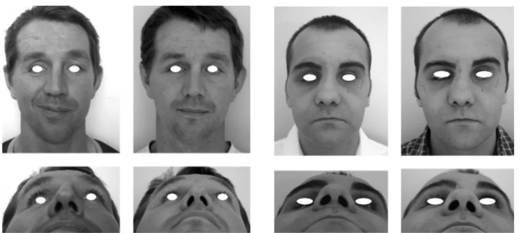
Fig. 8 : Patient 1. The postoperative pictures (right) in comparison to preoperative pictures (left) show restoration of antero-posterior pro-jection of the zygomatic body and the facial width.
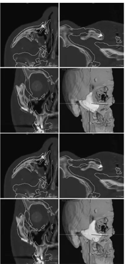
Fig. 9 : Patient 2. The postoperative pictures (right) in comparison to preoperative pictu-res (left) show pictu-restoration of antero-posterior projection of the zygomatic body and the facial width. Right prosthetic eyeball remains postoperatively retrusive despite orbital volume reconstruction.
Fig. 10 : Patient 3. The postoperative pictures (top and centre bottom) show facial symmetry restoration whereas the preoperative CT scan (bot-tom right and left) points out zygomatic displacement.
DISCUSSION
Development of augmented reality techniques seems to be able to help surgical treatment of late or acute OZMC deformities resulting from displaced or comminuted fractures. Preoperative, 3D multiplanar or surfa-cic reconstruction from CT scan data set is helpful in evaluating the extent and the way of post traumatic deformi-ties. Then, 3D planning system based on 3D virtual models allows segmenta-tion of the skeletal region of interest and interactive repositioning in order to assess different alternatives and retain the best to achieve skeletal sym-metry. Finally, surgical procedure can be carried out as planned preoperative-ly using 3DNS control and guidance. Thus, several suggestions were made addressing whether zygomatic reposi-tioning or orbital reconstruction, plan-ning or operative assistance for surgi-cal treatment [2, 3, 4].
The method we present includes all steps of a comprehensive computer aided protocol from diagnostic to intraoperative assistance, for both zygomatic and orbital reconstruction in primary or secondary treatment. Diagnostic, planning and simulation were automated as much as possible to shorten allocated time and get the pro-cess friendly. All 3D skeletal and soft tissue models, fractured zygoma or non-injured orbit are semi-automati-cally segmented. Zygoma repositioning is automatically planned as the regis-tration of segmented injured zygoma to the target skull. The corresponding mathematical transformation is equi-valent to the repositioning movement to apply. So far, manual shift with 6 degrees of freedom in a virtual envi-ronment was suggested [4] but seemed time consuming, difficult to achieve in
the same time reduction of all 4 zygo-matic buttresses and inaccurate in case of dislocated fracture which does not allow visual control of buttresses align-ment. Targets for both zygomatic and orbital reconstruction are defined sym-metrically from the healthy side around the midsagittal plane. Landmarks-based definition of the facial midsagittal plane without any further correction can give rise to a shift in target skull definition. So, in correction, we suggest to apply a dou-ble registration first rigid second elas-tic between symmetrical hemiskull computed from the healthy side and regions of interest of the native skull surrounding OZMC.
The planned procedure components including 3D native skull, target skull, target orbit and zygoma models as well as mathematical transformation are directly transferred to the operating theatre. OZMC reconstruction outlines can be check similar to the planned ones scanning bone surface with the calibrated pointer, but zygomatic trac-king makes proper repositioning easier and shorter. So far, only surface scan-ning control was performed [4] what means zygomatic osteosynthesis has to be taken down if repositioning is not checked correct.
Primary or secondary OZMC recons-truction was addressed by others work-groups, because it synthesizes require-ments and difficulties of computer-assisted preoperative planning and intraoperative assistance in cranio-maxillofacial plastic and reconstructive surgery. However, none of them dealt with orbital and zygomatic reconstruc-tion in the same time while they are anatomically and physiopathologically closely linked. Since no pretraumatic imaging data is available, all groups are
agreed to plane reconstruction symme-trically from the healthy side around a midsagittal plane. However, to avoid inaccuracy due to landmarks based definition of the midsagittal plane, either an automatic extraction based on matching of homologous surface areas using an iterative closest point optimization can be used [5] or, as we suggest, a correction by matching the theoretical symmetrical skull to the native skull around the region of inter-est. Planning of Zygoma repositioning to the target skull was mainly manual what reserves the method for modera-tely displaced zygomatic fracture without buttress dislocation whereas large displacement, dislocated fracture and secondary treatment are probably the best indications for computed-assisted OZMC reconstruction. Some groups ruled out direct bone guiding
fixing DRF onto the segmented bone which should be moved, to use as intra operative assistance the only surface scanning with a calibrated pointer to check reconstruction outline accurate [3]. In our opinion, in selected recons-tructive problems, direct bone gui-ding can be useful to achieve more easily repositioning as preoperatively planned.
This pilot study on the computer-assis-ted management of post traumatic OZMC deformities show virtual plan-ning for orbital boundaries reconstruc-tion or zygomatic reposireconstruc-tioning, as well as intraoperative guiding and verifica-tion of the surgical procedure in accor-dance with the planned one can be suc-cessfully carried out. First results seem promising specially in difficult case of late deformities.
BIBLIOGRAPHY
[1] MAUBLEU S., MARECAUX C., CHABANAS M. et al.
Computer-aided planning for zygomatic bone reconstruction in maxillofacial traumatology. Proceedings of the Second International Conference Surgetica’2005: Computer-Aided Medical Interventions: tools and applications.
Sauramps Medical Eds., 2005, pp. 405-411.
[2] MARMULLA R. NIEDERDELL-MANN H. et al.
Surgical Planning of Computer-Assisted Repositioning Osteotomies.
Plast. Reconstr. Surg., 1999: 104: 938-944.
[3] GELLRICH N.C., SCHRAMM A., HAMMER B. et al.
Computer-Assisted Secondary Recons-truction of Unilateral Posttraumatic Orbital Deformity.
Plast. Reconstr. Surg. 2002, 110: 1417-1429.
[4] WESTENDORFF C.,
GÜLICHER D., DAMMANN F. et al.
Computer-Assisted Surgical Treatment of Orbitozygomatic Fractures.
J Craniofac.Surg., 2006, 17: 837-842.
[5] DE MOMI E., CHAPUIS J., PAPPAS I. et al.
Automatic extraction of the mid-facial plane for cranio-maxillofacial surgery planning.
Int. J. Oral Maxillofac. Surg 2006, 35 : 636-642.