HAL Id: tel-01685238
https://tel.archives-ouvertes.fr/tel-01685238
Submitted on 16 Jan 2018HAL is a multi-disciplinary open access archive for the deposit and dissemination of sci-entific research documents, whether they are pub-lished or not. The documents may come from teaching and research institutions in France or abroad, or from public or private research centers.
L’archive ouverte pluridisciplinaire HAL, est destinée au dépôt et à la diffusion de documents scientifiques de niveau recherche, publiés ou non, émanant des établissements d’enseignement et de recherche français ou étrangers, des laboratoires publics ou privés.
Biomarqueurs du risque cardio-métabolique dans les
pathologies respiratoires chroniques : impact de la prise
en charge
Ingrid Jullian-Desayes
To cite this version:
Ingrid Jullian-Desayes. Biomarqueurs du risque cardio-métabolique dans les pathologies respiratoires chroniques : impact de la prise en charge. Médecine humaine et pathologie. Université Grenoble Alpes, 2017. Français. �NNT : 2017GREAV020�. �tel-01685238�
THÈSE
Pour obtenir le grade de
DOCTEUR DE LA COMMUNAUTÉ UNIVERSITÉ
GRENOBLE ALPES
Spécialité : Physiologie-Physiopathologie-Pharmacologie
Arrêté ministériel : 25 mai 2016
Présentée par
Ingrid JULLIAN-DESAYES
Thèse dirigée par le Dr Marie JOYEUX-FAURE et co-dirigée par le Pr Jean-Louis PEPIN
Préparée au sein du Laboratoire HP2 INSERM U1042 dans l'École Doctorale Chimie et Sciences du Vivant
Biomarqueurs du risque
cardio-métabolique dans les pathologies
respiratoires chroniques :
Impact de la prise en charge
Thèse soutenue publiquement le 24 avril 2017, devant le jury composé de :
Pr Jean-Paul JANSSENS
Professeur associé et Médecin-adjoint agrégé,
Hôpitaux universitaires Genève, président et rapporteur
Pr Frédéric GAGNADOUX
PU-PH, Université Angers, rapporteur
Dr Jean-Christian BOREL
PhD, Université Grenoble Alpes, examinateur
Dr Pierrick BEDOUCH
MCU-PH, Université Grenoble Alpes, examinateur
Dr Marie JOYEUX-FAURE
MCU, Université Grenoble Alpes, directeur de thèse
Pr Jean-Louis PEPIN
2
R e m e r c i e m e n t s
Mes remerciements vont tout d’abord au Docteur Marie JOYEUX-FAURE, qui depuis mon premier stage d’internat aux essais cliniques a toujours été là. Merci pour ton encadrement tout au long de ces travaux de Master 2 et de thèse de sciences. Tu es bien plus qu’une directrice de thèse et j’espère que nous continuerons à travailler ensemble.
Je tiens aussi à remercier le Professeur Jean-Louis PEPIN pour son encadrement et ses nombreuses idées de projets.
Je remercie également tous les membres du jury, le Docteur Pierrick BEDOUCH ainsi que le Professeur Frédéric GAGNADOUX et le Professeur Jean-Paul JANSSENS pour avoir accepté d’être les rapporteurs de ce travail.
Merci au Docteur Jean-Christian BOREL d’avoir accepté d’être membre de ce jury, ce fût un réel plaisir de travailler avec toi sur l’étude BioSOH.
Merci à l’équipe du laboratoire EFCR, Andry, Amina, Sandrine.
Je remercie également les Docteurs Damien VIGLINO et Bruno REVOL pour leur collaboration.
Bien sûr je n’oublie pas mes co-internes Mélanie, Anne-Laure, Laure, Virginie, et surtout
Audrey. Heureusement que tu étais là pour continuer la routine pendant que j’étais dans mes
recherches Pubmed ou tableaux Excel…
A mes parents, je vous dois tellement que je ne sais par où commencer. Merci pour votre
soutien tout au long de ces années d’études, qui n’aurait probablement pas été réalisables sans vous. Merci d’avoir été là dans les nombreux moments de doute.
3
Résumé/Abstract
4
Résumé
Le syndrome d’apnées obstructives du sommeil (SAOS) est associé à de nombreuses co-morbidités métaboliques et cardiovasculaires. L’hypoxie intermittente chronique, une des composantes du SAOS, induit des mécanismes intermédiaires délétères tels que stress oxydatif, inflammation, insulino-résistance ou encore dyslipidémie, à l’origine de ces comorbidités. Ces mécanismes intermédiaires sont également communs à d’autres pathologies respiratoires chroniques telles que la bronchopneumopathie chronique obstructive (BPCO) et le syndrome d’obésité hypoventilation (SOH).
L’hypoxie intermittente et les mécanismes intermédiaires associés sont aussi à l’origine de l’existence et de la progression de la stéatopathie métabolique (« non alcoholic fatty liver disease »). Ce lien entre pathologies respiratoires chroniques et atteinte hépatique est un mécanisme essentiel mais plus récemment étudié des co-morbidités dans le SAOS et la BPCO. Différents biomarqueurs cardiométaboliques ont donc été étudiés dans ces pathologies respiratoires chroniques à la fois pour caractériser les co-morbidités et l’atteinte systémique et pour apprécier l’impact de différentes thérapeutiques. La première partie de cette thèse sera consacrée à une revue systématique des différents biomarqueurs cardiométaboliques liés à chacune de ces 3 pathologies respiratoires chroniques : SAOS, BPCO et SOH.
Le traitement du SAOS par pression positive continue (PPC) a un effet bénéfique sur les symptômes fonctionnels liés à cette pathologie. Cependant, l’impact de la PPC sur d’autres conséquences cardio-métaboliques délétères du SAOS reste encore à démontrer par des essais randomisés contrôlés, notamment sur l’atteinte hépatique.
Dans la seconde partie de cette thèse, nous détaillerons l’impact de la PPC sur les différents marqueurs cardiométaboliques du SAOS à l’aide d’une revue systématique puis d’une étude randomisée contrôlée sur l’impact de la PPC sur les marqueurs d’atteinte hépatique.
Par ailleurs, les patients atteints de SAOS, BPCO ou SOH reçoivent du fait de leur polypathologie (multimorbidité) des traitements médicamenteux multiples qui visent à contrôler au mieux les co-morbidités. Il est donc primordial de considérer la prise en charge globale de ces patients du point de vue de leurs traitements instrumentaux (PPC et ventilation non invasive) mais aussi en considérant l’impact des traitements médicamenteux associés. En effet, les traitements médicamenteux peuvent interférer avec la sévérité de la pathologie elle-même et impacter les biomarqueurs liés aux comorbidités associées. La troisième partie de cette thèse sera consacrée à l’étude d’un antihypertenseur chez le patient SAOS et envisagera l’influence des médicaments sur la pertinence de l’usage des bicarbonates comme marqueurs diagnostiques du SOH.
En conclusion, nous insisterons sur la nécessité d’une prise en charge intégrée multi systémique et d’une prise en charge personnalisée de ces patients.
Mots clés :
Biomarqueurs cardiométaboliques, syndrome d’apnées obstructives dusommeil, bronchopneumopathie chronique obstructive, syndrome d’obésité hypoventilation, pression positive continue, médicaments concomitants
5
Abstract
Obstructive sleep apnea (OSA) is associated with related metabolic and cardiovascular comorbidities. Chronic intermittent hypoxia the hallmark of OSA induces deleterious intermediary mechanisms such as oxidative stress, systemic inflammation, insulin resistance and dyslipidemia. Cardiovascular and metabolic comorbidities are also key features of other chronic respiratory diseases such as chronic obstructive pulmonary disease (COPD) and obesity hypoventilation syndrome (OHS). Chronic hypoxia and deleterious intermediary mechanisms also trigger occurrence and progression of non-alcoholic fatty liver disease. This link between chronic respiratory diseases and liver injury is observed through modifications of specific liver biomarkers in OSA and COPD. A variety of cardio-metabolic biomarkers have been studied for stratification of cardio-metabolic risk and assessing treatment impact in chronic respiratory diseases. The first part of this PhD thesis is a systematic review of cardio-metabolic biomarkers in 3 respiratory diseases: OSA, COPD and OHS.
Continuous positive airway pressure (CPAP) the first line therapy for OSA improves symptoms and quality of life. However, CPAP effects on cardio-metabolic consequences remain still debated. In the second part of the PhD thesis, we will address CPAP impact on different cardio-metabolic biomarkers and more specifically in markers of liver injury by reporting original results of a randomized controlled trial (RCT).
Polypharmacy is usual in patients with OSA, COPD or OHS. Beyond CPAP or non-invasive ventilation treatment, it is essential to address the contribution of associated medications. Indeed, pharmacological treatments can interfere with the severity of the disease and control of associated comorbidities. The third part of the thesis will present a RCT evaluating Bosentan in hypertensive OSA patients and will present how medications for comorbidities decrease bicarbonate diagnosis value for OHS.
We will conclude by underlining the crucial importance of personalized medicine and integrated care in chronic respiratory diseases.
Key-words:
Cardio-metabolic biomarkers, obstructive sleep apnea syndrome, chronicobstructive pulmonary disease, obesity hypoventilation syndrome, continuous positive airway pressure, concomitant medications
6
Table des matières
Travaux de publications internationales ... 8
Communications orales ... 8
Liste des figures et tables ... 9
Liste des abréviations ... 10
Introduction générale ... 11
Partie I : Biomarqueurs dans les maladies respiratoires chroniques ... 14
1. Biomarqueurs cardiométaboliques du SAOS ... 15
1.1 Physiopathologie du SAOS ... 15
1.2 SAOS et comorbidités associées ... 16
1.3 Marqueurs cardiometaboliques du SAOS ... 17
1.4 Biomarqueurs d'atteinte hépatique et SAOS ... 20
2. Biomarqueurs hépatiques et BPCO : Etude BPCO-NASH ... 23
2.1 Physiopathologie de la BPCO ... 23
2.2 Comorbidités associées ... 23
2.3 Biomarqueurs hépatiques et BPCO ... 24
3. Bicarbonates et SOH : Etude BioSOH ... 54
3.1 Physiopathologie du SOH ... 54
3.2 Diagnostic du SOH ... 54
3.3 Prévalence du SOH : Etude BioSOH ... 55
Partie II : Pression positive continue et biomarqueurs cardiométaboliques du SAOS ... 85
1. Effet de la PPC sur les biomarqueurs du SAOS ... 86
1.1 Pression positive continue ... 86
1.2 Effets de la pression positive continue ... 86
1.3 Données des essais randomisés sham contrôlés ... 87
2. PPC et biomarqueurs d'atteinte hépatique ... 106
2.1 Données de la littérature ... 106
2.2 Etude PPC-NASH ... 107
Partie III : Traitements médicamenteux et pathologies respiratoires chroniques ... 118
1. Bosentan et SAOS ... 119
7
1.2 Bosentan ... 119
1.3 Effet du bosentan sur l’hypertension artérielle chez le patient SAOS : Etude BOSAS 120 2. Influence des médicaments sur les bicarbonates ... 129
3. Influence des médicaments sur le SAOS ... 141
4. Perspectives : Projet OSFP/OPTISAS ... 165 Conclusion ... 171 Références bibliographiques ... 174
8
Travaux de publications internationales
Publication 1 : Non-alcoholic fatty liver disease in chronic obstructive pulmonary disease. Viglino D, Jullian-Desayes I, Minoves M, Aron-Wisnewsky J, Leroy V, Zarski JP, Tamisier R, Joyeux-Faure M,Pépin JL. Sous presse dans l’Eur Respir J………..………..page 26 Publication 2 : Prevalence of obesity hypoventilation syndrome in obese. Borel JC, Guerber F, Jullian-Desayes I, Joyeux-Faure M, Arnol N, Taleux N, Tamisier R, Pépin JL. Sous presse dans
Respirology. ………...…..page 58
Publication 3 : Impact of obstructive sleep apnea treatment by continuous positive airway pressure on cardiometabolic biomarkers: a systematic review from sham CPAP randomized controlled trials. Jullian-Desayes I, Joyeux-Faure M, Tamisier R, Launois S, Borel AL, Levy P, Pepin JL. Sleep Med Rev. 2015;21:23-38..……….………..page 89 Publication 4 : Impact of effective versus sham continuous positive airway pressure on liver injury in obstructive sleep apnoea: Data from randomized trials. Jullian-Desayes I, Tamisier R, Zarski JP, Aron-Wisnewsky J, Launois-Rollinat SH, Trocme C, Levy P, Joyeux-Faure M, Pepin JL.
Respirology. 2016;21(2):378-85..……….………..page 108
Publication 5 : Comparison of continuous positive airway pressure and bosentan effect in mildly hypertensive patients with obstructive sleep apnoea: A randomized controlled pilot study. Joyeux-Faure M, Jullian-Desayes I, Pepin JL, Cracowski JL, Baguet JP, Tamisier R, Levy P, Godin-Ribuot D, Launois SH. Respirology. 2016;21(3):546-52..………...……..page 121 Publication 6 : Drugs influencing acid base balance and bicarbonate concentration readings. Jullian-Desayes I, Borel JC, Guerber F, Borel AL, Tamisier R, Levy P, Schwebel C, Pepin JL, Joyeux-Faure M. Expert Rev Endocrinol Metab online: 24 Feb 2016...………..page 131 Publication 7 : Impact of concomitant medications on obstructive sleep apnoea. Jullian-Desayes I, Revol B, Chareyre E, Camus P, Villier C, Borel JC, Pepin JL, Joyeux-Faure M. Br J Clin
Pharmacol. 2017;83(4):688-708………...….page 143
Communications orales
Impact of continuous positive airway pressure on liver injury induced by obstructive sleep apnea: Data from randomized controlled trials. R.Tamisier, I.Jullian-Desayes, J.P Zarski, J.Aron-Wisnewsky, S.Launois, N.Arnol, K.Clement, P.Levy, M.Joyeux-Faure, J.L Pepin. (ERS Amsterdam 2015)
Non-alcoholic fatty liver disease (NAFLD): a new mechanism for co-morbidities in chronic obstructive pulmonary disease (COPD)? D.Viglino, I.Jullian-Desayes, R.Tamisier, M.Perrin, M.Joyeux-Faure, JL.Pepin. (ERS Londres 2016)
Impact de la PPC sur l'évolution des biomarqueurs cardiométaboliques des patients SAOS : revue des essais randomisés contrôlés par sham PPC. I.Jullian-Desayes, M. Joyeux-Faure, R. Tamisier, S.Launois, AL Borel, P.Levy, JL Pepin. (Congrès sommeil Lille 2014 & CPLF Lille 2015) Pression positive continue et stéatose hépatique liée au SAOS : données d’essais randomisés contrôlés. I.Jullian-Desayes, R.Tamisier, J.P Zarski, S.Launois, N.Arnol, C.Trocme, P.Faure, P.Levy, M.Joyeux-Faure, J.L Pepin. (CPLF Lille 2015)
9
Liste des figures
Figure 1. L’augmentation du stress oxydatif dans le SAOS et ses conséquences
cardiométaboliques.
Figure 2. Hypoxie intermittente chronique et NAFLD
Figure 3. Flow-chart de l’étude BioSOH.
Figure 4. Design de l’étude à partir des données d’OPTISAS.
Liste des tables
Tableau I. Interprétation des scores du Fibromax®.
10
Liste des abréviations
BPCO Bronchopneumopathie chronique obstructive
CRP Protéine C réactive
HbA1c Hémoglobine glyquée A1c
HDL High-density lipoproteins
HOMA Homeostasis Model Assessment
IL Interleukine
IMC Indice de masse corporelle
LDL Low-density lipoprotein
NAFLD Non-alcoholic fatty liver disease
NASH Non-alcoholic steatohepatitis
PPC Pression positive continue
SAOS Syndrome d’apnées obstructives du sommeil
SOH Syndrome d’obésité hypoventilation
TNF-α Tumor necrosis factor alpha
11
Introduction générale
12 Les pathologies respiratoires chroniques telles que le syndrome d’apnées obstructives du sommeil (SAOS) [1], la bronchopneumopathie chronique obstructive (BPCO) [2] ou encore le syndrome d’obésité hypoventilation (SOH) [3] sont associées à un risque élevé de morbi-mortalité cardio-métabolique. L’hypoxie intermittente qui est le stimulus prédominant au cours du SAOS et du SOH induit des mécanismes intermédiaires délétères tels que stress oxydatif, inflammation de bas grade, insulinorésistance ou encore dyslipidémie, à l’origine de ces comorbidités. Ces mêmes mécanismes intermédiaires sont des acteurs importants de la survenue de l’atteinte multi systémique dans la BPCO. Ces mêmes mécanismes sont capables d’induire une atteinte hépatique plus récemment étudiée mais qui joue un rôle majeur dans les co-morbidités cardio-métaboliques des maladies respiratoires chroniques.
Les conséquences cardiovasculaires et métaboliques du SAOS sont nombreuses ; comprenant l’hypertension artérielle, le diabète de type 2, le syndrome métabolique, la stéatose hépatique, l’accident vasculaire cérébral, la fibrillation auriculaire ou encore l’insuffisance cardiaque [1,4,5]. Ces comorbidités ou la pathologie respiratoire en elle-même sont à l’origine de perturbations de biomarqueurs sanguins et urinaires [6,7]. En effet, on note chez les patients SAOS des modifications du profil lipidique (élévation des taux de triglycérides, diminution du cholestérol HDL…), glucidique (développement d’une insulinorésistance) [8] et inflammatoire (augmentation de la protéine C réactive (CRP), du facteur de nécrose tumorale (TNF)-α, des interleukines (IL)-6 et -8). Dans la BPCO, il existe aussi une inflammation systémique à l’origine du développement de pathologies cardiovasculaires ; les patients BPCO ont un risque 2 à 5 fois plus élevé d’arythmies, d’insuffisance cardiaque ou d’infarctus du myocarde. Comme dans le SAOS on observe chez les patients BPCO une forte prévalence d’hypertension (entre 28,5% et 64,7%), de syndrome métabolique (21 à 53%) ou de certaines de ses composantes telles que diabète de type 2 (16,8 à 28,5%), ou hypertriglycéridémie (19,7%) [9]. Ce lien entre BPCO et comorbidités cardiométaboliques se manifeste par des
13 modifications des taux d’IL-6 et -8, ou encore d’hémoglobine glyquée (HbA1c) [9]. Enfin le SOH est lui aussi associé à des comorbidités métaboliques et cardiovasculaires [10]. Il est important de noter que la plupart des patients porteurs de ce syndrome sont obèses morbides et atteints d’un SAOS sévère. Le SOH serait associé à une prévalence élevée d’hypertension artérielle pulmonaire (58% chez les SOH versus 9% chez les SAOS) ou d’insuffisance cardiaque (risque 9 fois plus élevé) [11,12]. En effet, on retrouve dans l’étude de Castro-Añón et al. une prévalence plus élevée d’hypertension, de dyslipidémie, d’arythmies, ou encore d’insuffisance cardiaque chez les SOH comparés aux SAOS [13]. Dans une première partie, nous détaillerons donc les biomarqueurs de retentissement des trois pathologies respiratoires chroniques : SAOS, BPCO et SOH.
Nous pourrons alors aborder l’impact de la prise en charge de la pathologie respiratoire chronique sur ces biomarqueurs. Dans une deuxième partie, nous étudierons l’effet de la pression positive continue (PPC) sur les modifications des taux de biomarqueurs sanguins et urinaires chez le patient SAOS, en ciblant tout particulièrement les biomarqueurs d’atteinte hépatique par une étude randomisée contrôlée.
De plus, les traitements médicamenteux, administrés de façon concomitante lors de ces pathologies respiratoires chroniques, peuvent eux aussi interférer avec les biomarqueurs de ces pathologies ou des comorbidités associées [14]. La dernière partie sera consacrée à l’étude d’un antagoniste de l’endothéline (le bosentan) administré chez le patient SAOS. Nous analyserons aussi l’influence des médicaments sur les bicarbonates, marqueurs diagnostiques du SOH. Enfin, nous terminerons par une revue de l’impact des traitements médicamenteux concomitants sur le SAOS.
14
Partie I : Biomarqueurs dans les pathologies respiratoires chroniques
15 Dans cette première partie, nous détaillerons les biomarqueurs de retentissement des 3 pathologies respiratoires chroniques : le SAOS, la BPCO et le SOH. Nous aborderons particulièrement les modifications des biomarqueurs d’atteinte hépatique dans le SAOS et la BPCO. Nous étudierons aussi la signification des variations du taux de bicarbonates, comme marqueurs diagnostiques du SOH.
1. Biomarqueurs cardiométaboliques du SAOS
1.1 Physiopathologie du SAOS
Le SAOS est une répétition de collapsus complets (apnées) ou partiels (hypopnées) des voies aériennes supérieures durant le sommeil. Il est défini par un nombre d’apnées et d’hypopnées supérieur à 5 par heure, en association avec des signes cliniques [15].
La prévalence du SAOS est élevée en Europe et en Amérique du Nord, variant de 9 à 38% selon les études [16]. Cette prévalence augmente avec l’âge, l’indice de masse corporelle (IMC) ou encore le sexe (de 13 à 33% chez les hommes versus 6 à 19% chez les femmes). Le SAOS induit une hypoxie intermittente chronique nocturne responsable de mécanismes intermédiaires tels que stress oxydatif, inflammation systémique ou encore activation du système sympathique à l’origine de comorbidités cardiométaboliques, comme illustré par la Figure 1 [17].
17 cérébral [18,19]. En 2004, Coughlin et al. ont aussi montré une incidence du syndrome métabolique significativement plus élevée chez les patients SAOS que chez les patients non SAOS (87 versus 35%) [20].
La physiopathologie du SAOS est aujourd’hui partiellement connue et semble être multifactorielle incluant les divers mécanismes évoqués précédemment : activation du système sympathique, stress oxydatif et inflammation, dysfonction endothéliale, anomalies de la coagulation ou encore composantes du syndrome métabolique (dyslipidémie, insulinorésistance, obésité) [18,19].
Différents biomarqueurs cardiométaboliques ont été étudiés pour caractériser et stratifier le risque au cours de cette pathologie respiratoire chronique. Les modifications de leurs taux sont liées à l’évolution du SAOS ainsi qu’à son traitement.
1.3 Marqueurs cardiometaboliques du SAOS
1.3.1 Métaboliques
Le syndrome métabolique est défini par un tour de taille ≥ 94 cm pour les hommes et 80 cm pour les femmes, associé à deux des facteurs suivants :
- Taux élevé de triglycérides (≥ 1,7 mmol/L) ;
- Faible taux de cholestérol HDL (< 1,03 mmol/L chez les hommes, < 1,29 mmol/L chez les femmes) ;
- Hypertension artérielle (Pression systolique ≥ 130 mmHg ou diastolique ≥ 85 mmHg) ;
18 - Taux élevé de glycémie veineuse à jeun ≥ 5,6 mmol/L ou diagnostic de diabète de
type 2.
• Profil lipidique
En 2014, une méta-analyse incluant 64 études a montré que les taux de cholestérol total, de LDL cholestérol et de triglycérides sont significativement augmentés chez les patients SAOS. A l’inverse les taux de HDL cholestérol sont eux diminués [21].
• Métabolisme glucidique
Il existe une association entre SAOS et insulinorésistance / diabète de type 2 avec un effet dose réponse selon la sévérité du SAOS [22,23]. Cette association est indépendante de l’obésité, bien que cette condition augmente l’intolérance au glucose. La prévalence du diabète de type 2 chez les patients SAOS est de 30% et celle de l'intolérance au glucose de 20% [18].
Ces modifications du métabolisme glucidique se manifestent par une augmentation des taux de glucose et d’insuline. Les taux d’hémoglobine glyquée (HbA1c) sont environ 3,69% plus élevés chez les diabétiques avec un SAOS sévère que chez les non apnéiques. Cette insulinorésistance se manifeste aussi par une élévation de l’index HOMA (Homeostasis Model assessment) proportionnellement à la sévérité du SAOS [24].
19
1.3.2 Marqueurs inflammatoires et de stress oxydant
Les taux de CRP et d’IL-6 sont significativement élevés chez les patients SAOS, et cela proportionnellement à la sévérité du SAOS. Le TNF-α est aussi augmenté chez les patients SAOS et est corrélé avec un risque cardiovasculaire élevé [18,25]. De plus, il existe dans le SAOS un déséquilibre entre production de substances oxydantes et capacités anti-oxydantes [26,27]. Comme indiqué dans la Figure 1, ce stress oxydatif induit une hyperactivité sympathique, une inflammation [5] et par conséquent une atteinte vasculaire se traduisant par une dysfonction endothéliale et de l’athérosclérose. Ainsi, on note par exemple chez les patients SAOS une élévation des concentrations d’isoprostanes [28].
1.3.3 Marqueurs d’activation sympathique
De nombreuses études ont montré une activation du système sympathique chez les patients SAOS [5] se manifestant par une augmentation des taux de catécholamines plasmatiques et urinaires [18,29]. La réponse sympathique à l’hypoxie intermittente s’exerce au niveau vasculaire et cardiaque, participant à une élévation de la pression artérielle et à une diminution de la variabilité de la fréquence cardiaque, à l’origine d’une augmentation du risque cardiovasculaire [30,31].
1.3.4 Biomarqueurs de la coagulation
L’augmentation du risque cardiovasculaire chez les patients SAOS peut aussi être due aux anomalies de la coagulation. Les taux de facteurs de coagulation activés et de D-dimères sont
20 augmentés chez les patients SAOS non traités comme rapporté par Robinson et al. [32], ce qui témoigne d’un rôle de l’état d’hypercoagulabilité dans le risque cardiovasculaire associé au SAOS. On note également une augmentation de la viscosité sanguine, des taux de fibrinogène et une activation des plaquettes chez les patients SAOS.
1.4 Biomarqueurs d’atteinte hépatique et SAOS
1.4.1 La stéatose hépatique non alcoolique
La prévalence de l’obésité et des maladies chroniques associées augmente actuellement. De ce fait, la prévalence de la stéatose hépatique non alcoolique (NAFLD pour non-alcoholic fatty liver disease), d’insulinorésistance, de diabète de type 2 et du syndrome métabolique augmente [33]. La prévalence de la NAFLD se situe entre 6 et 33% dans la population générale [34]. La NAFLD se caractérise par une accumulation de triglycérides dans le foie qui peut évoluer en stéatohépatite non alcoolique (NASH pour non-alcoholic steatohepatitis), laquelle est une association de stéatose, ballonisation et infiltration inflammatoire des hépatocytes. Cette NASH peut ensuite se transformer en fibrose, cirrhose ou encore évoluer vers un carcinome hépatocellulaire [35].
La physiopathologie est à ce jour non entièrement connue, plusieurs facteurs tels que l’insulinoresistance, l’inflammation, le stress oxydant ou la lipotoxicité semblent être impliqués dans le développement de cette stéatose (Figure 2). Un flux d’acides gras libres provenant d’une lipolyse du tissu adipeux dans l’obésité s’accumulent dans le foie et participent à la lipogenèse. Cela induit une activation de la β-oxydation et une production d’espèces oxygénées réactives comme de métabolites lipotoxiques, contribuant au développement de lésions hépatiques. L’hypoxie intermittente chronique active la lipolyse
21 avec un afflux plus important d’acides gras libres. Elle induit de plus une inflammation hépatique et bloque la β-oxydation mais provoque un dysfonctionnement mitochondrial exacerbant la production d’espèces oxygénées réactives et de stress oxydant [36].
Figure 2. Hypoxie intermittente chronique et NAFLD (extrait de Aron-Wisnewsky et al.[36])
Les flèches rouges représentent les voies physiopathologiques connues conduisant à la NAFLD : augmentation du flux d’acides gras libres provenant des adipocytes, lesquels s’accumulent dans le foie et induisent une augmentation de la lipogénèse qui secondairement provoque une β-oxydation ainsi qu’une production de
radicaux libres et de métabolites lipotoxiques contribuant aux lésions hépatiques.
Les flèches bleues représentent les mécanismes activés lors du syndrôme d’apnées du sommeil et donc de l’hypoxie intermittente chronique : augmentation de la lipolyse du tissu adipeux provoquant un flux d’acides
gras, une inflammation hépatique, un blocage de la β-oxydation mais une dysfonction mitochondriale exacerbant la production de radicaux libres et de stress oxydatif.
22
1.4.2 Diagnostic de la NAFLD
A ce jour l’examen histologique après biopsie reste la référence pour le diagnostic de la NAFLD. Cependant des tests non invasifs ont récemment été développés. L’élastographie [37] ou encore des algorithmes à partir de marqueurs sériques sont utilisables pour diagnostiquer une NAFLD, NASH et fibrose [38,39]. Par exemple, le Fibromax®, que nous avons utilisé (publications 1 et 4). Il s’agit d’une combinaison de 3 tests : SteatoTest®, NashTest® et FibroTest®, évaluant respectivement la stéatose, la NASH et la fibrose selon un score allant de 0 à 1, lequel correspond à un degré d’atteinte hépatique (Tableau I).
Test Score Stade Interprétation
FibroTest® 0.75-1.00 0.73-0.74 0.59-0.72 0.49-0.58 0.32-0.48 0.28-0.31 0.22-0.27 0.00-0.21 F4 F3-F4 F3 F2 F1-F2 F1 F0-F1 F0 Cirrhose
Fibrose portale et périportale avec de nombreux septa Fibrose portale et périportale avec de rares septa Fibrose perisinusoidale sans septa
Pas de fibrose SteatoTest® 0.69-1.00 0.57-0.68 0.38-0.56 0.00-0.37 S2-S3 S2 S1 S0 Stéatose sévère >32% Stéatose modérée 6-32% Stéatose minime 1-5% Pas de stéatose 0% NashTest® 0.75 0.50 0.25 N2 N1 N0 NASH certaine NASH possible Pas de NASH
Tableau I. Interprétation des scores du Fibromax® (adapté de Munteanu et al. [40])
1.4.3 NAFLD et SAOS
Dans le SAOS, 60% des patients sont obèses [41]. Or l’obésité est connue pour être liée à la survenue de NAFLD [33]. Par ailleurs, il existe une association indépendante de l’obésité entre l’hypoxie chronique intermittente survenant lors du SAOS et la NAFLD. Türkay et al.
23 ont observé une stéatose (évaluée par ultrasons) plus sévère en présence de SAOS [42]. La prévalence de la stéatose chez 226 patients obèses atteints de SAOS était de 61,5% alors qu’elle est entre 6 et 33% dans la population générale [43]. Une étude sur une cohorte de 100 patients obèses morbides avec une NAFLD/NASH diagnostiquée par biopsie a montré un effet dose réponse de l’hypoxie intermittente sur la sévérité des lésions [44]. Récemment, une étude multicentrique [45] a montré que la sévérité du SAOS est associée aux marqueurs sanguins de stéatose (index de stéatose), de cytolyse (ALAT) et de fibrose (FibroMeter®) chez 1285 patients SAOS. De plus, on retrouve dans cette étude une association entre sévérité du SAOS et risque plus élevé de stéatose, cytolyse et fibrose.
2. Biomarqueurs hépatiques et BPCO : Etude BPCO NASH
2.1 Physiopathologie de la BPCO
La BPCO est une pathologie respiratoire chronique caractérisée par une obstruction ventilatoire souvent irréversible. Il s’agit de la 3ème cause de mortalité dans le monde. Elle est principalement associée au tabagisme et peut évoluer vers l’insuffisance respiratoire chronique. Sa prévalence est estimée à 7,6% en Europe [46].
2.2 Comorbidités associées
Dans une cohorte de 213 patients atteints de BPCO, 54% d’entre eux présentaient au moins 4 comorbidités [47]. Les comorbidités les plus souvent en cause sont la fonte musculaire, la cachexie, le cancer pulmonaire, l’hypertension artérielle pulmonaire, l’infarctus du myocarde,
24 l’hyperlipidémie, le syndrome métabolique, l’ostéoporose, le SAOS, la dépression ou encore l’arthrose [9]. Les mécanismes en cause sont encore incomplètement décrits mais le stress oxydatif et l’inflammation, ajoutés aux facteurs de risques tels que le tabac ou la sédentarité, semblent être impliqués.
La BPCO s’accompagne d’une élévation des taux de CRP, fibrinogène, IL-6, IL-8, leucocytes et TNF-α en dehors des exacerbations ce qui témoigne d’un état d’inflammation systémique chronique. L’élévation simultanée des taux de CRP, fibrinogène et leucocytes est associée à un risque de comorbidités incluant infarctus du myocarde, insuffisance cardiaque, diabète de type 2, cancer pulmonaire et pneumonie, 2 à 4 fois supérieur dans la BPCO.
2.3 Biomarqueurs hépatiques et BPCO
BPCO et NAFLD possèdent des facteurs de risques communs : tabac, sédentarité, syndrome métabolique. Comme nous l’avons vu, il existe une relation entre SAOS et NAFLD [43]. Les mécanismes intermédiaires en cause sont entre autres l’inflammation systémique et le stress oxydant [36], que nous retrouvons aussi dans la BPCO [9]. De plus, BPCO et SAOS sont des maladies chroniques fréquentes qui peuvent coexister (« overlap syndrome »). Une forte prévalence de BPCO chez des patients présentant des lésions hépatiques a précédemment été rapportée par Minakata et al. mais aucune étude n’avait déterminé la prévalence de la NAFLD chez les patients BPCO [48].
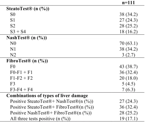
Notre objectif était donc d’établir la prévalence des lésions hépatiques dans une cohorte de patients BPCO. Cent onze patients BPCO, avec une consommation d’alcool < 30 g/jour pour les hommes et 20 g/jour pour les femmes et exempts d’hépatite B/C ou cancers, ont été inclus. Un Fibromax® ainsi que le dosage de marqueurs inflammatoires (leptine, adiponectine, résistine, CRP, TNF- α, IL-6) ont été réalisés pour chaque patient.
25 L’étude témoigne d’une prévalence élevée de lésions hépatiques chez les patients BPCO. Parmi les 111 patients, nous avons observé 41,4%, 36,9% et 61,3% de stéatose, stéatohépatite et fibrose hépatique, respectivement.
Les facteurs de risque de stéatose révélés par l’analyse multivariée sont le sexe masculin, l’IMC élevé, le SAOS non traité, l’index HOMA reflétant l’insulinorésistance et enfin la sévérité de la BPCO.
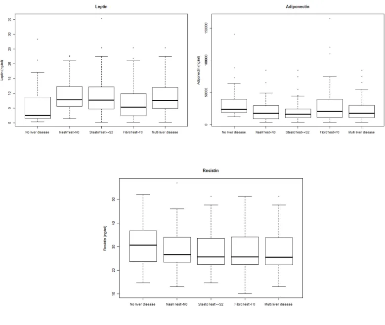
De plus, la stéatose est associée à un taux élevé de TNF-α après ajustement sur l’âge, le sexe et l’IMC. De même que la leptine est plus élevée chez les patients présentant une NASH ou plusieurs lésions hépatiques que chez ceux n’ayant aucune lésion hépatique.
26
Publication 1
“Non-alcoholic fatty liver disease in chronic obstructive pulmonary disease”
Viglino D, Jullian-Desayes I, Minoves M, Aron-Wisnewsky J, Leroy V, Zarski JP, Tamisier R, Joyeux-Faure M,Pépin JL.
27
Non-alcoholic fatty liver disease in chronic obstructive
pulmonary disease
Damien Viglino,1,2$ Ingrid Jullian-Desayes,1,3$ Mélanie Minoves,1,3 Judith
Aron-Wisnewsky,4,5,6 Vincent Leroy,7,8 Jean-Pierre Zarski,7,8 Renaud Tamisier,1,3
Marie Joyeux-Faure,1,3#, Jean-Louis Pépin.1,3#*
1 HP2 laboratory, INSERM U1042, University Grenoble Alps, Grenoble, France
2 Emergency Department and Mobile Intensive Care Unit, Grenoble Alps University Hospital, Grenoble, France
3 EFCR laboratory, Grenoble Alps University Hospital, Grenoble, France
4 Institute of Cardiometabolism and Nutrition, ICAN, Assistance Publique Hôpitaux de Paris, Pitié-Salpêtrière Hospital, Paris, France
5 Nutriomics Team, UMR_S 1166, INSERM , Paris, France.
6 Nutriomics Team UMR_S 1166, UPMC University Paris 06, Sorbonne Universités, Paris, France.
7 Hepatogastroenterology Department, Digidune, Grenoble Alps University Hospital, Grenoble, France
8 INSERM U823, IAPC Institute for Advanced Biosciences, University Grenoble Alps, Grenoble, France
$ These authors contributed equally to this work # These authors contributed equally to this work
* Corresponding author: Jean-Louis PEPIN Tel: 33 476 765 516 / Fax: 33 476 765 586
CHU Grenoble CS 10217 38043 Grenoble Cedex 9 Email: JPepin@chu-grenoble.fr
28 Pr Jean-Louis PEPIN acts as guarantor for the study and is accountable for all aspects of the work and ensures that questions related to the accuracy or integrity of any part of the work are appropriately investigated and resolved.
Authors’ Contribution
Conception and design: DV, IJD, RT, MJF and JLP; data acquisition, analysis and interpretation: all the authors; drafting and revising the manuscript for important intellectual content: all the authors; with a major contribution of JLP as principal investigator.
All authors read and approved the final version of the manuscript.
Conflicts of interest: None of the authors declare a conflict of interest in relation to this
publication
Role of the Funding Source:
“Air liquide" foundation, Endowment fund “AGIR pour les maladies chroniques”, and “Mutualia” provided unrestricted funding for the management of the cohort. This research was conducted with support from AstraZeneca. This work was also supported by the French National Research Agency in the framework of the " Investissements d’avenir” program (ANR-15-IDEX-02)
Short Running head: Non-alcoholic fatty liver disease in COPD
Take home message: Non-alcoholic fatty liver disease is highly prevalent in COPD patients
and might participate towards co-morbidities.
29
Abstract
Rationale: Nonalcoholic fatty liver disease (NAFLD) is independently linked to
cardio-metabolic morbidity and mortality. Low grade inflammation, oxidative stress and ectopic fat, common features of chronic obstructive pulmonary disease (COPD), might contribute to the development of NAFLD.
Objectives: To investigate the prevalence of NAFLD and to evaluate the relationship
between various types of liver damage and COPD severity, co-morbidities and circulating inflammatory cytokines.
Methods and Measurements: Validated noninvasive tests (Fibromax®: SteatoTest®,
NashTest® and FibroTest®) were used to assess steatosis, nonalcoholic steatohepatitis (NASH) and liver fibrosis. Patients underwent an objective assessment of COPD co-morbidities, including sleep studies. Biological parameters included a complete lipid profile and inflammatory markers.
Main results: In COPD patients the prevalence of steatosis, NASH and fibrosis were
respectively 41.4%, 36.9% and 61.3%. In multivariate analysis, SteatoTest® and FibroTest® were significantly associated with gender, body mass index (BMI), untreated sleep apnea, insulin resistance and COPD GOLD stage for SteatoTest®. Patients with steatosis had higher TNF-α levels and those with NASH or a combination of liver damages had raised leptin levels after adjustment for age, sex and BMI.
Conclusion: NAFLD is highly prevalent in COPD and might contribute to cardio-metabolic
co-morbidities.
Key words: COPD, non-alcoholic fatty liver disease, steatosis, nonalcoholic steatohepatitis,
30
Introduction
Nonalcoholic fatty liver disease (NAFLD) is a worldwide pandemic with 25% prevalence [1,2]. NAFLD is characterized by hepatic triglyceride accumulation (>5%), known as steatosis and the absence of significant alcohol consumption or secondary causes, mainly viral hepatitis [3]. While some patients remain at the stage of steatosis with or without mild inflammation [4], others progress to nonalcoholic steatohepatitis (NASH), defined as the presence of steatosis, hepatocyte ballooning and inflammatory infiltration [4,5]. NASH may also be accompanied by liver fibrosis, which accounts for the disease’s prognosis [6,7], that can further progress towards cirrhosis and hepatocarcinoma [8,9]. NAFLD has a complex and two-way relationship with metabolic syndrome. Not only is there a cross-sectional association between metabolic syndrome (Mets) and NAFLD but NAFLD may also be a precursor of MetS [10–12]. It has been reported that NAFLD diagnosis almost doubles the risk of incident MetS in the next few years [12]. NAFLD combines with metabolic co-morbidities such as obesity, type 2 diabetes, hyperlipidemia and hypertension, leading to a two-fold increase in liver-specific and overall mortality [2]. In addition, recent studies suggest that NAFLD may be considered as a novel independent cardiovascular risk factor [13].
Metabolic syndrome is common in chronic obstructive pulmonary disease (COPD) patients [14,15] with a prevalence ranging from 23% to 53%, mainly depending upon GOLD grades [16–18] and the patient’s inflammatory status [19–21]. COPD is strongly associated with major cardiovascular risk factors and COPD patients exhibit a two to five times higher risk of ischaemic heart disease, cardiac arrhythmias or heart failure [22]. There is increasing evidence that inflammatory visceral fat depots are significant contributors to systemic inflammation and COPD-related cardio-metabolic co-morbidities [19,23,24]. Interestingly, the literature reports a higher prevalence of all-cause liver diseases in patients with COPD, but with only a small number of patients with NAFLD [25]. Therefore, the specific links between NAFLD and COPD need to be confirmed and further evaluated.
NAFLD has been poorly studied in COPD patients, although there is a strong mechanistic rationale supporting the hypothesis of increased NAFLD prevalence in COPD patients. That oxidative stress and systemic inflammation reported in some COPD subtypes might participate towards reactive oxygen species production and inflammation in the liver, known as the spill-over theory, still remains debated. Patients with COPD have increased visceral
31 fat, a known inflammatory and lipolitic fat deposit [24,26]. Thus, the subsequent increase in free fatty acids accumulating in the liver might lead to NAFLD development in COPD. Nocturnal hypoxia subsequent to COPD per se, or related to the obstructive sleep apnea that is frequently associated with it, might also trigger the development of NAFLD [9,27,28]. Fibromax® is a non-invasive test for NAFLD screening that has been previously validated against liver biopsies [29] and is proposed by European guidelines [7]; the algorithm uses the association of sex, age, weight, height, and numerous serum biomarkers. Fibromax® uses SteatoTest®, NashTest®, and FibroTest® for the non-invasive evaluation of liver steatosis, NASH, and liver fibrosis, respectively. The aims of our study were (1) to use non-invasive blood tests (Fibromax®) to evaluate the prevalence of steatosis, NASH, and fibrosis in a well phenotyped cohort of COPD patients, and (2) to evaluate the relationship between steatosis, NASH, and fibrosis, and COPD severity, co-morbidities and circulating inflammatory cytokines.
Methods
Ethics and consent
Patients included in this study were part of two on going COPD cohorts attending Grenoble University Hospital. The studies were conducted in accordance with applicable good clinical practice requirements in Europe, French law, International Council on Harmonisation E6 recommendations, and the ethical principles of the Helsinki Declaration. These studies were approved by an independent ethics committee (Comité de Protection des Personnes, Grenoble IRB0006705). The possibility of later analysis of the patient’s blood sample including exploring their inflammatory status and hepatic features (this ancillary study) was initially mentioned in the protocol and was also approved and authorized. These studies have been registered on the ClinicalTrials.gov site (NCT00404430 and NCT01195064).
Patients
The vast majority of patients were included during out-patient consultations. In-patients referred for an acute exacerbation of COPD (n=7; 6%) represented a small percentage of the
32 study population. Participation in the prospective cohort was proposed to consecutive patients but the rate of refusal was high owing to complex phenotyping requiring additional blood samples, cardiovascular phenotyping and in-lab polysomnography. Patients aged over 18 years presenting with mild to severe COPD were eligible. We excluded patients with cancer, with daily alcohol consumption ≥ 20g for women and ≥ 30g for men, or with viral hepatitis. All patients with a positive NashTest® (stage ≥ N1, see “Procedures” for explanation) were checked to exclude secondary hepatic fat accumulation owing to significant alcohol consumption, use of steatogenic medication or hereditary disorders [1].
Procedures
Comorbidities were assessed based on predefined cut-offs [19] with objective clinical measurements, laboratory analyses (see below) and a systematic sleep study.
At study enrolment fasting serum samples were collected from all patients, frozen and stored at -80°C until analyses were conducted.
Metabolic parameters
Ten biochemical markers including liver enzymes, lipid profile, fasting glycaemia and insulin were measured in order to characterize metabolic comorbidities and calculate Fibromax® scores: α2-macroglobulin, apolipoprotein A1, haptoglobin, γ-glutamyltransferase (GGT), total bilirubin, alanine aminotransferase (ALT), aspartate aminotransferase (AST), fasting blood glucose, triglycerides (TG) and total cholesterol. Then, anonymized data were sent to the calculation centre (Biopredictive, Paris, France) to obtain Fibromax® scores blinded to the severity of COPD and comorbidities. Insulin resistance was assessed using the Homeostatic Model Assessment for Insulin Resistance (HOMA-IR), calculated using the following formula: insulinemia X glucose / 22.5 with glucose in mmol/L [30].
Fibromax® test
NAFLD screening through ultrasonography [31] together with the exclusion of competing etiologies [3,32] is indeed recommended at least in certain groups at high risk [33]. In this clinical study, prevalence was based on noninvasive Fibromax®. Fibromax® combines three tests FibroTest®, SteatoTest® and NashTest® [29,34]. FibroTest® includes α2-macroglobulin, apolipoprotein A1, haptoglobin, GGT, and total bilirubin values, age and gender. It provides a quantitative estimate ranging from 0.00 to 1.00 with higher values
33 corresponding to greater probability of lesions. Scores correspond to a METAVIR stage [35] as follows: F0 (0.00-0.21); F0-F1 (0.22-0.27); F1 (0.28-0.31); F1-F2 (0.32-0.48); F2 (0.49-0.58); F3 (0.59-0.72); F3-F4 (0.73-0.74) and F4 (0.75-1.0) [34]. The test gives an area under the ROC curve (AUC) of 0.89 [29]. This value appears better for the diagnosis of advanced fibrosis and cirrhosis than the validated NAFLD fibrosis score (NFS, AUC: 0.76) [29]. SteatoTest® combines FibroTest® parameters with height and weight plus ALT, serum fasting glucose, TG and cholesterol. SteatoTest® scores range from 0.00 to 1.00 determining four steatosis stages from S0 to S4: S0 (0.00-0.37) for no steatosis; S1 (0.38-0.56) minimal steatosis < 5%; S2 (0.57-0.68) moderate steatosis between 6 and 32%; and severe steatosis S3-S4 (0.69-1.0) corresponding to over 32% steatosis. SteatoTest® appears better (AUC = 0.80) for the diagnosis of steatosis than conventional markers, like GGT or ALT [29]. Finally, the NashTest® score is calculated from weight, height, AST, serum fasting glucose, TG and cholesterol in addition to age, gender and FibroTest® components. Thus, NashTest® distinguishes three NASH categories: N0 (0.00-0.25) for absence of NASH; N1 (>0.25-0.50) for borderline NASH and N2 (>0.50-0.75) for NASH [29,34]. The cut-off values of NashTest® for the diagnosis of NASH appear appropriate for our cohort (AUC=0.80) [29]. FibroTest® ≥ F0-F1, SteatoTest® ≥ S2 and NashTest® ≥ N1 were considered positives as previously applied by Minville et al. [27]. This choice is justified by the fact that any degree of liver fibrosis and/or inflammation is considered as a significant pathological condition, while mild steatosis has usually little impact on liver prognosis. When at least two types of liver damage were associated, the patient was assigned to a “combination of liver diseases” group.
Inflammatory markers
Serum levels of adiponectin, leptin, resistin and TNF-α were measured using multiplex assays (Merck). Interleukin-6 (IL-6) was measured by an immunoassay technique and ultra-sensitive C-reactive protein (usCRP) by an automated immunonephelometry technique.
Data analysis
Continuous data are presented as mean and standard deviation and categorical data as frequency and percentage.
The prevalence of positive Fibromax® tests is expressed as percentages. The relationship (odds ratio (OR)) between SteatoTest® or NashTest® and phenotyping characteristics were
34 analysed by logistic regression. The relationship between FibroTest® and phenotyping characteristics were analysed by linear regression. In univariate logistic regression, when a continuous variable was not log-linear, a new variable was created from the quartiles or the median value. Independent parameters were included in the multivariate model when significance was ≤ 0.05 in the univariate model. Inflammatory markers were not included in multivariate analyses but were investigated as exploratory variables to gain pathophysiological insights.
The inflammatory parameters are presented as boxplots between patients without liver disease and patients with a positive SteatoTest®, positive NashTest®, positive FibroTest® or combined liver diseases.
All statistical analyses were performed using SAS 9.4 software (SAS Institute Inc, USA).
Results
Patients’ characteristics
Of the 132 eligible COPD patients, twenty-one were excluded due to their daily alcohol consumption (≥ 20g for women and ≥ 30g for men). As a result, 111 COPD patients with available serum samples were included. Patients’ characteristics are given in Table 1. The included patients were predominantly men (77.5%, with a mean age of 63.8 ± 8.7 years, and a mean BMI of 25.1 ± 4.5 kg/m². 74.8% had GOLD stages 1-2 and 25.2% had GOLD stages III-IV COPD).
Prevalence and risk factors of liver disease in COPD
Stages of FibroTest®, SteatoTest® and NashTest® in the entire COPD population are reported in the online supplemental Table S1. The prevalence of liver disease was: 41.4% of moderate to severe steatosis (stages ≥ S2), 36.9% of borderline NASH and NASH (stages ≥ N1), and 61.3% of fibrosis (stages ≥ F0-F1) and is presented by COPD GOLD grade in Table 2. Prevalence of a combination of different types of liver damage is reported in the online supplement Table S1 and we observed that 17% of patients fulfilled to the criteria of all three studied abnormalities. Clinical characteristics and comorbidities of subjects without liver
35 abnormalities or with steatosis, NASH and fibrosis individually, and combined are detailed in online supplement Table S5.
Steatosis
In univariate analysis, steatosis was significantly associated with male gender, high BMI, current smoking, untreated OSA, dyslipidemia, insulin resistance (expressed by HOMA-IR) and post-bronchodilator FVC (see online supplement Table S2). In multivariate analysis, male gender, high BMI, untreated OSA, HOMA-IR and COPD severity were independent risk factors (Table 3A).
NASH
Age, BMI, current smoking, dyslipidemia and post-bronchodilator FVC were linked with a positive NashTest® in univariate analysis (online supplement Table S3). In multivariate analysis, only BMI was a risk factor for NASH (Table 3B).
Fibrosis
In univariate analysis, the various degrees of fibrosis severity were significantly associated with age, insulin resistance and untreated OSA. Women, non-diabetic and non-hypertensive patients were less at risk of developing fibrosis (online supplement Table S4). In multivariate analysis age, male gender, untreated OSA and insulin resistance (HOMA-IR) were risk factors of having a positive FibroTest® (Table 3C).
Adipokines, Inflammatory status and liver disease in COPD
Levels of adipokines (leptin, adiponectin, resistin), and inflammatory markers (TNF-α, IL-6 and usCRP) in the following 5 groups (no liver disease, steatosis, NASH, fibrosis and a combination of different types of liver damage) are shown in Figures 1 and 2, and in online supplement Table S5. Patients with steatosis had higher TNF-α levels than patients with no liver disease (p=0.016) after adjustment for age, gender and BMI. COPD patients with a combination of histological liver alterations also had higher leptin levels compared to those with no liver damage (p=0.047) after adjustment for age, sex and BMI. Finally, after the same adjustments, patients with a positive NashTest® had significantly higher leptin levels than patients without liver damage (p=0.039).
36
Discussion
To our knowledge, this study is the first to investigate the prevalence and severity of NAFLD using noninvasive blood tests in a relatively large group of well-characterized COPD patients. The prevalence in COPD patients of steatosis, NASH and fibrosis was respectively 41, 37 and 61%. Liver steatosis, NASH and fibrosis were primarily linked with the metabolic co-morbidities of COPD (i.e. obesity and insulin resistance) and systemic inflammation. There is an increase in awareness regarding the various COPD clinical subtypes [19,36]. The concomitant chronic diseases and systemic comorbidities of COPD represent a major challenge for risk prediction and complicate integrated care delivery. Clusters of co-morbidities, essentially cardiovascular and metabolic, have been shown to be associated with COPD [37] and linked to an increased risk of mortality [14,38]. Among these comorbidities, liver diseases are hugely under reported and certainly underestimated. Divo et al. [14] prospectively followed 1,664 patients with COPD for a median of 51 months with a systematic record of 79 comorbidities. Liver cirrhosis was reported in 2.5 % patients and associated with twofold higher mortality. While liver cirrhosis was attributed to alcohol and smoking it potentially might originate from the progression of steatosis to cirrhosis in the context of the metabolic syndrome associated with COPD. Using a system network analysis comparing 1,969 COPD and 316 non-COPD patients it was demonstrated that comorbidities encountered in COPD patients are interlinked [39]. This was the case for hepatitis, liver cirrhosis and liver cancer and again the suggested mechanism was linked to substance abuses. As stated by the authors, such a network analysis provides information about common risk factors or disease mechanisms that are difficult to discern when studied individually. We suggest that this cluster of liver diseases is more likely to be attributable to components of metabolic syndrome and the general reduction in physical activity. The limitation of the two studies by Divo et al. [14,39], as acknowledged by the authors themselves, was that the ascertainment of comorbidities was obtained by questioning the patient and from medical records. This is a sensitive method for capturing a large number of comorbidities but lacks objective measures that would improve specificity [19] and also allow severity to be graded. The strengths of our study were to focus on NAFLD and exclude other liver diseases with other etiologies and to quantify NAFLD related alterations and the severity of liver disease using biological markers with established cut-offs that have been previously validated against liver biopsies.
37 Among limitations, our study did not include a control group allowing a comparison between the prevalence rate in the general population and our COPD patients. Prevalence in the general population has been recently established through a meta-analyse reporting a combination of imaging (ultrasound, computed tomography scan and magnetic resonance imaging / spectroscopy), liver biopsy, and blood testing (elevated liver enzymes or fatty liver Index, etc.) data [2]. We did not perform liver biopsy, the gold standard for confirming NAFLD diagnosis and providing prognostic information. However, it is an invasive and costly procedure, prone to minor side effects such as pain, or more severe complications including a risk of death of 0.03% [29]. Moreover, intra and inter-observer variabilities exist [40]. Thus, several advantages of these noninvasive tests should be stressed: compared to biopsy, they are cheaper, repeatable, and lead to less inter-observer variability, severe adverse events, refusal and bias [29]. Non-invasive diagnosis and quantification of NAFLD mainly rely on biomarkers or imaging techniques [7]. Both strategies have limitations and lack sensitivity or specificity. As liver biopsies were not appropriate in our clinical research setting, we decided to use Fibromax® which is represents one of the best compromises for non-invasive assessment of NAFLD having been previously validated against liver biopsies [29] and which is now proposed by European guidelines to non-invasively screen for NAFLD [7]. In future studies of COPD, the combination of biomarkers / scores and transient elastography with liver ultrasonography (not performed in the current study) might confer additional diagnostic accuracy. Indeed, liver ultrasound is a first-line imaging technique used to identify hepatic steatosis in patients with NAFLD [3,31,32] and plays also a major role in the early diagnosis of hepato-cellular carcinoma in at risk populations. The strong prevalence of steatosis, NASH and fibrosis observed in our study needs to be replicated in different COPD cohorts and using different diagnostic tools. This prevalence also needs to be compared with that of the general population (with age- and smoking-matched controls) using the same noninvasive tests.
Many factors such as insulin resistance, low-grade inflammation, oxidative stress and lipotoxicity are involved in the onset and progression of NAFLD. It appears that COPD and NAFLD share common intermediary mechanisms. Our analyses showed an independent role of BMI and insulin resistance in the development of NAFLD in COPD patients. Hepatic steatosis develops in the context of increased triglyceride flux into the liver from visceral adipose tissue [41]. Furthermore, the fat inflammation, which has been demonstrated in COPD [42], drives the development of NAFLD. Excessive and inflamed visceral fat is
38 associated with circulating cytokines including leptin and TNF-α [43,44]. We found higher leptin levels in COPD patients with NASH or with a combination of liver lesions compared to COPD patients without liver damage. A continuous dynamic cross-talk between adipokines with a positive or negative impact on NAFLD resulting in an overall beneficial or detrimental effect has been described [45]. TNF-α might play an initial role in the occurrence of steatosis while leptin is known to exert a pro-steatotic and-pro fibrotic action [45,46]. TNF-α levels were significantly higher in our COPD patients with steatosis compared to those with no liver injury. TNF-α plays a crucial role in human and animal NAFLD [47]. Rosiglitazone has been shown to lower adipose tissue inflammation with a reduction in plasma leptin and TNF-α levels and less severe NAFLD [48]. The role of chronic intermittent hypoxia (CIH) in the development of NAFLD (for review see [9]) is clearly established both in rodent models and in sleep apnea patients. In mice, CIH increases the expression of key lipogenic, inflammatory and fibrotic transcription factors [49]. In line with this, in our COPD cohort that underwent systematic sleep studies, untreated OSA was significantly associated with steatosis and fibrosis. Finally, low physical activity is a major determinant of NAFLD [50] and might play a key role in COPD. However we did not objectively measure physical activity in our study and further studies are required to in order to provide evidence for this highly probable link.
Conclusion and perspectives
Owing to its deleterious consequences and independent impact on mortality [2] NAFLD should be included among COPD co-morbidities. Insulin resistance, systemic inflammation, and CIH seem to play a role in the development of NAFLD in COPD patients. Biomarkers of liver damage may allow specific COPD endotypes to be detected leading to personalized treatments [36]. As physical activity is the first line treatment of NAFLD [50–52], pulmonary rehabilitation could be expected to have a positive effect on NAFLD in COPD patients. In the obese inflamed COPD phenotype, life style intervention, with cautious weight loss might also improve NAFLD.
39
Acknowledgments
The authors are grateful to Marion Perrin for statistical analyses (INSERM U1042, HP2 Laboratory, Grenoble Alps University Hospital, France), Candice Trocmé (Biochemistry, Toxicology and Pharmacology Department, Grenoble Alps University Hospital, France), for biochemical analyses and Alison Foote (Grenoble Alps University Hospital, France) for English editing.
References
1. Chalasani N, Younossi Z, Lavine JE, Diehl AM, Brunt EM, Cusi K, Charlton M, Sanyal AJ. The diagnosis and management of non-alcoholic fatty liver disease: practice Guideline by the American Association for the Study of Liver Diseases, American College of Gastroenterology, and the American Gastroenterological Association. Hepatol. Baltim. Md 2012; 55: 2005–2023.
2. Younossi ZM, Koenig AB, Abdelatif D, Fazel Y, Henry L, Wymer M. Global epidemiology of nonalcoholic fatty liver disease-Meta-analytic assessment of prevalence, incidence, and outcomes. Hepatol. Baltim. Md 2016; 64: 73–84.
3. Nascimbeni F, Pais R, Bellentani S, Day CP, Ratziu V, Loria P, Lonardo A. From NAFLD in clinical practice to answers from guidelines. J. Hepatol. 2013; 59: 859–871. 4. Bedossa P, Tordjman J, Aron-Wisnewsky J, Poitou C, Oppert J-M, Torcivia A, Bouillot J-L, Paradis V, Ratziu V, Clément K. Systematic review of bariatric surgery liver biopsies clarifies the natural history of liver disease in patients with severe obesity. Gut 2016; 5. Bedossa P, Poitou C, Veyrie N, Bouillot J-L, Basdevant A, Paradis V, Tordjman J, Clement K. Histopathological algorithm and scoring system for evaluation of liver lesions in morbidly obese patients. Hepatol. Baltim. Md 2012; 56: 1751–1759.
6. Rinella ME. Nonalcoholic fatty liver disease: a systematic review. JAMA 2015; 313: 2263–2273.
7. European Association for the Study of the Liver (EASL). Electronic address: easloffice@easloffice.eu, European Association for the Study of Diabetes (EASD), European Association for the Study of Obesity (EASO). EASL-EASD-EASO Clinical Practice Guidelines for the management of non-alcoholic fatty liver disease. J. Hepatol. 2016; 64: 1388–1402.
8. Cohen JC, Horton JD, Hobbs HH. Human fatty liver disease: old questions and new insights. Science 2011; 332: 1519–1523.
9. Aron-Wisnewsky J, Clement K, Pépin J-L. Nonalcoholic fatty liver disease and obstructive sleep apnea. Metabolism. 2016; 65: 1124–1135.
40 10. Lonardo A, Ballestri S, Marchesini G, Angulo P, Loria P. Nonalcoholic fatty liver disease: a precursor of the metabolic syndrome. Dig. Liver Dis. Off. J. Ital. Soc. Gastroenterol. Ital. Assoc. Study Liver 2015; 47: 181–190.
11. Ballestri S, Nascimbeni F, Romagnoli D, Lonardo A. The independent predictors of non-alcoholic steatohepatitis and its individual histological features.: Insulin resistance, serum uric acid, metabolic syndrome, alanine aminotransferase and serum total cholesterol are a clue to pathogenesis and candidate targets for treatment. Hepatol. Res. Off. J. Jpn. Soc. Hepatol. 2016; 46: 1074–1087.
12. Ballestri S, Zona S, Targher G, Romagnoli D, Baldelli E, Nascimbeni F, Roverato A, Guaraldi G, Lonardo A. Nonalcoholic fatty liver disease is associated with an almost twofold increased risk of incident type 2 diabetes and metabolic syndrome. Evidence from a systematic review and meta-analysis. J. Gastroenterol. Hepatol. 2016; 31: 936–944.
13. Targher G, Byrne CD, Lonardo A, Zoppini G, Barbui C. Non-alcoholic fatty liver disease and risk of incident cardiovascular disease: A meta-analysis. J. Hepatol. 2016; 65: 589–600.
14. Divo M, Cote C, de Torres JP, Casanova C, Marin JM, Pinto-Plata V, Zulueta J, Cabrera C, Zagaceta J, Hunninghake G, Celli B. Comorbidities and risk of mortality in patients with chronic obstructive pulmonary disease. Am J Respir Crit Care Med 2012; 186: 155–161.
15. Cebron Lipovec N, Beijers RJHCG, van den Borst B, Doehner W, Lainscak M, Schols AMWJ. The Prevalence of Metabolic Syndrome In Chronic Obstructive Pulmonary Disease: A Systematic Review. COPD 2016; 13: 399–406.
16. Sin DD, MacNee W. Chronic obstructive pulmonary disease and cardiovascular diseases: a “vulnerable” relationship. Am. J. Respir. Crit. Care Med. 2013; 187: 2–4.
17. Van Eeden S, Leipsic J, Paul Man SF, Sin DD. The relationship between lung inflammation and cardiovascular disease. Am. J. Respir. Crit. Care Med. 2012; 186: 11–16. 18. Marquis K, Maltais F, Duguay V, Bezeau A-M, LeBlanc P, Jobin J, Poirier P. The metabolic syndrome in patients with chronic obstructive pulmonary disease. J. Cardpulm. Rehabil. 2005; 25: 226–232; discussion 233–234.
19. Vanfleteren LEGW. Does COPD stand for “COmorbidity with Pulmonary Disease”? Eur. Respir. J. 2015; 45: 14–17.
20. Fabbri LM, Luppi F, Beghé B, Rabe KF. Complex chronic comorbidities of COPD. Eur. Respir. J. 2008; 31: 204–212.
21. Tkacova R. Systemic inflammation in chronic obstructive pulmonary disease: may adipose tissue play a role? Review of the literature and future perspectives. Mediators Inflamm. 2010; 2010: 585989.
![Figure 2. Hypoxie intermittente chronique et NAFLD (extrait de Aron-Wisnewsky et al. [36])](https://thumb-eu.123doks.com/thumbv2/123doknet/14253602.488500/22.892.135.825.284.790/figure-hypoxie-intermittente-chronique-nafld-extrait-aron-wisnewsky.webp)
![Tableau I. Interprétation des scores du Fibromax® (adapté de Munteanu et al. [40])](https://thumb-eu.123doks.com/thumbv2/123doknet/14253602.488500/23.892.90.804.528.843/tableau-interprétation-scores-fibromax-adapté-munteanu-al.webp)