HAL Id: inserm-00463814
https://www.hal.inserm.fr/inserm-00463814
Submitted on 22 Mar 2010HAL is a multi-disciplinary open access archive for the deposit and dissemination of sci-entific research documents, whether they are pub-lished or not. The documents may come from teaching and research institutions in France or abroad, or from public or private research centers.
L’archive ouverte pluridisciplinaire HAL, est destinée au dépôt et à la diffusion de documents scientifiques de niveau recherche, publiés ou non, émanant des établissements d’enseignement et de recherche français ou étrangers, des laboratoires publics ou privés.
Mineralocorticoid receptor overexpression in embryonic
stem cell-derived cardiomyocytes increases their beating
frequency.
Damien Le Menuet, Mathilde Munier, Géri Meduri, Say Viengchareun, Marc
Lombès
To cite this version:
Damien Le Menuet, Mathilde Munier, Géri Meduri, Say Viengchareun, Marc Lombès. Mineralocor-ticoid receptor overexpression in embryonic stem cell-derived cardiomyocytes increases their beating frequency.: cardiac MR and pacemaker channel HCN1. Cardiovascular Research, Oxford University Press (OUP), 2010, 87 (3), pp.467-75. �10.1093/cvr/cvq087�. �inserm-00463814�
Mineralocorticoid Receptor Overexpression in Embryonic Stem Cell
1
Derived Cardiomyocytes Increases Their Beating Frequency
2
Damien Le Menuet1 ,2,, Mathilde Munier1,2, Geri Meduri1,3, Say Viengchareun1,2, and Marc 3
Lombès1,2,4, 4
5
1- INSERM, U693, Le Kremlin-Bicêtre, F-94276, France;
6
2- Univ Paris-Sud 11, Faculté de Médecine Paris-Sud, UMR-S693, Le Kremlin-Bicêtre,
F-7
94276, France;
8
3- Service de Génétique Moléculaire, Pharmacogénétique et Hormonologie, Le
Kremlin-9
Bicêtre Assistance Publique-Hôpitaux de Paris, Hôpital de Bicêtre F-94275, France;
10
4- Service d’Endocrinologie et Maladies de la Reproduction, Assistance Publique-Hôpitaux
11
de Paris, Hôpital de Bicêtre, Le Kremlin-Bicêtre, F-94275, France.
12 13 Corresponding author: 14 Marc LOMBES 15
Inserm U693, Faculté de Médecine Paris-Sud 16
63, rue Gabriel Péri 17
94276 Le Kremlin Bicêtre Cedex 18 France 19 Tel 33 1 49 59 67 09 20 Fax 33 1 49 59 67 32 21 Email: marc.lombes@u-psud.fr 22 23 Word count: 5636 24
Running title: cardiac MR and pacemaker channel HCN1 25
Abstract
26 27
Aims
28
Cardiac Mineralocorticoid Receptor (MR) activation triggers adverse cardiovascular 29
events that could be efficiently prevented by mineralocorticoid antagonists. To gain 30
insights into the pathophysiological role of MR function, we established embryonic 31
stem (ES) cell lines from blastocysts of transgenic mice overexpressing the human 32
MR driven by its proximal P1 or distal P2 promoter and presenting with 33
cardiomyopathy, tachycardia and arrhythmia. Cardiomyocyte differentiation allowed 34
us to investigate the molecular mechanisms contributing to MR-mediated cardiac 35
dysfunction. 36
37
Methods and Results
38
During cardiac differentiation, wild-type (WT) and recombinant ES cell cultures and 39
excised beating patches expressed endogenous MR along with cardiac gene 40
markers. The two-fold increase in MR protein detected in P1.hMR and P2.hMR 41
cardiomyocytes led to a parallel increase of the spontaneous beating frequency of 42
hMR-overexpressing cardiomyocytes compared to WT. The MR-mediated 43
chronotropic effect was ligand-independent, could be partially repressed by 44
spironolactone and was accompanied by a significant 2- to 4-fold increase in mRNA 45
and protein levels of the pacemaker channel HCN1, generating depolarizing If 46
currents, thus revealing a potential new MR target. This was associated with 47
modification in the expression of HCN4, the inward rectifier potassium channel Kir2.1 48
and the L-Type dependent calcium channel Cav1.2. 49
Conclusion
51
We demonstrate that the amplification of MR signaling in ES-derived cardiomyocytes 52
has a major impact on cardiomyocyte contractile properties through an important 53
remodelling of ion channel expression contributing to arrhythmias. Our results 54
highlight the prominent role of MR function in cardiac physiology and support the 55
benefit of MR antagonists in the management of cardiac dysfunctions. 56
Introduction
57
The Mineralocorticoid Receptor (MR or NR3C2) is a nuclear receptor exerting 58
various pleiotropic actions on a wide series of target tissues. MR acts as a ligand-59
dependent transcription factor, and is involved in numerous physiological processes 60
and pathological conditions1. MR is expressed in many components of the 61
cardiovascular system such as blood vessels2, endothelial cells3, cardiomyocytes4,
62
vascular smooth muscle cells5 and macrophages6. The importance of cardiac MR 63
has been strikingly underscored by several direct and indirect evidences. Indeed, 64
RALES and EPHESUS clinical trials have demonstrated the major benefit of 65
mineralocorticoid antagonist (spironolactone and eplerenone) administration on the 66
heart failure patient’s survival 7-8. MR can be activated both by mineralo- and
67
glucocorticoid hormones but selectivity-conferring mechanisms, mostly dependent 68
upon the cellular context1, have been described. Most notably in tight epithelia, the
69
large excess of glucocorticoids is metabolized into inactive compounds by the 11 70
beta hydroxysteroid dehydrogenase type 2 (11βHSD2) enzyme9, preventing an illicit
71
occupation of the receptor. Although glucocorticoids are most likely the natural MR 72
ligands in cardiomyocytes, which lack 11βHSD2, they seem to be unable to fully 73
activate the receptor10. Indeed, Transgenic mice ectopically expressing 11βHSD2 in 74
cardiomyocytes exhibit cardiac hypertrophy, fibrosis and heart failure, but no 75
hypertension, this phenotype being reversed by eplerenone11. These findings in an 76
animal model where the receptor is almost exclusively activated by aldosterone 77
underline the deleterious effect of inappropriate MR activation. 78
On the other hand, MR gene inactivation in the mouse leads to early post-natal 79
lethality caused by salt loss12 which can be rescued by daily NaCl injections followed
80
by high salt diet13. MR-/- mice have no cardiovascular abnormalities, notwithstanding 81
the expected activation of the renin-angiotensin system14, thus excluding a crucial
82
role of MR during cardiac development. 83
In order to better understand MR-dependent pathophysiological processes in vivo, 84
our group and others have exploited alternative strategies of MR overexpression in 85
the heart of transgenic mice15-16. In particular, we have generated a murine model in 86
which the proximal P1 promoter of the human MR (hMR) was used to drive the 87
expression of its own cDNA. P1.hMR animals express the transgene in most MR 88
target tissues including kidney, brain and heart. Interestingly, two extensively studied 89
mouse lines exhibited a moderate dilated cardiomyopathy associated with 90
arrhythmia, but without hypertension or cardiac fibrosis15 in contrast with aldosterone-91
high salt diet animal models17. In another mouse model, conditional MR
92
overexpression in the heart triggers cardiac hypertrophy and life-threatening 93
ventricular arrhythmias16. All these in vivo studies underlined a specific role of MR in
94
cardiomyocyte contractile properties. However, it cannot be excluded that transgene 95
expression might induce some adaptive and compensatory mechanisms secondary 96
to various feedback regulatory loops prevailing in vivo. The utilization of 97
cardiomyocytes isolated from animal models would thus facilitate analysing the 98
specific MR effects, regardless of the compensatory factors. Along similar lines, it 99
has been also reported that MR/aldosterone have major effects on cardiomyocyte 100
contraction frequency associated with an increased expression of T-type (Cav3.2) 101
and L-type (Cav1.2) calcium channels, consequently augmenting Ca2+ currents in
102
isolated rat ventricular myocytes18,19. However, such cell-based systems are quite 103
difficult to obtain in rodents, usually give poor yield, and often lead to highly variable 104
results. 105
To better comprehend MR actions in the heart, we decided to use an alternative 106
approach that consists in deriving cardiomyocytes from transgenic animals which 107
allows a fine tuned control of experimental conditions. A more effective strategy, 108
already validated by several groups (20 for review) is based on the utilization of the 109
embryonic stem (ES) cells that can be indefinitely expanded at the undifferentiated 110
stage and, under appropriate conditions, are able to differentiate into cell types 111
originating from the three germ layers (endoderm, mesoderm, ectoderm) including 112
cardiomyocytes21.
113
In this study, we established ES cell lines derived from hMR-overexpressing mice 114
that can undergo highly efficient differentiation into cardiomyocytes. Our cell-based 115
models permit not only to investigate a potential involvement of MR and/or 116
aldosterone during cardiomyocyte development but also to discriminate MR-117
dependent actions from those induced by various ligands. Herein, we show that hMR 118
overexpression leads to an increase of the beating frequency in ES derived-119
cardiomyocytes. We demonstrate that this is associated with an increase of the 120
expression of the pacemaker channel HCN1 and with an altered expression of 121
calcium and potassium cardiac ion channels involved in cardiomyocyte contractility. 122
Materials and Methods
123 124
Derivation and culture of ES cell lines
125
To generate ES cell lines, P1 or P2.hMR females were crossed with males from the 126
129 strain (Charles River, L’Arbresle, France) and checked daily for vaginal plugs. 127
Mice were cared according to the Guide for the Care and Use of Laboratory Animals 128
published by the US National Institutes of Health (NIH Publication No. 85-23, revised 129
1996). The animal facility was granted approval (N° B94-043-12), with an 130
authorization to experiment on living animals (75-978, ML) given by the French 131
Administration. At day 3.5 post-coitum, females were sacrificed, the uterus removed 132
and flushed with culture medium. Blastocysts were picked up with a mouth pipette 133
and plated in a 6 cm Petri dish on a SNL feeder cell layer. After 3 more days each 134
inner cell mass was recovered and plated in a well of a 24-well plate with SNL. 135
Cultures were split 1:1 every 3 days and an ES cell line was successfully derived in 136
approximately 20% of the attempts. Undifferentiated ES cells were cultured and 137
amplified as previously described22. See supplemental methods for details.
138 139
Cardiac differentiation
140
Cardiac differentiation was based on the hanging drop method23. Drops containing 141
400 cells were grown hanging on a Petri dish lid for 3 days in ES cell medium 142
containing 20% fetal calf serum. The embryoid bodies (EB) were then cultured for 2 143
days in the same medium (alternatively the serum was dextran-coated charcoal 144
(DCC) stripped of steroids for hormone experiments) complemented with 1 µM 145
ascorbic acid and 0.5% DMSO (cardiac differentiation medium). EB were then 146
seeded in gelatinized Petri dishes or culture plate wells and allowed to differentiate 5 147
to 15 days. Spontaneously beating areas were excised with a scalpel blade. 148
149
RT-PCR
150
RNA from ES, EB and beating patches were extracted with the Trizol reagent 151
(Invitrogen, Cergy Pontoise, France) according to the manufacturer’s instructions. 152
After DNase I treatment, one µg total RNA was reverse-transcribed with the 153
MultiScribe reverse transcriptase kit (Applied Biosystems, Courtaboeuf, France). 154
Semi-quantitative PCR were performed using the Taq Polymerase kit (Invitrogen) 155
and real-time quantitative PCR with the Power SYBR Green® PCR Master mix 156
(Applied Biosystems). For the latter, a standard curve was obtained with serial 157
dilutions of an amplicon subcloned in pGEMTeasy vector (Promega, Charbonnières, 158
France). Primers are listed in Supplemental Table 1. 159
160
Immunocytochemistry
161
Excised beating patches were digested in DMEM with 1 mg/ml collagenase (Sigma) 162
for 30 min with occasional shaking in a microcentrifuge tube, then spun down at 2000 163
rpm for 5 min. Collagenase was removed and patches were digested for 20 min in 164
trypsin-EDTA (invitrogen), spun down, resuspended with cardiac differentiation 165
medium, plated in 0.1% gelatin-coated LabTek (Nunc, Rochester, NY, USA) or 166
coverslips and incubated overnight. See supplemental methods for details. 167
168
Western blot
169
Total protein extracts were prepared from wild type and transgenic cardiomyocytes, 170
15 µg of proteins from cardiomyocyte homogenates were processed for 171
immunoblotting. See supplemental methods for details. Immunoblots were blocked 172
with TBST 0.5% non fat milk and incubated overnight with 1/1000 anti-MR 39 N24 or 173
1/300 anti-HCN1 (AB5884, Millipore) or 1/15,000 anti-α-tubulin (Sigma) as an internal 174
standard. Quantitative analysis was performed using QuantityOne software (Bio-Rad 175
Laboratory, Inc., Hercules, CA). 176
177
Statistical analysis
178
Results represent mean ± SEM with at least 6 independent determinations for each 179
condition. Statistical analyses were performed using a non parametric Mann-Whitney 180
test (Prism4, Graphpad Software, Inc., San Diego, CA). 181
Results
182
Generation of WT and hMR-overexpressing ES cell lines
183
We have previously generated transgenic mouse models using 1.2 kb of the proximal 184
(P1) and 1.8 kb of the distal (P2) hMR promoter to drive the hMR cDNA expression 185
as previously described15, 25. Figure 1A illustrates the schematic representation of the 186
transgenes inserted into P1.hMR and P2.hMR transgenic mouse genome. Both 187
constructs contained an untranslated region as well as part of the human βglobin 188
sequences to stabilize the transgene transcripts. As expected, we confirmed by RT-189
PCR using species-specific primers that the recombinant hMR was expressed in the 190
heart of both transgenic P1.hMR and P2.hMR mouse lines (Fig 1B), demonstrating 191
that these animals were suitable to investigate the functional impact of cardiac MR. 192
P1 and P2.hMR female mice were backcrossed with strain 129 males characterized 193
by a high success rate of ES cell derivation from blastocysts26 (Fig 1C, see 194
Supplemental methods). Several ES cell lines were established and genotyped as 195
wild type (WT), P1.hMR or P2.hMR (Fig 1D). Further experiments were mostly 196
performed on one representative ES cell line of each genotype. 197
198
MR expression during cardiac differentiation of ES cells
199
We optimized a protocol of cardiac differentiation based on the hanging drop 200
method20. The different steps of ES cell cardiac differentiation are represented in Fig 201
2A. As shown in the upper panel (ES), undifferentiated ES cells grew as compact 202
clusters on the feeder cell layers. Suspension culture of 400 undifferentiated ES cells 203
per drop led to standardized size embryoid bodies (EB) of approximately 200 µm in 204
diameter at day 3 (EB, middle upper panel). These spherical-shaped structures 205
contained cells that are able to differentiate into the three germ layers. From day 5, 206
cultures were grown in adherence in gelatinized Petri dishes and from day 7 onward 207
spontaneously beating areas arose, enlarging with time (D18 culture, middle bottom). 208
Beating areas containing a tight network of cardiomyocytes were excised with a 209
scalpel blade around day 16 to 18 (Patch, bottom panel) and were further used for 210
gene expression studies or immunodetection experiments. We defined an index of 211
cardiac differentiation by assessing the percentage of cultures originating from a 212
single EB presenting spontaneously beating cardiomyocytes. Approximately 70% of 213
EB presented beating areas at day 18. No significant variation in the cardiac 214
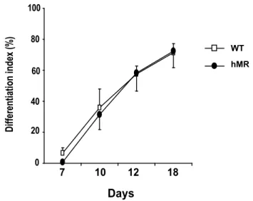
differentiation efficacy has been found among cell lines (see Fig 4). 215
The temporal expression pattern of early and late cardiac marker genes was 216
analyzed concomitantly with recombinant (hMR) and endogenous MR (mMR) by RT-217
PCR. Fig 2B presents data obtained from day 0 to 7 cultures and Fig 2C shows data 218
from day 18 excised patches. Transgene expression in the recombinant cells was 219
detected at all stages of cardiac differentiation and, importantly, a strong signal was 220
found in excised patches. Of note, the progressive appearance of early (NKx2.5) and 221
late (αMHC) cardiac marker gene expression was observed along differentiation as 222
early as day 4 and 7, in all cell lines while late markers such as αMHC and Troponin 223
T transcripts were expressed in day 18 excised patches. Quantification of mMR 224
mRNA levels by qPCR showed an approximately ten-fold expression increase in day 225
18 cardiomyocytes than in earlier differentiation stages (D0 undifferentiated ES cells 226
or D7 EB) (Fig 2D). 227
We next analyzed MR expression at the protein level in cardiac differentiation. We 228
used an anti-MR antibody recognizing both the endogenous murine MR and 229
recombinant hMR24. Western blot analyses of day 12 cardiomyocyte cultures 230
revealed an approximately two-fold increase in MR expression in the P1.hMR 231
cardiomyocytes as compared to the WT cells (Fig 3A and 3B). Confocal microscopy 232
imaging confirmed the coexpression of MR and α-sarcomeric actin in the 233
cardiomyocytes of each genotype detected by double immunolabelling (Fig 3C). Note 234
the nucleocytoplasmic distribution of MR and the stronger signal in P1.hMR and 235
P2.hMR cardiomyocytes. Thus, these ES cell-derived cardiomyocytes provided an 236
effective cell-based system to investigate the functional consequences of hMR 237
overexpression in cardiomyocytes. 238
239
MR overexpression causes an increase in cardiomyocyte beating frequency
240
We first tested a potential effect of MR overexpression and aldosterone exposure on 241
cardiac differentiation efficiency (See Fig 4 and Supplemental Fig S1). Since there 242
was no modification of the differentiation index compared to control conditions, we 243
excluded a major impact of MR signaling in early cardiac development. This 244
assumption seems to be supported by the lack of morphological alterations in the 245
cardiovascular system of MR KO newborn mice12. 246
In order to better understand the role of MR in cardiac function, we examined the 247
influence of hMR overexpression on cardiomyocyte contractile properties (Fig 5). The 248
beating rate of day 14-18 cultures was assessed by video capture. We found a highly 249
significant increase of the beating frequency in transgenic P1.hMR and P2.hMR 250
cardiomyocytes compared to WT (Fig 5A, WT: 1.09±0.2 Hz n=33, P1.hMR: 1.8±0.3 251
Hz n=12, P2.hMR: 1.7±0.5 Hz, n=31, P<0.005, and see Supplemental videos). This 252
increase in the spontaneous beating frequency of hMR-overexpressing 253
cardiomyocytes has been reproduced in several differentiation experiments and has 254
been confirmed in two different ES cell lines with the same genotype. Owing to the 255
chronotropic effect of β-adrenergic stimulation, we observed a significant increase in 256
the beating frequency of both WT and transgenic cardiomyocytes upon isoproterenol 257
exposure (Fig 5B). A 2.2-fold induction of isoproterenol-induced stimulation was 258
found in the WT cardiomyocytes while the amplitude of catecholamine–stimulated 259
beating frequency was only 1.4-fold in P1.hMR and P2.hMR cardiomyocytes, 260
suggesting that amplification of MR activation might somehow compromise 261
cardiomyocyte β-adrenergic signaling. Under these experimental conditions, the 262
adrenergic-stimulated beating frequency remained significantly higher in hMR-263
overexpressing cardiomyocytes than in WT ES derived cells, providing additional 264
support for a primary role of MR on cardiomyocyte contractile properties. 265
An important issue was whether the increase in the hMR-driven beating frequency 266
depended upon the presence of the ligand. To test this hypothesis, ES cells were 267
submitted to the cardiac differentiation process using DCC serum, in the presence or 268
absence of 10 nM aldosterone for 24 h before video capture. As shown in Fig 5C, 269
beating rate analysis indicated that, hMR overexpression in cardiomyocytes caused a 270
striking chronotopic effect (WT=0.25 ± 0.075 Hz, P1.hMR=1.0 ± 0.25 Hz, P<0.05), 271
even in steroid-free medium. On the other hand, aldosterone treatment induced a 272
significant increase of the beating frequency of WT cardiomyocytes 273
(WT+Aldo=0.91±0.5 Hz) compared to untreated cells but was unable to accelerate 274
further the already higher spontaneous contraction of P1.hMR cardiomyocytes. 275
Interestingly, the beating frequency of P1.hMR cardiomyocytes differentiated in DCC 276
medium could be reduced with 100 nM spironolactone (Fig 5D), suggesting that MR 277
blockade might reverse the MR-mediated positive chronotropic effect. Taken 278
together, these data show that MR overexpression per se is a potent regulator of 279
cardiomyocyte chronotropy, and that MR-induced increase of cardiomyocyte 280
contractility is at least partially independent of the ligand. 281
282
hMR overexpression alters cardiomyocyte ion channel expression
283
Cardiomyocyte contractions are tightly regulated by many ion channels and pumps27.
284
We therefore investigated the expression of several key regulators of cardiac 285
contractility to decipher the underlying mechanisms associated with the positive 286
chronotropic phenotype of MR-overexpressing cardiomyocytes. The 287
hyperpolarization-activated cyclic nucleotide gated potassium channels (HCN) were 288
especially good candidates since they participate to the occurrence of the pacemaker 289
currents (If) initiating the depolarization process28. We showed that the HCN1 290
channel mRNA level almost doubled in excised beating patches of P1.hMR 291
cardiomyocytes compared to WT controls (Fig 6A). Western blot analysis confirmed 292
a 4-fold increase in HCN1 channel expression in hMR-overexpressing 293
cardiomyocytes (Fig 6B), providing evidence for a direct relationship between the 294
expression of this pacemaker channel and the MR-increased cardiomyocyte beating 295
rate. 296
We next analyzed the relative abundance of other calcium and potassium channels 297
(Fig 7). Unexpectedly, we found that mRNA levels of another pacemaker channel, 298
HCN4, were repressed by 75% in hMR-overexpressing cardiomyocytes (Fig 7A), 299
suggesting a counter-regulatory mechanism to dampen the depolarization process 300
due to HCN1 up-regulation. Interestingly, we also demonstrated a 2-fold increase of 301
the inward rectifier potassium ion channel Kir2.1 expression in P1.hMR 302
cardiomyocytes (Fig 7B), which was accompanied by a parallel increase of the L-303
Type voltage dependent calcium channel Cav1.2 mRNA levels (Fig 7C). Collectively, 304
hMR overexpression in ES cell-derived cardiomyocytes leads to a major alteration of 305
the expression of several ion channels, all involved in cardiomyocyte contractility. 306
These results give a rationale for the faster contraction frequency observed in hMR-307
overexpressing cardiomyocytes strongly supporting the notion that MR signaling is a 308
pivotal regulator of cardiomyocyte function. 309
Discussion
310
In the present work, we have successfully developed new cellular models to study 311
cardiac MR function by means of ES cell-derived cardiomyocytes. We show that 312
hMR overexpression during cardiac differentiation leads to a sharp increase of 313
spontaneous cardiomyocyte beating frequency associated with the increase of the 314
pacemaker channel HCN1 expression as well as that of the L-type voltage 315
dependent calcium channel Cav1.2 and the inward-rectifier potassium ion channel 316
Kir2.1. 317
Myocardial contraction originates from the sino-atrial node and Purkinje fibers, where 318
HCN pacemaker channels initiate the spontaneous depolarization of cardiomyocytes 319
by the If current followed by the action of T-type and L-type calcium currents29.
320
Cardiomyocyte differentiation from ES cells may generate cells that exhibit these 321
electrical properties along with that of the atrium30, thus providing interesting
cell-322
based models to investigate the implication of key factors involved in cardiomyocyte 323
functions. 324
Although MR activation and aldosterone action in the heart seem to play critical roles 325
in the pathogenesis of several cardiac diseases, the precise molecular events 326
leading to cardiac hypertrophy, fibrosis and arrhythmia remain elusive and both the 327
relative contribution of MR and the exact nature of the endogenous activating ligands 328
are far from being well understood31. Several recent studies provided interesting new 329
insights in this field. Aldosterone exposure was shown to drastically increase 330
contraction frequency of isolated rat ventricular myocytes, associated with an 331
increase of the expression of L-type channel Cav1.2 and T-type channel Cav3.2 18-19.
332
This result paralleled the data obtained with our ES cell-derived cardiomyocytes. 333
Similarly, aldosterone treatment was reported to accelerate the spontaneous beating 334
rate of neonatal rat ventricular cardiomyocytes by increasing If currents due to 335
enhanced expression of HCN4 channel at both mRNA and protein level32. 336
Our work strongly supports a central role of MR in cardiomyocyte contractibility and 337
ion channel expression. This should be discussed in view of previous studies 338
demonstrating that the electrical remodeling of Ca2+ and K+ currents and the 339
modification of channel expression in an experimental myocardial infarction rat model 340
occur prior to cardiomyocyte hypertrophy and are prevented by MR antagonists33. As 341
a whole, our data show that MR overexpression in ES cell-derived cardiomyocytes 342
partially mimics aldosterone effects, inducing a positive chronotropic effect 343
associated with alterations of the expression of several ion channels, however our 344
results differ from previous reports under many aspects. 345
Interestingly, the increased beating frequency associated with MR overexpression 346
was also observed using DCC medium as early as day 3 of differentiation, 347
suggesting that cardiac MR is at least partially activated in a ligand-independent 348
manner. Under such experimental conditions, we showed that spironolactone 349
reduced spontaneous contraction of MR-overexpressing cardiomyocytes, providing 350
additional supports for the beneficial effect of anti-mineralocorticoid compounds on 351
MR-related cardiac arrhythmias. This finding is reminiscent of a recent proposal of a 352
ligand-independent activation of MR by Rac1 GTPase, responsible for deleterious 353
renal consequences, linking activation of MR signaling in podocytes, renal failure and 354
proteinuria34. In any case, the exact mechanisms involved in the cardiac
MR-355
mediated activation remain to be further investigated, most notably because of major 356
pharmacological perspectives. 357
ES cell-derived cardiomyocyte models likely differ from the neonatal ventricular 358
cardiomyocytes exploited by other groups, accounting for some differences between 359
our work and other studies. Murine ES cell cardiac differentiation lasts approximately 360
two and half weeks and we hypothesize that ES cell-derived cardiomyocytes might 361
represent an earlier stage of differentiation than isolated neonatal ventricular 362
myocytes. Indeed, a recent study reported the temporal expression pattern of ion 363
channels involved in contractility during ES cell cardiac differentiation35. It was shown 364
that the pacemaker cells maintained the expression of HCN channels during cardiac 365
differentiation while a ventricular-like phenotype was associated with a slight increase 366
in Kir2.1 rectifier potassium channel expression. We propose that our model of ES 367
cell-derived cardiomyocytes might correspond to an intermediate differentiation stage 368
exhibiting a pacemaker cell phenotype. 369
One of our main findings is that hMR overexpression in ES-derived cardiomyocytes 370
leads to the up-regulation of HCN1 pacemaker channel expression while the HCN4 371
channel expression is down-regulated. This could represent a compensatory 372
mechanism due to HCN1 up-regulation. It has also been proposed that in murine ES 373
cell-derived cardiomyocytes, HCN1 is a fast component while HCN4 is a slow 374
component of the If current36. This could potentially explain the resulting positive
375
chronotropic effect we observed. Of note, the conditional deletion of HCN4 in adult 376
mouse revealed that HCN4 is not directly involved in heart rate acceleration but 377
rather provides a depolarization reserve37, excluding a pivotal role of HCN4 in 378
autonomous cardiomyocyte contractility. 379
However, as expected, we could not find any difference in HCN1 expression in the 380
heart of the parental WT and P1.hMR adult mice (data not shown) since HCN1 is 381
predominantly, if not exclusively, expressed in the sinus node and because it is well 382
established that the atrial HCN1 expression gradually diminished in the postnatal 383
period 38. This finding strengthens the advantage of our in vitro ES system to unravel
384
MR-mediated ionic channel remodeling during cardiomyocyte differentiation. 385
HCN channels are cyclic nucleotide-gated channels whose activity is dependent 386
upon the intracellular cAMP concentrations that are regulated by the beta-adrenergic 387
signaling39. Interestingly, the heart beating rate of embryos with a HCN4 mutant that 388
is unable to bind cAMP was not accelerated upon adrenergic stimulation, providing a 389
functional linking between pacemaker channel HCN4 and catecholamine 390
responses40. The down-regulation of HCN4 in hMR-overexpressing cardiomyocytes
391
is associated with a decrease of β1-adrenergic receptor expression (Supplemental 392
Fig S2). We believe that both molecular events may account for the attenuated 393
responses to isoproterenol stimulation. 394
The mechanisms by which MR regulates the expression of HCN1, as well as other 395
Cav1.2 and Kir2.1 ion channels remain to be defined. However, we identified several 396
corticosteroid response element half-sites within the 0.9 kb of mouse HCN1 promoter 397
(MatInspector online software, Genomatix), suggesting the possibility of a direct 398
transcriptional control of ion channel genes by MR that remains to be studied. 399
Beside the obvious role of HCN1 channel in the control of cardiomyocyte beating 400
frequency, we cannot exclude the involvement of other molecular mechanisms in the 401
positive chronotopic effect of MR overexpression such as modifications in the cardiac 402
calcium handling through alteration of the intrinsic functional properties of ryanodine 403
receptor as recently proposed41.
404
In conclusion, we demonstrate that amplification of MR activation in ES-derived 405
cardiomyocytes leads to chronotropic responses associated with cardiac ion channel 406
alterations contributing to arrhythmias. Our results underscore the pivotal role of MR 407
in the homeostasis of cardiac contractibility and provide further support for the benefit 408
of mineralocorticoid antagonist treatments in the management of cardiac 409
dysfunctions42. 410
Figure legends
411 412
Figure 1
413
Generation of ES cell lines.
414
A) Schematic representation of P1.hMR and P2.hMR transgenes. The HindIII-AvaII (-415
969, +239) fragment of P1 promoter and the SspI-SspI (-1682, +123) fragment of P2 416
promoter have been used to target the expression of hMR cDNA in transgenic 417
mice15. B) Endogenous (mMR) and recombinant MR (hMR) mRNA expression in the
418
heart of WT, P1 and P2.hMR mice were detected by RT-PCR with species specific 419
primer set. Amplification of the β-actin was used as an internal control. C) The ES cell 420
lines were derived from blastocysts recovered 3.5 days post coitum from P1 or 421
P2.hMR females crossed with the 129 strain males. D) Genotyping of various ES cell 422
lines using the rapsn as an internal genomic PCR control. 423
424
Figure 2
425
Characterization of ES cell lines during cardiac differentiation.
426
A) ES cell differentiation: Top panel, (ES), undifferentiated ES cells; Middle top panel, 427
(EB), D3 embryoid bodies; Middle bottom panel, D18 culture; Bottom panel, excised 428
spontaneously beating patch. Scale bar = 50µm. B and C) RT-PCR of hMR, mMR 429
and cardiac marker genes (B) on Day 0, 4, 7 cultures and (C) on D18 excised 430
beating patches; αMHC: α myosin heavy chain; TropoT: troponin T. D) Relative 431
expression of endogenous mouse MR expression at D0 and D7 cultures and D18 432
excised beating patches measured by quantitative real-time PCR. Results are 433
expressed in amol mMR/ fmol 18S transcripts, and are means ± SEM of triplicates 434
from WT, P1.hMR and P2.hMR differentiation. 435
436
Figure 3
437
hMR overexpression in ES cell-derived cardiomyocytes.
438
A) Western blot on WT and P1.hMR cardiomyocyte lysates with anti-MR 39N 439
antibody and α-tubulin used as an internal loading control. A specific band for MR is 440
detected at ~130 kDa. B) MR protein levels were quantified and normalized to those 441
of α-tubulin using QuantityOne software (BioRad). Results are means ± SEM of 6 442
independent determinations and are expressed relative to MR expression measured 443
in WT, arbitrarily set at 1. Statistical significance: *, P<0.05. C) Confocal imaging of 444
immunofluorescence experiments with α−sarcomeric actin antibody (green, left 445
panels) and MR antibody (red, middle panels), double labeling (merged, right 446
panels), in WT, P1.hMR, P2.hMR enzymatically dissociated beating patches. X40 447 magnification. 448 449 Figure 4 450
Index of cardiac differentiation is not modified by MR ovexpression.
451
Cardiac differentiation index of WT and hMR-overexpressing (hMR) cultures, means 452
± SEM of 4 and 5 experiments, respectively. Data represent the percentage of 453
cardiac differentiation that contains beating areas in a 24-well plate over time (day 0, 454
7, 10, 12, 18). Each differentiation arose from a single EB. 455
456
Figure 5
457
hMR overexpression increases cardiomyocyte beating frequency.
458
Beatings are recorded by video capture for more than 30 s between days 14 and 18 459
(see Supplemental Videos). A) WT, P1 and P2 cardiomyocyte differentiation cultures. 460
(WT: 1.09 ± 0.2 Hz, n=33; P1.hMR: 1.8 ± 0.3 Hz, n=12, P2.hMR: 1.7 ± 0.5 Hz, n=31; 461
***, P<0.005). B) Effect of β−adrenergic stimulation. Cardiomyocytes were exposed 462
to 1 µM isoproterenol (Iso) for 15 min. **, P<0.01. C) Effect of 10 nM aldosterone 463
treatment (Aldo) or vehicle on the beating frequency of WT and P1.hMR 464
cardiomyocytes grown in steroid-stripped medium; **, P<0.01; *, P<0.05. D) Effect of 465
100 nM spironolactone treatment (spiro) or vehicle on WT and P1.hMR 466
cardiomyocytes in steroid stripped medium. *, P<0.05. **, P<0.01. 467
468
Figure 6
469
Pacemaker channel HCN1 expression is increased in P1.hMR cardiomyocytes.
470
A) Relative expression of HCN1 transcripts in excised beating patches of WT and 471
P1.hMR Day 16 cardiomyocytes was determined by qPCR. Results, normalized by 472
the amplification of 18S RNA, are means ± SEM of 3 experiments performed in 473
triplicate and are expressed relative to the WT value arbitrary set at 1.**, P<0.01. B) 474
Western blot analysis of HCN1 expression in WT and P1.hMR day 12 475
cardiomyocytes with HCN1 antibody, α-Tubulin was used as a loading control. 476
Results are means ± SEM of 6 independent determinations and are expressed 477
relative to HCN1 expression in WT, arbitrarily set at 1. *, P<0.05. 478
479
Figure 7
480
Altered expression of ion channels in hMR-overexpressing cardiomyocytes.
481
Relative expression of ion channel transcripts in WT and P1.hMR Day 16 482
cardiomyocytes was determined by qPCR. Results normalized by the amplification of 483
18S RNA are means ± SEM of 2 or 3 experiments performed in triplicate and are 484
expressed relative to the WT value arbitrarily set at 1. A) Relative HCN4 expression, 485
***, P<0.005. B) Relative Kir2.1 expression, *, P<0.05. C) Relative Cav1.2 486
expression, **, P<0.01. 487
References
488
1. Viengchareun S, Le Menuet D, Martinerie L, Munier M, Pascual-Le Tallec L, 489
Lombes M. The mineralocorticoid receptor: insights into its molecular and 490
(patho)physiological biology. Nucl Recept Signal 2007;5:e012. 491
492
2. Meyer WJ, 3rd, Nichols NR. Mineralocorticoid binding in cultured smooth 493
muscle cells and fibroblasts from rat aorta. J Steroid Biochem 1981;14:1157-1168. 494
495
3. Lombes M, Oblin ME, Gasc JM, Baulieu EE, Farman N, Bonvalet JP. 496
Immunohistochemical and biochemical evidence for a cardiovascular 497
mineralocorticoid receptor. Circ Res 1992;71:503-510. 498
499
4. Lombes M, Alfaidy N, Eugene E, Lessana A, Farman N, Bonvalet JP. 500
Prerequisite for cardiac aldosterone action. Mineralocorticoid receptor and 11 beta-501
hydroxysteroid dehydrogenase in the human heart. Circulation 1995;92:175-182. 502
503
5. Nichols NR, Hall CE, Meyer WJ, 3rd. Aldosterone binding sites in aortic cell 504
cultures from spontaneously hypertensive rats. Hypertension 1982;4:646-651. 505
506
6. Rickard AJ, Morgan J, Tesch G, Funder JW, Fuller PJ, Young MJ. Deletion of 507
mineralocorticoid receptors from macrophages protects against 508
deoxycorticosterone/salt-induced cardiac fibrosis and increased blood pressure. 509
Hypertension 2009;54:537-543. 510
511
7. Pitt B, Zannad F, Remme WJ, Cody R, Castaigne A, Perez A et al. The effect 512
of spironolactone on morbidity and mortality in patients with severe heart failure. 513
Randomized Aldactone Evaluation Study Investigators. N Engl J Med 1999;341:709-514
717. 515
516
8. Pitt B, Remme W, Zannad F, Neaton J, Martinez F, Roniker B et al. 517
Eplerenone, a selective aldosterone blocker, in patients with left ventricular 518
dysfunction after myocardial infarction. N Engl J Med 2003;348:1309-1321. 519
520
9. Funder JW, Pearce PT, Smith R, Smith AI. Mineralocorticoid action: target 521
tissue specificity is enzyme, not receptor, mediated. Science 1988;242:583-585. 522
523
10. Yoshimoto T, Hirata Y. Aldosterone as a cardiovascular risk hormone. Endocr 524
J 2007;54:359-370. 525
526
11. Qin W, Rudolph AE, Bond BR, Rocha R, Blomme EA, Goellner JJ et al. 527
Transgenic model of aldosterone-driven cardiac hypertrophy and heart failure. Circ 528
Res 2003;93:69-76. 529
530
12. Berger S, Bleich M, Schmid W, Cole TJ, Peters J, Watanabe H et al. 531
Mineralocorticoid receptor knockout mice: pathophysiology of Na+ metabolism. Proc 532
Natl Acad Sci U S A 1998;95:9424-9429. 533
13. Bleich M, Warth R, Schmidt-Hieber M, Schulz-Baldes A, Hasselblatt P, Fisch 535
D et al. Rescue of the mineralocorticoid receptor knock-out mouse. Pflugers Arch 536
1999;438:245-254. 537
538
14. Hubert C, Gasc JM, Berger S, Schutz G, Corvol P. Effects of mineralocorticoid 539
receptor gene disruption on the components of the renin-angiotensin system in 8-540
day-old mice. Mol Endocrinol 1999;13:297-306. 541
542
15. Le Menuet D, Isnard R, Bichara M, Viengchareun S, Muffat-Joly M, Walker F 543
et al. Alteration of cardiac and renal functions in transgenic mice overexpressing 544
human mineralocorticoid receptor. J Biol Chem 2001;276:38911-38920. 545
546
16. Ouvrard-Pascaud A, Sainte-Marie Y, Benitah JP, Perrier R, Soukaseum C, 547
Cat AN et al. Conditional mineralocorticoid receptor expression in the heart leads to 548
life-threatening arrhythmias. Circulation 2005;111:3025-3033. 549
550
17. Sun Y, Zhang J, Lu L, Chen SS, Quinn MT, Weber KT. Aldosterone-induced 551
inflammation in the rat heart : role of oxidative stress. Am J Pathol 2002;161:1773-552
1781. 553
554
18. Lalevee N, Rebsamen MC, Barrere-Lemaire S, Perrier E, Nargeot J, Benitah 555
JP et al. Aldosterone increases T-type calcium channel expression and in vitro 556
beating frequency in neonatal rat cardiomyocytes. Cardiovasc Res 2005;67:216-224. 557
558
19. Maturana A, Lenglet S, Python M, Kuroda S, Rossier MF. Role of the T-type 559
calcium channel CaV3.2 in the chronotropic action of corticosteroids in isolated rat 560
ventricular myocytes. Endocrinology 2009;150:3726-3734. 561
562
20. Boheler KR, Czyz J, Tweedie D, Yang HT, Anisimov SV, Wobus AM. 563
Differentiation of pluripotent embryonic stem cells into cardiomyocytes. Circ Res 564
2002;91:189-201. 565
566
21. Wobus AM, Boheler KR. Embryonic stem cells: prospects for developmental 567
biology and cell therapy. Physiol Rev 2005;85:635-678. 568
569
22. Gu P, LeMenuet D, Chung AC, Mancini M, Wheeler DA, Cooney AJ. Orphan 570
nuclear receptor GCNF is required for the repression of pluripotency genes during 571
retinoic acid-induced embryonic stem cell differentiation. Mol Cell Biol 2005;25:8507-572
8519. 573
574
23. Hescheler J, Fleischmann BK, Lentini S, Maltsev VA, Rohwedel J, Wobus AM 575
et al. Embryonic stem cells: a model to study structural and functional properties in 576
cardiomyogenesis. Cardiovasc Res 1997;36:149-162. 577
578
24. Viengchareun S, Kamenicky P, Teixeira M, Butlen D, Meduri G, Blanchard-579
Gutton N et al. Osmotic Stress Regulates Mineralocorticoid Receptor Expression in a 580
Novel Aldosterone-Sensitive Cortical Collecting Duct Cell Line. Mol Endocrinol 2009; 581
23:1948-1962.
582 583
25. Le Menuet D, Zennaro MC, Viengchareun S, Lombes M. Transgenic mouse 584
models to study human mineralocorticoid receptor function in vivo. Kidney Int 585
2000;57:1299-1306. 586
587
26. Kawase E, Suemori H, Takahashi N, Okazaki K, Hashimoto K, Nakatsuji N. 588
Strain difference in establishment of mouse embryonic stem (ES) cell lines. Int J Dev 589
Biol 1994;38:385-390. 590
591
27. Chandler NJ, Greener ID, Tellez JO, Inada S, Musa H, Molenaar P et al. 592
Molecular architecture of the human sinus node: insights into the function of the 593
cardiac pacemaker. Circulation 2009;119:1562-1575. 594
595
28. Baruscotti M, Barbuti A, Bucchi A. The cardiac pacemaker current. J Mol Cell 596
Cardiol 2009. 597
598
29. DiFrancesco D. Funny channels in the control of cardiac rhythm and mode of 599
action of selective blockers. Pharmacol Res 2006;53:399-406. 600
601
30. Maltsev VA, Wobus AM, Rohwedel J, Bader M, Hescheler J. Cardiomyocytes 602
differentiated in vitro from embryonic stem cells developmentally express cardiac-603
specific genes and ionic currents. Circ Res 1994;75:233-244. 604
605
31. Mihailidou AS, Loan Le TY, Mardini M, Funder JW. Glucocorticoids activate 606
cardiac mineralocorticoid receptors during experimental myocardial infarction. 607
Hypertension 2009;54:1306-1312. 608
609
32. Muto T, Ueda N, Opthof T, Ohkusa T, Nagata K, Suzuki S et al. Aldosterone 610
modulates I(f) current through gene expression in cultured neonatal rat ventricular 611
myocytes. Am J Physiol Heart Circ Physiol 2007;293:H2710-2718. 612
613
33. Perrier E, Kerfant BG, Lalevee N, Bideaux P, Rossier MF, Richard S et al. 614
Mineralocorticoid receptor antagonism prevents the electrical remodeling that 615
precedes cellular hypertrophy after myocardial infarction. Circulation 2004;110:776-616
783. 617
618
34. Shibata S, Nagase M, Yoshida S, Kawarazaki W, Kurihara H, Tanaka H et al. 619
Modification of mineralocorticoid receptor function by Rac1 GTPase: implication in 620
proteinuric kidney disease. Nat Med 2008;14:1370-1376. 621
622
35. Yanagi K, Takano M, Narazaki G, Uosaki H, Hoshino T, Ishii T et al. 623
Hyperpolarization-activated cyclic nucleotide-gated channels and T-type calcium 624
channels confer automaticity of embryonic stem cell-derived cardiomyocytes. Stem 625
Cells 2007;25:2712-2719. 626
627
36. Barbuti A, Crespi A, Capilupo D, Mazzocchi N, Baruscotti M, DiFrancesco D. 628
Molecular composition and functional properties of f-channels in murine embryonic 629
stem cell-derived pacemaker cells. J Mol Cell Cardiol 2009;46:343-351. 630
37. Herrmann S, Stieber J, Stockl G, Hofmann F, Ludwig A. HCN4 provides a 632
'depolarization reserve' and is not required for heart rate acceleration in mice. EMBO 633
J 2007;26:4423-4432. 634
635
38. Schweizer PA, Yampolsky P, Malik R, Thomas D, Zehelein J, Katus HA et al. 636
Transcription profiling of HCN-channel isotypes throughout mouse cardiac 637
development. Basic Res Cardiol 2009;104:621-629. 638
639
39. Biel M, Wahl-Schott C, Michalakis S, Zong X. Hyperpolarization-activated 640
cation channels: from genes to function. Physiol Rev 2009;89:847-885. 641
642
40. Harzheim D, Pfeiffer KH, Fabritz L, Kremmer E, Buch T, Waisman A et al. 643
Cardiac pacemaker function of HCN4 channels in mice is confined to embryonic 644
development and requires cyclic AMP. EMBO J 2008;27:692-703. 645
646
41. Gomez AM, Rueda A, Sainte-Marie Y, Pereira L, Zissimopoulos S, Zhu X et al. 647
Mineralocorticoid modulation of cardiac ryanodine receptor activity is associated with 648
downregulation of FK506-binding proteins. Circulation 2009;119:2179-2187. 649
650
42. Boldt LH, Rolf S, Huemer M, Parwani AS, Luft FC, Dietz R et al. Optimal heart 651
failure therapy and successful cardioversion in heart failure patients with atrial 652 fibrillation. Am Heart J 2008;155:890-895. 653 654 655 656
Funding
656
This work was supported by the European Section of Aldosterone Council (ESAC) (to 657
ML and DL), the Programme National de Recherche Reproduction Endocrinologie 658
(PNRRE), the Institut National de la Santé et de la Recherche Médicale (Inserm) and 659
the Université Paris-Sud 11. 660
661
Acknowledgments
662
We would like to thank Philippe Leclerc (Institut Biomédical de Bicêtre I2B, IFR 663
Bicêtre) for his help in confocal imaging and Meriem Messina for her technical help 664
in plasmid preparations. 665
666
Conflict of interest: none declared.
667 668 669 670 671
A
Figure 1
B
1β 1α 2 hMR Gene 5’ 3’ P2 1.8 kb P2.hMR UTR hMR cDNA 5’glob 5’glob P1 1.2 kb P1.hMR UTR hMR cDNA 5’glob 5’globSspI SspI HindIII AvaII
β ’ ’ 5 hMR mMR β-Actin WT P1.hMR P2.hMR
C
X P1 or P2.hMR WT 129 Blastocysts ES CellsD
WT P1.hMR P1.hMR P1.hMR Control P2.hMR Control WT P2.hMR WTFigure 2
0 4 7 WTA
Relative mMR expression (mMR/18S) 0 mMR expression ES EB D18 culture PatchB
0 4 7 P1.hMR 0 4 7 P2.hMR hMR mMR NKx2.5 α-MHC β-ActinC
D
hMR mMR α-MHC β-Actin TropoT WT P1.hMR P2.hMR 7 18 Days 0 0.05 0.10 0.15 DaysP2.hMR WT P1.hMR α−sarcomeric actin MR Merged
C
50 kDa 100 kDa 150 kDa 1 2 3 P1.hMR 1 2 3 WT MR α-TubulinA
B
Relative MR protein level
(MR/ α-T ubulin) P1.hMR WT
*
MR protein level 0 0.5 1.0 1.5 2.0 2.5Figure 4
Differentiation index (%) 7 10 12 18 Days 0 20 40 60 80 100 hMR WTA
0 WTB
P1.hMR P2.hMR Beating frequency***
***
2.0 1.0 1.5 0.5 Frequency (Hz) β-Adrenergic stimulation 3 1 2 0 Frequency (Hz) WT P1.hMR P2.hMR-
+-
+-
+ Iso**
**
Figure 5
Aldosterone effects**
*
0 1.0 1.5 0.5 Frequency (Hz) Aldo WT P1.hMR Spiro WT P1.hMR-
+-
+-
+-
+D
Spironolactone effectsC
Frequency (Hz) 0 0.2 0.6 1.0 1.4**
*
A
B
HCN1 expressionFigure 6
**
WT P1.hMR 0 Relative HCN1 expression (HCN1/18S) 2 1 50 kDa 1 2 3 WT α-Tubulin HCN1 1 2 3 P1.hMR 2 4 0 6Relative HCN1 protein level
(HCN1/ α-T ubulin) WT P1.hMR HCN1 protein level
*
100 kDaA
B
HCN4 expressionFigure 7
WT P1.hMR***
0 Relative HCN4 expression (HCN4/18S) 1.0 0.5 Kir2.1 expression WT P1.hMRRelative Kir2.1 expression
(Kir2.1/18S) 0 2 1
*
WT P1.hMRRelative Cav1.2 expression
(Cav1.2/18S) 0 2 1
**
Cav1.2 expressionC
0 20 40 60 80 100 0 Differentiation index (%) 7 11 17 Days
Supplemental Figure S1
EtOH Aldoβ1-AdR expression
Supplemental Figure S2
WT P1.hMR
Relative
β1-AdR expression (β1-AdR/18S)