In Vi•ro Analysis of thL iobin-Uddin,
-iJ&rayJ Gireenmicd and
;litinol
Blood Clot Filtersby
Martin Raymond Prince
'I
Sumitted in Partial Fulfillment of the tc.uirent. s for the
Degree of C[laster of Science
at the
Massachusetts T.nstitute of Tch••ibloy
nOctober
lc iartin Raymond Prince
The author hereby gra nts to M.I.T. permission to rerroduce and to distribute copies of this docu1ent in whole or in part.
Signature of Author
Departtment f Mechanical Engineering
August 28, 1981
Certified by
. / Professor &kesis SupervisorMorris Simon
Certc- ,ified by _--L__ U
Professor Robert M1ann Thesis Suoervisor
cce-ted by..
Herbert B. Richardson •-fi Chairman, Mechanicali Engineering
rch
ve.S
MASSACHUT.. ,...LOF 3-,C;NO.9Y2
JUL 301982 LIB RTR1S
Blood Clot Filters
by
Martin Raymond Prince
Submitted to the Department of Mechanical Engineering on May 20, 1982 in partial fulfillment of the
requirements for the Degree of Master of Science in Mechanical Engineering
Abstract
The Mobin-Uddin, Kimray Greenfield and Nitinol Inferior Vena Cava (IVC) interruption devices were analyzed in an in
vitro simulation of the human inferior vena cava to evaluate the
correctness of filter positioning, the clot capturing
effectiveness, interference with blood flow and security of
filter anchoring. This simulation reproduced the temperature, pressure, flow rate and vena cava sizes normally encountered in
the human. It also included a movie camera and specially
oriented mirrors to document three simultahe-ous projections of
the filter delivery and the arrival of an embolus at the filter for later slow notion analysis. The Kimray Greenfield filter
tended to assume a less effective tilted orientation; in large
cavae it allowed 7mm diameter emboli to pass through. The vMobin-Uddin Umbrella captured emboli well but presented a
significant obstruction to flow and demonstrated a tendency to dislodge in large vena cava and migrate with the fluid stream. The most effective device was the Nitinol filter which was
well-oriented, captured clots effectively and shoýwed negligible flow interference. The in vitro performance of the Kimray
Greenfield and Mobin-Uddin filters reflects observations reported in the clinical literature. The Mitinol filter is still experimental and has not yet been used in humans but its superior in vitro performance is encouraging.
Thesis Supervisors: Morris Simon
Professor of Radiology Harvard Medical School Robert Mann
Professor of Mechanical Engineering Massachusetts Institute of Technology
I wish to thank the members of the Nitinol filter research team at Beth Israel Hospital for providing guidance and support
in this study. Professor Morris Simon, M.D., project director,
spent endless hours teaching me research and design skills. He
along with his colleague, Aubrey Palestrant, M.D., gave me
continual encouragement and helpful advice especially in the clinical and radiologic aspects of pulmonary embolism.
Professor Robert Mann, Ph.D., my research advisor at M.I.T., afforded me invaluable direction in the conduct and reporting of engineering research. Israel Soibelman was a reliable and
competent assistant. Finally, I wish to thank the
M.I.T./Harvard, Medical Engineering and Medical Physics program in the Health Sciences and Technology Division for providing
Introduction
ach year an estimated 630,000 Americans suffer
signifitant health problems when large clots that form in the veins o' the lower limbs or pelvis break loose and migrate to the lunds (Figure 1). For 200,000 Americans these clots, called pulmona y emboli, will be fatal (1). These emboli plug
pulmona y arteries blocking the flow of blood into the lungs.
This re ult is so dangerous that clinicians are willing to take substantial risks to vigorously treat patients for whom there is-a high robability of emboli migrating to the lungs (Table 1).
The standard treatment for pulmonary embolism,
anticoa ulation therapy, effectively reduces the mortality but carries a significant risk of bleeding complications (2).
Occasio ally these clots can be removed with surgery (3) or with thrombo ytic (clot dissolving) drugs (4), in time to save a
patient s life. An alternative form of treatment in patients
preclud d from receiving anticoagulation is to modify the shape of the
en ot
largest vein leading from the legs to the lungs ( he inferior vena cava (IVC)) so that clots, whichmigrate from the lower part of the body, are arrested in the IVC
and can r ot reach the lungs. This is presently accomplished by
three t chniques:
a) direct abdominal surgery on the IVC to subdivide its lumen by suture rraterial or external clips (5) (Figure 2A);
of a transvenous filter (Figure 2B) via an incision in the vein wall (6-6) (Figure 3); or
c) percutaneous catheterization of the femoral or other
peripheral vein (without surgery) and insertion of a memory wire (Nitinol) filter into the IVC via this catheter (9) (Figures 2B
& 4)
Each of the present clinical procedures involves
significant problems. IVC interruption by the direct abdominal surgical approach is effective in preventing pulmonary embolism but there is a 14% mortality rate associated with using general anesthesia on these acutely ill patients (10). Surgery for the indirect insertion of transvenous devices (see Figure 3) is much less dangerous primarily because it involves only local
anesthesia. Despite this advantage, complications such as filter migration, vein wall perforation, filter misplacements, infection, filter breakage, and the like have all been described in the clinical literature (9,11-19). Furthermore, clinicians still await a well controlled analysis of the ability of these filters to capture emboli in the human.
The transcatheter, memory wire, Nitinol filter is still experimental and has not yet been approved for clinical use. However, the catheterization procedure is clearly much simpler than neck, groin, or abdominal surgery. If, in addition, the transcatheter device proves to be effective in capturing emboli, it would constitute a considerable advance in the treatment of pulmonary embolism. To provide more information on this issue
experimental transcatheter devic1, the R:itinol filter, with two transcaval devices currently used clinically, the Greenfield Filter and the Mobin-Uddin Umbrella.
In Vivo Studies Versus In Vitro Simulation
The first methodological consideration was the relative value, at this stage of research/development, of in vivo versus in vitro analysis. In vivo studies using experimental animals to evaluate these devices have not been carried out easily
(20-23). Appropriate research animals are expensive and animal
experiments require substantial preparation time; thus, only a limited amount of data can be collected in this way. Another
problem is that the x-ray studies used to evaluate implanted devices provide only a limited view of what's happening in the IVC. Further, each data point must be derived from a different animal with different IVC anatomy, cava caliber, flow rate, and other factors. For these reasons, it is not possible to
adequately control experimental conditions nor to provide
appropriate data for statistical analysis. The most important limitation on experiments with dogs, the classic cardiovascular research animal, is that they do not, in the case of the IVC, provide a completely realistic simulation of human anatomy and physiology. For example, when these devices are squeezed into a 13mm diameter dog IVC they may have very different shapes than
when they expand to fill a 28mm diameter human IVC (Figure 5 & 6)
Scientific evaluation of an IVC interruption device in human subjects is not practical. The number, size; shape and
clinical presentation of emboli found in humans are typically unpredictable. Furthermore the clinical background of each patient is unique. For obvious ethical reasons, the controlled injection of emboli into human subjects is not acceptable. The angiographic procedures which would be necessary to evaluate these devices might generate other medical problems such as allergic reactions to the X-ray contrast materials, infection,
internal bleeding, thrombus formation, and even loss of limbs. These risks would be compounded by the poor general health of most patients for whom IVC interruption would be appropriate. This study overcame many of the problems just described by evaluating the performance of IVC interruption devices in an in vitro simulation of the human IVC described previously (24). The simulated IVC was made completely of transparent materials
to allow clear visualization of the filters during
experimentation. The caval tissue was simulated with cellulose dialyzer tubing in three sizes within the range of observed human IVC sizes. Supporting apparatus controlled temperature, pressure and flow rate of the experimental fluid (saline in
these experiments). A system of mirrors and a 16mam rmovie camera were set up to document three' simultaneous views of filter
were delivered to the simulated IVC via an access port. Figures 7 and u are diagramatic illustrations of the simulation. This
apparatus was used to conduct a comprehensive in vitro analysis of the three previously mentioned filtering devices: the Kimray Greenfield, [Mobin-Uddin and Nitinol Filters.
-Exoerimental Desiicn
Overview: This experiment examined four parameters of
filter performance relevant to clinical application: i) filter
orientation in the IVC, 2) clot capturing ability, 3) filter
interference with blood flow and 4) security of filter
anchoring. Filter orientation was recorded immediately after delivery; clot capturing ability was determined by introducing
canine blood clots of standardized sizes into the test system; filter interference with flow was measured as the pressure gradient across the filter; filter anchoring strength was
measured by positioning the simulated vena cava vertically and
attaching weights to the filter within.
Before these parameters could be evaluated, appropriate cava
sizes and experimental flow rates were established. The former were determined by examining human venocavograms. Experimental flow rate was determined by adjusting the flow rate until the experimental emboli traveled at the same rate as emboli studied
in vivo. Details of these two procedures and the experimental
methods will be described in the following sections.
Determination of Cava Size: Since IVC interruption devices
tend to have a more compact shape in smaller cavae, cava size would be expected to influence the test parameters. To
determine the mean and standard deviation of human caval
follow-up x-ray films of patients at Beth Israel Hospital and MIiassachusetts General Hospital with KG filters were examined for a) cava diameter at site of hooks and b) filter length. The magnification factor for the filter was calculated from the measured filter length with equation (1):
measured filter length
magnification factor = --- (1)
actual filter length Vena Cava diameter was calculated with equation (2)
actual measured vena- cava diameter
vena cava = --- (2)
diameter magnification factor
Thus since the actual length of filter limbs for the Greenfield filter is known to be 4.6cm these equations can be combined to form equation (3):
actual measured vena cava diameter
vena cava = --- X 4.6cm (3)
diameter measured filter length
Caval cross-sections were assumed to be nearly circular due to the uniform pressure of filter hooks on the inner wall of the vena cava. Additional support for this assumption came from computerized tomographic scans of human patients with vena cava filters taken at the Massachusetts General Hospital which all showed the vena cava to be circular in cross-section at the level of the filter hooks (25). Results are shown in Figure 9. Selection of experimental vena cava diameters was limited to the sizes of cellulose dialyzer tubing available. The three sizes acquired, 15nmm, 20rmm and 283mm are reasonably representative of
the range of observed human vena cava sizes.
Determination of Flow P Rate: It is difficult to estimate the average blood flow rate through the infrarenal vena cava in the population of patients threatened by pulmonary emboli. In a normal, healthy adult total cardiac output at rest is 5 liters
per minute (although it may increase to 15 liters per minute with exercise). It is estimated that during the resting state twenty percent of the cardiac output flows to the lower body
(below the kidney level) (26). Patients diagnosed as being at high risk for pulmonary emboli are usually hospitalized and at
rest. Therefore, their infrarenal IVC flow rate is one liter per minute.
Unfortunately this flow rate in a 20 mm diameter cava was insufficient to propel the experimental emboli to the filter at a perceptible rate. More realistic motion of emboli was
obtained by increasing the flow to two liters/minute or 100 cm/sec. This clot motion was compared to cine films of
opacified blood clots delivered to dogs and seemed comparable. The fluid velocity was standardized and appropriate volume flow
rates were calculated for the other sizes of cavae as shown in Table 2.
: :. : ::;:.:: :: i;
Table 2
Tubing Cava Volume
Flat Width Diameter Area Velocity Flow Rate
(mm) (mm) (mm) (mm/sec) (i/min)
24 15 133 100 1.1
32 20 314 100 2.0
44 28 616 100 3.6
Evaluation of Filter Orientation and Anchring Securitv:• For optimal clinical functioning the filters are designed to be positioned along the central axis of the vena cava without tilt. The filters must also be securely anchored to prevent migration because a) movement toward the heart and lungs could be life threatening and b) backward migration, away from the heart and lungs, could perforate the vena cava, and possibly damage
adjacent critical structures such as the aorta.
To evaluate filter orientation, MU and KG filters were purchased from Beth Israel Hospital's clinical service and loaded into appropriate delivery systems (Figure 3). The
delivery capsule was then inserted into the simulated IVC. The filters were released by withdrawing the capsule from around the filter as recommended in the clinical literature (27) . Nitinol filters were also delivered to the simulated IVC but via a French 8 angiographic catheter (see Figure 4). After each delivery the orientation (tilted or central) was noted. To evaluate filter anchoring security the simulated IVC was
the filter and filled slowly with water until the filter began sliding in the simulated vena cava tubing. The weight of the cup and water was measured and recorded as the filter holding force (Table 3). This procedure, including filter delivery and holding force measurement, was repeated ten times for each type of filter in each of the three sizes of simulated venae cavae.
Evaluation of Clot Capture and Flow -Obstruction: The flow velocity was set to 100mm/sec as shown in Table 2. Temperature was adjusted to body temperature and the manometer measuring
fluid pressure just upstream from the filter was set to zero. The filter was delivered into the simulated vena cava. A
standardized blood clot, made previously from canine blood drawn into glass tubing and left at room temperature for more than one hour to ensure completion of the clotting process, was then
introduced into the fluid flow 20 cm upstream from the filter. Arrival of this clot at the filter was carefully observed and recorded as follows:
+ captured completely
+- clot is captured but more than 1 cm protrudes through the filter
clot passed completely through the filter The +- category was adopted because protruding fragments of captured clots sometimes broke loose becoming, at least in part, an uncaptured embolus.
causing it to migrate downstream or obstruct flow through the filter so that no additional clots could reach the filter.
These events were recorded as:
M migration of filter
I insufficient flow to bring clot to filter Finally, pressure upstream from the filter was recorded. Since the manometer was initially set to zero, this pressure was the pressure gradient across the filter and thus reflected the severity of flow obstruction due to the filter and contained clot.
Two types of experiments were conducted. One involved delivery of a single 10 cm long clot to the filter. The other involved delivery of a set of five, two cm long clots in
succession. Capture status and pressure changes were recorded for each individual clot.
Determination of Test Clot Diameter: The normal, healthy
human body has a tremendous excess of pulmonary capacity
utilising only a fraction of the vascular bed at rest (28).
Thus, for a clot to be lethal, it must block most of the pulmonary arterial blood supply. For this reason, many
clinicians feel that a clot is generally greater than 7mm in
diameter when fatal (29). However several smaller clots,
embolizing in rapid succession, can have the equivalent effect of a single large clot. Also , a patient who already has
seriously compromised pulmonary function may have a reduced
i ---·T1~r3s*a~
pulmonary reserve capacity. In such a case a single smaller embolus could be fatal. Indeed, the p&tients most commonly treated to prevent pulmonary embolism have prior heart or lung
a disease and may have sustained a previous pulmonary embolus that
has further compromised their pulmonary function. For these reasons, capturing clots smaller than 7mm in diameter is
desirable. This does not mean that very small clots, those less
than 4mm in diameter, have to be captured. These are readily
cleared from the pulmonary arteries by normal biological
mechanisms in the lungs and do little or no harm (4). To
summarize, then, an effective IVC filter should capture all
clots 7mmP or larger, most 4-7mm clots, but may let smaller clots
through to avoid becoming unnecessarily occluded. In line with
this reasoning this study is based upon testing the various
Results: Orientlti n and Anchoring Security
The three filters are designed to be oriented on the
central axis of the vena cava and to lock into the caval wall. Each of the filters was analyzed with regard to these two
parameters and is described by first defining its optimal filtering orientation, second, examining the probability of delivery into that orientation and third, by evaluating, the
filter anchoring security..
Greenfield Filter: The optimal filtering orientation for this device is with the apex located on the central axis of the vena cava and the six limbs evenly spaced around the caval
circumference (Figure 17). Any deviation from this optimum produces an asymmetry which increases the size of some spaces and decreases others. This allow:,s more and larger clots to pass through the filter. Delivery of the KG filter into the optimal orientation occurred in only 8 of 30 deliveries while 22 were tilted. (Table 3)
Three causes of filter tilt were observed:
I) Capsule Weight: During delivery the capsule
containing the filter (see Figure 3) was heavy and tended to lie on the posterior surface of the caval
filter was pushed out of the capsule, the apex emerged last and thus tended to hug the bottom of the cava in a tilted orientation (Figure 10).
2) Filter spring energy: As the filter emerged from
the capsule it reached a critical point, at which the elastic energy stored in the filter was suddenly
released and the filter sprung out into the vena cava
(Figure 10C). Even the most careful operator would
have difficulty controlling this.
3) Filter Weight: Even if the Greenfield filter was
delivered into the optimal central orientation, it was not likely to remain so for more than a few moments because the tilted orientation was thermodynamically
rore
stable (Figure 10D). This was due to the weightof the filter itself, the lack of a centering support and the larger circle formed by the legs when tilted.
It was also observed that the arrival of a clot would
knock the filter into a tilted orientation. Thus, the filter's lowest energy state was in a tilted position. During clot capture trials of the Greenfield filter in
a central orientation a support was necessary to hold the filter in that orientation.
security in the 15mm and 20=mm diameter cavae but this force
dropped off dramatically in the 20m'm cava. It w"-as also observed that all of the hooks engaged the cava in the smaller sizes as compared with only a few hooks in the largest vena cava. Failure to engage the wall seemed to be caused by an inappropriate hook angle as shown in Figure 11. An appropriate hook angle is
calculated in Figure 12 and was found to have about twice the holding force as the existing hook in 28mm diameter cavae.
Ilobin-Uddin Umbrella: Since this device incorporates a silastic membrane between its tines, its performance was
unaffected by slight tilting. Excessive tilting was undesirable because the filter would fail to span the caval lumen and would allow clots to slip through gaps between the filter and the vena
caval wall. The sudden filter release from the capsule, the
capsule resting against the posterior caval wall, and
thermodynamic instability factors tended to result in a tilted
orientation after delivery similar to that observed in the case of the KG filter. However, excessive tilting was prevented by
the centering effects of the attached guidewire.
After delivery of the filter, two significant problems were noted. First, the filter was not large enough to engage the
walls in the 28mrm vena cava. It was therefore difficult to unscrew, its delivery guidewire because the filter was not
secured; the filter rotated with the guidewire as it was turned and did not easily separate. Once the filter did release it
usually fell on its side and rolled downstream. For clot
capture studies the hooks had to be engaged by hand. Second, in the smaller cava, the silastic plastic sheet between the spokes formed deep folds as the spokes came closer together to
accommodate the smaller cava size. These folds constituted channels larger than the holes in the membrane and permitted experimental clots to pass. The filtering qualities of the device were thus determined by the channel size, rather than by the size of holes in the mesh.
Holding forces for the MU filter were even larger than for the KG filter in the 15mmr and 20mm diameter cavae but there was practically no anchoring security for the filter in the 28mm cava. Thus, delivering the Liobin-Uddin Umbrella filter into a 28mm or larger vena cava would carry a high risk of embolizing the filter itself.
Nitinol Filter: As with the KG filter, this filter is designed to function optimally with the apex centered and the limbs evenly spaced. However, the Nitinol filter has an
additional orienting element at its apex, the mesh of
overlapping loops, which centers the device automatically even if the limbs were tilted slightly. The Nitinol filter was
easily delivered into this optimal orientation through the lumen of a French 8 angiographic catheter as shown in Figure 4. Of the 30 deliveries, all resulted in appropriate orientations although some were tilted slightly particularly in the 20mm
diameter cava (see Table 3).
Nitinol filter holding forces in the 15mm and 20mm diameter
cavae were comparable to those of the Mobin-Uddin and Greenfield
filters but security was substantially higher in the 28mm cava.
More significantly, yield force resulted in the filter slipping
O* along the cava as opposed to the caval tearing observed when excessive force was applied to the other devices. The
experimental Nitinol filter hooks were not needle sharp and pressed into, but not through, the vena cava wall. In the
smaller vena cava sizes the mesh provided additional anchoring
security.
ResultSlCot Cap turing _AIbii ty
Greenfield Filter: This filter captured clots the least
effectively, allowing 7rmm diameter clots through in all three
cava sizes (Figures 15-21). In its most common orientation,
tilted, it let
~
50% of the clots at least part way through.Clots passing part way through were considered potentially as
dangerous as clots passing all the way through because the
downstream, unprotected, protruding portion could break off and embolize to the lungs. It could also serve as a nidus for
further clot formation on the downstream side of the filter, a possible source of lethal emboli.
This filter, when empty, captured small clots well since the
limbs guided them to the acex where the mesh was finer. However, as soon as the apex filled with clot the flow was
diverted to the wider peripheral spaces and all additional clots passed through. The observation that the later clots tended to pass through the filter is evident in Figures 17-21.
Sometimes, after its initial capture, a clot would be jarred free by the impact of another clot arriving at the filter.
Similarly with a single long clot the leading end sometimes filled the apex and forced the trailing end to somersault
through a larger peripheral space. The momentum could pull the
rest of the clot through along with it.
This analysis suggests that if large peripheral holes exist, clots may first plug all of the small central holes diverting the flow and further clots through the remaining large openings at the periphery.
Hgobin-Uddin Umbrella: The IMobin-Uddin membrane was fairly
impervious to all test clots since its holes were only 3mm in
diameter. Some 4mm diameter clots, however, passed through the
channels between the tines created by the infolding of the
silastic in accommodating smaller cavae. Other clots forced the' Mobin-Uddin to tilt excessively creating a space between the
membrane and the caval wall. Occasionally large pressure gradients across the filter for~ced 4mm clots through the 3mm
effective in capturing clots in the two smaller sized venae cavace (F'igures 22 & 23)
In the 28mm vena cava, however, the filter did not engage the caval wall securely (Figure 24). Its orientation frequently changed with clot impact.
Nitinol Filter: This filter has both a set of anchoring limbs and an umbrella-like mesh, both of which function as clot capturing elements. This combination would be expected to
capture clots as effectively than the other devices and, in fact, the Nitinol filter captured nearly all clots in all three vena cava sizes (Figures 25-27). Paradoxically it captured
clots better in the 20mm cava than in the 15mm cava. This
resulted because the overlapping loops formed mieore uniform hole sizes at the 20mm diameter whereas some larger holes appeared in the slightly less uniform mesh in the 15mni diameter vena cava.
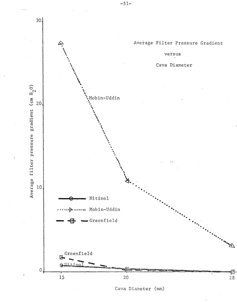
Results: Interference with Flow
Interference with flowý reflected the tendency of the filter to occlude the IVC and was measured as the pressure gradient across the filter device. The KG and KNitinol filters both showed no measurable pressure gradidnt without clots and only minimal gradients occurred after delivery of experimental clots
clots, created a measurable pressure gradient and after delivery
of clots the pressure gradient increased markedly and sometimes
exceeded the pressure limitations of the in vitro system
(Figures 22-24). The vena cava upstream of the filter usually
distended due to this pressure gradient while the downstream vena cava collapsed. If the filter was poorly secured, this often dislodged the filter and caused it to embolise. Many
times flow through the Mobin-Uddin became so reduced that it was
insufficient to carry additional clots to the filter. At other
times pressure gradients across the Mobin-Uddin became so large
Limitations of In Vitiro Simul•in
Obviously no model can perfectly simi.ulate the human body. Failure to simulate tissue response, immune mechanism, the reticulo-endothelial system, adjacent structures, and other uniquely biological phenomena restrict use of this system to evaluating mechanical effects only. Analysis of these
mechanical effects is limited by some of the simplifications in the system design. They can be enumerated as follows:
First, saline has different properties from blood, i.e. their densities and viscosities are different. These
differences may hve been compensated for by adjusting the flow velocity but one cannot be absolutely sure.
Flow in the vena cava is known to be pulsatile (29) but the simulation is strictly limited to uniform flow. Pulsatile flow might be expected to shake clots loose from the filter
increasing the probability they would pass through any of the larger openings in the filtering devices.
The experimental emboli made by allowing dog blood to sit stagnant in a glass tube for one hour are probably not exact replicae of human emboli which have bizarre shapes and are formed over days or weeks developing substantial organization.
The simulating device was fixed in a horizontal position Dut patients might assume a variety of postures that could influence filter performance.
Caval tissue is much tougher and more elastic than the
cellulose dialyzer tubing used in tle simulation. This lack of perfect simulation probably had a marked effect on filter
holding force determination (Table 3). Failure of the filter to stay secure in the vena cava was usually due to tearing of the cellulose dialyzer tubing. Human caval tissue would tear only when subjected to much higher forces.
Finally, humans also display much greater anatomical
variation than can be modeled with three sizes of cylindrical tubing. In fact, body scans show the IVC before filter
insertion to be oval in cross-section as opposed to a circular shape asuned by this model. A further complication is that Shuman subjects have curving vessels 'with numerous branches and surrounding structures.
Many of these deviations from the real life situation were purposely designed into the simulation to optimize visualization
and to generate a well controlled experiment. Some aspects could be improved. Lethal human blood clots could be acquired from autopsies and cut into standardized sizes. A pulsatille pump could generate pulsatile flow. Gelatin could be added to the saline to create a viscosity and density closer to that of blood without sacrificing visualization. Perhaps most
importantly, the circulation modelling parameters could be studied during human surgical procedures or on ex:perimental ~ animals to determine optimal values fdr in vitro system
Na
-Wi 21aimn
Despite the problems enumerated above, this simulation goes a long way toward evaluating the relative utility of three types of intracaval devices. The validity of this comparison is
verified by noting clinical ovservations that correlate with observations made in the in vitro simulation.
The MU was observed to occlude readily and generate large pressure gradients in vitro while the KG showed minimal pressure gradients and never occluded. Clinically the MU has a 73%
incidence of occlusion and venous stasis (a condition caused by interference of venous flow out of a limb) while it is only 5% for the KG (13)
In 28mm diameter cavae in vitro the 11U was observed to dislodge and migrate. Twenty-eight incidences of 1MU migration have been reported clinically in 2215 deliveries (28).
Tilting of the KG filter seen in vitro has been reported clinically (14,15) , was seen in 50% of Beth Israel hospital KG filter patients and occurred in 63% of in vitro KG filter
Celiveries.
'The MIU and KG hooks are effective in preventing downstream migration but offer little resistance to migration distally, awCay from the heart. Distal rigration of the MU and KG filter has been documented clinically (15,2).
this si.mulation is a good model for predicting the clinical properties of new IVC blood clot filters.
Clinical ImnlicatiQns
Filter clot capturing ability and anchoring security diminished with increasing vena cava diameter. In the 28mm diameter cava the KG filter allowed most 7mm diameter clots to pass and the MU migrated. A decision to place a KG filter in a patient with a cava 28 mm or more in diameter should therefore be taken with caution and the MU is contraindicated. Future
research might explore ways of retaining filter function in a wider range of cava diameters or develop multiple sizes of
filters so appropriate filters will be available for every patient threatened by pulmonary embolism.
Many clinicians report that a 7mm diameter embolus is
likely to be lethal or, at the vey least, clinically significant (resulting in physiologic changes which can be detected by
routine clinical exam). But the KG let 70% of 7mm diameter clots through dispite its low reported recurrence rate for pulmonary embolism clinically (3). This observation can be
explained in three ways. First, the present in vitro simulation may be unrealistic and 7mm diameter clots may not in fact pass
through the KG filter. Second, the clinical findings reported may not reflect the true incidence of recurrent pulmonary
embolisms with the KG filter. Finally, clinical trials of the KG filter may have yielded inaccurate results for the following
m±grating emboli, (2) more than one type of therapy was
employed, e.g. filter plus anticoagulation, (3) there was
inadequate patient follow-up, (4) there was insufficient
follow-up time for the clinical studies to be conclusive. While
the present analysis of the KG filter was as precise as
possible, the results of this study do not permit complete resolution of these issues. But a filter that captures clots better than the KG in vitro is likely to capture clots
effectively clinically.
Optimal filter design in terms of maximizing clot capturing
ability while minimizing filter pressure gradients was achieved
by the Nitinol filter which had the most uniform mesh.
Redesigning the MU and KG filters to have more uniform meshes
ýwould probably improve their performance significantly. The MU
could have a fish net like structure in place of the silastic membrane with occasional holes, thus reducing its obstructive
effects. The KG could have the six tines branch to form 12 at
the periphery where most clots currently get through.
The most significant implication of this research is that the favorable performance of the flitinol filter warrants its further evaluation. This study shoýwed it to anchor securely and
to capture emboli effectively without obstructing the IVC; all this better than the two filters currently used clinically. In addition, the Nitinol filter is inserted without surgery. If it
Summary and ConclusioDns
An in vitro comparison was made of three filters designed to
prevent clinically significant and especially'potentially lethal
emboli from reaching the lungs. Two of the filters, the Kimray
Greenfield and J1obin-Uddin, are currently used clinically; the third, the Nitinol (a memory wire device), is experimental. The
filters were delivered into an artificial vena cava where
measurements of clot capturing ability, flow interference
effects, anchoring security and orientation were obtained. The
-Mlobin-Uddin captured clots well in small and average size cavae
but was very occlusive. In large cavae the problem of
Mobin-Uddin filter migration was severe. The Kimray Greenfield
was not very occlusive but tended to fall into a tilted
orientation and allowed large clots to pass, particularly after
occlusion of the small spaces in the apex. The Nitinol filter
captured clots well in all caval sizes, displayed the smallest occlusive effect and was remarkably stable. Since the Nitinol
filter can be inserted into the vena cava through the lumen of a catheter, while the Greenfield and Mobin-Uddin require surgery, it is clearly the easiest and safest of the three devices to
deliver.
While the present simulation of blood flow in the vena cava did not completely parallel that of the human, it was
sufficiently close to permit a reasonable evhluation of the
Nitinol filter warr-ants further research on its biologic aspects through studies on both animals and selected human subjects.
1. Dalen JE and Alpert JS: Natural History of Pulmonary Embolism. Prog Card Dis 17:259-270, 1975.
2. Kakkar VV: The Current Status of Low-Dose Hleparin in the Prophylaxis of Thrombophlebitis and Pulmonary Embolism. World J Surg 2:3-18, 1978.
3. Greenfield LJ and Zocco JJ: "Intraluminal Management of Acute Massive Pulmonary Thromboembolism," Journal of
Thoracic and Cardiovascular Surgery 77:402-410, March, 1979. 4. Sabiston DC et Al: Ann Surg 185:699-711, 1977.
5. Bernstein EF: The Role of Operative Inferior Vena Caval Interruption in the Management of Venous Thromboembolism. Wiorld J Surg 2:61-71, 1978.
6. Mobin-Uddin K, McClean R, Jude JR: A New Catheter Technique of Interuption of Inferior Vena Cava for Prevention of
Pulmonary Embolism. Am Surg 35:889-894, 1969.
7. liobin-Uddin K et Al: Transvenous Caval Interruption with Umbrella Filter. N Eng J 1led 286:55-58, 1972.
8. Greenfield, LJ, McCurdy, JR, Brown, PP, Elkins, RC: A 1New Intracaval Filter Permitting Continued Flow and Resolution of• Emboli," Surgery, 73:599-606, April, 1973.
9. Simnon 8: A vena Cava Filter Using Thermal Shape Miemory Alloy. Radiology 125:89-94, 1977.
10. Simon 8 and Palestrant AM: Transvenous Devices for the
7-7777-77-777----lianagement of Pulmonary Embolism.
11. Philli)s, HN, idrich, C, and Johnson, C: erforation o the Inferior Vena Cava by the Kim-Ray Greenfield Filter. Surgery 233-5, 1980.
12. Sautter, RD, Myers, 7WO, Lawton, BR: Experience with Vena
Cava Filter 1Migration. (letter to the editor), JAIA
219:1217, 1972.
13. Cimochowski GE, Evans RH, Zarins CK, Lu CT, Demeester TR: Greenfield filter versus Iiobin-Uddin umbrella The
continuing wuest for the ideal method of vena caval interruption. J Thor Cardiovasc Surg 79:353-365, 1980.
14. Wingerd
1,
Bernhard VI, Macaddison F, Towne JB: Comparison of Caval Filters in the iManagement of Venous Thromboembolism. Arch Surg 113:1264-1268,1978.15. Berland LL, Maddison FE, Bernhard Vii: Radiologic Follow-up of Vena Cava Filter Devices. AJR 134:1047-1052, 1980.
16. Mobin-Uddin K, Utley JR, Bryant LR: The Inferior Vena Cava Umbrella Filter. Prog Card Dis 17:391-399, 1975.
17. Sher .MHP1: Complications in the Application of the Inferior Vena Cava Umbrella Technique. Arch Surg 103:688-690, 1971. 18. Gaston EA: Incorrect Placement of Intracaval Prothesis for
Pulmonary Embolism. JAd1A 214:2338, 1970.
19. Sauters RD: Experience with Vena Cava Filter Miigration. JAVA 219:1217, 1972.
20. Schroeder TM, Elkins RC, Gr-eenfield LJ: Entr apment of Sized• • Emboli by the Ki·A-Greenfield Intracaval Filter. Surgery
83:435-439, 1978.
21. Brown PP, Peyton iID, Elkins PC, Creenfield LJ: Experimental Comparison of a New Intracaval Filter with the M'obin-Uddin Umbrella Device. Card Surg Suppl II to vols. 49 &
50:272-276, 1974.
22. Mobin-Uddin, K, Smith, PE, Hartinez,LD, Lombardo, CR, Jude, JR: A Vena Cava Filter for the Prevention of Pulmonary Embolus. Surg Forum, 18:209, 1967.
23. Gianturco, C, Anderson, JH, and Wallace, S: A New Vena Cava Filter: Experimental Animal Evaluation. Radiology
137:835-837, 1980.
24. Prince iDR: Simulation of the Human Inferior Vena Cava for Evaluating IVC Interruption Devices. Bachelor's Thesis in M•echanical Engineering, M.I.T., 1980.
25. Novelline, PA: personal communication, Massachusetts General Hospital, Boston, Oct. 1, 1981.
26. Guyton AC: Textbook of Medical SchoL1 PthysiQ vIgy, B Saunders Co. Philadelphia 1976, p.251.
27. Correct technique for introducing filters
28. West JB: Respiratory Physiology - the essentials2nd edition, Williams & Wilkins, Baltimore, 1979.
29. Gardner AR1I1: Inferior Vena Caval Interruption in the Prevention of Fatal Pulmonary Embolism. Amer Heart J
95:679-G82, 1978.
30. Castleman LS: Biocompatibility of Nitinol Alloy as an Implant Miaterial. J Biomed M!at Res 10:695-731, 1976.
.H
r--4
CD
04O rd
S 0H 0 0 Z Cd 0 Cd Ici UCO CO 4-i O0
4-J
to to ac D CQdd 'H rd 42H 0 ato t o'H ' >54 UL-4- Cr, C Cd 'H 'H m 0 'H Cd OH-0 Cd 4J E to L co 0 CO d 't 40 -H -. O o d 'H O 0 ct S E v H O o OP-3 cti0 Cd Cd 0 S * H Cd OO F-4 o U 4-i 4 0~ 0 Cd k 0 00 0 ~ Cd 4-i H Cd Cd H 0 Lo OL C -H 0 "' 05 0 ~-r Lr 4-J Cd 0 4-J CdQ)
kH
4-1 0 z C rd 0 H 0O CD 0 Cd 0 rd 0 .H Cd0 LH 0 Cd4-J
0 4-J ICd H U 4-i O .-t ., 0'H 42 O U 'H CO rd L)0 -4-10 S 0 H 0 4.. -H 0 U tO Cd U0 Cd Cd 0 0 U 4( 4-J ,-CO .,-CO toC-) -H a 42 H0 4 O 0 o 4 H 42 0 Et a,-ý4 4_1 O H 0 CI 0 S e o 0 QJ H-{ co O o CO 0 o4-J 0 H0 I ~ r-__ _~ __~ _ ____C..
--- m 1115·-·-~i --
---- -I
I
-i
IFilter Deliveries
* the Mobin-Uddin Umbrella was not walls in the 28mm diameter cava.
simply rolled on downstream.
large enough to engage the cava For every delivery the filter Table 3
Mobin-Uddin Greenfield Nitinol
IVC diam (cm) 15 20 28 15 20 28 15 20 28 Orientation: % tilted 60 90 * 60 80 50 40 70 0 % central 40 10 * 40 20 50 60 30 ý100 Holding Force mean (gm) 83 117 0 77 55 34 91 55 49 standard 20 33 0 12 6 22 25 21 37 deviation # hooks engaged 6 5 0 6 4 2 0 0 0
Physiology
1) Clot forms in lower body
2) Clot breaks loose and migrates toward lungs
3) Clot plugs pulmonary artery
Anticoagulant therapy
prevents new clot formation
IVC interruption prevents large clots from reaching
the lungs
Surgery removes clot
Thrombolytic drugs lyse clots Figure 1 -Pulmonary Embolism: Physiology and Treatment.
sutures
DEWEESE FILTERclips
MORETZ ADAMS-DEWEESE MILES aiFigure 2A Direct surgical techniques for interrupting the IVC
intracaval devices
Mob in-Uddin ad·iPd··bl-s MU delivery capsule KG delivery capsule delivery catheterInterruption devices that fit inside the IVC
'"
1-
---L "-11 aýLNAA 4--.
TECHNIQUE FOR INSERTING THE KINRAY GREENFIELD FILTER
A) Load filter into capsule.
'
I
U
B) Expose internal jugular vein
Withdraw capsule.
---.mmwmlmpý
-a '-~---__
A) Insert and ini C' i-•• K ' / I I f 1 'I
B) Insert guidewire through ne and into femoral vein.
C) Remove needle leaving in vein.
guidewire
Insert catheter into femoral vein by sliding over guidewire.
Figure 4a. Transcatheter approach to IVC interruption. - ·--- - --- - - -- ----·--I-~-is 7----~ --"~~ --Ce
-/
I L I I r r r r I ID) Attach filter storage and feeder device.
Advance Nitinol filter into catheter (in its straight wire, low temperature form
E) Push filter through catheter and into the IVC.
(It recovers its high tempera filter configuration instantl upon contact with blood at bo
temperature).
F) Remove catheter and apply compression at site of needle puncture for 10 minutes to prevent bleeding.
Figure 4b. Transcatheter approach to IVC interruption.
(
af
k
--- - -- I T- - -~Wb~4RICC~-"~-- ---- ----~-~-~-TI -c~--- ~-~ ---\ •k
i I rAverage human Large human GREENFIELD
t-
2Omm -4
4- 28m.m
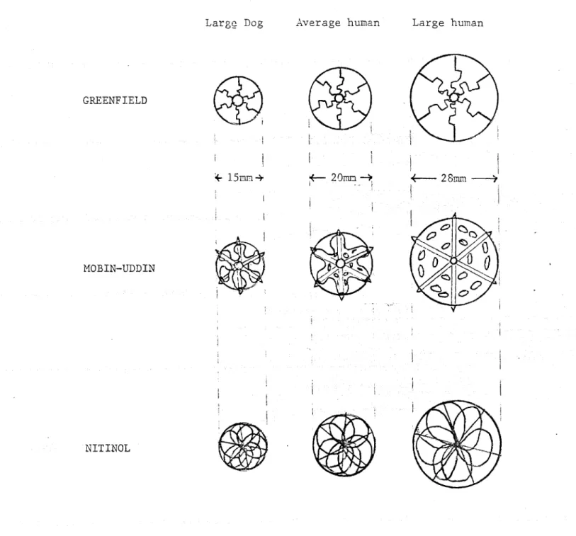
i . , MOBIN-UDDIN NITINOLFigure 5 Variations in filter mesh with changing vena cava diameter Large Dog
5
--2?~
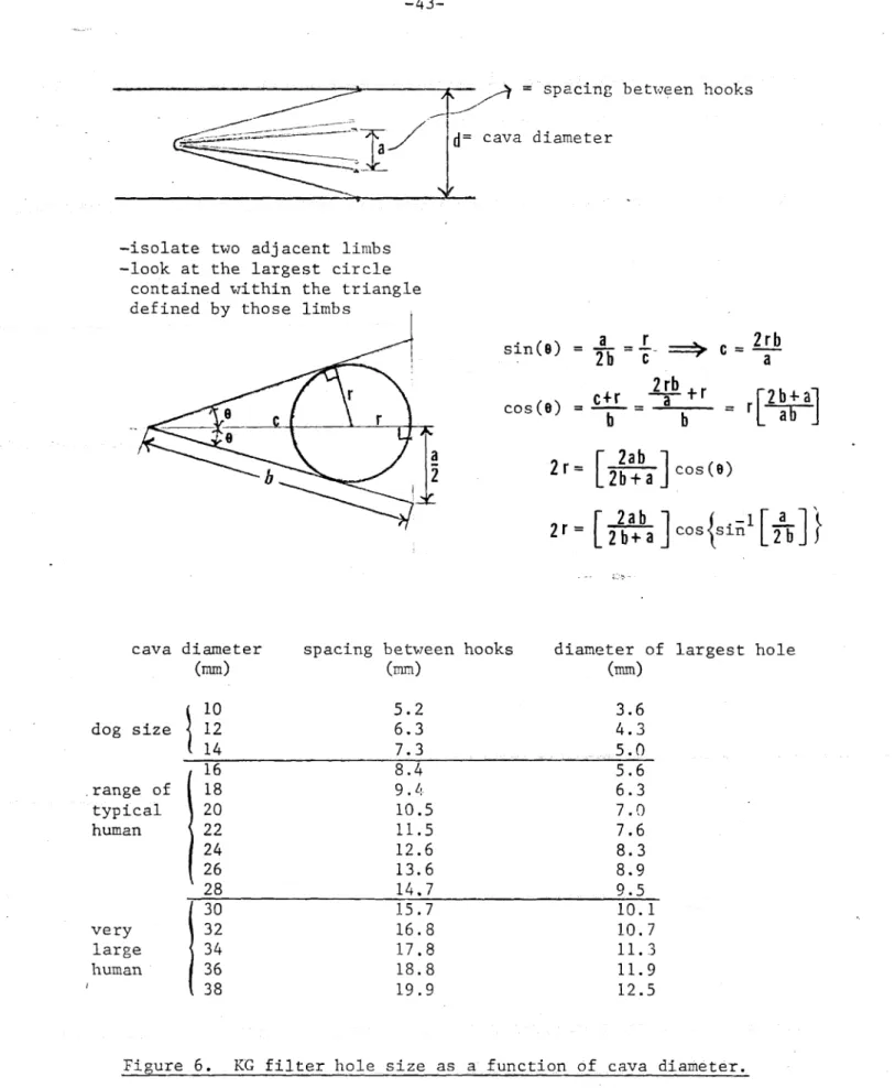
= = ~spacing s~ between hooks
d=cava diameter
-isolate two adjacent limbs -look at the largest circle
contained within the triangle
Amfq"MA
1-NT
fmInno'
sin(s)
sin() b c
a rc
=2rb=
2rb cos() = ab
r
2bba2r=-
2b+ab
cos(s)2r
2ab a S j2bta
b
2
r
2b+aCos
sin 2
b
cava diameter (mm))spacing between hooks
(mm) diameter of (mm) largest hole 10 12 14 5.2 6.3 7.3 3.6 4.3 5.0 16 8.4 5.6 18 9. 4 6.3 20 10.5 7.0 22 11.5 7.6 24 12.6 8.3 26 13.6 8.9 28 14,7 9.5 30 32 34 36 38 15.7 16.8 17.8 18.8 19.9 10.1 10.7 11.3 11.9 12.5
Figure 6. KG filter hole size as a function of cava diameter.
I
dog size .range of typical human very large humanL.-.&a
fI
-44-,)
co:
LU
cc.
cc
ILlw
7,FE
a
cU in U a4 m
-45-LC
04-J
O
a Cava Diameters patients with y Greenfield Filters 14 12 10 8 6 4 12 14 16 18 20 22 24 26 28r 28 30
Vena Cava Diameter (mm)
number mean cava diameter standard deviation Kimray Greenfield with venogram 20 20 3.8 Kimray Greenfield without venogram 45 19 3.2 total 65 20 3.4 .. . ,- •, .... .i. ... .---
Figure 9. Infrarenal inferior vena cava diameters in KG filter patients.
a L) 4-J
Cd
a) 44 0 15 3232
sa~c ,..~--- ---7---,~-~1---~-·--9~wl
A) Filter in capsule.
B) Capsule withdrawn exposing filter.
filter jumps out.
<Ir--A riia on
D) Capsule removed.
Figure 10. KG filter delivery.
,, - ~- --'; ~ --- i --··
(1-_;~~~---~---
I I ; l~-~kid
·-·I:-- -··-illF---_·A·· L- . 6-__ _·,,, -I ~ ~~Mp
-- I ' ''- i--!ip
---EXISTING HOOK DESIGN
hook not engaged
SUGGESTED HOOK DESIGN
both hooks engaged
note larger hook angle increases the probability of hook engaging
Analysis of Greenfield filter hooksi Figure 11.
Calculation of Optimal Greenfield Hook Angle
7,
hook angle =
limb length = b D= largest possible cava diameter
e
= angle between limb and cava wall S= sin)1 Dif oC :• then hook will not point into and engage cava
if -& > 90 hook will slip o'ut of cava wall
thus 90 > optimal oC >
let D = 40mm and b = 46rm
90 > c. 60
Figure 12. Determination of optimal KG filter hook angle.
Filter Holding Force versus Cava Diameter " Mobin-Uddin h" N44 N* *N% I' %, Sreenfield 'Til 100 8d a 60 .i 40o *20 20 -20 28 Cava Diameter (nm)
Figure 13. Filter holding force versus cava diameter.
12Q - : r --- -- = Nitinol ... .~... = Mobin-Uddin -- = Greenfield 3 15 ,1 IF ,
Average Filter Pressure Gradient versus Cava Diameter .Mobin-Uddin . r r r r 1 S cl ~3220, 4-4 a)
to
0 k 0) · rH t~ o 21 C) Greenfield U "two 15 20 Cava Diameter (mm)Figure 14.- Average filter pressure gradients versus cava diameter.
30. \9 I--0... Nitinol . .. ... ...- Mobin-Uddin -- -&• . .Greenfield 28 -- ~Raeaars~·--~*p~ll~Rp~--~---~--* r
100 -90 80 70 H60 5Q 0 0 040 30 20 .... obin-Uddin Nitinol
% of Clots Capdtured Completely
ver ~us Cava Diameter
\ reenfield (cent ral orientation)
SGreenfield -,tilted orientation) Nitinol .--- = .Mobin-Uddin ' - --. --- = Greenfield-central - - 7 -- = Greenfield-tilted )c 0 15 20 28 Cava Diameter (mrm) Figure 15. Comparative clot capture performance.
Nitinol U.-... - r 1%
a
".Mobin-Uddin S. Ile OF lot ogoo °°r+°o BI'Ir 90-. 80- 70-ý4 50 -0 rH Q (CiH
40 0K
% of 7mm diameter clots captured versus Cava Diameter --- Nitinol -·. At.. Mobin-Uddin -- J-- -- Greenfield I 5 20n I
nfl
\Greenfield 30 20 10 0 Ii?/ Cava Diameter (nrm)Figure 16. Comparative clot capture performance (7mm clots only).
28 ussF.runarru~~l-u4+rr~·l~'~cupe~i~i4~* i+Ld~~··~;sulu~a~~YI,~ r.. _i ~oaa~- uraqn i
-·---fp
.
op
1•OGE.1EIE L5D
15mm
Length 10cm 2cm Diameter 7mm 4mm 7mm 4mm Order 1 2 3 4. 5 1 2 3 4 5 P 4 * * * 3 3 5 3 * * * * * * + 60 70 100 80 70 50 90 78 80 80 30 40 20 50 - 40 30 0 20 $20 40 10 18 10 20 40 40 20 26 0 0 0 0 10 10 0 4 10 0 30 20 60 24I
0
0
0
0
0
0
0
0
0
0
0
0
0
0
0 0 0 0 0 0 0 0 0 0 0 0 0 0 P pressure in cm of H20+ %clots captured completely
+- %clots captured partially
- %clots passing completely through filter I %cases of insufficient flow
M %cases of filter migration
* pressure never exceeded 5cm of H20
. . .. . . "- • -- ,, • . .... • :_ • -:; . . . : L • '": : .• . ::,! : ~ • -'. . .' ' ...
Figure 17.
---GA
EELmEIRD
2,O.mm
Length 10cm 2cm Diameter 7mm 4mm 7mm 4rmm Order 1 2 3 4 5 1 1 2 3 4 5 Z P * * * * * 2 2 1 * * * * * 90 20 75 70 55 55 40 59 70 40 60 30 10 42 +- 10 80 25 25 25 30 45 30 20 40 10 40 30 28 0 0 0 5 20 15 15 11 10 20 30 30 60 30 1 0 0 0 0 0 0 01 0 0 0 0 0 0 0 M 0 0 0 0 0 0 0 0 000 0 0 0 0 0 P pressure in cm of H20+ %clots captured completely
+- %clots captured partially
- %clots passing completely through filter
I %cases of insufficient flow
M %cases of filter migration
* pressure never exceeded 5cm of H20 Figure 18. ^"-"L-~ p II ·--··I----~---i
---TLLLED
2DmJLwn
Length 0cm 2cm Diameter 7ram 4mm 7mm 4mm Orde1 2 3. 4 5 1 2 3 4 5 Z + 80 0 80 20 30 20 30 36 0 0 0 0 0 0 " 20 90 20 70 30 70 50 48 40 40 10 40 50 6 36 0 10 0 10 40 10 20 16 60 60 90 60 50 64 I 0 0 0 0 0 0 0 0 0 0 0 0 0 0 M 0 0 0 0 0 0 0 0 0 0 0 0 0 0 0 P pressure in cm of H20+ %clots captured completely
+- %clots captured partially
- %clots passing completely through filter
I %cases of insufficient flow M %cases of filter migration
* pressure never exceeded 5cm of H20
• - . . _• _.. " ' .... ' . . . . . . .. : ! ... ' :" . . < " i k_ • • • i •-_- v -.. . Figure 19.
I
r I.
GREE NFI"
·-.---~---c~-I-~--._2i
mL
Length 10cm 2cm Diameter 7mram 4mm 7mm 4mm 1 2 3 4 5 1 2 3 4 55 Order + 0 0 20 20 30 10 10 18 10 10 0 0 0 4 + - 80 100 70 60 30 40 10 42 80 80 50 40 90 68 - 20 0 10 20 40 50 80 40 10 10 50 60 10 28 I 0 0 0 0 0 0 0 0 0 0 0 0 0 0 M 0 0 0 0 0 0 0 0 0 0 0 0 0 0 P pressure in cm of H20+ %clots captured completely
+- %clots captured partially
- %clots passing completely through filter I %cases of insufficient flow
M %cases of filter migration
* pressure never exceeded 5cm of H20 2 Figure 20.
I
4I
C-- ~----.?~n~a~R"P*IIPI~·l~l-- C---l· -- - ---~P1S~1GRIED
.ll_D_28_mm_
ri-mu
Length
10cm
2cm
Diameter 7mm 7mm 4mm Order 1 2 3 .. 4 5 1 2 3 4 5 P 7 ** J * * *_ * * *1*' + 0 010 10 0 4 0 0 0 0 0 0 +- 1001100 50 30 10 20 20 26 100 90 50 30 50 64 0 0 50 70 80 70 80 70 0 10 50 70 50 36S
0
0
0
0
0
o
0
0
0
0
0 o
0
0
M 0 0 0 0 0 0 0 0 0 0 0 0 0 0 P pressure in cm of H20+ %clots captured completely +- %clots captured partially
- %clots passing completely through filter
I %cases of insufficient flow
M %cases of filter migration
* pressure never exceeded 5cm of H 0 2 Figure 21.
--·rr
---15nmm
Length 10cm 2cm Diameter 7mm 4mm 7mm 4mm Order1 2 3 .. 4 5 1 1 2 3 4 5Order
__-I
_ P 33 20 16 30 33 35 35 30 5 29 29 35 35 27 + 90 100 100 80 20 10 0 42 100 100 .20 20 0 48 + - 10 0 0 0 0 0 0 0 0 0 0 0 0 00 0 0 0 0
0 0
0
0 0
O
0
I 0 0 0 20 80 90 100 58 0 0 80 80 10 52 M 10 10 0 00 0 00 0 0 0 ' 0 0 00 P pressure in cm of H20 -+ %clots captured completely+- %clots captured partially
- %clots passing completely through filter I %cases of insufficient flow
M %cases of filter migration
* pressure never exceeded 5cm of H20
Figure 22.
--20m_
-19.II3~
Length 10cm 2cm Diameter 7mm 4mm 7mm 4mrm Order 1 2 3..4 5 2 1 2 3 4 5 P 20 7 6 12 12 13 11 * * 7 9 12 6 + 100 100 100100 90 10 10 62 100 100 90 60 301 76 - 0 0 0 0 10 0 0 2 0 0 0 10 4 0 0 0 0 0 10 0 2 0 0 0 30 0 6 I 0 0 0 0 0 80 9D 34 0 0 0 10 60 14M
0 0
0
0
10
0
0
2
0
0
0
0
0
0
P pressure in cm of H20. + %clots captured completely+- %clots captured partially
- %clots passing completely through filter
I %cases of insufficient flow
M %cases of filter migration
* pressure never exceeded 5cm of H20
. . . ... .. . . .... : . • .:.. . . : • _ .. . ..•. .·_ : • . -: .. _ . . >:; . .. .• u . -i ' i f -. ._• • .: i Figure 23. - -• L -:.-•••.. . ••-"... . .
B-
.I
N IN
A-
D.
-·C I r~l··~-3lpl~sl~·a~a~a~~-C
0 0Z
l C
163 "b
Length 10cm 2cm
Diame ter 7mm 4ramm 7mm 4mm
Order 1 2 3 -4. 5 1 2 3 4 5
x
P 28 3 0 2 2 2 2 2 0 0 1 1 0 0 + 30 160 70 40 10 20 10 30 40 30 10 10 10 20. 20 10 0 0 0 0 0 0 0 0 -20 20 10 10 0 0 0 30 40 20 30 24 10 20 20 20 30 20 I 0 0 0 0 0 0 0 0 0 0 0 0 0 0 M 50 30 30 30 50 60 60 46 50 50 50 50 50 50 P pressure in cm of H20+ %clots captured completely +- %clots captured partially
- %clots passing completely through filter I %cases of insufficient flow
M %cases of filter migration
Figure 24.
M.0 N
-m
D~atP
98mm
T--Length 10cm 2cm Diameter 7mm 4mm 7mm 4mm Order 1 2 3. 4 5 2 1 2 3 4 5 I
P
**
*4*
4
2
*
*
*
*
*
*
+ 1100 100 100 100 100 100 100 90 80 90 70 100 86+-
0 0 0 0 0 0 0 0 0 0 0 10 0 2 0 0 0 0 0 0 0 0 10 20 10 20 0 12 'I 0 o 0 0o 0 0 0 0 0 0 0 0 0 M, 0 0 0j0
0 0 0 0 0 0 0 0 0 0 P pressure in cm of H20+ %clots captured completely
+- %clots captured partially
- %clots passing completely through filter I %cases of insufficient flow
M %cases of filter migration
* pressure never exceeded 5cm of H20
.. WN W-..
Figure 25. `CI-C-I--I
/7 Length 10cm 2cm Diameter 7rnn Amrmn 7mm 4mm Order
I
2 3.4. 5
X1
2 3
4 5
P
*
*
*
*
*
122.1
0.8
*
*
*
*
* * + 100 100 100 100 100 100 100 100 100 100 .90 100 100 98 +- 0 0 0 0 0 0 0 0 0 0 0 0 0 0 0 o 0 0 0 0 0 0 0 0 10 0 0 2 I 0 0 0 0 0 0 O- 0 0 0 0 0 0 0 M 0 0 0 0 0 0 0 0 0 0 0 0 0 0 P pressure in cm of H20-+ %clots captured completely
+- %clots captured partially
- %clots passing completely through filter I %cases of insufficient flow
M %cases of filter migration
* pressure never exceeded 5cm of H20
~~IY9~~13LL~~~ -~ - I-- - - ~ IIr ~ ~ - .'--
Figure 26.
;--·- _~ ,,,,~~e -
-- ---
Length 10cm 2cm Diameter 7mm 4rmm 71m 4mm Order1 2 3.4 5 1 2 3 4 5 P -* * * * * * * * .*- * * * * + 100 80 100 100 100 100 100 100 100 90 100 100 100 98 0 + 20 0 0 0 0 0 0 0 10 0 0 0 2 - 0 0 0 0 0 0 0 0 0 0 0 0 0 0 0 0 0 0 0 0 0 0 0 0 0 0 0 0 M 0 0 0 0 0 0 0 0 0 0 0 0 0 0 P pressure in cm of H20
+ %clots captured completely
+- %clots captured partially
- %clots passing completely through filter
I %cases of insufficient flow M %cases of filter migration
* pressure never exceeded 5cm of H 0 2 Figure 27.
~~NJ