HAL Id: hal-03035522
https://hal.archives-ouvertes.fr/hal-03035522
Submitted on 25 May 2021HAL is a multi-disciplinary open access archive for the deposit and dissemination of sci-entific research documents, whether they are pub-lished or not. The documents may come from teaching and research institutions in France or abroad, or from public or private research centers.
L’archive ouverte pluridisciplinaire HAL, est destinée au dépôt et à la diffusion de documents scientifiques de niveau recherche, publiés ou non, émanant des établissements d’enseignement et de recherche français ou étrangers, des laboratoires publics ou privés.
Effects of chronic masitinib treatment in APPPS1dE9
transgenic mice modeling Alzheimer’s disease
Tengfei Li, Elodie Martin, Yah-Se Abada, Céline Boucher, Aurélia Cès, Ihsen
Youssef, Grégory Fenaux, Yona Forand, Annaelle Legrand, Nadkarni
Nachiket, et al.
To cite this version:
Tengfei Li, Elodie Martin, Yah-Se Abada, Céline Boucher, Aurélia Cès, et al.. Effects of chronic ma-sitinib treatment in APPPS1dE9 transgenic mice modeling Alzheimer’s disease. Journal of Alzheimer’s Disease, IOS Press, 2020, 76 (4), pp.1339-1345. �10.3233/jad-200466�. �hal-03035522�
1 TITLE
Effects of chronic masitinib treatment in APPPS1dE9 transgenic mice modeling Alzheimer’s disease
RUNNING TITLE
Mastocyte inactivation in Alzheimer’s disease AUTHORS Tengfei Li 1# Elodie Martin 1# Yah-se Abada 1 Céline Boucher 1 Aurélia Cès 1 Ihsen Youssef 1 Grégory Fenaux 2 Yona Forand 2 Annaelle Legrand 2 Nadkarni Nachiket 3,4 Marc Dhenain 3,4 Olivier Hermine 5 Patrice Dubreuil 2 Cécile Delarasse 1,6 Benoît Delatour 1* # Co-authors
2
* Corresponding author: Benoît Delatour. ICM Institut du Cerveau et de la Moelle épinière, CNRS UMR7225, INSERM U1127, Sorbonne Universités, Team "Alzheimer's and Prion
diseases", 47 bd de l’hôpital, 75013 Paris, France. benoit.delatour@upmc.fr
1 ICM Institut du Cerveau et de la Moelle épinière, CNRS UMR7225, INSERM U1127,
Sorbonne Université, Hôpital de la Pitié-Salpêtrière, Paris, France
2 CRCM, [Signaling, Hematopoiesis and Mechanism of Oncogenesis, Equipe Labellisée Ligue
Contre le Cancer], Inserm,U1068; Institut Paoli-Calmettes; Aix-Marseille Univ, UM105; CNRS, UMR7258, Marseille, France.
3 Centre National de la Recherche Scientifique (CNRS), Université Sud, Université
Paris-Saclay UMR 9199, Neurodegenerative Diseases Laboratory, 18 Route du Panorama, F-92265 Fontenay-aux-Roses, France
4 Commissariat à l’Energie Atomique et aux Energies Alternatives (CEA), Direction de la
Recherche Fondamentale (DRF), Institut François Jacob, MIRCen, 18 Route du Panorama, F-92265 Fontenay-aux-Roses, France
5 INSERM UMR1163 and CNRS URL 8254, Imagine Institute, Paris Descartes
University-Sorbonne Paris Cité, Department of Hematology, Necker Children's Hospital, APHP, Paris, France.
3 ABSTRACT AND KEYWORDS
Background: Masitinib is a selective tyrosine kinase inhibitor that modulates mast cells activity. A previous phase II study reported a cognitive effect of masitinib in patients with Alzheimer’s disease.
Objective: We aimed to shed light on the mode of action of masitinib in Alzheimer’s disease. Methods and results: We demonstrated here that chronic oral treatment of APPPS1dE9 transgenic mice modeling Alzheimer’s disease restored normal spatial learning performance while having no impacts on Aß loads nor on neuroinflammation. However, masitinib
promoted a recovery of synaptic markers. Complete genetic depletion of mast cells in APPPS1dE9 mice similarly rescued synaptic impairments.
Conclusion: These results underline that masitinib therapeutic efficacy might primarily be associated with a synapto-protective action in relation with mast cells inhibition.
Keywords : Alzheimer’s disease; mast cells; masitinib; cognition; synapses; drug evaluation, preclinical
4 INTRODUCTION
Alzheimer’s disease (AD) is a neurodegenerative pathology that affects 40 million people worldwide. There are currently no available therapies to stop or even slow down AD neurodegeneration [1].
In recent years, a number of preclinical and clinical observations have drawn attention to the role of mast cells (MCs) in the physiopathology of AD. A randomised, placebo-controlled, phase 2 study was performed with masitinib (AB1010) [2], a selective tyrosine kinase
inhibitor with a known inhibitory action on the survival, differentiation and degranulation of MCs through targeting the c-Kit receptor [3]. Masitinib given orally as an adjunct therapy was found to slow down cognitive decline in AD patients [2] and a multicenter phase III study was subsequently initiated in patients with mild to moderate AD (NCT01872598).
Besides playing a key role in innate immunity, MCs have been involved in different neurological conditions [for review see 4, 5] and the beneficial effect of masitinib in brain pathologies (e.g. multiple sclerosis, amyotrophic lateral sclerosis) has been exemplified [6-8]. As described above, masitinib treatment mitigates cognitive symptoms in AD patients but its exact mode of action remains to be determined. Inhibition of MCs at the blood-brain barrier vicinity may reduce permeability and penetrance of proinflammatory mediators.
Alternatively, chymotrypsin-like protease released by MC can catabolize the amyloid precursor protein to generate Aß-containing fragments [9] and inhibition of MCs may therefore lower Aß pathology.
The aim of the present study was to better clarify the effects of masitinib in AD using transgenic mice (APPPS1dE9 model) modeling the disease and displaying brain amyloid pathology [10], neuroinflammation [11] and synaptic loss [12]. To evaluate the hypothesis that the masitinib effects observed after treatment are related to MCs inhibition we
5
generated a new model of APPPS1dE9 mice expressing a Wsh mutation leading to a specific depletion of MC populations.
MATERIAL AND METHODS
Animals and treatments
Transgenic APPPS1dE9 mice co-express human APP and PS1 genes with AD familial
mutations on a C57BL/6J background [10]. APPPS1dE9 (TgAD) mice as well as wild-type (WT) littermates were purchased from Jackson Laboratory and bred in-house.
For the present study, 12 month-old TgAD and WT male mice were treated with masitinib (AB1010, AB Science) at 75 mg/kg/day or vehicle (distilled water) in a single dose by oral gavage for 2.5 months, 6 days/week (WT-Vehicle, n= 10; WT-masitinib, n= 10; TgAD-Vehicle, n= 8; TgAD-masitinib, n= 9).
Mast cell-deficient APPPS1dE9 mice were produced by mating APPPS1dE9 males with female mice harboring a c-Kit receptor mutation (homozygous Wsh line 13) [13] at the Centre de Recherche en Cancérologie (Marseille, France). Ten month-old male (M) and female (F) mice were used (WT, n= 6 M + 4 F; Wsh, n= 5 M + 4 F; TgAD, n= 5 M + 5 F; TgAD/Wsh, n= 7 M + 8 F).
Animals were held in accordance with the French animal welfare act and the EU legislation (council directive 2010/63/EU). The present research project was positively evaluated and authorized by the regional ethics committee (Project N°04311.03) and all protocols were carried out according to ARRIVE guidelines.
Cognitive evaluation
Learning and memory of mice were evaluated using the Morris Water Maze (MWM) task as described in supplementary methods.
6
Histology
All mice were sacrificed at 10 or 14 months of age. They were transcardially perfused with phosphate buffered saline following deep anesthesia with sodium pentobarbital. Their brains were collected and cut by half. The right hemisphere was immediately frozen in liquid nitrogen and then transferred at 80°C before subsequent biochemical analysis. The left hemisphere was fixed by immersion in 4% phosphate buffered paraformaldehyde at 4°C for 48 h and then cryoprotected in 2% DMSO - 20% glycerol PBS at 4°C, and finally sliced on a freezing microtome (serial 40 µm thick sections).
Amyloid load assessment was performed following Congo red staining according to Puchtler's method [14]. For each APPPS1dE9 mouse a whole series of coronal sections spanning the entire rostro-caudal extent of the brain were stained and subsequently analyzed.
Immunohistochemistry was performed on other batches of serial sections using the standard ABC (Avidin-Biotin Complex) detection method with diaminobenzidine as chromogen. Used antibodies were a polyclonal anti-Iba1 antibody (Wako 1:3000), a polyclonal anti-GFAP antibody (Dako 1:5000) or a monoclonal anti-synaptophysin antibody SY38 (Millipore, 1:1000) that were incubated overnight at 4°C.
All histological sections were scanned with a NanoZoomer 2.0-RS slide scanner (Hamamatsu
Photonics, pixel size 0.25 µm2) and image analysis was performed using different methods
(supplementary data).
Biochemical analysis
Aβ peptides, IL-1β and chemokines dosages were performed according to protocols described in supplementary methods.
7
Statistical analyses
All data were expressed as mean ± SEM and analyzed with student t-tests or ANOVA using GraphPad Prism 6 (GraphPad Software, La Jolla, CA, USA) or Statistica 10-13 (StatSoft, Inc., Tulsa, OK, USA) software packages. Statistical significance was defined as P <0.05. In ANOVA designs, multiple comparisons relied on post-hoc Bonferroni tests. All neuropathological and biochemical measures were normalized to reference groups. This allowed to take into account variability between discrepant factors (e.g. brain region analyzed) to better assess the effect of main factors of interest (i.e. treatment and genotype).
RESULTS
Cognitive function
We first addressed the effects of masitinib treatment on the cognitive performance of TgAD mice trained in the Morris water maze task.
The analysis of navigation strategies (Figure 1A) underlined that all but the TgAD-Vehicle group succeeded to develop spatial navigation strategies with continuous training. As a noticeable observation TgAD mice treated with masitinib reached levels of performance very close to those obtained by WT mice. ANOVA confirmed these observations underlining a significant effect of the group factor (F(3,32)= 4.3; p<.025). Post-hoc comparisons indicated that TgAD-Vehicle mice performed a reduced number of spatial trials in comparison to WT-Vehicle mice (p<.025) while WT and TgAD mice treated with masitinib performed equally (p>.99). Besides, TgAD-masitinib mice performed better than TgAD-Vehicle mice (p<.05). The learning accuracy, assessed by the distance travelled in the maze, paralleled navigation strategy data (Figure 1B). ANOVA results indicated an effect of the group factor (F(3,32)= 7.023; p<.001). Post-hoc tests showed that TgAD mice were strongly impaired in comparison
8
to WT mice in the vehicle condition (p<.001) but not following treatment with masitinib (p>.99). TgAD-masitinib mice performed indeed better than TgAD-Vehicle mice as they displayed lower travelled distance (p<.05).
During the probe test (Figure 1C) WT mice showed a clear preference for the target
quadrant in both vehicle and masitinib drug conditions indicating memory for the platform location (t-tests to compare percentage of time in the target quadrant to 25% chance level, p<.05 for both WT groups). On the contrary, transgenic mice did not perform above the chance level (all p-values > 0.7). A two-way ANOVA confirmed a genotype effect on memory retention performance with TgAD mice being globally impaired as compared to WT mice (F(1, 31)= 6.30, p<.025) with no significant effect observed for the treatment factor or treatment x genotype interaction (Fs<1).
Brain amyloidosis and neuroinflammation
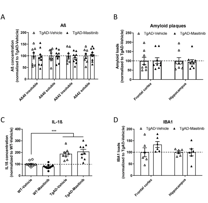
Next, we explored if the beneficial effect of masitinib on the cognitive deficits of TgAD mice were associated with a decrease of Aß accumulation and brain inflammation.
In 14 month-old TgAD mice, that display numerous aggregated amyloid deposits, no
differences between vehicle- and masitinib-treated mice were observed in terms of Aß brain concentrations, regardless of peptide isoform (Aß 40, Aß 42) or conformation (soluble, insoluble) (all ts(15)<0.61; ps>.55; see Figure 2A). Also amyloid loads assessed from brain sections stained with Congo red did not show any differences between TgAD-Vehicle and TgAD-masitinib mice in frontal and hippocampal areas (all ts(15)<0.25; ps>.81; Figure 2B). As anticipated, both transgenic groups showed highly increased IL-1ß concentrations in comparison with WT mice confirming a neuroinflammatory background in TgAD mice (WT-Vehicle vs TgAD-(WT-Vehicle: t(16)= 5.20; p<.0001; WT-(WT-Vehicle vs TgAD-masitinib: t(17)= 5.00; p<.0001; Figure 2C). Analysis did not however reveal any difference between the two TgAD
9
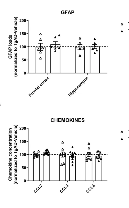
groups (t(15)= 0.70; ns), indicating no effect of masitinib treatment on the proinflammatory IL-1ß cytokine levels. In addition, brain levels of different chemokines (CCL-2, CCL-3, CCL-4) were found to be comparable in the two transgenic groups (All ts(15)<2.117; ps>.051; Figure S1B).
The two TgAD groups under vehicle or masitinib treatment showed comparable IBA1 loads in brain tissue underlining an overall constant number of microglial cells (comparison of IBA1 loads in TgAD-Vehicle vs TgAD-masitinib groups, in frontal and hippocampal areas: all
ts(10)<1.44; ps>.18; Figure 2D). Similarly, no difference in GFAP loads were observed in the two transgenic groups (All ts(10)<0.54; ps>.84) indicating equivalent levels of astrocytocis (Figure S1A).
Synaptic integrity
To evaluate possible correlates of pro-cognitive effects of masitinib treatment in TgAD mice, we assessed the drug’s effect on synaptic markers (Figure 3A). As expected, a strong
decrease in synaptophysin immunoreactivity was detected in TgAD-Vehicle mice when compared with WT-Vehicle animals (t(15)= 4.4; p<.0005), confirming synaptic anomalies in this transgenic line. Chronic treatment with masitinib significantly increased by 20% synaptophysin immunoreactivity in TgAD mice (comparison Tg-Vehicle vs TgAD-masitinib: t(15)= 2.92; p<.01). No difference were observed between WT-Vehicle vs TgAD-masitinib mice (t(16)= 2.11; ns) indicating recovery of synaptic integrity in treated Tg mice.
To assess the hypothesis that masitinib protective effect on synapse is related to MCs inhibition, we generated a new model of APPPS1dE9 mice in which c-Kit expression was knocked-down leading to a complete depletion of MC populations. This allowed us to investigate the consequences of mast cell deficiency in TgAD mice. Paralleling observations in 14 month-old mice (see above), a lower but still significant decrease in synaptophysin
10
immunoreactivity was evidenced in younger (10 month-old) TgAD mice as compared with WT littermates (t(17)= 2.111; p<.05; see Figure 3B). Mast cell deficiency in TgAD mice promoted an increase of synaptophysin immunoreactivity (comparison TgAD vs TgAD/Wsh: t(22)= 2.80; p<.025), mimicking the effects of pharmacological treatment with masitinib and allowing a full recovery of synaptic integrity in this genotype (comparison TgAD/Wsh vs WT: t(23)= 0.56; ns).
DISCUSSION
The functions of MCs, initially focused to the immune system and in particular to allergic responses, have been broadened in recent years. MCs are now viewed as key actors in different pathologies including neurological conditions. The mechanism of action of masitinib leading to cognitive improvement in AD [2] remains however unidentified and several pathways could be involved relying on blockage of MC activity, and/or inhibition of different kinases [15].
In the present study we investigated the effects of oral masitinib treatment in APPPS1dE9 transgenic mice that develop amyloid plaques and are cognitively impaired. We showed that a 2-month chronic treatment mitigates spatial learning impairment in 14-month old animals, replicating therefore the clinical amelioration described by Piette and collaborators in AD patients [2]. In addition to an amelioration of cognitive phenotypes, APPPS1dE9 mice treated with masitinib also demonstrated synaptic protection, as previously demonstrated in a rat model of amyotrophic lateral sclerosis [16]. These effects were not however paralleled by a reduction of Aß brain levels or of histologically-assessed amyloid plaques loads. In addition, we did not find evidence that chronic treatment with masitinib modified brain dosage of IL-1ß cytokine, a key pro-inflammatory mediator expressed in the APPPS1dE9 mice used in the
11
present study [11] and pro-inflammatory chemokines remained similarly unaffected after drug treatment. Also, microglial and astrocytic densities were not modified by masitinib treatment. These observations argue against the hypothesis of an effect of masitinib based on a strong modulation of brain amyloidosis or of neuroinflammation.
Importantly, a synapto-protective action of masitinib was evidenced in APPPS1dE9 mice. This effect was mimicked in mice depleted in MCs (APPPS1dE9/Wsh model) strongly suggesting that targeting MCs was critical in the mode of action of masitinib. To our knowledge this is the first indication of a deleterious effect of MC on synapses in an AD background.
Therapeutic effect of masitinib may be directly related to lower secretion of specific
mediators that would be toxic for the synapses. Indeed, MCs are at the source of a vast array of cytokines and other molecules known to impact on synaptic structure and function [17, 18]. MCs can behave as an early sensor of Aß accumulation in the brain [19] and trigger the production of neuro- and synapto-toxic factors. Reducing the release of such mediators is expected to be beneficial. Further investigations are needed to unravel the detailed mechanisms involved.
In summary, our study indicates that masitinib mitigates synaptic pathology and cognitive anomalies in APPPS1dE9 mice, through an MC-dependent mechanism. These results provide new experimental support and compelling biological rationale for the use of masitinib in the treatment of Alzheimer’s disease.
12
ACKNOWLEDGMENTS (INCLUDING SOURCES OF SUPPORT)
The behavioral studies and the histological studies of this work were carried out respectively on the PHENOPARC platform and the HISTOMICS platform of the ICM and we thank all technical staff involved.
CONFLICT OF INTEREST/DISCLOSURE STATEMENT
OH and PD are cofounders and shareholders of AB Science. Other authors declare that they have no competing interests.
This work was supported by BPI France, INSERM, CNRS, Université SorbonneUniversity, and program “Investissements d'avenir” ANR-10-IAIHU-06 (IHU-A-ICM). The funders had no role in the study design, data collection and analysis, decision to publish, or manuscript
13 FIGURE LEGENDS
Figure 1 Masitinib mitigates spatial learning deficits in TgAD mice.
Analyzed data of the Morris water maze task during the spatial learning phase and during the memory probe test.
A: Number of spatial trials performed during the training phase. B: Total distance travelled during training.
C: Percent time spent in the target quadrant during the memory probe test. All values are expressed as mean ± SEM. *P<.05, **P<.01.
Figure 2 Masitinib does not impact brain amyloidosis and inflammation in TgAD mice
A: Biochemically-assessed brain Aß (soluble and insoluble) concentrations in TgAD mice. B: Histologically-assessed Amyloid loads of TgAD mice in the frontal cortex and
hippocampus.
C: Brain (whole hemisphere) dosage of IL-1ß in WT and TgAD mice D: IBA1 loads of TgAD mice in the frontal cortex and hippocampus. Data expressed as mean ± SEM.
*** P<.0001.
Figure 3 Masitinib and mast cell-deficiency induce a recovery of synaptic markers in TgAD mice
A: Relative optical density of synaptophysin immunoreactivity in the hippocampus of WT and TgAD mice treated with Vehicle or masitinib (left part) and representative microphotographs illustrating synaptophysin immunoreactivity levels in the four studied groups (right part). B: Relative optical density of synaptophysin immunoreactivity in the hippocampus of WT and TgAD mice with or without mast cells depletion induced by the Wsh mutation (left part) and representative microphotographs illustrating synaptophysin immunoreactivity levels in the four studied groups (right part).
14 Data expressed as mean ± SEM.
15 REFERENCES
[1] Cummings J (2017) Lessons Learned from Alzheimer Disease: Clinical Trials with
Negative Outcomes. Clin Transl Sci, 1-6.
[2] Piette F, Belmin J, Vincent H, Schmidt N, Pariel S, Verny M, Marquis C, Mely J,
Hugonot-Diener L, Kinet J-P, Dubreuil P, Moussy A, Hermine O (2011) Masitinib as an adjunct therapy for mild-to-moderate Alzheimer's disease: a randomised,
placebo-controlled phase 2 trial. Alzheimers Res Ther 3, 16-16.
[3] Dubreuil P, Letard S, Ciufolini M, Gros L, Humbert M, Castéran N, Borge L, Hajem B,
Lermet A, Sippl W, Voisset E, Arock M, Auclair C, Leventhal PS, Mansfield CD, Moussy A, Hermine O (2009) Masitinib (AB1010), a potent and selective tyrosine kinase
inhibitor targeting KIT. PLoS One 4, e7258-e7258.
[4] Hendriksen E, van Bergeijk D, Oosting RS, Redegeld FA (2017) Mast cells in
neuroinflammation and brain disorders. Neurosci Biobehav Rev 79, 119-133.
[5] Jones MK, Nair A, Gupta M (2019) Mast Cells in Neurodegenerative Disease. Front
Cell Neurosci 13, 171-171.
[6] Petrov D, Mansfield C, Moussy A, Hermine O (2017) ALS Clinical Trials Review: 20
Years of Failure. Are We Any Closer to Registering a New Treatment? Front Aging
Neurosci 9, 68-68.
[7] Trias E, Ibarburu S, Barreto-Nunez R, Varela V, Moura IC, Dubreuil P, Hermine O,
Beckman JS, Barbeito L (2017) Evidence for mast cells contributing to neuromuscular
pathology in an inherited model of ALS. JCI Insight 2.
[8] Vermersch P, Benrabah R, Schmidt N, Zéphir H, Clavelou P, Vongsouthi C, Dubreuil P,
Moussy A, Hermine O (2012) Masitinib treatment in patients with progressive
16
[9] Nelson RB, Siman R, Iqbal MA, Potter H (1993) Identification of a chymotrypsin-like
mast cell protease in rat brain capable of generating the N-terminus of the Alzheimer
amyloid beta-protein. J Neurochem 61, 567-577.
[10] Jankowsky JL, Fadale DJ, Anderson J, Xu GM, Gonzales V, Jenkins NA, Copeland NG, Lee MK, Younkin LH, Wagner SL, Younkin SG, Borchelt DR (2004) Mutant presenilins specifically elevate the levels of the 42 residue beta-amyloid peptide in vivo:
evidence for augmentation of a 42-specific gamma secretase. Hum Mol Genet 13,
159-170.
[11] Martin E, Boucher C, Fontaine B, Delarasse C (2017) Distinct inflammatory
phenotypes of microglia and monocyte-derived macrophages in Alzheimer's disease
models: effects of aging and amyloid pathology. Aging Cell 16, 27-38.
[12] Zhang C, Kuo C-C, Moghadam SH, Monte L, Campbell SN, Rice KC, Sawchenko PE, Masliah E, Rissman RA (2016) Corticotropin-releasing factor receptor-1 antagonism mitigates beta amyloid pathology and cognitive and synaptic deficits in a mouse
model of Alzheimer's disease. Alzheimers Dement 12, 527-537.
[13] Duttlinger R, Manova K, Chu TY, Gyssler C, Zelenetz aD, Bachvarova RF, Besmer P (1993) W-sash affects positive and negative elements controlling c-kit expression: ectopic c-kit expression at sites of kit-ligand expression affects melanogenesis.
Development 118, 705-717.
[14] Puchtler H, Sweat F, Levine M (1962) On the binding of Congo red amyloid. J
Histochem Cytochem 10, 355-364.
[15] Folch J, Petrov D, Ettcheto M, Pedrós I, Abad S, Beas-Zarate C, Lazarowski A, Marin M, Olloquequi J, Auladell C, Camins A (2015) Masitinib for the treatment of mild to
17
[16] Trias E, Ibarburu S, Barreto-Núñez R, Babdor J, Maciel TT, Guillo M, Gros L, Dubreuil P, Díaz-Amarilla P, Cassina P, Martínez-Palma L, Moura IC, Beckman JS, Hermine O, Barbeito L (2016) Post-paralysis tyrosine kinase inhibition with masitinib abrogates neuroinflammation and slows disease progression in inherited amyotrophic lateral
sclerosis. J Neuroinflammation 13, 177-177.
[17] Skaper SD, Facci L, Kee WJ, Strijbos PJ (2001) Potentiation by histamine of
synaptically mediated excitotoxicity in cultured hippocampal neurones: a possible
role for mast cells. J Neurochem 76, 47-55.
[18] Skaper SD, Facci L, Romanello S, Leon A (1996) Mast cell activation causes delayed neurodegeneration in mixed hippocampal cultures via the nitric oxide pathway. J
Neurochem 66, 1157-1166.
[19] Harcha Pa, Vargas a, Yi C, Koulakoff aa, Giaume C, Saez JC (2015) Hemichannels Are Required for Amyloid -Peptide-Induced Degranulation and Are Activated in Brain
Figure
1
A
B
C
W T-Vehic le W T-Mas itinib TgAD -Veh icle TgAD -Ma sitini b 0 2 4 6 8 Navigation strategies * * WT-Vehi cle WT-Ma sitin ib TgAD -Veh icle Tg AD-Mas itini b 0 20 40 60 80 Acquisition performance ** *Figure
2
Aß40 inso luble Aß40 sol uble Aß42 i nsol uble Aß4 2 so luble 0 50 100 150 200 Aß TgAD-Vehicle TgAD-Masitinib Fron tal co rtex Hippo cam pus Am yl oi d l o ad s (n o rm al ize d to T g A D -V eh ic le )A
B
C
D
WT-V ehic le WT-M asit inib Tg AD-Vehicle TgAD-M asit inib IL 1ß c o n ce n tr at io n (n or mal ized t o W T -V eh icle )Figure 3
WT-Ve hicle W T-Mas itinib TgAD-Ve hicle TgA D-M asiti nib 0 50 100 150Effect of masitinib treatment *** ** WT Wsh TgA D TgAD -Wsh Sy na p toph y si n RO D (n o rm a li ze d t o W T -W T )