Antigens of a Human Breast Carcinoma Cell Line (BT 20). I.
Synthesis of Serum Proteins, Membrane-Associated Antigens, and
Oncofetal-Associated Antigens
1.2J. Hurlimann 3. 4 and R. Dayal 3, 5
ABSTRACT-The synthesis of various products by a human mammary cell line (BT 20) was studied by Incorporation of "C-labeled amino acids, choline, glucosamlne, or galactosamine Into nondlalyzable materials. These products had molecular weights ranging from less than 12,300 daltons to more than 200,000
daltons. They were analyzed by Immunoelectrophoresls and double diffusion In agar. Among the synthesized products, the following proteins were identified: ~2-glycoproteln I, lX2HS-glyco-protein, (wlipoproteln, actin, ~2-mlcroglobulln, carclnoembryonlc antigen, three oncofetal-associated antigens, and various eryth-rocyte membrane-associated antigens (namely, glycophorin). Synthesis of milk proteins was not detectable. Only the protein moiety of the glycophorln molecule seemed to be synthesized. The ~2-mlcroglobulin was synthesized in an unbound state as well as bound to a glycoprotein whose relationship with the transplantation or tumor antigens must be determined. The three oncofetal-associated antigens were also synthesized in vitro by human fetal tissues and neoplastic and dysplastic human mam-mary tissues.-J Natl Cancer Inst 61: 677-686, 1978.
Tumor-associated antigens are present on breast carcinoma cells. The evidence supporting the existence of such antigens is based largely on the demonstration of lymphocytes and antibodies against carcinoma cells and carcinoma extracts in patients with breasl carci-noma (1-7). Extracts from breast carcinoma and from a breast carcinoma cell line (MC.F-7) have also been shown to inhibit the migration of leukocytes from patients with breast carcinoma (8-10). Several attempts have been made to purify the tumor-associated anti-gens, but the results have been too different to allow any comparison (11-16). Essentially, these diverse re-sults can be explained by the heterogeneity of the starting material and the difficulty in the solubilization of membrane-associated antigens. However, investiga-tors have shown that several proteins are released in the culture fluids or can be isolated from cells of various tumor cell lines from human or animal origin and that some of these proteins are tumor-associated antigens (17-22).
Isolation of various products synthesized by breast carcinoma cell lines could be important, inasmuch as these products might be used as carcinoma markers. Also, the purification of breast carcinoma tumor-associated antigens released in the culture fluids by these cell lines would be useful for immunodiagnosis and productlon of specific antisera.
In this study, we have attempted to analyze the products synthesized by a human mammary cell line (BT 20) for two reasons: 1) The starting material consisted only of tumor cells and was constant and reproducible; 2) several products released in the culture fluids of this cell line were soluble, therefore making it
unnecessary to use solubilization techniques which are difficult and denaturing. We selected the BT 20 cell line because it seemed to have kept the properties of a breast carcinoma. These cells have an ultrastructure similar to that of the original tumor (23). When transplanted into nude mice. they produce tumors morphologically similar to the original mammary carcinoma (24). They can be used as target cells in cytotoxicity tests (4).
We studied the synthesis of several products in this cell line by incorporation of labeled amino acids. choline, galactosamine, and glucosamine into nondia-lyzable materials. These products were analyzed by immunoelectrophoresis and double diffusion in agar with antisera against serum, milk, and membrane-associated and oncofetal-membrane-associated antigens.
MATERIALS AND METHODS
Antigens.-Human milk was obtained from the Department of Gynecology and Obstetrics, University of Lausanne. Free secretory component was prepared from human milk by the method of Brandtzaeg (25). Lactoferrin was isolated from human milk (26). Lact-albumin was purified according to the method of Kaplanas and Antanavichyns (27). The milk fraction rich in P2-microglobulin used as carrier for autoradio-graphs was isolated from milk. Human lactoserum was fractionated on a Sephadex G-200 column. The last fraction was chromatographed on a DEAE-cellulose column equilibrated with 0.01 M (pH 7.0) phosphate buffer. After the first protein peak had been obtained, the remaining proteins were eluted by a linear NaCl gradient in the starting buffer. The fractions rich in Pr
ABBREVIATIONS usm: BOFA=~-oncofetal antigen;
CEA=carcinoem-bryonic antigen; FCS=fetal calf serum; SDS=sodium dodecyl sulfate; SP=specific for pregnancy.
1 Received July 14. 1977; revised December 14. 1977; accepted
March 17. 1978.
2 Supported in part by a grant from the Swiss National Founda-tion for Scientific Research.
3 Department of Pathology. University of Lausanne. Medical
School. lOll Lausanne. Switzerland; and Department of Pathology. Hopital Cantonal Universitaire. CH-lOll Lausanne. Switzerland. 4 Address reprint requests to Or. Hurlimann at the Hopital Cantonal Universitaire.
5 We thank Or. L. Ozzello. Department of Pathology. University of Lausanne. for his advice and Mrs. Michele Nicolet and Mrs. Simone Burki for their technical help.
678 Hurlimann and Dayal
microglobulin were pooled. Acid-precipitated casein from human milk was chromatographed on a DEAE-cellulose column by the method of Nagasawa et al. (28). Fraction 5 was used, which corresponds to a fraction of the /3-casein group. Spectrin and glyco-phorin were gifts from Dr. C. Bron, Department of Biochemistry, University of Lausanne. CEA and BOFA were gifts from Dr.
J.
P. Mach, Department of Bio-chemistry, University of Lausanne. Actin was a gift from Dr. G. Gabbiani, Department of Pathology, University of Geneva, Geneva, Switzerland. Human placenta lactogen and /3ISP-glycoprotein were obtained from Behringwerke, Marburg, Federal Republic of Germany. Human ferritin (/3-fetoprotein) and human serum negative for /3-fetoprotein were obtained from Bioware Inc., Wichita, Kansas.Antisera.-Antisera against free secretory
compo-nent, lactoferrin, milk, casein, and lactalbumin were produced in rabbits weighing 2.5-3 kg. The rabbits were immunized im each week with 500 p,g of protein in I ml saline in emulsion with an equal part of Freund's complete adjuvant. They were bled I week after receiving the third injection.
To render them specific, the antisera were absorbed with various proteins from human milk and/or human serum. Each of these antisera was tested by immuno-electrophoresis and by double diffusion .. in agar against human serum and human milk. Antiserum against free secretory component gave one line against lactoserum with a cathodal spur corresponding to the bound secretory component. Antisera against lactalbumin, casein, and lactoferrin gave a single line against milk. The antimilk serum gave several lines against milk but no lines against human serum.
Antisera against BT 20 cells were obtained by immu-nization of rabbits with BT 20 cells cultured during 9 days without FCS and prepared as described later in
"Tissue cultures" (sample 4). The immunization
method was as described for milk proteins. The unab-sorbed anti-BT 20 antisera did not reveal human milk or serum proteins nor did they reveal FCS proteins when tested by double diffusion in agar and by immunoelectrophoresis with various concentrations of human serum, human milk, and FCS.
Antisera against spectrin and glycophorin were gifts from Dr. C. Bron, Department of Biochemistry, Uni-versity of Lausanne. Two antiglycophorin antisera were used. One of the antisera was obtained in a rabbit by immunization with glycophorin isolated according to the method of Tillack et al. (29). The other was obtained in a rabbit by immunization with a glyco-phorin fraction obtained upon electrophoresis of this protein in SDS polyacrylamide gel. The 2 antisera were absorbed with polymerized human plasma and blood groups A and B (30). Both antisera revealed only the glycophorin line in immunoelectrophoresis with red blood cell ghosts solubilized in Nonidet P-40. Al-though the first of these antisera perhaps contained antibodies against actin, spectrin, and band 3 of red blood cell ghosts electrophoresed in SDS-polyacryla-mide gel, the second seemed to be specific for
glyco-J
NATL CANCER INSTphorin (31). The antiserum specific for spectrin was obtained by immunization of rabbits with a spectrin fraction obtained by electrophoresis of red blood cell stroma in SDS-polyacrylamide 5% gel. This antiserum was specific for spectrin as shown by two-dimensional immunoelectrophoresis with red blood cell stroma.
Antiserum against BOFA was a gift from Dr.
J.
P. Mach. It has been characterized by Fritsche and Mach (32). Serum from a patient with chronic aggressive hepatitis was used as an antiactin antiserum and was obtained from Dr. G. Gabbiani. The specificity of this antiserum for actin has been shown by immunofluores-cence and immunodiffusion (33).The following antisera were obtained commercially: swine antiserum polyvalent against human serum (Re-search Institute for Immunology, Prague, Czechoslo-vakia); rabbit antisera specific for al-fetoprotein, /3ISP-glycoprotein, placental lactogen, /32-glycoprotein I. a 2HS-glycoprotein, ai-lipoprotein, ceruloplasmin. transferrin, haptoglobin, hemopexin, ai-acid glyco-protein, /32-glycoprotein Ill, and /3-lipoprotein (Beh-ringwerke, Marburg); rabbit antisera specific for eryth-rocyte membrane, /32-microglobulin, and CEA (Dako-patts, Copenhagen, Denmark); and rabbit antiserum specific for ferritin (Bioware Inc.).
Tissue cultures.-The BT 20 cell line, originating
from an infiltrating ductal breast carcinoma, was obtained from Dr. L. Ozzello. These cells were grown as monolayers in 25-cm2 plastic flasks (Falcon Plastics, Oxnard, CaliL) in medium RPMI-1640 supplemented with 2% FCS (Gibco-Biocult, Glasgow, Scotland). The BT 20 cultures were controlled three times for myco-plasma during the study. They were mycomyco-plasma-free at the beginning but were mycoplasma-positive after I year. New BT 20 cells free of mycoplasma wne obtained for our study. The material used for our study was obtained from mycoplasma-free cultures as well as from contaminated cultures.
For incorporation studies, the cells, 24 hours after passage, were washed with Hanks' balanced salt solu-tion without Ca2+ and Mg2+ (Gibco-Biocult) and cul-tured with 3 ml of the adequate radioactive medium for 72 hours. Thereafter, the medium was collected (sample I), changed to RPMI-1640 medium without FCS, supplemented with I p,g hydrocortisone hydrogen succinate/ml and 20 p,g crystalline insulin/ ml, and cultured for 72 hours. This procedure was repeated once (samples 2 and 3). After the last collection of culture fluid, the cells were harvested in 3 ml of phosphate-buffered saline (pH 7.2) by their being scraped with a rubber policeman. The cell suspension was frozen and thawed twice and sonicated fOl I minute in the cold at an output power of 3S watts (Sonifier B 10; Branson Sonic Power Co., Danbul\, Conn.); it constituted sample 4.
Several radioactive media were used. The\' consisted of a modified Eagle's nutrient mixture to which were added I p,g hydrocortisone hydrogen succinate/m\. 20 p,g crystalline insulin/ml, 121 p,g crystalline sodium penicillin G/mJ. and 25 mM N-2-hydrox\'ethvlpipera-zine-N'-2-ethanesulfonic acid. To this basic
tion various labeled products were added: 1) 0.5 JoLCi L-[14C]lysine/ml plus 0.5 JoLCi L-[,4C]isoleucine/ml (1,200-1,270 mCi/mmole); 2) 0.5 JoLCi L-[14C]arginine/ml plus
0.5 JoLCi L-(14C]valine/ml (>225 mCi/mmole); 3) 0.5 JoLCi
[methy/-,4C]choline/ml (40-60 mCi/mmole); 4) 0.5 JoLCi
n-[ 14C]galacLOsamine hydrochloride/m I (>45 mCiI mmole); and 5) 0.5 JoLCi n-[14C]glucosamine hydrochlo-ride/ml (>200 mCilmmole). All radiolabeled products were from Radiochemical Centre, Amersham, Bucking-hamshire, England.
Several flasks of BT 20 cells were incubated only in medium RPMI-1640 without FCS. The cells were treated as the cells incubated in radiolabeled medium. In several cullures, the radiolabeled medium and the RPMI-1640 medium were supplemented with 1) 50, 5, 0.5, and 0.05 JoLg ovine prolactin/m I (NIH-P-SI0; kindly supplied by the National Institute of Arthritis and Metabolic Diseases, Bethesda, Md.); 2) 5, 1, and 0.1 JoLg concanavalin A/ml (Calbiochem, La Jolla, Calif.); 3) 10, 5, and 1 JoLI phytohemagglutinin P/ml (Difco Laboratories, Detroit, Mich.); and 4) 10, 5, and 1 JoLI pokeweed mitogen/ml (Grand Island Biological Co., Grand Island, N.Y.).
Placentas, neoplastic and dysplastic human breast tissues, as well as liver, submandibular glands, intes-tine, spleen, and lungs from human fetus were cullured in roller tubes as described previously (34). After cullure, all fluids were dialyzed against 0.005 M (pH 8.6) barbital buffer and lyophilized. Before lyophiliza-tion, the radioactivity of a IO-JoLI sample was counted on a filler paper in 2,5-diphenyloxazole-l,4-bis[2-(5-phenyloxazolyl)]benzene toluol scintillation solution. The incorporation of the radioactive products into nondialyzable material was calculated by a comparison with the radioactivity of the medium before dialysis. The lyophilized powders from cullures with radio-labeled medium were dissolved in 0.3 ml of distilled water, and the solutions were analyzed by autoradiog-raphy of double diffusion and immunoelectrophoresis.
To 50 JoLI of several concentrated cullure fluids from cullures with RPMI-1640 medium, 10 JoLI of a solution containing 2.5 JoLCi of [59Fe]ferrous citrate (>4 mCi Fe/mg; Eidgenossisches Institut fur Reaktorforschung, Wurenlingen, Switzerland) was added. The solutions were analyzed by autoradiography of electrophoresis in agarose and immunoelectrophoresis.
Double diffusion in agar.-The micromethod was
used in 2% agar in 0.025 M barbital buffer, pH 8.2.
SyntheSiS of Antigens by BT 20 Cells 679
Immunoelectrophoresis.-The micromethod of
Scheidegger (35) was used with slight modifications for the LKB apparatus 6800 (LKB Products, Stockholm, Sweden). In double diffusion in agar and in immunoelectrophoresis, concentrated culture fluids were added with a carrier to the antigen wells. Various carriers and antisera were used according to the kind of protein to be demonstrated.
Electrophoresis in agarose.-Electrophoresis was
made in agarose placed on a film of polyester (Readyfilm; Mullener, Spreitenbach, Switzerland) in 0.05 M barbital buffer (pH 8.6).
Polyacrylamide gel electrophoresis.-Cullure fluids
were analyzed by electrophoresis in a 6% polyacryla-mide slab gel containing 0.1 % SDS in 0.02 M phosphate buffer (pH 7.0). Several markers were also electro-phoresed for an evaluation of the molecular weights: bovine serum albumin, ovalbumin (Pentex Inc., Kan-kakee, IlL), and cytochrome c (Sigma Chemical Co., Saint Louis, Mo.). Each cullure fluid was run twice on the gel. In one case the gel was dried and autoradio-graphed. In the other case it was cut along the length in 2-mm sections. Each fragment was digested over-night at 65° C in a 15% NCS (Amersham/Searle Corp., Arlington Heights, Ill.) solution in ammonia and then counted in scintillation liquid.
Autoradiography.-The immunoelectrophoresis and
the double diffusion slides, the agarose electrophoresis slides, and dried SDS-polyacrylamide gel were placed in contact with photographic film (Kodak professional Royal Pan 400 ASA) for 14 or 42 days at room temperature.
Trichloroacetic acid precipitation.-Twenty
micro-liters of several radioactive cullure fluids were incu-bated with 25 JoLI of normal human serum to which was added 125 JoLI of 15% trichloroacetic acid. The precipi-tate formed was washed twice with 15% trichloroacetic acid by repeated centrifugation. The washed precipitate was solubilized in 50-100 JoLI of 2 M NaOH and then transferred onto a filler paper. After drying, the filler paper was counted in scintillation liquid.
RESULTS
Incorporation of 14C-Labeled Products Into Non-dlalyzable Material
BT 20 cells incorporated radiolabeled products into
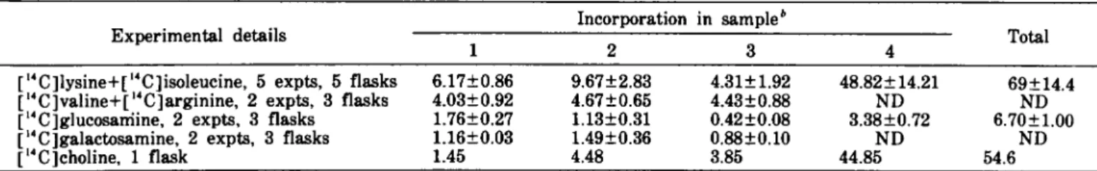
TABLE I.-Incorporation of 14C-labeled products into nondialyzable material"
Experimental details
1 [1'C]lysine+C'C]isoleucine, 5 expts, 5 flasks 6.17±O.86 C4C]valine+[ 14 C]arginine, 2 expts, 3 flasks 4.03±O.92 [14C]glucosarriine, 2 expts, 3 flasks 1.76±O.27 [14 C ]galactosamine, 2 expts, 3 flasks 1.16±O.O3
C4C]choline, 1 flask 1.45 Incorporation in sampleb 2 3 9.67±2.83 4.31±1.92 4.67±O.65 4.43±O.88 1.13±O.31 O.42±O.O8 1.49±O.36 O.88±O.lO 4.48 3.85 4 48.82±14.21 ND 3.38±O.72 ND 44.85 Total 69±14.4 ND 6.70±1.00 ND 54.6
" The incorporation is expressed as the percentage of the initial radioactivity. The percentage is a mean of several experiments±sD. ND=not done.
680 Hurllmann and Dayal
nondialyzable material in amounts varying according to the product used and to the period of culture (table 1). Precipitable by trichloroacetic acid was 84%±3.6% of the radioactivity of the nondialyzable material.
With lysine and isoleucine used as 14C-labeled prod-ucts, no significant difference in incorporation was seen between the BT 20 cells cultured in the radio-labeled medium with or without concanavalin A, phytohemagglutinin, pokeweed mitogen, or ovine pro-lactin.
Analysis of the culture fluids by autoradiography of electrophoresis in agarose revealed various bands, de-pending on the labeled products used and on the sampling time (fig. 1). Four bands were more anodal than albumin. Autoradiographs of SDS-polyacrylamide gel electrophoresis showed 14-19 bands varying from products that did not penetrate the gel [0 products having a molecular weight less than 12,300 daltons (cytochrome c). Several spots were common to the 4 samples; others were present in 1 or 2 samples only (fig. 2). These observations were particularly outstand-ing in the profiles of the graphs on radioactivity counts (text-fig. I). Two peaks corresponding to a molecular weight of 55,000 and 28,000 daltons were present in all 4 samples. Two peaks with molecular weights of 17,000 and 9,000 daltons were found in the first 3 samples, a peak of 85,000 daltons was found in all samples with the exception of sample 3, and a peak of 115,000 daltons was found in samples I and 2.
Characterization of Products
The products synthesized by BT 20 cells were ana-lyzed by autoradiography of double diffusion and immunoelectrophoresis slides. These products could be classified as serum proteins, milk proteins, membrane-associated proteins, and fetal proteins according to the antisera and carriers used.
Serum Proteins
In autoradiographs with normal human serum as carrier and with a swine antiserum polyvalent against serum proteins, 3-5 labeled lines were seen in culture fluid samples I and 2 from cells cultured with [14C]ly-sine and [14C]isoleucine. These lines had an 0/- and
p-electrophoretic mobility in agar. No lines were visible in culture fluid samples 3 and 4. When the culture fluids were analyzed with the same antiserum but without carrier, none had any visible labeled line.
For further characterization of these labeled lines, the culture fluids were analyzed with normal human serum as carrier against all the antisera specific for serum proteins mentioned in "Materials and Methods." Only culture fluid samples I and 2 gave labeled precipitation lines against 3 of the antisera used, namely, anti-p2-glycoprotein I, anti-0/2HS-anti-p2-glycoprotein, and anti-O/r lipoprotein (fig. 3c). No lines were visible in culture fluids from cells cultured with choline or glucosamine. No change in the above results was seen in culture fluids from cells cultured in the same medium
supple-J
NATL CANCER INSTmented with concanavalin A, phytohemagglutinin, pokeweed mitogen, and prolactin.
Milk Proteins
No evidence was seen of labeled lines corresponding to free secretory component, lysozyme. casein. or lact-albumin. With rare culture fluids. the immunoprecipi-tation line corresponding to lactoferrin was labeled. However. this line was not visible when the culture fluid was used alone. Autoradiographs of electrophore-sis in agarose and immunoelectrophoreelectrophore-sis of culture fluids did not reveal lactoferrin.
Membrane-Associated Proteins
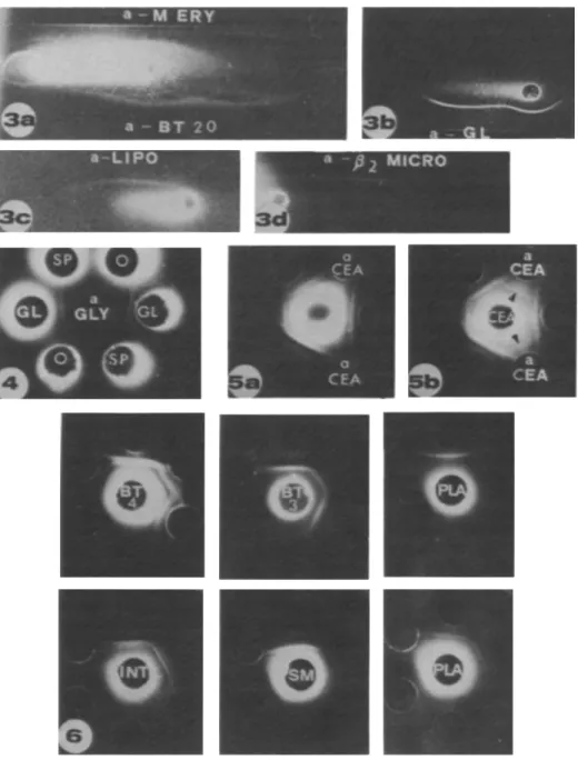
Three to 5 labeled lines were revealed in all 4 culture fluids with an antiserum specific for erythrocyte mem-brane (fig. 3a). A labeled line corresponding to glyco-phorin was seen only when a carrier was used with culture fluids from cells cultured with 14C-Iabeled amino acids. No labeled line corresponded to spectrin (fig. 4). However. with spectrin. glycophorin. or actin as carriers and antisera against spectrin and glyco-phorin. a labeled line not superposable on the precipi-tation line of the carrier was seen. With the system actin-antiactin. a labeled line at the limit of visibility corresponded to the precipitation line of the carriel:. Whether the actin-Iabeled line was identical to that revealed by antispectrin and antiglycophorin sera could not be determined by double diJfusion in agar.
A labled line corresponding to Prmicroglobulin was seen with or without the use of a P2-microglobulin carrier (fig. 3d). This labeled line was visible in samples I and 2 from cells cultured in a medium with [14C]lysine plus [i4C]isoleucine or [i4C]valine plus [14C]-arginine but not from cells cultured in a medium with [14C]glucosamine. [14C]galactosamine. or [14C]choline. In some of the samples I and 2 analyzed without carrier. a second line. parallel to the P2-microglobulin line and closer to the antigen well. was seen. This second labeled line was present in culture fluids from cells cultured in a medium with [14C]lysine. [14C]galac-tosamine, or in some cases with [14C]choline.
Fetal Proteins
A labeled line corresponding to CEA was seen only when CEA was used as carrier (figs. 5a. 5b). Synthesis of O/l-fetoprotein. anodal isoferritin. or BOF A was not detectable.
In all culture fluids from BT 20 cells cultured with 14C-Iabeled amino acids and analyzed without a carrier. 1-3 radiolabeled lines were seen with 6 rabbit antisera against BT 20 cells. These lines corresponded to proteins that a~e also synthesized by human adult and fetal tissues (fig. 6). They were detected in all culture fluids from 5 placentas and from 17 fetal tissues originating from 6 different fetuses. in 9 culture fluids from 10 dysplastic breast tissues. and in 6 culture fluids from 11 breast carcinomas. Double diffusion in agar
~~ 0 0 ~ ~~ ID ID
~
E
2000 V VV Vv
~:l
~ ~ 0 0 1500 ~~~
~...
~ ID 0 t; cL V VV V V ..; ~ :> ~ 1000 >- 0 ~ ~ ~ et: 0 0 et 500 500 et: 10 20 30'"
10 20 30'"
FRACTION NUMBER ~ ~ 0 0 15001
5000 et ell~m
!!...
>-~ >-~ ID 0 U 0 0 v vv v v et !!~ ~ ~~
ell ID OlD :E V VV v v :E cL ~ 4000 ";1000 ~ !:: >-> 2: t; ~ et 0 0 0 0 :500:3CXXl
10 20 30 40 10 20 30 40 FRACTION NUMBERTEXT.FIGURl. I.-Curves of the radioactivity counts of SDS poly-acrylamide gel electrophoresis. Ten microliters of concentrated cul-ture fluid from BT 20 cells culcul-tured with [14C]lysine and p4C]iso-leucine was applied per geL The positions of the markers are indi-cated. Slices were cut to 2 mm in length. Top left: sample I; top right: sample 2; bottom left: sample 3; bottom right: sample 4. OV A=ovalbumin; BSA=bovine serum albumin; CYT=cytochrome c; C.P.M.=counts per min.
revealed a complete identity between these lines and the lines of culture fluids from BT 20 cells. The absorption of the anti-BT 20 antisera by FCS and double diffusion in the presence of FCS did not change the results. No labeled lines were visible in culture fluids from BT 20 cells cultured in a medium with [14C]choline, [14C]glucosamine, or [14C]galactosamine.
DISCUSSION
BT 20 cells incorporate various labeled products into nondialyzable materials that are released in culture over a 9-day period. The difference between the radio-activity in the nondialyzable materials and that in the precipitate obtained by trichloroacetic acid is probably
Synthesis of Antigens by BT 20 Cells 681
due to the presence of small peptides, Human breast cancer cells have been reported to have concanavalin A receptors (36). We have, therefore, tried to stimulate the BT 20 cells with concanavalin A and other lectins. These attempts, as well as attempted stimulation with prolactin, were unsuccessful.
Several of the synthesized products have been identi-fied. Of the 5 serum proteins, 3 have been characterized as ~rglycoprotein I, a2HS-glycoprotein, and arlipo-protein, We previously showed that the labeling of these precipitation lines is not due to a nonspecific fixation of radiolabeled material to the immune precip-itate (34). These serum proteins are also synthesized by dysplastic and neoplastic human mammary tissues (34), and some of them probably correspond to glycopro-teins whose concentrations have been found elevated in the sera of patients with breast carcinoma (37, 38). Although these serum proteins have been found in high concentrations in various diseases and are thought to be produced by the liver (39), their synthesis by BT 20 cells necessitates a reconsideration of the role of the breast carcinoma itself in their production.
Milk proteins were not detected in any of the culture fluids from the different labeled media used. We did not take into account the rare labeling of lactoferrin, inasmuch as we did not find any synthesized products able to fix 59Fe.
Absence of synthesis of milk proteins could be due to several factors. Cells for the in vitro synthesis of these proteins possibly require a complex culture medium as noted by Flaxman (40) that does not correspond to our conditions of culture. Also, BT 20 cells may have lost some specific characteristics of differentiated mammary epithelium that have been kept in another cell line, MCF-7 (41). However, most probably the original carcinoma cells were unable to synthesize milk pro-teins. This had been shown to be so for many breast carcinomas. Casein was found by immunofluorescence in cells of 5 of 20 infiltrating ductal carcinomas of no special type (34). Monaco et al. (42) reported that only 8 of 47 breast cancer tumors were positive for casein and that none of the 7 breast cancer cells tested synthesized casein in vitro. Kleinberg (43) also showed that only 4 of 19 breast cancer tissues cultured in vitro synthesized lactalbumin. This absence of synthesis of milk proteins has also been demonstrated in cultures of rat and mouse mammary carcinoma cells (44). To-gether, these results suggest that most breast carcinoma cells in vitro and in vivo have lost their potential to synthesize milk proteins and that milk proteins are poor markers for most breast carcinomas.
Several proteins present on the erythrocyte mem-brane are synthesized by the BT 20 cells, One of them is glycophorin, the major protein of the erythrocyte membrane described by Marchesi et al. (45). With an antiserum specific for erythrocyte membrane, the radio-labeled lines are visible without the use of a carrier; however, the line revealed by an antiserum specific for glycophorin is labeled only when glycophorin is used as carrier. In contrast to the other carriers used in this study, glycophorin precipitated by its specific
anti-682 Hurllmann and Dayal
serum captures unspecifically various labeled products. However, in these cases the radioactive line is just visible. The strong radiolabeled line of glycophorin with BT 20 culture fluids is in favor of a specific labeling. The necessity of using a carrier for the demonstration of glycophorin is perhaps due to the fact that the BT 20 cells synthesize only a part of the molecule of glycophorin as it exists in red blood cells. This molecule having only some of the antigenic determinants of glycophorin is probably not specific to the erythrocyte membrane but could be present on various other cells. This would further explain the immunologic cross-reaction between glycophorin and ai, a human cell-surface antigen (46).
The in vitro synthesis of actin confirms the results obtained by immunofluorescence, which revealed the presence of actin in cells from several carcinomas and particularly from breast carcinomas (47, 48). The ab-sence of spectrin synthesis is in accord with the results obtained by Hiller and Weber (49) with cultures of HeLa cells and fibroblasts. The samples of spectrin and glycophorin used are probably contaminated with small amounts of actin, and antisera specific for spectrin and one antiserum specific for glycophorin also reveal the antigenic determinants of actin. This could explain the fine labeled line obtained with these antisera when actin, spectrin, or glycophorin is used as carrier inasmuch as the labeled line is not superposable to the precipitation line. However, this hypothesis could not be proved by double diffusion in agar with the actin-antiactin system due to a labeling at the limit of visibility and the difficulty of having an adequate concentration of all the antisera and carriers on the same slide.
BT 20 cells synthesize ,B2-microglobulin as do many other tumor cell lines (50). However, the detection of a second labeled line with an antiserum specific for
,B2-microglobulin warrants further study. The position of this line near the antigen well and its curvature suggest that the line corresponds to a product heavier than
,Br
microglobulin. Whereas the usual line of ,B2-micro-globulin is labeled only with 14C-Iabeled amino acids, the supplementary line is labeled also with (14C]glu-cosamine. These two observations taken together sug-gest that the supplementary line corresponds to,Br
microglobulin associated with a glycoprotein which could be a transplantation antigen or a tumor-specific antigen. Thomson et al. (16) reported that antigenic tumor material from human breast cancer was associ-ated with ,Brmicroglobulin, and Nakamuro et al. (51) demonstr<¥ed ,Brmicroglobulin in an unbound state and bound to HLA in several human cell lines.The BT 20 cells synthesize 4 fetal proteins, one of which is CEA. However, it is not possible to specify whether this protein is the CEA originally described by Gold and Freedman (52) or a breast carcinoma mem-brane-associated antigen that shares antigenic deter-minants with CEA as reported by Kuo et al. (11). Three other fetal proteins are detected by the anti-BT 20 cell antisera. These antisera detect only these proteins and no proteins specific to the BT 20 cells. Some or all of
J
NATL CANCER INSTthese proteins could correspond to products synthesized by mycoplasma that contaminated some cultures. How-ever, this does not seem to be the case inasmuch as the anti-BT 20 antisera show the same radiolabeled lines when tested with either the contaminated culture fluids or the mycoplasma-free culture fluids. Moreover, the same labeled lines are obtained with the use of antisera against BT 20 cells with culture fluids from various human tissues cultured for 48 or 72 hours only, in which there is little probability of a mycoplasma contamination.
We have not evaluated the relationship between the 3 other fetal proteins and most of the numerous fetal proteins described to date (32, 53-59). However, we can prove that these fetoproteins are neither al-fetoprotein, BOF A, or ferritin nor components due to cultivation in
medium containing FCS (60). The cultures were main-tained for 9 days without FCS, with two changes of medium, and desorption of FCS occurs at 3 days (61). Also, the antisera specific for BT 20 cells, which reveal these fetal proteins, do not precipitate any constituents of FCS. Moreover, both absorption of the anti-BT 20 cells antisera by FCS and double diffusion in the presence of FCS did not change the results.
Both BT 20 cells as well as neoplastic and dysplastic human mammary tissues synthesize these fetal antigens in relatively large amounts, as demonstrated by the intensity of the radiolabeled lines. These proteins can be classified in the group of oncofetal-associated anti-gens described by Alexander (62). The synthesis of these oncofetal-associated antigens could be due to a derepression caused either by tumor transformation of the cells or by the tissue culture conditions, inasmuch as Parker et al. (63) have demonstrated that normal tissues can express new fetal antigens in vitro. Thus the in vivo importance of these oncofetal antigens remains to be determined.
The results obtained are promising in that many products which are probably important as character-istics of the breast carcinoma cells are synthesized as soluble antigens in vitro by BT 20 cells. Isolation and purification of these products are in progress in our laboratory; the preparation of antisera against these products could lead to their identification in various tissues.
REFERENCES
(1) Esn:s NC. MORSE PA. Hl1MPHREY LJ: Antibody studies of sera from patients with breast cancer. Surg Forum 25: 121-123. 1974
(2) GORSKY Y. VANKY F. SOUTZEANIT D: Isolation from patients with breast cancer of antibodies specific for antigens associ-ated with breast cancer and other malignant diseases. Proc Natl Acad Sci USA 73:2101-2105. 1976
(3) PRIORI ES. SEMAN G. DMOCHOWSKI HS. et al: Immunofluores-ence studies on sera of patients with breast carcinoma. Can-cer 28:1462-1471. 1971
(4) SINKOVICS JG. REEVES WJ. CABINESS JR: Cell- and antibody-mediated immune reactions of patients to cuhured cells of breast carcinoma. J Natl Cancer Inst 48:1145-1147. 1972 (5) DELI.A PORTA G. CANEVARI S. FOSSATI G: Immune responses
to tumour and embryo cells in patients with mammary carci-noma. Br J Cancer 28 (suppl 1):103-107. 1973
(6) BALDWIN RW. EMBLETON MJ. JONES JS. el al: Cell-mediated and humoral immune reactions to human tumours. Int J Cancer 12:73-83. 1973
(7) FOSSATI G. CANEVARI S. DELLA PORTA G. et al: Cellular
im-munity 10 human breast carcinoma. Int J Cancer 10:391-396.
1972
(8) COCHRAN AJ. SPILG WG. MACKIE RM. et al: Postoperalive de-pression of tumour-directed cell-medialed immunity in pa-lients with malignant disease. Br Med J 4:67-70. 1972 (9) McCoy JL. JEROME LF. ANDERSON C. el al: Leukocyte
migra-lion inhibimigra-lion by soluble extracls of MCF-7 tissue culture cell line derived from breast carcinoma. J Natl Cancer Insl 57: 1045-1049. 1976
(10) McCoy JL. JEROME LF. DEAN JH. et al: Inhibition of
leuko-cyte migralion by lumor-associated antigens in soluble ex-IraCls of human breasl carcinoma. J Natl Cancer Insl 53:11-17. 1974
(11) Kuo T. RosAI J. TILLACK TW: Immunological studies of mem-brane glycoproleins isolated from human breast carcinomas. Int J Cancer 12:532-542. 1973
(12) TAYLOR G. ODlLl JL: Tumour specific T-like antigen of hu-man breast carcinoma. Br J Cancer 24:447-453. 1970 (13) GENTILE JM. FLlCKINGER JT: Isolalion of a tumor-specific
anti-gen from adenocarcinoma of the breast. Surg Gynecol Obstet 135:69-73. 1972
(14) lACHRAU RE. BLACK MM. DION AS. et al: Prognostically sig-nificant protein components of human breast cancer tissues. Cancer Res 36:3143-3146. 1976
(15) BARTORELLI A. ACClNNI R: Immunological and radioimmuno-logical sludies of membrane antigen(s) from human breast carcinomas and non-Iumoral breast tissues. I. Experientia 33: 85-87. 1977
(16) THOMSON DM. GOLD P. FREEDMAN SO. et al: The isolation and charaClerizalion of lumor-specific antigens of rodent and human lumors. Cancer Res 36:3518-3525. 1976
(17) POSKrrr PK. POSKITf TR. WALLACE JH: Release in culture medium of membrane-associaled. tumor-specific antigen by B-16 melanoma cells. Proc Soc Exp Bioi Med 152:76-80. 1976 (18) BYSTRYN JC: Release of lumor-associated antigens by murine
melanoma cells. J Immunol 116:1302-1310. 1976
(19) HICKOK DF. MILLER L: Evidence for tumor-specific antigens in filtered lissue culture media from human cancer cells. In Vitro 10:157-166. 1974
(20) SINGH I. TSANG KY. BLAKEMORE WS: Isolation and panial purificalion of plasma membrane-associaled antigens from human OSleosarcoma (TE-85) cells in tissue culture. Cancer Res 36: 4130-4136. 1976
(21) GRIM M EA. SILVER HK. ROTH JA. et al: Delection' of tumor-associated antigen in human melanoma cell line supernatants. Int J Cancer 17:559-564. 1976
(22) STUHLMILLER GM. SElGLER HF: Enzymatic susceptibililY and spontaneous release of human melanoma tumor-associated antigens. J Natl Cancer Ins!. 58:215-221. 1977
(23) LASFARGUES EY. OZZELLO L: Cultivation of human breast car-cinomas. J Natl Cancer Inst 21:1131-1147. 1958
(24) OZZELLO L. SoRDAT B. MERENDA C. el al: Transplantalion of a human mammary carcinoma cell line (BT 20) into nude mice. J NaIl Cancer Insl 52:1669-1672. 1974
(25) BRANDTZAEG P: Human secretory component. I. Purification of
free secretory component from colostrum. Scand J Immunol 3:579-588. 1974
(26) QUERINJEAN P. MAssoN PLo HEREMANS JF: Molecular weighl. single-chain structure and aminoacid composition of human lacloferrin. Eur J Biochem 20:420-425. 1971
(27) KAPLANAS RI. ANTANARICHYNS AI: The isolation and purifica-lion of a-Iaclalbumin. a component of lactose synthetase. Bio-chemislry 40:493-495. 1975
(28) NAGASAWA T. RYOKI T. KIYOSAWA I. et al: Sludies on human
casein. I. FraClionation of human casein by dielhylaminoelhyl
cellulose column chromatography. Arch Biochem Biophys 121:502-507. 1967
(29) TIl.LACK TW. SCOTf RE. MARCHESI VT: The struClure of erYlhrocyte membrane sludied by freeze-etching. 11. Localiza-lion of receplors for PHA and influenza virus 10 Ihe
intra-Synthesis of Antigens by BT 20 Cells 683
membranous particles. J Exp Med 135:1209-1227. 1972
(30) BRON C. GALLAGHER G: Immunochemical studies of human red cell membrane proteins excreted in normal urine. Immu· nochemistry 11:409-415. 1974
(31) BERTHOUD D. BRON C: Antigenicity of human glycophorin constituents. Experientia 32:765. 1976
(32) FRITSCHE R. MACH J: Identification of a new oncofoetal anti-gen associated wilh several types of human carcinomas. Na-ture 258:734-737. 1975
(33) CHAPONNIER C. KOHUR L. GABBIANI G: Fixation of human anti-actin autoantibodies on skeletal muscle fibres. Clin Exp Immunol 27:278-284. 1977
(34) HURLlMANN J. L1CHAA M. OZZELLO L: In vitro synthesis of im-munoglobulins and other proteins by dysplastic and neo-plastic human mammary tissues. Cancer Res 36:1284-1292. 1976
(35) SCHEJDEGGER
11:
Une micromethode deJ'immunoelectro-phorese. Int Arch Allergy Appl Immunol 7:103-110. 1955 (36) MATOSKA J. SIRACKY J: Concanavalin A receptors on the
sur-faces of human breast cancer cells in organ culture. J Natl Cancer Inst 58:21-28. 1977
(37) MACBETH RA. BEKESI JG: Plasma glycoproteins in various dis-ease states incl uding carcinoma. Cancer Res 22: 1170-1176. 1962
(38) ROSATO FE. SELTZER MH: Serum protein-bound fucose and carcinoma of the female breast. Am J Surg 118:61-64. 1969 (39) ROBERTS JG. KEYSER JW. BAUM M: Serum a-I-acid
glycopro-tein as an index of dissemination in breast cancer. Br J Surg 62:816-819. 1975
(40) FLAXMAN A: In vitro studies of the normal mammary gland. J Invest Dermalol 63:48-57. 1974
(41) ROSE HN. MCGRATH CM: a-Lactalbumin production in hu-man mammary carcinoma. Science 190:673-675. 1975 (42) MONACO ME. BROUZERT DA. TORMEY DC. et al: Casein
pro-duction by human breast cancer. Cancer Res 37:749-754. 1977 (43) KLElNBERG DL: Human a-lactalbumin: Measurement in serum and in breast cancer organ cultures by radio immunoassay. Science 190:276-278. 1975
(44) TURKINGTON RW. RIDDLE M: Expression of differentiated func-tion by mammary carcinoma cells in vilro. Cancer Res 30: 127-132. 1970
(45) MARCHESI VT. TILLACK TW. JACKSON RL. el al: Chemical characterization and surface orientation of the major glyco-protein of the human erythrocyte membrane. Proc Natl Acad Sci USA 69: 1445-1449. 1972
(46) MOORE EE. JONES C, PUCK TT: Cell surface antigens. IV. Im-munological correspondence between glycophorin and the a. human cell surface antigen. Cylogenet Cell Genel 17:89-97. 1976
(47) GABBIANI G. TRENCHER P. HOLBOROW EJ: Increase of contrac-tile proteins in human cancer cells. Lancet 2:796-797. 1975 (48) GABBIANI G, CsANK·BRASSERT J. SCHNEEBERGER JC. et al:
Con-tractile proleins in human cancer cells. Am J Pathol 83:457-474. 1976
(49) HILLER G, WEBER K: Spectrin is absent in various tissue culture cells. Nature 266:181-183. 1977
(50) NILSSON K. EVRIN PE. BERGGARD I. el al: Involvement of
lymphoid and non-lymphoid cells in Ihe production of
/l2-microglobulin-a homologue of Ihe con slant domains of IgG. Nature [New Bioi] 244:44-45. 1973
(51) NAKAMURO K. TANIGAKI N, PRESSMAN D: Common antigenic structures of HLA antigens. VII. Seleclive combinalion bind-ing of /lrmicroglobulin with HLA large component in cul-lured human cell lines. Immunology 32:139-150. 1977 (52) GOLD P. FREEDMAN SO: Demonstration of tumor-specific
anti-gens in human colonic carcinomata by immunological and absorption lechniques. J Exp Med 121:439-459. 1965 (53) EDYNACK EM. LARDlS MP. VKANA M: Antigenic changes in
human breasl neoplasia. Cancer 28:1457-1461. 1971 (54) EDYNACK EM. OLD LJ. VRANA M, et al: A fetal antigen
associ-ated with human neoplasia. N Engl J Med 286:1178-1183. 1972
(55) HAKKINEN I, VIIKARI S: Occurrence of fetal sulphoglycoprotein antigen in the gaslric juice of palienls with gastric diseases.
684 Hurlimann and Dayal
Ann Surg 169:277-281. 1969
(56) KLAVINS JV. MESA-TEJADA R. WEISS M: Human carcinoma antigens cross reacting with anti-embryonic antibodies_ Na-ture [New Bioi] 234:153-154. 1971
(57) TAKAHASHI A. YACHI A. ANZAI T. et al: Presence of a unique serum protein in sera obtained from patients with neoplastic diseases and in embryonic and neonatal sera. Clin Chim Acta 17:5-12. 1967
(58) HARRIS R. VIZA D. TODD R. et al: Detection of human leukae-mia associated antigens in leukaemic serum and normal em-bryos. Nature 233:556-557. 1971
(59) TAT,\RINOV YS. KALASHNIKOV VV: New human embryonic anti-gen and its presence in some adenocarcinomas. Nature 265:
J
NATL CANCER INST638-639. 1977
(60) IRIE RI". IRIE K. MORTON DL: Natural antibody in human serum ID a neoantigen in human cultured cells grown in fetal bovine serum. J Natl Cancer Inst 52:1051-1058. 1974
(61) KERBEL RS. BLAKESLEE D: Rapid adsorption of a fetal calf serum component by mammalian cells in culture. A potential source of artifacts in studies of antisera to cell-specific anti-gens. Immunology 31:881-891. 1976
(62) ALEXANDER P: Foetal "antigens" in cancer. Nature 235:137-140. 1972
(63) PARKER GA. HVATT C. ROSENBERG SA: Normal adult murine cells in tissue culture express fetal antigens. Transplantation 23:161-163. 1977
FIGIIRE l.-Autoradiographs of agarose elec-trophoresis of culture fluids from BT 20 cells cultured with [14C]lysine plus [14C]isoleucine, samples 1-4 (SI-54). At bottom. electrophore-sis of normal human serum (NHS) as refer-ence for electrophoretic mobilities.
FI(;I'RE 2.-Autoradiographs of SDS poh-acrylamide gel electrophoresis of culture fluids from BT 20 cells cultured with [14C]h-sine plus [14C]isoleucine. From top to bot-tom: samples 1-4. Markers are indicated: CYT=cytochrome c; OVA=ovalbumin; BSA=bovine serum albumin; DIM=dimer.
Synthesis of Antigens by BT 20 Cells 685
FIGURE 3.-Autoradiographs of immunoelectrophoresis of culture fluids from BT 20 cells. a) No carrier. antierythrocyte membrane (a-M ERY) and anti-BT 20 cell serum (a-BT 20). b) Glycophorin as carrier and antiglycophorin serum (a-GL). c) Human serum as carrier and anti-a2-lipoprotein serum (a-LIPO). d) p2-microglobulin as carrier and anti-P2-microglobulin serum (a-p2 MICRO). Anode is on left.
FIGI'RE 4.-Autoradiograph of double diffusion in agar. In an/er well. antiserum specific for glycophorin (a-GLY); in peripheral wells. cul-ture fluids from 2 different culcul-tures of BT 20 cells alone (0) or with glycophorin (GL) or spectrin (SP) as carriers.
FIGURE 5.-Autoradiographs of double diffusion in agar. In anler well. culture fluid from BT 20 cells alone (fig. 5a) or with CEA as carrier (fig. 5b). In peripheral wells. anti-BT 20 cell sera and anti-CEA sera (a CEA). Labeled line of CEA is visible with CEA as carrier (arrowheads).
FIGURE 6.-Autoradiographs of double diffusion in agar. Peripheral wells contain 3 different anti-BT 20 cell sera. Cenler well contains samples 3 and 4 from BT 20 cells (BT 3 and BT 4). from 2 different placentas (PLA). and from fetal intestine (INT) and fetal submandibular gland (SM).