2007/77
Does a specific MR imaging protocol with a supine-lying
subject replicate tarsal kinematics seen during upright
standing?
aBildet ein spezifisches MR-Verfahren mit ru¨cklings liegendem Probanden
die tarsale Kinematik unter stehenden Bedingungen nach?
Peter Wolf1,*, Alex Stacoff1, Anmin Liu2,
Anton Arndt3,4, Chris Nester2, Arne Lundberg3
and Edgar Stuessi1
1Institute for Biomechanics, ETH Zurich, Zurich,
Switzerland
2Centre for Rehabilitation and Human Performance
Research, University of Salford, Salford, UK
3Karolinska Institute, Stockholm, Sweden
4The Swedish School of Sport and Health Sciences,
Stockholm, Sweden
Abstract
Magnetic resonance (MR) imaging is becoming increas-ingly important in the study of foot biomechanics. Spe-cific devices have been constructed to load and position the foot while the subject is lying supine in the scanner. The present study examines the efficacy of such a newly developed device in replicating tarsal kinematics seen during the more commonly studied standing loading con-ditions. The results showed that although knee flexion and the externally applied load were carefully controlled, subtalar and talo-navicular joint rotations while lying dur-ing MR imagdur-ing and when standdur-ing (measured opto-electrically with markers attached to intracortical pins) did not match, nor were they systematically shifted. Thus, the proposed MR protocol cannot replicate tarsal kine-matics seen during upright standing. It is concluded that specific foot loading conditions have to be considered when tarsal kinematics are evaluated. Improved replica-tion of tarsal kinematics in different postures should comprehensively consider muscle activity, a fixed hip position, and a well-defined point of load application.
Keywords: intracortical pins; magnetic resonance
imag-ing; subtalar joint kinematics; talo-navicular joint kine-matics; tarsal bones.
Zusammenfassung
Die Magnetresonanz- (MR) Tomographie gewinnt in der Fußbiomechanik immer mehr an Bedeutung. Um den Fuß
aThe first author received a conference award for his poster
enti-tled Transmission within the tarsal gearbox at the Combined Annual Meeting of DGBMT, O¨ GBMT and SGBT (Zurich, Swit-zerland, 2006). The methods section of this poster was based on the non-invasive approach presented in this paper. *Corresponding author: Dr. Peter Wolf, ETH Ho¨nggerberg HCI
E451, 8093 Zurich, Switzerland Phone: q41-44-6336186 Fax: q41-44-6331124 E-mail: pwolf@ethz.ch
positionieren und belasten zu ko¨nnen, wa¨hrend der Pro-band ru¨cklings im Tomographen liegt, wurden spezifische Aufbauten konstruiert. Die vorliegende Studie pru¨ft die Effektivita¨t eines derartigen, neu entwickelten Aufbaus hinsichtlich der Imitation der tarsalen Kinematik, die sich unter den u¨blicherweise untersuchten stehenden Bedin-gungen ergibt. Die Ergebnisse zeigten, dass trotz sorg-fa¨ltiger Kontrolle der Knieflexion und der a¨ußeren Last die Rotationen des unteren Sprunggelenks sowie talo-navi-kularen Gelenks wa¨hrend dem Liegen im MR-Tomo-graphen nicht mit denen wa¨hrend des Stehens (opto-elektrisch gemessen anhand von im Knochen fixierten Dra¨hten) u¨bereinstimmen, wobei die Ergebnisse auch nicht systematisch verschoben sind. Das vorgeschlage-ne MR-Verfahren ist daher nicht in der Lage, die tarsale Kinematik wa¨hrend des Stehens abzubilden. Folglich sind bei Betrachtungen der tarsalen Kinematik die spezifischen Belastungen des Fußes zu bedenken. Eine verbesserte Imitation der tarsalen Kinematik in verschie-denen Ko¨rperhaltungen sollte sorgfa¨ltig die Aktivita¨t der Muskulatur, eine fixierte Hu¨fte sowie einen exakt defi-nierten Kraftangriffspunkt beru¨cksichtigen.
Schlu¨sselwo¨rter: Kinematik des talo-navikularen Gelenks; Kinematik des unteren Sprunggelenks; Kno-chenschrauben; Magnetresonanztomographie; tarsale Knochen.
Introduction
In the past, kinematics of the tarsal bones (calcaneus, cuboid, navicular and talus) have been examined by either two- and three-dimensional X-ray stereophoto-grammetry w3, 8–10, 23x or by opto-electrical registration of markers on intracortical pins w1, 13, 14, 18–20x. How-ever, these methods are invasive or ionising and cannot be used routinely in living subjects. The alternative approach, using skin mounted markers, is also problem-atic because the talus is inaccessible, and the cuboid and navicular are too small to mount the three required markers. Furthermore, motion of the bones relative to the skin limits the validity of kinematic data derived from skin-mounted markers w11, 22, 24x.
Magnetic resonance (MR) imaging overcomes these methodological limitations, provides equally accurate motion data w25x, and is becoming increasingly popular for investigation of tarsal kinematics w12, 15, 17, 21x. However, MR imaging requires that the subject is supine, whereas it is desirable to study the foot under conditions
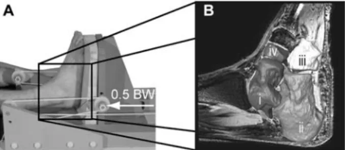
Figure 1 (A) Foot positioning and loading device of the MR imaging procedure. A load of half bodyweight was applied axi-ally under the heel. (B) MR image with 3D reconstructed tarsal bones: (i) talus, (ii) calcaneus, (iii) cuboid, and (iv) navicular.
of standing or walking. In this context, an MR imaging procedure was developed to enable spatial foot positions and loading to be controlled whilst supine in the MR scanner w26x. The MR imaging protocol involves loading the plantar surface of the foot using a horizontal loading device while the subject is supine in the MR scanner. This allows investigation of tarsal kinematics under near-bodyweight loading conditions while the foot is pronated and supinated using wedged platforms. The open ques-tion is whether or not this MR imaging protocol ade-quately matches a standing foot loading condition. Thus, the aim of this study was to evaluate the new MR pro-tocol by comparing tarsal kinematics during foot prona-tion and supinaprona-tion measured using (1) the MR imaging protocol and (2) intracortical pins during standing. The hypothesis was that the tarsal kinematics from the MR protocol would match those measured during standing.
Materials and methods
Subjects
The study was conducted on three male volunteers with-out signs of musculoskeletal diseases aged 28, 33, and 55 years, 175, 180, and 182 cm high, and weighing 71, 75, and 80 kg, respectively. Informed written consent in accordance with the guidelines of the local research Ethics Committee was obtained from all subjects.
MR procedure
Subjects lay on the MR table and their right foot was fixed into the foot-loading and -positioning device w25, 26x. A load of 0.5=body weight was applied to the board under the right foot, simulating relaxed standing. Three different wooden blocks were placed under the foot to control foot position: a flat block (neutral), a 158 wedged ‘‘pronating’’ block (10.88 eversion, 3.38 dorsiflexion, 9.88 abduction), and a 158 wedged ‘‘supinating’’ block (10.88 inversion, 3.38 plantarflexion, 9.88 adduction), as shown in Figure 1A. The subject’s foot was aligned on the blocks according to the longitudinal axis of the foot defined by the second toe and the most posterior aspect of the heel. The extent of the pronation and the ratio of frontal to transverse to sagittal plane rotation was based on: (i) the commonly reported 108 of calcaneal eversion during the initial stance phase of running w6, 19x; and (ii) on an
approximated subtalar axis with an orientation of 418 relative to the transverse plane and 178 to the sagittal plane w7, 16x.
Imaging was performed on a 3-T whole-body MR unit (Intera 3T, Philips Medical Systems, Eindhoven, The Netherlands) with two synergy coil elements (Sense Flex M, Philips Medical Systems). A 3D T1-weighted gradient echo sequence with the following parameters was used: repetition time, 16 ms; echo time, 4 ms; flip angle, 118; field of view, 200 mm; acquisition matrix, 288=273; Fourier interpolation, 512=512 pixels; and 1.4-mm-thick over-continuous slices with 50% slice overlapping. Thus, the resolution of the reconstructed images was 0.39=0.39=0.7 mm3(Figure 1B). For each subject and
test condition, 130 sagittal slices were acquired during approximately 9 min.
3D reconstruction of the tarsal bones (Figure 1B) was performed semi-automatically by one operator using AMIRA software (v.3.5, Konrad-Zuse Zentrum fu¨r Infor-mationstechnik Berlin, Germany). The resulting surface points were imported into MatLab (v.7.0, MathWorks, Natick, MA, USA). An iterative closest-point algorithm w4x was used to register the surface point cloud of each bone obtained in the neutral position with those during pro-nation and supipro-nation. This algorithm can be summarised as follows. First, the matching points of a reference posi-tion (in the present study, the neutral foot posiposi-tion) and of a final position (in the present study, the two foot excursions) are computed based on the minimum dis-tance. Second, the corresponding points are registered by a least-square singular value decomposition. Third, the resulting transformation is applied to the reference point cloud. This iteration is terminated when the change in mean square error of the distances between the ref-erence point cloud and the point cloud of the final posi-tion falls below a defined threshold w4x.
The overall coordinate transformations were then used to calculate tarsal joint motion expressed as finite helical axis rotations. These were projected onto the axes of the reference bone under consideration of the helical axis ori-entation w27x. The overall error introduced by this MR data processing was less than 0.058, whereas the differ-ence for repeated joint rotations was less than 38 w26x.
Opto-electrical registration
Intracortical pins (1.6 mm in diameter) were inserted under local anaesthetic into the calcaneus, cuboid, navicular, and talus, and a reflective marker triad was attached to each (Figure 2). (Pins were inserted into other bones for other studies; one of them describing the pro-cedure in more detail has recently been published w2x.) Kinematic and kinetic data were collected using a ten-camera opto-electrical system (Qualysis, Go¨teborg, Swe-den) at 240 Hz and a force plate (Kistler, Wintherthur, Switzerland). Analysis of stance times and ground reac-tion forces with and without these pins in place revealed that gait was not remarkably modified by the presence of the pins. Coefficients of multiple correlation for tibial motion and ground reaction forces were moderate to high, and the duration of stance phases was not signifi-cantly slower during running with pins compared to run-ning without pins w2x. Thus, it was assumed that insertion
Figure 2 (A) Anterior view of standing on the pronation block. The marker triads of the intracortical pins of the talus, cuboid, and navicular are emphasised. (B) Posterior view of standing on the supination block. The calcaneal marker triad is highlighted.
Figure 3 Subtalar joint rotations in response to quasi-static foot (A) pronation and (B) supination.
Results of repeated measurements of the inserted intracortical pins are shown as boxplots, and results of MR imaging as cir-cles. Lying supine (during MR imaging) and standing upright (with intracortical pins) resulted in different rotations, since the results of the MR imaging procedure were not consistently within the lower and upper quartile of the intracortical pin measure-ments.
of the pins did not significantly affect the kinematics, par-ticularly not during the standing positions used in the present study.
Technical coordinate frames (a non-anatomical coor-dinate system directly calculable from the 3D location of the marker arrays) for each bone were determined in the global coordinate system. The neutral standing trial was matched to the conditions in the MR imaging by repro-ducing the neutral foot block position. Individual bone motion relative to the neutral bone location was deter-mined for the pronation and supination foot positions. The contralateral foot stood on the neutral block, allow-ing straight leg standallow-ing and a neutral pelvis position. Each foot excursion was repeated five times. The sub-jects descended from the blocks between the trials.
Applying the same method as used in the MR protocol, joint rotations were computed using the helical axis approach w27x. The analysis focused on the subtalar and talo-navicular joints, since the rotations between other tarsal bones were found to be small during the foot ex-cursions described (calcaneus-cuboid, 2–68, navicular-cuboid, -28 w25x).
Results
Rotations of the calcaneus relative to the talus in response to quasi-static foot pronation and supination are presented in Figure 3. Rotations determined with the MR procedure (circles) were only occasionally within the inter-quartile range of the five standing trials measured with intracortical pins, and no systematic shift was pres-ent. Similar results were found for the talo-navicular joint. No subject showed consistently comparable rotations when lying supine and when standing upright (Figure 4).
Discussion and conclusion
This study examined the feasibility of using an MR load-ing device to replicate the quasi-static tarsal kinematics seen in upright posture.
The results showed that tarsal joint rotations while lying supine in the MR scanner did not systematically match with corresponding rotations during standing (Figures 3 and 4). Differences between the median standing (opto-electrical measurement) and the lying supine (MR
imag-ing) results were of up to 108 in magnitude, which is at least twice the measurement error of both methods. Thus, although knee flexion and the externally applied load were carefully controlled during lying and standing, the motion of the tarsal joints in the MR protocol did not imitate those in an upright posture.
There is a range of reasons why the tarsal kinematics differ. Clearly, we would expect some degree of differ-ence between how the tarsal bones move during the two experiments simply because they were carried out on dif-ferent days w5x. Also, subtle changes in hip position, in combination with slightly altered points of load applica-tion (particularly during the measurements with inserted intracortical pins), would contribute to the different tarsal joint rotations. Furthermore, in contrast to standing upright, lying supine does not require activity of the fol-lowing muscles inserting at the midfoot: tibialis anterior, tibialis posterior, peroneus brevis, and peroneus longus. Activity in these muscles may have resulted in tarsal bone rotations and thus may have also contributed to the differences observed.
Figure 4 Talo-navicular rotations in response to quasi-static foot (A) pronation and (B) supination.
Results of repeated measurements of the intracortical pin mark-er triads are shown as boxplots, and results of MR imaging as circles. Similar to the subtalar joint, lying supine during MR imaging and standing upright with intracortical pins resulted in different rotations.
In conclusion, despite using a specific foot loading device, the proposed MR protocol cannot replicate tarsal kinematics seen during upright standing. Tarsal kine-matics are influenced by many factors; consequently, improved replication of tarsal kinematics in different pos-tures would require (i) additional consideration of muscle activity; (ii) external foot positioning and loading; and (iii) greater constraints on the hip position and point of load application. In general, specific foot loading conditions have to be considered when tarsal kinematics are meas-ured and compared.
Acknowledgements
The authors wish to thank the subjects participating in this study, Richard Jones for assistance during opto-electrical measure-ment of the intracortical pins and Roger Luechinger for his supervision during MR imaging. The study was financially sup-ported by the International Society of Biomechanics (Disserta-tion Grant 2005).
References
w1x Arndt A, Westblad P, Winson I, Hashimoto T, Lundberg A. 2004. Ankle and subtalar kinematics measured with intra-cortical pins during the stance phase of walking. Foot Ankle Int 2004; 25: 357–364.
w2x Arndt A, Wolf P, Liu P, et al. Intrinsic foot kinematics meas-ured in vivo during the stance phase of slow running. J Biomech 2007; doi:10.1016/j.jbiomech.2006.12.009. w3x Benink RJ. The constraint mechanism of the human
tar-sus. Acta Orthop Scand 1985; 56 (Suppl 215): 49–68. w4x Besl PJ, McKay ND. A method for registration of 3-D
shapes. IEEE Trans Pattern Anal Mach Intell 1992; 14: 239–256.
w5x Carson MC, Harrington ME, Thompson N, O’Connor JJ, Theologis TN. Kinematic analysis of a multi-segment foot model for research and clinical applications: a repeatability analysis. J Biomech 2001; 34: 1299–1307.
w6x Cornwall MW, McPoil TG. Influence of rearfoot postural alignment on rear foot motion during walking. Foot 2004; 14: 133–138.
w7x Isman RE, Inman VT. Anthropometric studies of the human foot and ankle. Bull Prosthet Res 169; 11: 97–129. w8x Lundberg A, Goldie I, Kalin B, Selvik G. Kinematics of the
ankle foot complex – plantarflexion and dorsiflexion. Foot Ankle Int 1989; 9: 194–200.
w9x Lundberg A, Svensson OK, Bylund C, Goldie I, Selvik G. Kinematics of the ankle foot complex. 2. Pronation and supination. Foot Ankle Int 1989; 9: 248–253.
w10x Lundberg A, Svensson OK, Bylund C, Selvik G. Kinematics of the ankle foot complex. 3. Influence of leg rotation. Foot Ankle Int 1989; 9: 304–309.
w11x Maslen BA, Ackland TR. Radiographic study of skin dis-placement errors in the foot and ankle during standing. Clin Biomech 1994; 9: 291–296.
w12x Mattingly B, Talwalkar V, Tylkowski C, Stevens DB, Hardy PA, Pienkowski D. Three-dimensional in vivo motion of adult hind foot bones. J Biomech 2006; 39: 726–733. w13x Reinschmidt C, van den Bogert AJ, Murphy N, Lundberg
A, Nigg BM. Tibiocalcaneal motion during running, meas-ured with external and bone markers. Clin Biomech 1997; 12: 8–16.
w14x Reinschmidt C, van den Bogert AJ, Lundberg A, et al. Tibiofemoral and tibiocalcaneal motion during walking: external vs. skeletal markers. Gait Post 1997; 6: 98–109. w15x Ringleb SI, Udupa JK, Siegler S, et al. The effect of ankle
ligament damage and surgical reconstructions on the mechanics of the ankle and subtalar joints revealed by three-dimensional stress MRI. J Orthop Res 2005; 23: 743–749.
w16x Root ML, Weed JH, Sgarlato TE, Bluth DR. Axis of motion of the subtalar joint. J Am Podiatr Med Assoc 1966; 56: 149–155.
w17x Siegler S, Udupa JK, Ringleb SI, et al. Mechanics of the ankle and subtalar joints revealed through a 3D quasi-stat-ic stress MRI technique. J Biomech 2005; 38: 567–578. w18x Stacoff A, Nigg BM, Reinschmidt C, van den Bogert AJ,
Lundberg A. Tibiocalcaneal kinematics of barefoot versus shod running. J Biomech 2000; 33: 1387–1395.
w19x Stacoff A, Nigg BM, Reinschmidt C, et al. Movement coupling at the ankle during the stance phase of running. Foot Ankle Int 2000; 21: 232–239.
w20x Stacoff A, Reinschmidt C, Nigg BM, et al. Effects of foot orthoses on skeletal motion during running. Clin Biomech 2000; 15: 54–64.
w21x Stindel E, Udupa JK, Hirsch BE, Odhner D, Couture C. 3D morphology of the rearfoot from MRI data: technical vali-dation and clinical description. Proc Soc Photo-opt Ins-trum Eng 1998; 3335: 479–487.
foot: a 2-D roentgen photogrammetry study. Clin Biomech 1998; 13: 71–76.
w23x van Langelaan EJ. A kinematical analysis of the tarsal joints. Acta Orthopaed Scand 1983; 54 (Suppl 204): 135– 265.
w24x Westblad P, Hashimoto T, Winson I, Lundberg A, Arndt A. Differences in ankle-joint complex motion during the stance phase of walking as measured by superficial and bone-anchored markers. Foot Ankle Int 2002; 23: 856– 863.
w25x Wolf P. Tarsal kinematics. PhD thesis. Zurich: ETH 2006. w26x Wolf P, Stacoff A, Luechinger R, Boesiger P, Stuessi E. An
MR imaging procedure to investigate tarsal bone mechan-ics. In: Proceedings of the 9th International Symposium on the 3D Analysis of Human Movement, University of Valen-ciennes, France 2006. http://www.univ-valenciennes.fr/ congres/3D2006/Abstracts/128-Wolf.pdf.
w27x Woltring HJ. 3-D attitude representation of human joints: a standardization proposal. J Biomech 1994; 27: 1399– 1414.


