Aminoglycosides and renal magnesium homeostasis in humans
5
0
0
Texte intégral
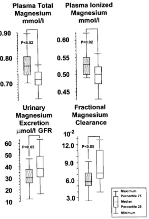
(2) Aminoglycosides and renal magnesium homeostasis in humans. three times daily, medication with pancreatic enzymes, and high-calorie diet. Before and at the end of the 14 days treatment period a 2-h urine specimen was collected after overnight fasting, sitting (more than 10 min) blood pressure (first and fifth sounds), and heart rate were measured, and mid-point blood was taken anaerobically with minimal stasis and without movement of the forearm. Haemoglobin, pH, carbon dioxide pressure, urea, albumin, and ionized calcium and magnesium were assessed in blood, and total magnesium, sodium, potassium, chloride, uric acid, phosphate, creatinine and osmolality in both blood and urine, and total calcium in urine. In addition urinalysis (glucose and protein) and the excretion of N-acetyl-b--glucosaminidase were assessed. Plasma and urine were also collected in a control group of 25 subjects (11 female and 14 males, aged between 4.1 and 19, median 12 years) with either nocturnal enuresis (n=16), dysfunctional voiding (n=5), or unstable bladder (n=4).. Data analysis All measurements were performed in duplicate. Haemoglobin (cyanhaemoglobin method ), uric acid (uricase–catalase assay), albumin (bromcresol purple method ), total calcium (cresolphthalein complexone method ), and magnesium (xylidil blue method ) [15], phosphate (ammonium molybdate method ), creatinine (kinetic alkaline picrate method ), urea (Bertheloth–urease assay) and N-acetyl-b--glucosaminidase (cresolsulphonphthaleinyl N-acetyl-b--glucosaminide assay) [16 ] were measured colorimetrically. Osmolality was assessed by freezing point osmometry, glycosylated haemoglobin A 1c by a latex immunoagglutination inhibition assay, and circulating amikacin by fluorescence polarization assay. Urinary glucose and protein were determined using commercially available dipsticks. The susceptibility of isolated Ps. aeruginosa to amikacin and ceftazidime was assessed by conventional disk diffusion techniques. Ion-selective electrodes were used for the measurement of sodium, potassium, chloride, ionized calcium, pH, carbon dioxide pressure, and ionized magnesium. Plasma bicarbonate concentration was calculated using the Henderson–Hasselbalch equation. Plasma ionized magnesium was analysed within 15 min of collection in blood drawn into silicone-free tubes (heparin 1000 U/l ) using a selective electrode, which has been recently characterized [11]. The electrode contains the neutral carrier-based membrane ETH 7025, which is incorporated in standard AVL electrode bodies by solvent casting (AVL 988–4/Mg Analyzer). The cell is provided with electrodes for sodium, calcium, pH, and magnesium together with a common reference electrode. The measuring cell is maintained at 37°C. Plasma or urinary electrolytes (P , U ) and creatinine (P , x x Cr U ) were used to calculate the fractional clearance (1) or Cr the excretion corrected for 1 litre of glomerular filtration rate (GFR) (2), using the following standard equations: U ×P x Cr (1) P ×U x Cr U ×P x Cr (2) U Cr The fractional clearance of magnesium and calcium was calculated from their plasma ionized concentrations. The maximal tubular reabsorption of phosphate was calculated from plasma (P ) or urinary ( U ) phosphate and plasma Ph Ph (P ) or urinary ( U ) creatinine as follows [17]: Cr Cr. 823. A. B. U ×P Ph Cr U Cr The urinary excretion of N-acetyl-b--glucosaminidase (unit, U/l ) was factored by creatinine (unit, mmol/l ). An estimate of aldosterone activity is the potassium concentration gradient between blood and nephron at the end of the cortical collecting tubule [18]. To assess the mentioned gradient and thereby the aldosterone activity, a non-invasive test has been designed. Consequently, the transtubular potassium concentration gradient was calculated from plasma and urinary potassium (P , U ; in mmol/l ) and osmolality (P , U ; K K osm osm in mmol/kg) by the equation [18]: P − Ph. U P K × osm U P osm K The results are expressed either as median and interquartile range or depicted as ‘box and whisker plot’ (boxes are median and interquartile ranges, vertical lines are ranges). The Wilcoxon matched-paired-signed rank test (nonparametric rank sum test for paired samples), the Wilcoxon–Mann–Whitney test (non-parametric rank sum test for two independent samples), and simple regressions with the non-parametric coefficient of correlation r were s used for analysis. A P value of <0.05 was accepted as statistically significant.. Results When studied before amikacin and ceftazidime, the group of 24 patients with cystic fibrosis slightly but significantly tended towards tachycardia, hypoalbuminaemia, hypocalcaemia, and hyperuricaemia ( Table 1). Plasma total (0.77 (0.74–0.81) vs 0.80 (0.78–0.83 mmol/l ) and ionized magnesium (0.53 (0.50–0.55) vs 0.54 (0.51–0.56) mmol/l ), the fractional magnesium clearance (0.0568 (0.0494–0.0716) vs 0.0498 (0.0394–0.0591)), and the urinary excretion of this ion (30.7 (26.5–38.0) vs 26.9 (20.1–31.0) mmol/l GFR) were not statistically different in cystic fibrosis patients and in control subjects. Plasma total (0.75 (0.72–0.78) vs 0.77 (0.75–0.81 mmol/1) and ionized (0.53 (0.50–0.55) vs 0.52 (0.51–0.55 mmol/l ) magnesium were similar in 16 cystic fibrosis patients with at least one treatment course with aminoglycosides in the past and in the eight patients without such a course. Systemic treatment with amikacin and ceftazidime, intensive physiotherapy, inhalation therapy, and highcalorie diet did not significantly ameliorate the noted tendency towards tachycardia, hypoalbuminaemia, hypocalcaemia and hyperuricaemia (Table 1). Also blood pressure, body weight, haemoglobin, plasma creatinine and urea, blood acid–base balance, and plasma and urinary sodium, potassium and phosphate were unchanged following systemic anti-pseudomonal treatment with amikacin and ceftazidime for 14 days. None of the patients had a change in plasma creatinine concentration of more than 15 mmol/l. Treatment with amikacin and ceftazidime significantly increased the urinary excretion rate of N-acetyl-b--glucosaminidase ( Table 1) and decreased plasma total magnesium (from 0.77 (0.74–0.81) to 0.73 (0.71–75), plasma ionized.
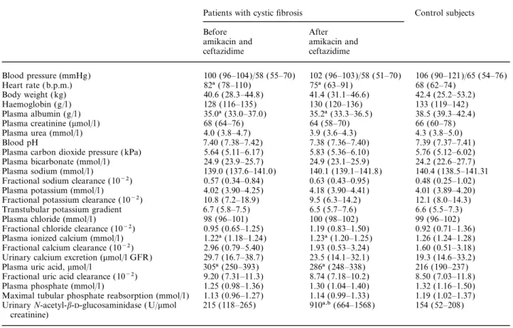
(3) 824. R. O. von Vigier et al.. Table 1. Clinical and laboratory findings in 24 patients (11 female and 13 male, aged between 9.0 and 19 years, median 14) with cystic fibrosis, before and after treatment with amikacin and ceftazidime for 14 days, and in a control group of 25 healthy subjects (11 female and 14 male, aged between 4.1 and 19 years, median 12). The results are given either as median (with interquartile range between brackets) or as relative frequency. Urinalysis failed to reveal pathological glucosuria and proteinuria in the 23 patients (both before and after amikacin and ceftazidime) and in the 25 control subjects Patients with cystic fibrosis. Blood pressure (mmHg) Heart rate (b.p.m.) Body weight (kg) Haemoglobin (g/l ) Plasma albumin (g/l ) Plasma creatinine (mmol/l ) Plasma urea (mmol/l ) Blood pH Plasma carbon dioxide pressure (kPa) Plasma bicarbonate (mmol/l ) Plasma sodium (mmol/l ) Fractional sodium clearance (10−2) Plasma potassium (mmol/l ) Fractional potassium clearance (10−2) Transtubular potassium gradient Plasma chloride (mmol/l ) Fractional chloride clearance (10−2) Plasma ionized calcium (mmol/l ) Fractional calcium clearance (10−2) Urinary calcium excretion (mmol/l GFR) Plasma uric acid, mmol/l Fractional uric acid clearance (10−2) Plasma phosphate (mmol/l ) Maximal tubular phosphate reabsorption (mmol/l ) Urinary N-acetyl-b--glucosaminidase ( U/mmol creatinine). Control subjects. Before amikacin and ceftazidime. After amikacin and ceftazidime. 100 (96–104)/58 (55–70) 82a (78–110) 40.6 (28.3–44.8) 128 (116–135) 35.0a (33.0–37.0) 68 (64–76) 4.0 (3.8–4.7) 7.40 (7.38–7.42) 5.64 (5.11–6.17) 24.9 (23.9–25.7) 139.0 (137.6–141.0) 0.57 (0.34–0.84) 4.02 (3.90–4.25) 10.8 (7.2–18.9) 6.7 (5.8–7.5) 98 (96–101) 0.95 (0.65–1.25) 1.22a (1.18–1.24) 2.96 (0.79–5.40) 29.7 (16.7–38.7) 305a (250–393) 9.20 (7.31–11.3) 1.25 (0.98–1.36) 1.13 (0.96–1.27) 215 (118–265). 102 (96–103)/58 (51–70) 75a (63–91) 41.4 (31.1–46.6) 130 (120–136) 35.2a (33.3–36.5) 64 (58–70) 3.9 (3.6–4.3) 7.38 (7.36–7.40) 5.83 (5.36–6.10) 24.9 (23.1–25.9) 140.1 (139.1–141.8) 0.63 (0.43–0.95) 4.18 (3.90–4.41) 9.5 (6.3–14.2) 6.5 (5.7–7.6) 100 (98–102) 1.19 (0.83–1.50) 1.23a (1.20–1.25) 1.93 (0.53–3.24) 23.5 (14.1–32.1) 286a (248–338) 8.74 (7.18–10.2) 1.30 (1.04–1.40) 1.14 (0.99–1.33) 910a,b (664–1568). 106 (90–121)/65 (54–76) 68 (62–74) 42.4 (25.2–53.2) 133 (119–142) 38.5 (39.3–42.4) 66 (60–78) 4.3 (3.8–5.0) 7.39 (7.37–7.41) 5.76 (5.12–6.02) 24.2 (22.6–27.7) 140.4 (138.5–141.31 0.48 (0.25–1.02) 4.01 (3.89–4.20) 12.1 (8.0–14.3) 6.6 (5.5–7.3) 99 (96–102) 0.92 (0.71–1.36) 1.26 (1.24–1.28) 1.60 (0.51–3.18) 19.3 (14.6–33.2) 216 (190–237) 8.50 (7.03–11.8) 1.32 (1.16–1.50) 1.19 (1.02–1.37) 154 (52–208). GFR, glomerular filtration; aP<0.05 vs control group; bP<0.02 vs before amikacin and ceftazidime.. magnesium (from 0.53 (0.50–0.55) to 0.50 (0.47–0.52) mmol/l ), as shown in Figure 1. The tendency towards hypomagnesaemia was associated with a significantly increased fractional magnesium clearance (from 0.0568 (0.0494–0.0716) to 0.0721 (0.0630–0.111)) and urinary magnesium excretion (from 30.7 (26.5–38.0) to 38.5 (31.5–49.0) mmol/l glomerular filtration rate (GFR)). In patients no correlation was observed between the changes in the urinary excretion of magnesium and those in the urinary excretion of N-acetyl-b--glucosaminidase.. Discussion When magnesium intake is curtailed or when there is intestinal magnesium malabsorption the normal kidney reduces magnesium excretion to very low values. When renal magnesium handling is impaired, hypomagnesaemia ensues because, unlike with calcium, equilibration with cellular stores does not occur for several weeks [9,10,19,20]. In the present study, treatment with amikacin plus ceftazidime for 14 days very often caused subtle changes in the renal magnesium homeostasis. However, the treatment was not associated with. changes in circulating creatinine and urea, a relatively common complication of aminoglycoside therapy [21]. Consequently the study demonstrates that in cystic fibrosis, systemic therapy with the aminoglycoside amikacin plus the cephalosporin ceftazidime causes mild and probably transient [1–5] hypomagnesaemia secondary to inappropriate renal magnesium wasting even in the absence of a rise in circulating creatinine and urea. In cystic fibrosis the drawbacks of hypomagnesaemia on respiratory symptoms have not been addressed. Data from the literature suggest that in humans, hypomagnesaemia may reduce respiratory muscle power and cause airflow obstruction [22]. Our findings of hypoalbuminaemia, hypocalcaemia, and hyperuricaemia in advanced cystic fibrosis before and at the end of systemic anti-pseudomonal therapy concur with the literature [23–26 ]. Hypoalbuminaemia and hypocalcaemia reflect the severity of exocrine pancreatic insufficiency that leads to malabsorption of fat, fat-soluble vitamins (such as vitamin D), and protein [23–25]. On the other hand, hyperuricaemia is secondary to the purine contamination of the pancreatic replacement therapy [26 ]. None of our cystic fibrosis patients was on treatment with diuretics, the drugs most frequently associated with hyperuricaemia [26 ]..
(4) Aminoglycosides and renal magnesium homeostasis in humans. Fig. 1. Plasma total and ionized magnesium, magnesium excretion rate corrected for 1 litre of GFR, and fractional magnesium clearance in 24 patients with cystic fibrosis before (shaded boxes) and at the end (clear boxes) of systemic treatment with amikacin and ceftazidime for 14 days.. Pseudomonas aeruginosa is a major cause of bronchopulmonary morbidity in cystic fibrosis [13,14]. Aggressive chest physiotherapy, nutritional management, and intravenous antibiotics for 14 days have been largely responsible for the increased life-span of patients with cystic fibrosis. In most centres the standard antibiotic regimen for acute exacerbation includes a b-lactam (e.g. ceftazidime) and an aminoglycoside (e.g. amikacin) [13,14]. Ceftazidime is a recognized anti-pseudomonal agent [27,28]. No report mentions a possible link between ceftazidime or other b-lactams and magnesium wasting. Aminoglycosides are a mainstay in the management of severe Gram-negative infection but are nephrotoxic [21]. Acute renal failure is a relatively common complication of aminoglycoside therapy that can occur even if drug levels are closely monitored [21]. Aminoglycosides undergo uptake into the proximal tubular cells, accumulate within lysosomes, and cause a histologically detectable damage and renal failure [21]. None of the patients included in this study developed acute renal failure. The results of our study concur with those of investigations in rats. In this animal aminoglycosides acutely decrease the tubular reabsorption of magnesium [6–8] and increase the excretion of N-acetyl-b--glucosaminidase [8]. However, glomerular filtration rate remains unaffected [6–8] and no abnormalities in renal tubular cell mor-. 825. phology are detectable [8]. Our data and the literature do not provide information on the nephron site and on the cellular site at which aminoglycosides interfere with the magnesium transport. N-acetyl-b--glucosaminidase is a large lysosomal enzyme that does not undergo glomerular filtration [21,29]. Since this enzyme is located predominantly within the proximal tubule, an increased excretion is generally interpreted as evidence of proximal tubular injury and has been shown to occur in humans 2–3 days after onset of aminoglycoside therapy [21,29]. In the experimental animal, however, an increased excretion of this enzyme occurs before the development of histologically detectable tubular injury, suggesting that aminoglycosides simply interfere with the cellular cycling of the enzyme [8]. In the present study the urinary magnesium excretion did not parallel that of N-acetyl-b--glucosaminidase. Furthermore data from the literature indicate that the reabsorption of magnesium predominantly occurs by paracellular diffusion in the thick ascending loop of Henle [19,20]. Recent studies disclosed the gene encoding paracellin-1, a protein found exclusively in the tight junctions of the thick ascending loop of Henle that mediates the paracellular reabsorption of magnesium and calcium [30]. Available data do not provide information on the possible interaction between aminoglycosides and the paracellular reabsorption of magnesium. Whatever the underlying mechanisms, the results of the present study indicate that in our patients, inappropriate renal magnesium wasting was brought about by amikacin. In cystic fibrosis some further causes of renal magnesium wasting deserve mention, including intestinal malabsorption, the use of diuretics, diabetes mellitus and aldosteronism [9,10]. In the present study, however, cystic fibrosis patients on treatment with diuretics or with diabetes mellitus were excluded. Furthermore, we failed to disclose signs consistent with aldosteronism, as indicated by the transtubular potassium gradient [18]. These factors probably account for the rather mild degree of magnesium deficiency noted in our patients after aminoglycosides. Hence we assume that aminoglycosides may cause a more severe magnesium deficiency in cystic fibrosis patients with poorly controlled intestinal malabsorption or secondary diabetes mellitus, in those treated with diuretics, or in those with aldosteronism. It behoves us to be alert for the possible occurrence of hypomagnesaemia among cystic fibrosis patients so that severely affected subjects can be given replacement. Hypokalaemia, hypocalcaemia, or hypophosphataemia sometimes occur in patients with severe hypomagnesaemia [9,10]. In the cystic fibrosis patients presented in this study mild renal magnesium wasting was not linked with hypokalaemia. A tendency towards hypocalcaemia, however, was observed both before as well as after treatment with amikacin and ceftazidime. It is therefore speculated that hypocalcaemia is related directly to cystic fibrosis, as discussed above. It has been known for many years that aminoglycosides sometimes cause renal magnesium wasting [1–5]..
(5) 826. This prospective study indicates that in cystic fibrosis treatment with amikacin plus ceftazidime for 14 days often causes renal magnesium wasting.. R. O. von Vigier et al.. 14. 15.. References 1. Werner CA, Tompsett R, Muschenheim G, McDermott W. The toxicity of viomycin in humans. Am Rev Tubercle 1951; 63: 49–61 2. Bar RS, Wilson HE, Mazzaferri EL. Hypomagnesemic hypocalcemia secondary to renal magnesium wasting: a possible consequence of high-dose gentamicin therapy. Ann Intern Med 1975; 82: 646–649 3. Kelnar CJH, Taor WS, Reynolds DJ, Smith DR, Slavin BM, Brook CGD. Hypomagnesaemic hypocalcaemia with hypokalaemia caused by treatment with high dose gentamicin. Arch Dis Child 1978; 53: 817–820 4. Wilkinson R, Lucas GL, Heath DA, Franklin IM, Boughton BJ. Hypomagnesaemic tetany associated with prolonged treatment with aminoglycosides. Br Med J 1986; 292: 818–819 5. Adams JP, Conway SP, Wilson C. Hypomagnesaemic tetany associated with repeated courses of intravenous tobramycin in a patient with cystic fibrosis. Respir Med 1988; 92: 602–604 6. Foster JE, Harpur ES, Garland HO. An investigation of the acute effect of gentamicin on the renal handling of electrolytes in the rat. J Pharmacol Exp Ther 1992; 261: 38–43 7. Garland HO, Birdsey TJ, Davidge CG et al. Effects of gentamicin, neomycin and tobramycin on renal calcium and magnesium handling in two rat strains. Clin Exp Pharmacol Physiol 1994; 21: 109–115 8. Parson PP, Garland HO, Harpur ES, Old S. Acute gentamicininduced hypercalciuria and hypermagnesiuria in the rat: doseresponse relationship and role of renal tubular injury. Br J Pharmacol 1997; 122: 570–576 9. Whang R. Clinical disorders of magnesium metabolism. Compr Ther 1997; 23: 168–173 10. Truttmann AC, Bettinelli A, Bianchetti MG. Me´ tabolisme du magne´ sium pour le clinicien: une mise au point actuelle et simple. Med Hyg 1997; 55: 551–553 11. Morger ID, Truttmann AC, von Vigier RO, Bettinelli A, Ramelli GP, Bianchetti MG. Plasma and ionized magnesium in tubular disorders with and without total hypomagnesemia. Pediatr Nephrol 1999; 13: 50–53 12. Saha H, Harmoinen A, Nisula M, Pasternack A. Serum ionized versus total magnesium in patients with chronic renal disease. Nephron 1998; 80: 149–152 13. Schaad UB, Wedgwood-Krucko J, Suter S, Kraemer R. Efficacy. 16. 17. 18.. 19. 20. 21. 22. 23. 24. 25. 26. 27. 28. 29.. 30.. of inhaled amikacin as adjunct to intravenous combination therapy (ceftazidime and amikacin) in cystic fibrosis. J Pediatr 1987; 111: 599–605 Fiel SB. Clinical management of pulmonary disease in cystic fibrosis. Lancet 1993; 341: 1070–1074 Mann CK, Yoe JH. Spectrophotometric determination of magnesium with sodium 1-azo-2-hydroxy-3-(2,4-dimethylcarboxanilido)-naphthalene-1∞-(2 hydroxybenzene-5-sulfonate). Anal Chem 1956; 28: 202–205 Noto A, Ogawa Y, Mori S et al. Simple, rapid spectrophotometric assay for urinary N-acetyl-b- glucosaminidase, with use of a new chromogenic substrate. Clin Chem 1983; 29, 1713–1716 Brodhel J, Krause A, Hoyer PF. Assessment of maximal tubular phosphate reabsorption: comparison of direct measurement with the nomogram of Bijvoet. Pediatr Nephrol 1988; 2: 183–189 Rodrı´guez-Soriano J, Ubetagoyena M, Vallo A. Transtubular potassium concentration gradient: a useful test to estimate renal aldosterone bio-activity in infants and children. Pediatr Nephrol 1990; 4: 105–110 de Rouffignac C. Renal magnesium handling. Adv Nephrol Necker Hosp 1997; 27: 317–342 Kelepouris E, Agus ZS. Hypomagnesemia: renal magnesium handling. Semin Nephrol 1998; 18: 58–73 Swan SK. Aminoglycoside nephrotoxicity. Semin Nephrol 1997; 17: 27–33 Dhingra S, Solven F, Wilson A, McCarthy DS. Hypomagnesemia and respiratory muscle power. Am Rev Respir Dis 1984; 129: 497–498 Dodge JA. Gastrointestinal tract and nutrition in cystic fibrosis: pathophysiology. J R Soc Med 1986; 79 [Suppl 12]: 27–31 Reisman J, Petrou C, Corey M, Stringer D, Durie P, Levison H. Hypoalbuminemia at initial presentation in patients with cystic fibrosis. J Pediatr 1989; 115: 755–758 Abman SH, Accurso FJ, Sokol RJ. Hypoalbuminemia in young infants with cystic fibrosis. J Pediatr 1990; 116: 840–841 Baldree LA, Stapleton FB. Uric acid metabolism in children. Pediatr Clin North Am 1990; 37: 391–418 Blumer JL, Stern RC, Klinger JD et al. Ceftazidime therapy in patients with cystic fibrosis and multiply-drug-resistant Pseudomonas. Am J Med 1985; 79 [Suppl. 2A]: 37–46 Moellering RC. Ceftazidime: a new broad spectrum cephalosporin. Pediatr Infect Dis J 1990; 4: 390–393 Hu¨ gli R, Artho G, Wiesmann UN, Peheim E, Schaad UB, Bianchetti MG. Untersuchung der Nephrotoxizita¨ t von Amikacin bei Patienten mit zystischer Fibrose. Schweiz Med Wochenschr 1992; 122: 930–935 Wong V, Goodenough DA. Paracellular channels! Science 1999; 285: 62. Received for publication: 15.6.99 Accepted in revised form: 18.1.00.
(6)
Figure
Documents relatifs
Les différentes méthodes de stabilisation de ces arcs (par la paroi, par transpiration, par injection tourbillonnaire d’un liquide ou d’un gaz ou par champ magnétique)
The temperature of the particle before its impact, the time-temperature evolution of the resulting splat and the flattening parameters (flattening time and flattening degree)
Turbulence in a toroidal magnetized plasma investigated by collective light scattering: plasma form factor and plasma diffusion... Turbulence in a toroidal magnetized
Lorsque tu auras terminé, compare ce que tu as noté avec ce qu’ont écrit tes ca- marades, puis rédige un résumé au dos de cette feuille qui expliquera de quoi est composé notre
Comme dans Ie cas d'un ressort ou d'un pendule, cette force de rappel est proportionnelle au depla- cement et donne naissance a une oscillation coherente de tous
These unstable phenomena are easily detected on the plasma glow emission: Regions with an enhanced emission appear stochastically or regularly at different places of the plasma..
Helium channeling dynamics as a function of time after plasma has been turned on ( t = 0 ) with a grounded metallic target located 2 cm downstream of the capillary outlet, for
Similar interactions like the merging or the splitting of regions of enhanced plasma emission were also observed very recently in plasma jets where plasma streams with a
