Improving in vivo folding and stability of a single-chain Fv
antibody fragment by loop grafting
Sabine Jung and Andreas Plu¨ckthun1
Biochemisches Institut der Universita¨t Zu¨rich, Winterthurerstr. 190, CH-8057 Zu¨rich, Switzerland
1To whom correspondence should be addressed
The complementary determining regions (CDRs) from the fluorescein-binding antibody 4-4-20, which yields almost no soluble protein in periplasmic expression in Escherichia
coli, were transplanted to the framework of the humanized
antibody 4D5. The resulting single-chain Fv fragment (scFv) 4D5Flu showed both a dramatic improvement in soluble expression, even at 37°C, and an improved thermo-dynamic stability. Antigen affinity was maintained upon this engineering by paying attention to crucial framework– CDR contacts. This demonstrates that the use of superior frameworks is a robust strategy to improve the physical properties of scFv fragments. We also report that the grafted version was selected in phage display over several competing variants of the same antibody with identical binding constant but poorer folding or stability properties. The selection required four panning rounds and a temper-ature of 37°C and we show that the underlying reason for this selection is a higher fraction of phages carrying functional scFv molecules.
Keywords: antibody engineering/CDR grafting/in vivo folding/
phage display/protein expression/protein stability
Introduction
A large number of therapeutic, diagnostic or research antibodies have been produced in recombinant form from Escherichia
coli (Plu¨ckthun, 1994). The minimal antigen-binding fragment,
the Fv fragment, is generally not very stable against dissociation and therefore single-chain Fv fragments (scFv) (Bird et al., 1988; Huston et al., 1988), in which both variable domains VL and VH are connected by a flexible linker, are frequently
used. Alternatively, the two domains have been linked by a disulfide bond (Glockshuber et al., 1990; Brinkmann et al., 1993; Reiter et al., 1994; Young et al., 1995). These small fragments are considered promising for medical applications because of superior tissue penetration, absence of side reactions involving the constant domains, as well as facile engineering of fusion proteins, such as scFv-coupled toxins, enzymes for prodrug activation (ADEPT) or the creation of multivalent or bispecific proteins (reviewed by Huston et al., 1993). However, the application of recombinant antibody fragments has often been limited by two major problems: variable and sometimes low expression yields of functional protein from E.coli and limited stability of the scFv fragments. Although the thermo-dynamic stability of the corresponding Fab fragments may be somewhat higher (Shimba et al., 1995), they are usually produced in E.coli at lower functional yields and do not have the same advantages of small size, especially in multivalent formats. Similarly, disulfide-linked Fv fragments are produced
at lower efficiencies than the corresponding Fv or scFv fragments (Glockshuber et al., 1990) and have mostly been prepared by refolding (Reiter et al., 1994; Young et al., 1995). Despite the advent of synthetic antibody libraries (Winter
et al., 1994; Vaughan et al., 1996), many antibodies derived
from hybridomas remain of great value when they are well characterized for their specific application and if they possess special features or recognize very valuable epitopes. Therefore, when such antibodies are to be converted into the scFv format and show insufficient stability and/or expression behavior, ways and means are needed to improve them. Furthermore, antibodies derived from phage libraries also do not necessarily have better than average expression behavior (Vaughan et al., 1996) and therefore would benefit from further engineering. We show in this study that both properties, in vivo folding and stability, can be simultaneously improved. While the expression yields of total (i.e. soluble and insoluble combined) protein tend to be satisfactory for most antibody fragments in strains and vectors optimized for expression, the yield of functional, soluble antibody fragments is often poor because of the formation of off-pathway aggregates during the in vivo fold-ing process.
A previous comparison between the Fv fragments of the antibodies 4D5 and McPC603 (Knappik and Plu¨ckthun, 1995) under identical conditions showed that the in vivo folding properties of antibody fragments are strongly sequence depend-ent. Therefore, changing the sequence of a given antibody towards the sequence of an antibody which folds better in vivo proved to be an adequate means of improving the in vivo folding yield of Fv, scFv and Fab fragments and thus their functional expression. In particular, that study demonstrated that only very few amino acids can dramatically alter the aggregation behavior of an antibody.
In this paper, we show that instead of ‘back-engineering’, another strategy can also be used, namely the grafting of the complementary determining regions (CDRs) of the antibody of interest onto a superior framework. For this purpose, we chose to investigate the transplantation of the loops of the antibody 4-4-20, an antibody with very high aggregation tendency (Mallender et al., 1996; Nieba et al., 1997), which yields almost no soluble protein in periplasmic expression, to the framework of 4D5, an antibody with very favorable folding properties (Carter et al., 1992a; Eigenbrot et al., 1993) which previously served as a model for modifying the sequence of McPC603 (Knappik and Plu¨ckthun, 1995). We wished to investigate whether the framework of 4D5 retains its favorable properties even when fitted with loops from an unrelated, very poorly folding antibody.
This grafting procedure has often been used to ‘humanize’ antibodies and antibody fragments by engineering the CDRs of a therapeutically interesting antibody derived from a hybridoma onto the framework of a human antibody (reviewed by Winter and Harris, 1993). Rather than for decreasing immunogenicity, we used this procedure here to convey stability and folding
quality to a bad folder by grafting its CDRs onto the framework of a superior folder. In this study, we characterized the resulting molecule biophysically and we also compared this construct with a related variant carrying a VH–VL interchain disulfide
bond in addition to the single-chain linker. Furthermore, we compared it to the ‘CDR donor’ wild-type scFv 4-4-20 and also to a surface-engineered mutant of the scFv 4-4-20, Flu4 (Nieba et al., 1997). We also show here that the superior, grafted molecule 4D5Flu can be selected in phage display (O’Neill and Hoess, 1995; Barbas and Burton, 1996) from other fluorescein-binding scFvs, even though they have the same binding constants.
Materials and methods Graft construction
Evaluation of the crystal structures of the Fv 4D5 (PDB file 1fvc) and the Fab fragment 4-4-20 (PDB file 4fab) was performed using the INSIGHT II program. In a separate least-squares superposition of the VL and the VH domains of
both antibodies, the CDR–framework contacts were checked. Framework residues required for antigen binding were identi-fied, as was a position where a disulfide bond can be introduced into the framework (see Results). The original design was carried out with the 4fab structure of the 4-4-20 Fab fragment and later comparison with the higher resolution structure (PDB file 1flr) showed no significant differences in the regions of interest for graft construction. First, the designed sequence for the scFv fragment 4D5FluSS (with the interdomain disulfide bond) was back-translated and constructed by gene synthesis (Prodromou and Pearl, 1992) from eight overlapping oligonu-cleotides of length between 41 and 82 bp, in the orientation VL–
linker–VH. The scFv fragment 4D5Flu without the interdomain
disulfide bond was then obtained by site-directed mutagenesis (Kunkel et al., 1987) of 4D5FluSS. The long linker (G4S)6
(used in all studies described here) was introduced by cassette mutagenesis using two restriction sites, AflII and BamHI, at both linker ends.
Plasmids and strains
For all expression experiments, the fluorescein-binding scFv fragments were cloned into the plasmid pIG6 (Ge et al., 1995) between the unique restriction sites EcoRV at the N-terminus and EcoRI at the C-terminus of the scFv. The original p185HER2 binding scFv 4D5 in the vector pLisc (Knappik
et al., 1993) was in the orientation VH–linker–VL. In all phage
display experiments the vector pAK100 was used (Krebber
et al., 1997) with the scFvs cloned into specific SfiI sites after
PCR amplification with primers containing the SfiI sites in add-on tails, thus creating an scFv–gene3 protein fusion in suppressor strains. Phage display experiments were carried out with E.coli strain XL1-Blue (Stratagene). For expression studies of soluble scFvs the E.coli strain JM83 (Yanisch-Perron
et al., 1985) was used and for large-scale production of the
scFv 4D5Flu the E.coli strain SB536 (Bass et al., 1996) was preferred.
Analysis of in vivo folding
The in vivo folding properties of the scFv were compared by determining the ratio of soluble to insoluble material accumulated in the periplasm during expression in parallel experiments. A 20 ml volume of 23YT medium containing 100µg/ml ampicillin was inoculated to an OD550of 0.08 with
an overnight culture grown at 24°C from a single colony of
E.coli JM83 transformed with a plasmid encoding the
respect-ive antibody fragment. The cultures were grown at 37°C and induced with 1 mM isopropyl-β-D-thiogalactopyranoside (IPTG) (final concentration) at an OD550 of 0.5. After 1 h
induction at 37°C the cells were harvested and periplasmic extracts were prepared precisely as described previously Skerra and Plu¨ckthun (1991) using Ready Lyse Lysozyme (Epicentre) and spheroblast buffer (200 mM Tris, pH 8.0, 500 mM sucrose, 0.5 mM EDTA). After centrifugation, the supernatant peri-plasmic fractions contained the soluble material. The pelleted insoluble fractions were resuspended in TBS (50 mM Tris, pH 7.5, 150 mM NaCl) and sonicated to disrupt cell particles and the DNA. Both fractions were normalized to OD550and
analyzed by reducing SDS–PAGE with subsequent Western blotting on PVDF membrane and immunostaining with M1-anti-FLAG antibody (Kodak) as the first antibody (Knappik and Plu¨ckthun, 1994) and an Fc-specific anti-mouse antiserum conjugated to horseradish peroxidase (Pierce) as second anti-body. For chemiluminescent detection the ECL kit (Amersham) was used.
Another measure for the in vivo folding behavior was the yield of protein expressed functionally in the periplasm at 24°C. The scFv 4D5Flu was expressed from the plasmid pIG6 in SB536 cells at 24°C for 3 h of induction and purified by immobilized metal–ion affinity chromatography (IMAC) and subsequent cation-exchange chromatography exactly as described by Nieba et al. (1997). The purified scFv was characterized by Coomassie-stained SDS–PAGE, competition ELISA, mass spectrometry and gel permeation chromatography (Superdex 75, Pharmacia SMART system). The concentration and yield were determined photometrically using an extinction coefficient calculated according to Gill and von Hippel (1989).
Physicochemical properties
The determination of Kd was carried out by fluorescence
quenching of fluorescein with stepwise addition of purified scFv as described by Nieba et al. (1997), using the same buffer (20 mM HEPES, pH 7.5, 150 mM NaCl). The excitation wavelength was 485 nm. Five emission spectra per scFv concentration were recorded from 495 to 530 nm and the emission data averaged at 510 nm were directly fitted to the equation
[Abtot]1[Agtot]1Kd [Abtot]1[Agtot]1Kd
–
√
(
)
2
– [Abtot][Agtot]
2 2
F5F01(F`–F0)
[Agtot]
where the dependent variable F is the fluorescence intensity of fluorescein at a concentration [Abtot] of scFv fragment as
independent variable, F0 is the fluorescence intensity in the
absence of antibody and F` that in the presence of saturating antibody. The parameters fitted were Kd and F`. All
fluores-cence intensities were corrected for dilution effects in the titration.
The thermodynamic stability was determined by equilibrium denaturation with urea as described by Nieba et al. (1997). Samples (1.7 ml) containing 8.5 µg of scFv in HBS buffer (20 mM HEPES, pH 7.4, 150 mM NaCl, 1 mM EDTA) with different concentrations of urea (4–9 M in 0.15 M steps) were prepared, incubated overnight at 10°C and equilibrated to 20°C prior to the measurements. Five fluorescence emission spectra of each sample were recorded from 325 to 365 nm at 20°C with excitation of the protein fluorescence at 280 nm, averaged and the emission maximum determined by a Gaussian fit.
The shift of the emission maximum with increasing urea concentration was used to calculate the fraction of unfolded scFv. The resulting curve was fitted according to Pace (1990).
Phage ELISA
To assay the amount of functionally displayed scFv on M13 phage, ELISAs were carried out. Single colonies were grown separately at 37°C in 2 ml 23YT medium containing 25µg/ ml chloramphenicol and 15 µg/ml tetracycline. At an OD550
of 0.5, 4 ml of 23YT medium containing 25 µg/ml chloram-phenicol, 15µg/ml tetracycline, 1010c.f.u. VCS helper phage
(Stratagene) and 1.5 mM IPTG were added. The cultures were allowed to produce phages overnight for 8 h at 37°C. Phages from 4.8 ml culture supernatant were precipitated with 1/4 volume of polyethylene glycol (PEG) solution (3.5 M ammo-nium acetate, 20% PEG 6000) at 4°C and the phage pellets were redissolved in 300µl of PBS (10 mM Na2HPO4–KH2PO4,
pH 7.2, 150 mM NaCl). ELISA plates (Nunc) were coated with 20µg/ml fluorescein isothiocyanate-coupled bovine serum albumin (BSA-FITC) in PBS at 4°C overnight and blocked with 4% skimmed milk in PBST (PBS with 0.05% Tween 20) for 2 h at room temperature. A defined number of phages (measured by titer) per well were preincubated with 2% skimmed milk in PBST in the absence and in the presence of 10µM fluorescein for 1 h at 4°C and then applied to the blocked ELISA wells. For detection, an anti-M13 antibody conjugated with horseradish peroxidase (Pharmacia) was used, the development was carried out with soluble BM blue POD substrate (Boehringer Mannheim) and signals were read at 405 nm after stopping the reaction with 0.1 M HCl.
Phage panning
For enrichment experiments, a 2 ml culture of XL1-Blue growing in the logarithmic phase in 23YT medium containing 15 µg/ml tetracycline and 1% glucose was infected at an OD550 of 0.5 with 2.53108 c.f.u. of each scFv-displaying
phage species at 37°C and diluted into 10 ml of 23YT medium containing 15µg/ml tetracycline and 1% glucose. After 1 h at 37°C, chloramphenicol was added to a final concentration of 25µg/ml. After a further 1 h at 37°C, 1012 c.f.u. VCS helper
phage were added and the culture was diluted into 100 ml of 23YT medium containing 25µg/ml chloramphenicol, 15µg/ ml tetracycline and 1 mM IPTG. The culture was then grown overnight at 37°C and, 2 h after infection with helper phage, kanamycin was added to a final concentration of 30 µg/ml. The cells were harvested and phagemid DNA was prepared from an aliquot of cells corresponding to 1 ml at an OD550of
5 (QIAGEN; QIAprep spin kit). The resulting plasmid mixture was analyzed by a StyI–EcoRI digest. Phage particles in the supernatant of the overnight culture were PEG precipitated (see above) and redissolved in 1 ml of PBS. Immunotubes (Nunc) were coated with 20µg/ml BSA-FITC in PBS overnight at 4°C and blocked with 4% skimmed milk in PBST for 2 h at room temperature. A 500µl volume of the phage solution containing 2% skimmed milk was applied for 2 h at room temperature. Tubes were washed 10 times with PBST and 10 times with PBS, then bound phages were eluted with 1 ml of 0.1 M glycine–HCl, pH 2.2, for 10 min. The eluate was neutralized with 60µl of 2 M Tris and the phages (typically 104–107c.f.u.) were used for reinfection.
Results
Graft construction
In order to test the hypothesis that the favorable in vivo folding properties and stability of the scFv 4D5 can be transferred to
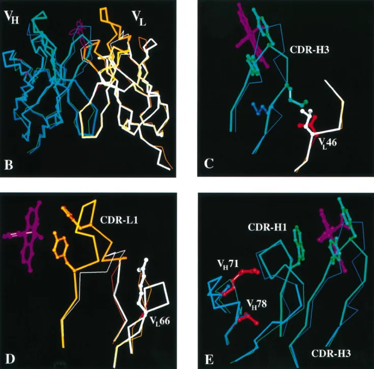
a poorly folding scFv, we grafted the antigen binding loops of the antibody 4-4-20 onto the 4D5 framework. All CDRs except CDR-L2 are involved in the binding of the antigen fluorescein (Herron et al., 1989; Whitlow et al., 1995). From the crystal structure of the antibody 4-4-20 we defined the minimal range of the loops necessary for binding as CDR–L1 27a–34, CDR– H1 28–35, CDR–L2 50–55, CDR–H2 50–58, CDR–L3 90–96 and CDR–H3 93–100c (Figure 1A; all numbers according to Kabat et al., 1987). We exchanged CDR–L2, even though it is not involved in binding, since we wanted to obtain an scFv derived from a ‘poor’ antibody in all CDRs for these studies. After superimposing both Fvs (Figure 1B), the framework region contacting the CDRs to be exchanged was carefully examined for additional residues directly involved in fluor-escein binding or supporting the orientation of the CDRs of scFv 4-4-20. Four 4-4-20 framework residues were identified that seemed to be important for the CDR orientation, where at the same time the respective residues from 4D5 appeared to be inappropriate. VL46 is valine in 4-4-20, but leucine in
4D5, which was expected to interfere with M99 in CDR–H3 of 4-4-20. To prevent that this clash would be avoided by a displacement of CDR–H3, the 4-4-20 residue valine was chosen instead of the 4D5 framework residue leucine (Figure 1C). The other residues considered were G66 in VL where the 4D5
residue arginine seemed to change the conformation of the framework loop contacting CDR1 of VL (Figure 1D) and
residues R71 and V78 in the core of VHsupporting CDR–H1,
which in turn could affect the position of CDR–H3 (Figure 1E). The last three residues had already been considered important for antigen binding by Carter et al. (1992b) in the construction of the final version of the 4D5 antibody, whose antigen is p185HER2.
Two further residues at the end of VL (L106 and L109)
were exchanged to obtain unique restriction sites compatible with the vector system used. In order to produce an scFv fragment, we used a long linker (G4S)6 of 30 amino acids
strictly to avoid diabody formation and we chose the orientation VL–VH to exploit the short FLAG sequence (Knappik and
Plu¨ckthun, 1994). The resulting sequence of scFv 4D5Flu is shown in Figure 1A. The protein has binding properties ident-ical with those of the parent antibody 4-4-20 (see below), demonstrating that the design was successful. However, since we directly synthesized the sequence given in Figure 1A, based on the considerations described above, we do not know whether all of the four framework residues from 4-4-20 would actually have been necessary. Additionally, a variant was constructed in which two cysteines were introduced in positions L100 and H44 into the scFv fragment (Brinkmann et al., 1993), to create a molecule which is both single-chain and disulfide-bonded (dsscFv) (Young et al., 1995), called 4D5FluSS. The details of the gene syntheses are given in Materials and methods.
In vivo folding
We then examined the in vivo folding behavior of various scFv constructs by periplasmic expression at 24 and 37°C. Figure 2 shows the soluble and insoluble scFv protein that accumulated in the periplasm after 1 h expression at 37°C, where the effect of the grafting can be observed most dramatically. In addition to the original 4D5 scFv, which yields almost exclusively soluble protein, only for the grafted scFv 4D5Flu is soluble protein found at all at 37°C, whereas for the wild-type 4-4-20, the mutant Flu4 (4-4-20 with mutation V84D in VH; Nieba et al., 1997) and the dsscFv 4D5FluSS the protein is completely
in the insoluble fraction. The same relative tendency can be seen at 24°C but the effect is less pronounced, as all antibodies yield some soluble protein to varying extents (data not shown). The mutation V84D in the scFv 4-4-20 has a strong positive influence on folding only at 24°C. The length of the linker (30- or 15mer) showed no effect on the ratio of soluble to insoluble protein and we will only discuss results from the 30mer linker, which gives rise to purely monomeric material (see below).
Purification yields after periplasmic expression at 24°C allow a direct comparison of in vivo folding properties. The grafted 4D5Flu could be purified after periplasmic expression by Ni-NTA chromatography and cation exchange with a yield of ~2.5 mg/l culture at 24°C. In contrast, the 4-4-20 wild-type scFv itself could not be purified at all under these conditions (Nieba et al., 1997), while the improved mutant Flu4 had given a yield of 1 mg/l. This shows that both the graft 4D5Flu and the point mutant Flu4 have improved in vivo folding characteristics compared with the 4-4-20 wild-type. Gel filtra-tion on a SMART system showed that the purified scFv 4D5Flu was indeed monomeric (data not shown).
Physicochemical properties
In the grafting procedure special precautions had been taken to preserve any framework residues from the antibody 4-4-20 that support the CDRs from 4-4-20. To test the functionality of the construct, we determined the binding constant of the
grafted 4D5Flu by measuring the fluorescence quenching of the hapten fluorescein by the antibody. The Kdof the grafted
scFv 4D5Flu was determined to be 2.2310–8 M, which is in
good agreement with the Kdof the scFv 4-4-20 (wild-type) of
2.3310–8M (Figure 3). These data show that in the construction of the graft the binding properties were maintained, without further rounds of engineering, which justifies the four 4-4-20 residues within the 4D5 framework introduced into the graft
a priori.
The thermodynamic stability was then measured by urea denaturation and the shift of the protein fluorescence emission maximum was used for the analysis of the data (Figure 4). The midpoint of the resulting curve was found to be at 6.4 M urea for the graft, compared with 4.1 M urea for 4-4-20 wild-type (Nieba et al., 1997) and therefore the graft is one of the most stable scFvs reported. With the caveat that both curves may not reflect true state behavior because of the two-domain nature of the scFv fragment, we estimate ∆G values
of 7 kcal/mol for the original 4-4-20 scFv and 12 kcal/mol for the grafted scFv. Thus, the 4D5 framework not only improves
in vivo folding, but also the thermodynamic stability of the
antibody.
Phage display
It has long been suspected that with phage display not only are good binders selected, but also scFvs are found with at least reasonable folding and stability properties (Deng et al.,
1994; Jackson et al., 1995). However, in many experiments on antibodies obtained from libraries and differing at the same time in affinity, stability, folding yield and even epitope, such conclusions are difficult to draw. We therefore decided to test this question in a direct experiment. In a competition between four scFvs with practically identical fluorescein binding con-stants but different physicochemical properties, the most stable and best folding scFv was selected. The competitors for the grafted 4D5Flu, which displays significantly enhanced stability and folding behavior at 37°C, were the scFv 4-4-20
(wild-Fig. 1. Amino acid sequence and structure comparison of grafted scFv 4D5Flu with its parental Fvs. (A) Amino acid sequences of the grafted scFv 4D5Flu in
the orientation VL–VHwith 30mer linker and the parental Fvs 4D5 (1fvc) and 4-4-20 (4fab). 4D5Flu sequence: black on white, amino acids from the
antibody 4D5 (framework residues); white on black, amino acids from the antibody 4-4-20 (CDR residues and selected framework residues); thin type, amino acids unrelated to either antibody 4D5 or 4-4-20 (FLAG, linker, Leu 106 at the end of VL); black on gray, framework amino acids exchanged for cysteines in
the dsscFv variant 4D5FluSS. Boxes in 4D5 sequence: CDR amino acids differing from 4-4-20 sequence. Boxes in 4-4-20 sequence: framework amino acids differing from 4D5 sequence. All residue numbering according to Kabat et al. (1987). (B)–(E) Superimposition of X-ray structures of the Fv 4D5 and the Fv part of the Fab 4-4-20. Blue, VHof 4D5; white, VLof 4D5; orange, VLof 4-4-20; green, VHof 4-4-20; purple, fluorescein; red, framework residues of
4-4-20 used in the grafts 4D5Flu and 4D5FluSS; thick lines, sequence parts used in the grafts 4D5Flu and 4D5FluSS.
type) with poor in vivo folding characteristics and mediocre stability, the engineered Flu4 (Nieba et al., 1997) with signific-antly better in vivo folding properties (at least at 24°C), but the same thermodynamic stability in urea denaturation as the wild-type, and the scFv 4D5FluSS with an engineered disulfide bond in the scFv fragment for further stabilization, but having roughly the same poor in vivo folding properties as the 4-4-20 (wild-type). After four rounds of panning and proliferation at 37°C, using an equimolar mixture of phage of each species at the outset, the grafted 4D5Flu was selected. This enrichment
Fig. 2. In vivo folding analysis of the scFv 4D5 and various
fluorescein-binding scFv fragments by comparison of soluble (s) and insoluble (i) fractions of periplasmic extracts on reducing SDS–PAGE and subsequent Western blotting and immunostaining with an M1-anti-FLAG antibody. MW marker: Rainbow (Amersham); only lysozyme band (14 kDa) is
immunostained. The periplasmic extracts were prepared from parallel cultures induced with 1 mM IPTG for 1 h at 37 °C. The scFv 4D5 produces more soluble than insoluble material, the scFv 4-4-20 and the 4-4-20 surface point mutant Flu4 produce no soluble material at 37°C. For the grafted scFv 4D5Flu with the 4D5 framework and the 4-4-20 CDRs the ratio of soluble to insoluble product is ~1:1, whereas the dsscFv version of the grafted antibody yields almost no soluble protein.
Fig. 3. Fluorescence quenching of fluorescein at 510 nm with purified
fluorescein-binding scFvs. (s) Titration curve with scFv 4-4-20 (wild-type); (j) titration curve with scFv 4D5Flu. Direct fitting of the curves results in Kds of 2.2310–8M for 4D5Flu and 2.3310–8M for 4-4-20.
can be directly followed by a restriction digest of the pool after each round, using a restriction site solely present in the 4D5Flu scFv, but not in one of the other three scFvs (Figure 5). This selection was not due either to a better growth of the cells expressing the scFv 4D5Flu or to a better proliferation of phage displaying 4D5Flu, compared with the competing phages. Only phages carrying 4D5FluSS have a lower phage titer (see below). A selection for growth and proliferation advantages could be excluded from a proliferation experiment over four rounds in which the panning steps were omitted and the phages harvested from the overnight culture were directly used for reinfection. In this experiment no enrichment could be seen (Figure 5). Moreover, a phage ELISA with phages
Fig. 4. Equilibrium denaturation with urea. Denaturation curves for scFvs
4-4-20 (wild-type) (s) and 4D5Flu (j). Midpoints were determined at 4.1 M urea for 4-4-20 and 6.4 M urea for 4D5Flu.
Fig. 5. Phage proliferation competition experiment. For each round of
phage proliferation, phagemids were prepared from cells harvested from overnight proliferation cultures. The phagemid pools were analyzed by restriction digest with EcoRI–StyI. StyI does not cut within the scFv gene of 4-4-20, Flu4 and 4D5FluSS, but once in the gene of 4D5Flu. Therefore, pAK100–4D5Flu yields a different restriction pattern (lane 1) than the phagemids encoding the other three competing scFvs, whose restriction pattern is represented by the digest of pAK100–4-4-20 (lane 2). MW marker: marker VI (Boehringer Mannheim) (lane 3). In the selection experiment with panning on BSA-FITC, 4D5Flu is selected from all its competitors after four rounds. In contrast, in the selection experiment without panning, no enrichment can be achieved.
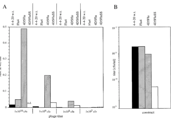
grown at 37°C, normalized for phage particles, showed that phages displaying the scFv 4D5Flu give by far the highest ELISA signals (Figure 6A). For comparable ELISA signals, roughly 100 times more phages of the 4-4-20 and Flu4 type and 10 times more phages of the 4D5FluSS type are needed than 4D5Flu type phages. Thus, far more 4D5Flu antibody is functionally displayed on phage than any of the other antibod-ies, most likely due to the higher stability and better folding characteristics of 4D5Flu at 37°C.
The total phage yields (Figure 6B) are comparable for the 4D5Flu, the 4-4-20 and the Flu4, with the exception of the
Fig. 6. (A) ELISA signals of phages displaying the scFvs 4-4-20 (black bars), Flu4 (bars horizontally hatched), 4D5Flu (bars diagonally hatched) and
4D5FluSS (white bars). For 4D5FluSS no ELISA data for a phage titer of 1010c.f.u. could be determined because of low phage yields. Phages displaying the
scFv 4D5Flu reach by far the highest ELISA signals. (B) Typical yield of phages displaying different scFvs from a 37°C culture. The titer of phages displaying the scFv 4D5FluSS is significantly lower (two orders of magnitude) than the titer of the phages displaying any of the other antibodies.
disulfide linked 4D5FluSS, which is almost two orders of magnitude lower. The 4D5FluSS aggregates more than the 4D5Flu parent in the in vivo folding analysis, presumably because of incorrect disulfide bond formation. The reproducibly low phage titers of phages displaying the 4D5FluSS are probably also due to this behavior. Thus, taking the total phage yields and the display rates determined in ELISA into account, it can be concluded that the thermodynamic stability and the expression behavior of scFvs play a crucial role for phage yield and display rate on phage and therefore also for the enrichment in phage display. In addition, with these data the power of the grafting approach was also demonstrated in a phage display selection experiment.
Discussion
The approach of improving an scFv with respect to its thermodynamic and in vivo folding properties by grafting its CDRs onto an scFv framework with superior properties has proved to be successful. Our results show that the antibody framework is a major, albeit not exclusive, determinant in the physicochemical properties of the resulting scFv fragment. Clearly, the framework of humanized 4D5 (VHsubgroup III,
Vκ subgoup I) has a number of favorable properties. This explains the high expression titers observed in the original studies of this antibody (Carter et al., 1992a) and in later studies with humanized antibody fragments using the same framework (Rodrigues et al., 1992; Eigenbrot et al., 1994; Zhu
et al., 1996). However, the CDRs do contribute, as expected,
to the physicochemical behavior (Ulrich et al., 1995) and in the present study the complete in vivo solubility of the original humanized 4D5 scFv at 37°C (Figure 2) was not mirrored by the graft.
We found that this graft is not only much less aggregation prone than the original 4-4-20 (wild-type), but also of signific-antly higher thermodynamic stability. Therefore, two
mechan-istically unlinked problems are solved in this graft at the same time and this may explain why the molecule shows a dramatic improvement also at 37°C. Thermodynamic stability and improved folding at 24°C are not directly coupled, since we found in two independent studies (Knappik and Plu¨ckthun, 1995; Nieba et al., 1997) that significant improvement of folding was not accompanied by an increased thermodynamic stability against urea denaturation. We currently cannot distin-guish whether the improved expression behavior at 37°C is just a reflection of even less aggregation of the graft than any of the other molecules tested in this study, or whether at 37°C the thermal denaturation of folded molecules already becomes such an important factor that the increased stability is simply a necessity for successful soluble expression at 37°C.
The unequivocal selection of the scFv 4D5Flu from the mixture of the fluorescein binding scFvs, all with the same binding constant, by phage display leads to several conclusions.
In vivo folding behavior and stability at the selection
temper-ature are the parameters with regard to which the grafted version performs much better than the wild-type and the other engineered forms. Phage ELISA demonstrated that this leads directly to an increase in the number of functional scFv molecules displayed by a given phage titer, due to stability and in vivo folding behavior of the respective scFv. Thus, not only are the best binders selected, but also thermodynamic stability and in vivo folding characteristics constitute a crucial factor in the selection by phage panning.
On the one hand, the diversity of libraries on phage may be reduced by these factors; on the other, scFvs with at least useful in vivo folding behavior and stability will preferably be selected, provided that such molecules are present in the library. It is clear, however, that the effect is subtle and it requires four rounds until the molecule is enriched completely from an equimolar mixture of competitors, some of which make essentially no soluble protein at all in expression studies.
Thus, in the absence of such a superior molecule in a library, a poor folder will be selected.
From a protein engineering perspective, there are now three strategies for improving the folding behavior of antibody fragments. Knappik and Plu¨ckthun (1995) showed that by a ‘back-engineering’ approach, single residues can be identified which determine the aggregation behavior of the molecule, acting synergistically, implying that they all lie on the same folding pathway. The residues were found to influence equally the folding behavior of Fv fragments, scFv fragments and Fab fragments, demonstrating that these observations are not due to the ‘unnatural’ Fv or scFv format, but to the intrinsic properties of the antibody variable domain. Moreover, the problematic residues identified are by no means uncommon, implying that there is no strong selective pressure optimizing folding in the animal cell. The fragments so obtained did not differ in thermodynamic stability in urea unfolding.
Nieba et al. (1997) sought a more general solution in identifying exposed hydrophobic residues at the variable/ constant interface. Using the same model system as described here, it was shown that mutations in the interface can improve folding behavior, again without changing the thermodynamic stability.
In a third approach, we investigated whether superior frameworks can be found as recipients for diverse antibody specificities. As a first step in this engineering approach, we examined whether the humanized 4D5 framework is a useful starting point by already possessing many favorable properties, such as a distinct lack of aggregation and high stability. With the CDRs from 4-4-20, which itself gives almost no soluble protein at all in periplasmic expression, the favorable properties remain, consistent with previous experiments, such as the humanization of 4D5 itself (Carter et al., 1992b; Rodrigues
et al., 1992), or grafts of McPC603 on the humanized 4D5
(H.Bothmann, K.Bauer, A.Knappik and A.Plu¨ckthun, unpub-lished data).
We have presented in this paper a simple and robust strategy for dramatically improving antibody folding, expression and stability. We believe that this is an accessible tool for any antibody which needs improvement, with the additional advant-age that it becomes humanized at the same time. Especially when the X-ray structure of the ‘CDR donor’ or at least a closely related sequence is available, grafting is feasible without a significant loss in binding strength, as we have shown here. The three strategies for improving folding (back-engineering, V/C surface engineering and loop grafting) are clearly not mutually exclusive, but lend themselves to future combined approaches.
Acknowledgements
We thank Lars Nieba for providing the data on the 4-4-20 scFv and Hendrick Bothmann and Drs Annemarie Honegger, Anke Krebber, Claus Krebber and Karl Proba for helpful discussions. This work was supported by the Schweizerische Nationalfonds grant 31-37717.93.
References
Barbas,C.F.,III and Burton,D.R. (1996) Trends Biotechnol., 14, 230–234. Bass,S., Gu,Q. and Christen,A. (1996) J. Bacteriol., 178, 1154–1161. Bird,R.E. et al. (1988) Science, 242, 423–426.
Brinkmann,U., Reiter,Y., Jung,S.H., Lee,B. & Pastan,I. (1993) PNAS, 90, 7538–7542.
Carter,P., Kelley,R.F., Rodrigues,M.L., Snedecor,B., Covarrubias,M., Velligan,M.D., Wong,W.L., Rowland,A.M., Kotts,C.E., Carver,M.E. et al. (1992a) Biotechnology, 10, 163–167,
Carter,P. et al. (1992b) Proc. Natl Acad. Sci. USA, 89, 4285–4289. Deng,S.J., MacKenzie,C.R., Sadowska,J., Michniewicz,J., Young,N.M.,
Bundle,D.R. and Narang,S.A. (1994) J. Biol. Chem., 269, 9533–9538. Eigenbrot,C., Randal,M., Presta,L., Carter,P. and Kossiakoff,A.A. (1993)
J. Mol. Biol., 229, 969–995.
Eigenbrot,C., Gonzalez,T., Mayeda,J., Carter,P., Werther,W., Hotaling,T., Fox,J. and Kessler,J. (1994) Proteins, 18, 49–62.
Ge,L., Knappik,A., Pack,P., Freund,C. and Plu¨ckthun,A. (1995) In Borrebaeck,C.A.K. (ed.), Antibody Engineering. A Practical Approach. IRL Press, Oxford, pp. 229–266.
Gill,S.C. and von Hippel,P.H. (1989) Anal. Biochem., 182, 319–326. Glockshuber,R., Malia,M., Pfitzinger,I. and Plu¨ckthun,A. (1990) Biochemistry,
29, 1362–1367.
Herron,J.N., He,X.M., Mason,M.L., Voss,E.W.,Jr and Edmundson,A.B. (1989) Proteins, 5, 271–280.
Huston,J.S. et al. (1988) Proc. Natl Acad. Sci. USA, 85, 5879–5883. Huston,J.S., McCartney,J., Tai,M.S., Mottola-Hartshorn,C., Jin,D., Warren,F.,
Keck,P. and Oppermann,H. (1993) Int. Rev. Immunol., 10, 195–217. Jackson,J.R., Sathe,G., Rosenberg,M. and Sweet,R. (1995) J. Immunol., 154,
3310–3319.
Kabat,E.A., Wu,T.T., Reid-Miller,M., Perry,H.M. and Gottesman,K.S. (1987), Sequences of Proteins of Immunological Interest. US Department of Health and Human Services, National Institutes of Health, Bethesda, MD. Knappik,A. and Plu¨ckthun,A. (1994) Biotechniques, 17, 754–761. Knappik,A. and Plu¨ckthun,A. (1995) Protein Engng, 8, 81–89.
Knappik,A., Krebber,C. and Plu¨ckthun,A. (1993) Biotechnology, 11, 77–83. Krebber,A., Bornhauser,S., Burmester,J., Honegger,A., Willuda,J.,
Bosshard,H.R. and Plu¨ckthun,A. (1997) J. Immunol. Methods, 201, 35–55. Kunkel,T.A., Roberts,J.D. and Zakour,R.A. (1987) Methods Enzymol., 154,
367–382.
Mallender,W.D., Carrero,J. and Voss,E.W.,Jr (1996) J. Biol. Chem., 271, 5338–5346.
Nieba,L., Honegger,A., Krebber,C. and Plu¨ckthun,A. (1997) Protein Engng,
10, 435–444.
O’Neill,K.T. and Hoess,R.H. (1995) Curr. Opin. Struct. Biol., 5, 443–449. Pace,C.N. (1990) Trends Biotechnol., 8, 93–98.
Plu¨ckthun,A. (1994) In Rosenberg,M. and Moore,G.P. (eds), The Pharmacology of Monoclonal Antibodies. Springer, Berlin, Vol. 113, pp. 269–315.
Prodromou,C. and Pearl,L.H. (1992) Protein Engng, 5, 827–829.
Reiter,Y., Brinkman,U., Webber,K.O., Jung,S.H., Lee,B. and Pastan,I. (1994) Protein Engng, 7, 697–704.
Rodrigues,M.L., Shalaby,M.R., Werther,W., Presta,L. and Carter,P. (1992), Int. J. Cancer (Suppl.), 7, 45–50.
Shimba,N., Torigoe,H., Takahashi,H., Masuda,K., Shimada,I., Arata,Y. and Sarai,A. (1995) FEBS Lett., 360, 247–250.
Skerra,A. and Plu¨ckthun,A. (1991) Protein Engng, 4, 971–979.
Ulrich,H.D., Patten,P.A., Yang,P.L., Romesberg,F.E. and Schultz,P.G. (1995) Proc. Natl Acad. Sci. USA, 92, 11907–11911.
Vaughan,T.J. et al. (1996) Nature Biotechnol., 14, 309–314.
Whitlow,M., Howard,A.J., Wood,J.F., Voss,E.W.,Jr and Hardman,K.D. (1995) Protein Engng, 8, 749–761.
Winter,G. and Harris,W.J. (1993) Trends Pharmacol. Sci., 14, 139–143. Winter,G., Griffiths,A.D., Hawkins,R.E. and Hoogenboom,H.R. (1994) Annu.
Rev. Immunol., 12, 433–455.
Yanisch-Perron,C., Vieira,J. and Messing,J. (1985) Gene, 33, 103–119. Young,N.M., MacKenzie,C.R., Narang,S.A., Oomen,R.P. and Baenziger,J.E.
(1995) FEBS Lett., 377, 135–139.
Zhu,Z., Zapata,G., Shalaby,R., Snedecor,B., Chen,H. and Carter,P. (1996) Biotechnology, 14, 192–196.

![[PDF] Cours pour débuter la programmation avec le langage ADA | Cours informatique](data:image/gif;base64,R0lGODlhAQABAIAAAP///wAAACH5BAEAAAAALAAAAAABAAEAAAICRAEAOw==)