DISTANT HETEROTOPIC CALLOSAL CONNECTIONS TO PREMOTOR
CORTEX IN NON-HUMAN PRIMATES
F. LANZ,aV. MORET,aR. AMBETT,aC. CAPPE,b E. M. ROUILLERaAND G. LOQUETa*
aDepartment of Medicine, Domain of Physiology and
Fribourg Cognition Center, University of Fribourg, Chemin du Muse´e 5, CH-1700 Fribourg, Switzerland
bCentre de recherche Cerveau et Cognition (CerCo),
CNRS, Universite´ de Toulouse – UPS, 31052 Toulouse, France Abstract—Cortico-cortical connectivity has become a major focus of neuroscience in the last decade but most of the connectivity studies focused on intrahemispheric circuits. Little has been reported about information acquired and pro-cessed in the premotor cortex and its functional connection with its homotopic counterpart in the opposite hemisphere via the corpus callosum. In non-human primates (maca-ques) lateralization is not well documented and its exact role is still unknown. The present study confirms in two maca-ques the existence of homotopic contralateral projections and completes the picture by further exploring heterotopic (non-motor) callosal projections. This was tested by inject-ing retrograde tracers in the premotor cortical areas PMv and PMd (targets). Our method consisted of identifying the connections with all the homo- and heterotopic cortical areas located in the contralateral hemisphere. The results showed that PMd and PMv receive multiple low-density labeled inputs from the opposite heterotopic prefrontal, parietal, motor, insular and temporal regions. Such unex-pected collection of transcallosal inputs from heterotopic areas suggests that the premotor areas communicate with other modalities through long distance low-density net-works which could have important implications in the understanding of sensorimotor and multimodal integration. Key words: neuroanatomy, cortical connectivity, corpus callosum, premotor cortex, non-human primate, multisensory integration.
INTRODUCTION
In placental mammals the corpus callosum is the main commissural structure (versus the anterior commissure) which connects homotopic and heterotopic regions of the cerebral cortex between the two hemispheres (e.g.
Innocenti, 1986; Innocenti, 1994, 1995; Aboitiz et al., 2003). The identification of the topography of these con-nections is still in progress in humans especially through
functional magnetic resonance imaging (Fabri et al., 2011; Phillips and Hopkins, 2012) and diffusion tensor imaging (Hofer and Frahm, 2006; Phillips and Hopkins, 2012). In non-human primates, the majority of brain con-nectivity data (see datasets established based on the work ofPaxinos et al., 2000; Van Essen, 2002; Dubach and Bowden, 2009; Rohlfing et al., 2012; Markov et al., 2014; Calebrese et al., 2015) originate from one hemi-sphere based on the assumption (though unproven) that lateralization does not play a key role in macaques’. The few available studies (e.g. Pandya and Vignolo, 1971) state that callosal connections predominantly link homo-topic cortical regions. This view has been questioned in the past decade (Clarke, 2003) and new evidence of numerous and widespread heterotopic callosal connec-tions have emerged in human studies. For example, in the visual cortex, heterotopic connections to the opposite hemisphere have been identified (Clarke and Miklossy, 1990; Clarke, 1994). Visual connections have also been reported from the inferior temporal cortex (associated with visual recognition) to the contralateral temporoparietal junction referred to as Wernicke’s area in humans (asso-ciated with speech comprehension) and to the inferior frontal gyrus (Broca’s area associated with speech pro-duction) (Di Virgilio and Clarke, 1997). In the motor cor-tex, corticocortical connectivity between motor areas and the other hemisphere has been identified in non-human primates (e.g.Pandya and Vignolo, 1971; Jenny, 1979; Rouiller et al., 1994; Liu et al., 2002; Marconi et al., 2003). In 2005, Boussaoud et al. showed that the premotor cortical areas PMd-c (F2) and PMd-r (F7) receive heterotopic inputs from contralateral pre-SMA (F6) and that PMd-r was strongly connected with pre-frontal cortex. According to those authors callosal afferent connectivity to PMv-c (F4) was broader than that to PMv-r (F5). Other authors (Marconi et al., 2003) reported that the major heterotopic callosal projection to F7 originated from F2 followed by weaker inputs from pre-SMA (F6), area 8 (FEF) and prefrontal cortex (area 46). The same authors showed that the heterotopic inputs to F2 mainly emanated from F7 followed by a smaller contingent com-ing from F5, F4, SMA-proper (F3) and F1. These reports suggest that premotor areas connectivity is composed of sets of heterotopic inputs originating from a mosaic of motor areas. Furthermore, in the premotor cortex, touch, vision and/or hearing inputs have been found (Weinrich and Wise, 1982; Weinrich et al., 1984; Graziano et al., 1997, 1999) contributing to sensorimotor transformation (Blanchard et al., 2013). Those heterotopic or
multisen-*Corresponding author. Fax: +41 26 300 97 34.
E-mail address:[email protected](G. Loquet).
http://doc.rero.ch
Published in "Neuroscience 344: 56–66, 2017"
which should be cited to refer to this work.
sory inputs would gain in being further studied since they provide a basis for underlying voluntary actions directed to a goal, more efficiently when more than one sensory modality is engaged (Stein and Meredith, 1993; Giard and Peronnet, 1999; Driver and Noesselt, 2008).
Most available tracing studies in monkeys (see above) on callosal motor connectivity were based on injections restricted to a specific cortical subarea (e.g. F2, F3, F4 or F5 in PM; F3 or F6 in SMA) or even to a limited body part (hand area in F1 and F3). In order to establish a more comprehensive callosal connectivity pattern, the present study is based on larger tracer injections covering a large part of the dorsal premotor cortex (PMd) and the ventral premotor cortex (PMv), respectively. We tested the hypothesis that both PMd and PMv receive significant direct heterotopic non-motor callosal inputs from the prefrontal, parietal and temporal lobes, which may amount up to 5% of the total callosal projections in each of these lobes.
EXPERIMENTAL PROCEDURES
Two non-human primates (Mk-CI Macaca mulatta and Mk-R9 Macaca fascicularis), 3 and 4 years old and weighing 3 and 4 kg, respectively, were re-used from a previous study on thalamocortical and corticothalamic projections (Cappe et al., 2007, 2009). The study was conducted according to both the guidelines of the National Institute of Health (Guide for the Care and Use of Labora-tory Animals, NIH Publication N°80-23, revised in 1996), those of the European Community (Guidelines for Ani-mals Protection and Use for Experimentation, 86/609/ EEC), and approved by local (Swiss) veterinary authori-ties (authorization N°156/04 and 156/02). All efforts were made to minimize the number of animals used and their suffering. The present work is based on the same injec-tions of four neuroanatomical retrograde tracers (see
Table 1) as described in a previous report (Cappe et al., 2009) but considered here for the callosal connectivity. Briefly, the animals were pre-medicated with ketamine (5 mg/kg, i.m.), Carprofen as an analgesic (Rymadil, 4 mg/kg, s.c.), antibiotics (Albipen, ampicillin 10%, 15– 30 mg/kg diluted 1:1 in saline, i.m.), atropine sulfate (0.05 mg/kg, i.m.) and dexamethasone (Decadron, 0.02–0.3 mg/kg/day diluted 1:1 in saline, i.m.). Then, the monkeys were anesthetized with propofol (0.1–0.3 mg/ kg/min, i.v.) and placed in a stereotaxic frame under asep-tic conditions. The skull and the dura mater on the left side were opened over the premotor cortex. In the frontal lobe, PMd and PMv were localized based on the position of the
central and arcuate sulci and the boundary between both areas was established based on the genu of the arcuate sulcus (Liu et al., 2002; Morel et al., 2005).
Injections of the tracers were executed by using 5- to 10-ll Hamilton syringes inserted perpendicularly to the cortical surface. Then, the dura mater, muscles and skin were sutured and the monkeys were treated for several days with an analgesic (Rymadil, 5 mg/kg, p.o.) and an antibiotic (Amoxicillin, 10 mg/kg, p.o.). Following a survival period of 2–3 weeks, the animals were deeply anesthetized, given a lethal dose of sodium pentobarbital (Vetanarcol 90 mg/kg i.p.) and were perfused transcardially with first 0.3 L saline (0.9%) then 3 L paraformaldehyde (4% in phosphate buffer 0.1 M, pH = 7.4), with a mixture (2 L) of paraformaldehyde 4% and sucrose 10% (in phosphate buffer) and finally with sucrose 20% and 30% (2 L in phosphate buffer).
The histological processing of the brain has also been described in detail in previous reports by Morel et al. (2005), Cappe et al. (2007)and Cappe et al. (2009). In summary, first the brain was sectioned in the frontal plane (40-l sections) on a freezing microtome. The sections were collected in five series among which one was imme-diately mounted on slides and stored in the refrigerator for fluorescent microscopy analysis. The plotting of labeled neurons with fluorescent and/or non-fluorescent tracers was done using the MicroBrightField Neurolucida System (Colchester, USA). Drawings of cortical contours in Nissl-stained sections were imported in Neurolucida’s system in order to be overlapped with the analyzed sections to iden-tify the cortical areas. Complete drawings including the plots of labeled cells were then exported to the software CorelDrawX6 (Version 16, 2012) and cell counting was performed.
The quantitative analysis was conducted by calculating for each tracer the percentage of cells labeled in one cortical area against the total number of cells labeled with this particular tracer in the whole hemisphere. Such percentage distribution as a function of a cortical area was represented first in histograms and grouped according to anatomical location. Second, the strength of the callosal connections between the multiple areas was represented in the form of a color weighted connectivity matrix as done by others (e.g.
Markov et al., 2014). This matrix used a logarithmic scale which was then translated into a positive scale where val-ues covered 3 equal ranges from 0 to 1.25 corresponding to sparse connections, from 1.25 to 2.5 corresponding to moderate connections and, greater than 2.5 correspond-ing to strong connections.
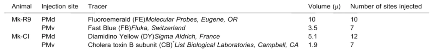
Table 1.Summary of injection sites, tracers, volumes and number of sites injected in the two macaques Mk-R9 and Mk-CI. Representations of the
injection sites are available inFigs. 1 and 2. *Due to an error of transcription inCappe et al. (2009), the tracer injected in PMv in Mk-CI is indeed CB and
not WGA, as indicated by mistake inCappe et al. (2009): in their legend ofFig. 1, ‘‘WGA” should be replaced by ‘‘CB”. The data are however not
affected, as theFig. 1ofCappe et al. (2009)indeed describes the data for PMv, derived from CB injection
Animal Injection site Tracer Volume (l) Number of sites injected
Mk-R9 PMd Fluoroemerald (FE)Molecular Probes, Eugene, OR 10 10
PMv Fast Blue (FB)Fluka, Switzerland 3.5 7
Mk-CI PMd Diamidino Yellow (DY)Sigma Aldrich, France 5.1 12
PMv Cholera toxin B subunit (CB)*List Biological Laboratories, Campbell, CA 1.9 7
RESULTS
Injection sites
In the present study, two out of the six cortical areas injected inCappe et al. (2009)were reinvestigated for cal-losal connectivity: the dorsal and the ventral premotor cor-tices (PMd and PMv). Repeated injections of different retrograde tracers extended anteriorly from the genu of the arcuate sulcus (Figs. 1–3) to the caudal end of the spur of the arcuate sulcus. The injections sites were in F7/F2 (learning-related area [Brasted and Wise, 2004] modulated by eye movement [Boussaoud, 1985]/guiding reaching area [Cisek and Kalaska, 2005]) and in F4/F5 (sensory guidance of movement [Graziano et al., 1994] and peripersonal space [Fogassi et al., 1996]/hand shap-ing durshap-ing graspshap-ing, vocalization [Coude` et al., 2011] and mirror neurons [Kohler et al., 2002]). Examples of the general distribution of retrogradely labeled neurons with FB, FE, DY and CB are presented inFig. 3. The relative position of each coronal section (as well as their esti-mated stereotaxic level in mm from interaural axis) is indi-cated on a schematic brain map based on the monkey brain atlas ofSaleem and Logothetis (2007).
Injections in PMd
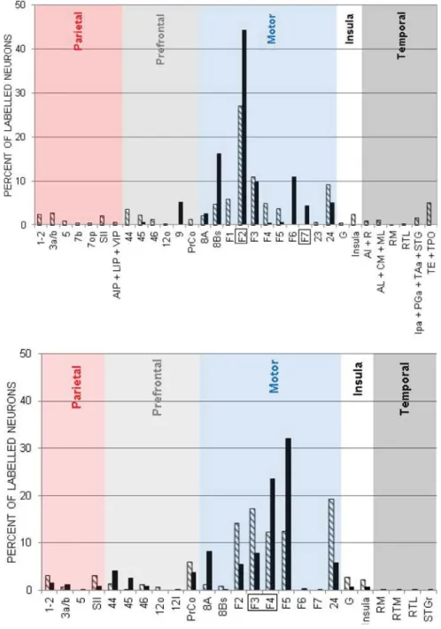
Fig. 4, upper panel, shows the distribution of retrograde labeling in the hemisphere contralateral to the injection of tracers FE and DY in PMd. The distribution of callosal inputs to PMd with respect to their lobe of origin is indicated in Table 2. In Mk-R9 the most abundant retrograde labeling was found in motor areas (69.1%). Less dense labeling was observed in the temporal lobe (areas TE + TPO; 9.1%), the parietal lobe (3a/b, 1–2, SII; 9.7%), the prefrontal cortex (8.4%, in particular areas 44 and 45) and the insular cortex (2.9%). In Mk-CI the main labeling has been obtained in motor areas (94.3%) followed by the prefrontal cortex (5.7%, area 9). In terms of origin of the callosal cortical projections, callosal inputs to PMd originate mainly from motor cortical areas. To a lesser extent, moderate transcallosal projections came from the temporal and the parietal lobe in one animal (Mk-R9) and from the prefrontal cortex (Mk-R9 & Mk-CI). Sparse callosal projections were identified coming from the insula (Fig. 4).
Injections in PMv
Fig. 4, lower panel, illustrates the histogram distribution of retrograde labeled cells in the hemisphere contralateral to the injection of two tracers (FB and CB) in PMv of the two monkeys. The distribution of callosal inputs to PMv with respect to their lobe of origin is indicated inTable 3. In Mk-R9 these injections have labeled cells mainly in motor areas (78%): area 24, SMA-proper (F3), F2, F4 and F5. A moderate labeling was found in the prefrontal (9.8%), parietal (7.0%) and insular (5.1%) regions. No retrograde labeling was observed in the temporal lobe. In Mk-CI, the main (homotopic) labeling was found in motor areas (83.4%): F5, F4, frontal eye field area 8A, F3, F2 and in the cingulate cortex (area 24). Less dense labeling was noticed in the other lobes: prefrontal,
parietal, insula and temporal lobes. Put into connectivity perspective callosal inputs to PMv mainly originated from contralateral premotor cortices. To a lesser extent, moderate to weak callosal inputs were identified from the FEF, the precentral operculum, somatosensory cortices (SII and 1–2), the area G (terminal plexus in gustatory cortex) and the insula. Finally, sparse callosal connections originated from a large palette of cortical areas (Fig. 4).
Connectivity matrices
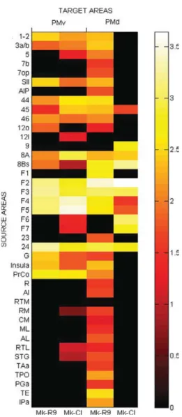
In order to assess more quantitatively the respective cortical areas of origin, connectivity matrices were established. Fig. 5 shows the individual connectivity matrices for Mk-R9 and Mk-CI. This matrix has been obtained based on the calculated ratio of labeled neurons with one marker in a specific cortical area relative to the total number of labeled neurons with the same marker over the hemisphere opposite to the injection site. These values have been turned into logarithms then translated to a positive scale in order to quantify connection weights. In Fig. 5, each column gives the calculated connection weight for each animal per area (PMv and PMd) and each row the origin of its transcallosal inputs originating from 41 separate cortical areas. According to the colorbar used, bright colors represented strong connections whereas dark colors represented weaker connections. The results show strong callosal homotopic connections especially at F4, F5, F2 levels. Other strong links with PMv and PMd were observed with heterotopic areas F2, F3, F4, F5 and 24. Moderate non-motor heterotopic connections are also well present in both monkeys but show less homogeneity in relation with PMd. Therefore only areas 8A and 8Bs can be noted as moderately connected to PMd whereas PrCo, the insula, the area G, the parietal and the prefrontal lobes show moderate connections to PMv. As far as sparse heterotopic connections are concerned, the sources to PMv originate essentially from the parietal and the prefrontal lobes in both animals. In contrast, for PMd, only one animal (Mk-R9) displays sparse heterotopic connections with the parietal and the prefrontal lobes but also with the temporal lobe.
DISCUSSION
Our results are in agreement with the hypothesis that both PMd and PMv receive multiple heterotopic non-motor callosal inputs from the contralateral prefrontal, parietal and possibly the temporal lobes. Those projections are few in quantity compared with the homotopic ones but represent a non-negligible amount (up to 5–7%) and therefore may be functionally relevant.
Connectivity of PMd and PMv
Across the two animals of different species, it appears that the homotopic projections (Figs. 4 and 5) are relatively consistent, suggesting that injections are comparable. Those repeated injections were executed in two different cortical subregions (F2/F7 and F4/F5;
see Figs. 1 and 2) and revealed strong homotopic corticocortical connections with the opposite hemisphere as described earlier by others (Pandya and Vignolo, 1971; Rouiller et al., 1994; Marconi et al., 2003). In
addi-tion to these major homotopic inputs linking bilaterally the F3 areas, the cingulate motor areas (Rouiller et al., 1994), F7, F2, F4, F5 (Boussaoud et al., 2005), smaller contin-gent of callosal inputs to PMd or PMv originating from
Fig. 1. (A) Upper left: the photomicrograph shows the PMd region of the left hemisphere of Mk-R9 injected with Fluoroemerald (FE). Below that,
sections S-15 to S-41 are examples of consecutive coronal slices through the PMd of the same animal displaying a reconstruction of the injected site. One can note that the injections covered an area from anteriorly to the genu of the arcuate sulcus (F7) till midway of the spur of the arcuate sulcus (F2). (B) The second photomicrograph from the same animal shows the PMv region of the left hemisphere injected with Fast Blue (FB). Examples of corresponding consecutive coronal sections through PMv are displayed as a reconstruction of the injected site. The injections started at the level of the caudal end of the principal sulcus and finished midway over the spur of the arcuate sulcus (therefore in F4 and F5). See list of abbreviations for the lettering.
contralateral heterotopic areas were described with PMd connected with area 46 of the prefrontal cortex, F6, F5, F4, F3, F2, F1 and area eight (Marconi et al., 2003). The present study reports the same patterns of homo and heterotopic callosal connections to PMd and PMv (see Figs. 4 and 5) and completes it with some other non-motor contingent of callosal projections to PMv (1–
2, 3a/b, SII, 44, 45, area 12, 24, G, Insula, PrCo) identified in both animals and to PMd (area 45, 24). The present observations mean that globally PM receives more con-tralateral non-homotopic projections than expected (Marconi et al., 2003; Boussaoud et al., 2005). We can add to these results the identification of sparse hetero-topic connections originating from the parietal cortex
Fig. 2. (A) The upper left photomicrograph shows the PMd region of the left hemisphere of Mk-CI injected with Diamidino Yellow (DY). Below that,
sections S-26 to S-49 are examples of consecutive coronal slices through the PMd of the same animal displaying a reconstruction of the injected site. The injections happened from anteriorly to the genu of the arcuate sulcus (F7) till midway of the spur of the arcuate sulcus (F2). (B) The second half of the page shows the PMv region of the left hemisphere of the same animal injected with Cholera toxin B subunit (CB). Examples of corresponding consecutive coronal sections through PMv are displayed to present the reconstruction of the injected site. With this marker, the injections were partly in F5 and partly in F4. For the lettering, see list of abbreviations.
(area 1–2, 3a/b, 5, 7, SII, AIP/LIP/VIP), the prefrontal cor-tex (44, 45, area 9, 24), the auditory corcor-tex (R, AI, RM, CM, ML, AL, RTL) or the temporal gyrus (STG, TAa, TPO, PGa, TE, IPa) to PMd in one animal (Mk-R9). Such differences between our two animals may be due to differ-ences in connectivity between the two species but also
differences at the injection site (e.g. marker spread, precise location of the injection, layers distribution) or because of the tracer type (e.g. sensi-tivity, specificity, single tracing). With-out bringing a definite answer to this point, one can say that our approach using injections covering a large part of the premotor cortex was appropri-ate since it allowed us to describe long-distance projections whose func-tional significance (though still unknown) is probably not related to the quantity of neurons of origin. Intrahemispheric versus callosal connections
In the motor system, reports demonstrated that intrahemispheric connectivity is formed by a series of interconnected areas working hierarchically (Keele et al., 1990; Grafton and Hamilton, 2007) and/or in parallel (Rizzolatti et al., 1998; Rizzolatti and Luppino, 2001). For example the neurons projecting to F1 were shown to originate mainly from PMd, PMv, SMA according to a somatotopic organization (Godschalk et al., 1984; Ghosh et al., 1987) and from other networks like the somatosensory areas 3a, 1–2, SII, the posterior parietal cortex and the cingulate cortex (Morecraft et al., 2012). Similarly PMd and PMv were shown to be connected to many other cortical areas located in the same hemisphere in the frontal and parietal lobes and to a lesser extent in the temporal lobe (Matelli et al., 1984; Barbas and Pandya, 1987; Kurata, 1991; Morecraft et al., 2012). Interest-ingly these two premotor areas have already been reported to receive inputs from associative ‘‘sensory” areas like MIP, AIP, 7a and 7b (Matelli et al., 1986; Ghosh and Gattera, 1995; Wise et al., 1997; Tanne´-Garie´py et al., 2002) known to have visual properties. Therefore these premotor areas were further investigated recently from a multisen-sory perspective (Lanz et al., 2013) in order to better understand their involvement in sensory-motor trans-formations. However, although these pathways and pro-cesses were generally found in one hemisphere because motor projections are mostly crossed when they reach the cortex (seeVan der Knaap and Van der Ham, 2011about the inhibitory theory through the corpus callo-sum to facilitate brain lateralization) it remains that during
Fig. 3. Examples of distribution of labeled neurons following injections of FE (green) and FB (blue) in
Mk-R9 (A) and DY (orange) and CB (dark blue) in Mk-CI (B), respectively in PMd and PMv. Upper left in both boxes shows sections level (1–3) indicated on a lateral view of a schematic macaque brain with aimed injection sites. The gray filled region marks out the block formed before histological work. Upper right in both boxes gives examples of injection site reconstructions (coronal sections taken fromFigs. 1 and 2). The gray filled regions indicate the injected cortical area according to the
parcellation obtained bySaleem and Logothetis (2007). Lower part of both boxes displays coronal
sections illustrating retrogradely labeled neurons in parceled cortex (drawing based on the monkey brain atlas) with lettering identifying the areas (see list of abbreviations for the meaning of the acronyms). Below each coronal section the rostrocaudal position of the section is tentatively given relative to the vertical plane passing through the interaural line like in the atlas.
bimanual tasks (e.g. opening a peanut) each hemisphere receives an afferent copy from the opposite hemisphere in order to confront inputs from both sides and perform
accurate actions (Brinkman, 1984; Geffen et al., 1994; Andres et al., 1999; Wahl and Ziemann, 2008; Liuzzi et al., 2011). In this context, a large connection via the corpus callo-sum with the opposite cortex was described by Rouiller et al. (1994)
between both SMAs (F3) which are consistent with the functional data (Kermadi et al., 1997, 1998) showing that these structures play a role in the control of bimanual coordinated movements (see Kermadi et al., 2000 for similar conclusions with the cingulate motor cortex, the posterior parietal cortex, F1 and PMd). The anatomical basis of such results have been confirmed in the present work (Figs. 4 and 5) but some other addi-tional projections originating from contralateral heterotopic areas state that a broader range of information (most likely inhibitory and excitatory) is transmitted to both PMd and PMv suggesting that PM might be part of a large sensorimotor and multisen-sory network stretched over the oppo-site hemisphere. For instance, a recent investigation in humans (Rousseau et al., 2016) concluded that the execution of a vertical hand movement activates a network that includes the contralateral primary motor and somatosensory cortices, PM, SMA, the anterior cerebellum, the cingulate cortex, the prefrontal cortex, the temporal gyrus, the hip-pocampi (bilateral), and the insula. These anatomical findings help describe the cortical network con-nected through the corpus callosum which is in position to contribute to sensorimotor and multisensory inte-gration across the two hemispheres. Multisensory processes
The present data when brought together in a connectivity matrix (Fig. 5) demonstrate that there is a general trend among the two animals characterized by a predominance of homotopic callosal links innervating PMv and PMd. However moderate and sparse projections form a heterotopic network which can be used as a substrate for multisensory integration. Indeed, recent studies support this view and showed various sensory inputs to the two studied premotor areas. Examples include visuomotor behavior (Nelissen et al., 2011; Takahara et al., 2012; Limanowski and Blankenburg, 2016), somatosensory representation (Disbrow et al., 2003; Wardak et al., 2016) and auditory discrimination (Lemus et al., 2009). Premotor fields seem
Fig. 4. Distribution of callosal neurons in cortical areas connected to PMd (upper graph) and PMv
(lower graph) from the opposite hemisphere. The explored cortical areas have been arbitrarily grouped for clarity’s sake. The lettering is explained in the abbreviations’ list. The hatched ( ) and
black (j) bars in the histogram correspond to the two different monkeys Mk-R9 and Mk-CI,
respectively.
Table 2.Summary of distribution of retrogradely labeled callosal inputs
to PMd Mk-R9 Mk-CIj Parietal 9.7% 0% Prefrontal 8.4% 5.7% Motor 69.1% 94.3% Insula 2.9% 0% Temporal 9.1% 0%
http://doc.rero.ch
therefore good candidates for passing information from one system to another and hence perform integrative pro-cesses. Moreover, we suggest that cortico-cortical cal-losal pathways could be added to the originally
described multisensory integration mechanisms formed by intrahemispheric cortico-cortical loops (Ghazanfar and Schroeder, 2006) and thalamocortical loops (Cappe et al., 2009). However, if we want to further understand the functional intricacy significance of these cortico-cortical callosal connections it will be necessary to exam-ine neural activity in situ and simultaneously on both sides of the brain.
Applied clinical relevance
The neurophysiological mechanisms of task-related modulation of neural networks have been studied by
Merchant et al. (2014). A cognitive task with maximum interactions shows local field potential changes in the cor-responding hemisphere as well as in the opposite hemi-sphere. A longer time lag was observed in the opposite hemisphere. Time lags for negative interactions were longer than for positive interactions in keeping with neu-roanatomical measurements (Merchant et al., 2014).
In the context of recovery from a cortical lesion, following a focal lesion in F1, it has been reported in non-human primates that adjacent cortical territories (Nudo and Milliken, 1996; Friel and Nudo, 1998) as well as interconnected regions (e.g. premotor cortex, Frost et al., 2003; Dancause et al., 2005) may play a role. Fur-thermore, when the monkeys were treated with anti-Nogo-A antibody (neutralizing axon growth inhibitors), the callosal connectivity of the premotor cortex was reor-ganized (Hamadjida et al., 2012). Our anatomical findings support the notion that post-lesional plasticity taking place in one hemisphere might trigger some adaptive changes of the callosal connectivity in case of a unilateral lesion of the premotor area (e.g. apraxia symptoms, Watson and Heilman, 1983).
In the context of sensory deprivation in humans suffering from chronic deafness, recent morphometric studies (Penhune et al., 2003; Kara et al., 2006) have reported an increase in the volume of the hand motor area, suggesting cross modal plasticity involving either hemispheres. One could deduce that this compensatory phenomenon of sensory substitution (see for example
Rauschecker, 1995; Von Melchner et al., 2000; Finney et al., 2003; Lomber et al., 2010; Barone et al., 2013) points toward the role of the corpus callosum in cortical plasticity. However the speculative nature of these phe-nomena in humans warrants further experiments to become relevant in clinical practice.
CONFLICT OF INTEREST
The authors declare that they have no conflict of interest.
Acknowledgments—The authors thank Laurent Bossy and Jac-ques Maillard for their highly valuable daily collaboration for the care of the monkeys in the animal facility, especially before, dur-ing and after the various interventions. This work was supported by the Swiss National Science Foundation – Switzerland to Prof. E.M. Rouiller [grant numbers 310000-110005, 31003A-132465 and 310030B-149643].
Fig. 5.Individual connectivity matrices for Mk-R9 and Mk-CI. Each
row represents one of the 41 separated source areas projecting to one of the two injected target areas under columns PMv or PMd. To better compare labeling distribution between monkeys each target area has been organized into two panels with Mk-R9 on the left and Mk-CI on the right. The labeled neurons are given in terms of
logarithmic values (log10(ratio)) transformed into positive numbers.
The colorbar uses the same scaling for both animals with bright colors representing strong connections (superior or equal to 2.5), medium colors representing moderate connections (between 1.25 and 2.5) and dark colors representing sparse connections (inferior to 1.25).
Table 3.Summary of distribution of retrogradely labeled callosal inputs
to PMv Mk-R9 Mk-CIj Parietal 7.0% 3.8% Prefrontal 9.8% 11.3% Motor 78.0% 83.4% Insula 5.1% 1.2% Temporal 0% 0.3%
http://doc.rero.ch
REFERENCES
Aboitiz F, Ide A, Olivares R (2003) Corpus callosum morphology in relation to cerebral asymmetries in the postmortem human. In: Zaidel E, Iacoboni M, editors. The parallel brain. London: The MIT Press. p. 33–46. Chapter 2.
Andres FG, Mima T, Schulman AE, Dichgans J, Hallett M, Gerloff C (1999) Functional coupling of human cortical sensorimotor areas during bimanual skill acquisition. Brain 122:855–870.
Barbas H, Pandya DN (1987) Architecture and frontal cortical connections of the premotor cortex (area 6) in the rhesus monkey. J Comp Neurol 256:211–228.
Barone P, Lacassagne L, Kral A (2013) Reorganization of the connectivity of cortical field DZ in congenitally deaf cat. PLoS One 8(4):e60093.
Blanchard C, Roll R, Roll JP, Kavounoudias A (2013) Differential contributions of vision, touch and muscle proprioception to the coding of hand movements. PLoS One 8(4):e62475.
Boussaoud D (1985) Primate premotor cortex: modulation of preparatory neuronal activity by gaze angle. J Neurophysiol 73 (2):886–890.
Boussaoud D, Tanne´-Garie´py J, Wannier T, Rouiller EM (2005) Callosal connections of dorsal versus ventral premotor areas in the macaque monkey: a multiple retrograde tracing study. BMC
Neurosci 6(67).http://dx.doi.org/10.1186/1471-2202-6-67.
Brasted PJ, Wise SP (2004) Comparison of learning-related neuronal activity in the dorsal premotor cortex and striatum. Eur J Neurosci 19(3):721–740.
Brinkman C (1984) Supplementary motor area of the monkey’s cerebral cortex: short- and long-term deficits after unilateral ablation and the effects of subsequent callosal section. J Neurosci 4(4):918–929.
Calebrese E, Badea A, Coe CL, Lubach GR, Shi Y, Styner MA, Johnson A (2015) A diffusion tensor MRI atlas of the postmortem rhesus macaque brain. Neuroimage 117:408–416.
Cappe C, Morel A, Rouiller EM (2007) Thalamocortical and the dual pattern of corticothalamic projections of the posterior parietal cortex in macaque monkeys. Neuroscience 146:1371–1387.
Cappe C, Morel A, Barone P, Rouiller EM (2009) The thalamocortical projection systems in primate: an anatomical support for multisensory and sensorimotor interplay. Cereb Cortex 19 (9):2025–2037.
Cisek P, Kalaska JF (2005) Neural correlates of reaching decisions in dorsal premotor cortex: specification of multiple direction choices and final selection of action. Neuron 45(5):801–814.
Clarke S (1994) Association and intrinsic connections of human extrastriate visual cortex. P R Soc London B 257:87–89.
Clarke S (2003) Complexity of human interhemispheric connections. In: Zaidel E, Iacobini M, editors. The parallel brain, commentary 2.1. London: The MIT Press. p. 47–49.
Clarke S, Miklossy J (1990) Occipital cortex in man: organization of callosal connections, related myelo- and cytoarchitecture, and putative boundaries of functional visual areas. J Comp Neurol 298:188–214.
Coude` G, Ferrari PF, Roda` F, Maranesi M, Borelli E, Veroni V, Monti F, Rozzi S, Fogassi L (2011) Neurons controlling voluntary vocalization in the macaque ventral premotor cortex. PLoS One 6 (11):e26822.
Dancause N, Barbay S, Frost SB, Plautz EJ, Chen DF, Zoubina EV, Stowe AM, Nudo RJ (2005) Extensive cortical rewiring after brain injury. J Neurosci 25:10167–10179.
Di Virgilio G, Clarke S (1997) Direct interhemispheric visual input to human speech areas. Hum Brain Mapp 5:347–354.
Disbrow E, Litinas E, Recanzone GH, Padberg J, Krubitzer L (2003) Cortical connections of the second somatosensory area and the parietal ventral area in macaque monkeys. J Comp Neurol 462:382–399.
Driver J, Noesselt T (2008) Multisensory interplay reveals crossmodal influences on ‘‘sensory specific” brain regions, neural responses, and judgments. Neuron 57:11–23.
Dubach MF, Bowden DM (2009). In: BrainInfo online 3D macaque brain atlas: a database in the shape of a brain. Chicago, IL: Soc Neur Ann Meet. Abstract No 199.5.
Fabri M, Polonra G, Mascioli G, Salvolini U, Manzoni T (2011) Topographical organization of human corpus callosum: an fMRI mapping study. Br Res 1370:99–111.
Finney EM, Clementz BA, Hickok G, Dobkins KR (2003) Visual stimuli activate auditory cortex in deaf subjects: evidence from MEG. NeuroReport 14:1425–1426.
Fogassi L, Gallese V, Fadiga L, Luppino G, Matelli M, Rizzolatti G (1996) Coding of peripersonal space in inferior premotor cortex (area F4). J Neurophysiol 76(1):141–157.
Friel KM, Nudo RJ (1998) Recovery of motor function after cortical injury in primates: compensatory movement patterns used during rehabilitative training. Somatosens Mot Res 15(3):173–189.
Frost SB, Barbay S, Friel KM, Plautz EJ, Nudo RJ (2003) Reorganization of remote cortical regions after ischemic brain injury: a potential substrate for stroke recovery. J Neurophysiol 89:3205–3214.
Geffen GM, Jones DL, Geffen LB (1994) Interhemispheric control of manual motor activity. Behav Brain Res 64(1–2):131–140.
Ghazanfar AA, Schroeder CE (2006) Is neocortex essentially multisensory? Trends Cogn Sci 10(6):278–285.
Ghosh S, Gattera R (1995) A comparison of the ipsilateral cortical projections to the dorsal and ventral subdivisions of the macaque premotor cortex. Somatosens Mot Res 12(3–4):359–378.
Ghosh S, Brinkmann C, Porter R (1987) A quantitative study of the distribution of neurons projecting to the precentral motor cortex in the monkey (M. fascicularis). J Comp Neurol 259:424–444.
Giard MH, Peronnet F (1999) Auditory-visual integration during multimodal object recognition in humans: a behavioral and electrophysiological study. J Cognitive Neurosci 11:473–490.
Godschalk M, Lemon RN, Kuypers HG, Ronday HK (1984) Cortical afferents and efferents of monkey postarcuate area: an anatomical and electrophysiological study. Exp Brain Res 56:410–424.
Grafton ST, Hamilton AF (2007) Evidence for a distributed hierarchy of action representation in the brain. Hum Movement Sci 26:590–616.
Graziano MSA, Yap GS, Gross CG (1994) Coding of visual space by premotor neurons. Science 266(5187):1054–1057.
Graziano MSA, Hu X, Gross CG (1997) Visuo-spatial properties of ventral premotor cortex. J Neurophysiol 77:2268–2292.
Graziano MSA, Reiss LAJ, Gross CG (1999) A neuronal representation of the location of nearby sounds. Nature 397 (4):428–430.
Hamadjida A, Wyss AF, Mir A, Schwab ME, Belhaj-Saif A, Rouiller EM (2012) Influence of anti-Nogo-A antibody treatment on the reorganization of callosal connectivity of the premotor cortical areas following unilateral lesion of primary motor cortex (M1) in adult macaque monkeys. Exp Brain Res 223(3):321–340.
Hofer S, Frahm J (2006) Topography of the human corpus callosum revisited - Comprehensive fiber tractography using diffusion tensor magnetic resonance imaging. Neuroimage 32:989–994.
Innocenti GM (1986) General organization of callosal connections in the cerebral cortex. In: Jones EG, Peters A, editors. Cerebral cortex, vol. 5. New York: Plenum. p. 291–354.
Innocenti GM (1994) Some new trends in the study of the corpus callosum. Behav Brain Res 64:1–8.
Innocenti GM (1995) Cellular aspects of callosal connections and their development. Neuropsychologia 33:961–988.
Jenny AB (1979) Commissural projections of the cortical hand motor area in monkeys. J Comp Neurol 188:137–146.
Kara A, Hakan Ozturk A, Kurtoglu Z, Umit Talas D, Aktekin M, Saygili M, Kanik A (2006) Morphometric comparison of the human corpus callosum in deaf and hearing subjects: an MRI study. J Neuroradiology 33:158–163.
Keele SW, Cohen A, Ivry R (1990) Motor programs: concepts and issues. In: Jeannerod M, editor. Attention and performance 13: motor representation and control. Hillsdale, NJ, USA: Lawrence Erlbaum Associates Inc.. Chapter 3.
Kermadi I, Liu Y, Tempini A, Rouiller EM (1997) Effects of reversible inactivation of the supplementary motor area (SMA) on unimanual grasp and bimanual pull and grasp performance in monkeys. Somatosens Mot Res 14:268–280.
Kermadi I, Liu Y, Tempini A, Calciati E, Rouiller EM (1998) Neuronal activity in the primate supplementary motor area and the primary motor cortex in relation to spatio-temporal bimanual coordination. Somatosens Mot Res 15(4):287–308.
Kermadi I, Liu Y, Rouiller EM (2000) Do bimanual motor actions involve the dorsal premotor (PMd), cingulate (CMA) and posterior parietal (PPC) cortices? Comparison with primary and supplementary motor cortical areas. Somatosens Mot Res 17 (3):255–271.
Kohler E, Keysers C, Umilta` MA, Fogassi L, Gallese V, Rizzolatti G (2002) Hearing sounds, understanding actions: action representation in mirror neurons. Science 2(297):846–848.
Kurata K (1991) Corticocortical inputs to the dorsal and ventral aspects of the premotor cortex of macaque monkeys. Neurosci Res 12:263–280.
Lanz F, Moret V, Rouiller EM, Loquet G (2013) Multisensory integration in non-human primates during a sensory-motor task. Frontier Hum Neurosci 7(799):1–15.
Lemus L, Hernandez A, Romo R (2009) Neural encoding of auditory discrimination in ventral premotor cortex. Proc Natl Acad Sci USA 106:14640–14645.
Limanowski J, Blankenburg F (2016) Integration of visual and proprioceptive limb position information in human posterior parietal, premotor, and extrastriate cortex. J Neurosci 36:2582–2589.
Liu J, Morel A, Wannier T, Rouiller EM (2002) Origins of callosal projections to the supplementary motor area (SMA): A direct comparison between pre-SMA and SMA-proper in macaque monkeys. J Comp Neurol 443:71–85.
Liuzzi G, Ho¨rniß M, Zimermann GerloffC, Hummel FC (2011) Coordination of uncoupled bimanual movements by strictly timed interhemispheric connectivity. J Neurosci 31 (25):9111–9117.
Lomber SG, Meredith MA, Kral A (2010) Cross-modal plasticity in specific auditory cortices underlies visual compensations in the deaf. Nat Neurosci 13:1421–1427.
Marconi B, Genovesio A, Giannetti S, Molinari M, Caminiti R (2003) Callosal connections of dorso-lateral premotor cortex. Eur J Neurosci 18:775–788.
Markov NT, Ercsey-Ravasz MM, Ribeiro Gomes AR, Lamy C, Magrou L, Vezoli J, Misery P, Falchier A, Quilodran R, Gariel MA, Sallet J, Gamanut R, Huissoud C, Clavagnier S, Giroud P, Sappey-Marinier D, Barone P, Dehay C, Toroczkai Z, Knoblauch K, Van Essen DC, Kennedy H (2014) A weighted and directed interareal connectivity matrix for macaque cerebral cortex. Cereb Cortex 24:17–36.
Matelli M, Camarda R, Glickstein M, Rizzolatti G (1984) Interconnections within the postarcuate cortex (area 6) of the macaque monkey. Brain Res 310:388–392.
Matelli M, Camarda R, Glickstein M, Rizzolatti G (1986) Afferent and efferent projections of the inferior area 6 in the macaque monkey. J Comp Neurol 251:281–298.
Merchant H, Crowe DA, Fortes AF, Georgopoulos AP (2014) Cognitive modulation of local and callosal neural interactions in decision making. Front Neurosci 245:1–10.
Morecraft RJ, Stilwell-Morecraft KS, Cipolloni PB, Ge J, McNeal DW, Pandya DN (2012) Cytoarchitecture and cortical connections of the anterior cingulate and adjacent somatomotor fields in the rhesus monkey. Brain Res Bull 87(4–5):457–497.
Morel A, Liu J, Wannier T, Jeanmonod D, Rouiller EM (2005) Divergence and convergence of thalamocortical projections to premotor and supplementary motor cortex: a multiple tracing study in macaque monkey. Eur J Neurosci 21:1007–1029.
Nelissen K, Borra E, Gerbella M, Rozzi S, Luppino G, Vanduffel W, Rizzolatti G, Orban GA (2011) Action observation circuits in the macaque monkey cortex. J Neurosci 31:3743–3756.
Nudo RJ, Milliken GW (1996) Reorganization of movement representations in primary motor cortex following focal ischemic infarcts in adult squirrel monkeys. J Neurophysiol 75: 2144–2149.
Pandya DN, Vignolo LA (1971) Intra- and interhemispheric projections of the precentral, premotor and arcuate areas in the rhesus monkey. Brain Res 26(2):217–233.
Paxinos G, Huang XF, Toga AW (2000) The rhesus monkey brain in stereotaxic coordinates. San Diego: Academic Press.
Penhune VB, Cismaru R, Dorsaint-Pierre R, Petitto LA, Zatorrec RJ (2003) The morphometry of auditory cortex in the congenitally deaf measured using MRI. Neuroimage:1215–1225.
Phillips KA, Hopkins WD (2012) Topography of the Chimpanzee
corpus callosum. PLoS One 7(2):e31941. http://dx.doi.org/
10.1371/journal.pone.0031941.
Rauschecker JP (1995) Compensatory plasticity and sensory substitution in the cerebral cortex. Trends Neurosci 18:36–43.
Rizzolatti G, Luppino G (2001) The cortical motor system. Neuron 31 (27):889–901.
Rizzolatti G, Luppino G, Matelli M (1998) The organization of the cortical motor system: new concepts. Electroencephalogr Clin Neuro 106:283–296.
Rohlfing T, Kroenke CD, Sullivan EV, Dubach MF, Bowden DM, Grant KA, Pfefferbaum A (2012) The INIA 19 template and neuromaps atlas for primate brain image parcellation and spatial normalization. Front Neuroinf 6:27.
Rouiller EM, Babalian A, Kazennikov O, Moret V, Yu X-H, Wiesendanger M (1994) Transcallosal connections of the distal forelimb representations of the primary and supplementary motor cortical areas in macaque monkeys. Exp Brain Res 102:227–243.
Rousseau C, Fautrelle L, Papaxanthis C, Fadiga L, Pozzo T, White O (2016) Direction-dependent activation of the insular cortex during vertical and horizontal hand movements. Neurosci 325:10–19.
Saleem KS, Logothetis NK (2007) A combined MRI and histology atlas of the rhesus monkey brain in stereotaxic coordinates. Elsevier: Academic Press.
Stein BE, Meredith MA (1993) The merging of the senses. Cambridge, MA: MIT Press.
Takahara D, Inoue KI, Hirata Y, Miyachi S, Nambu A, Takada M, Hoshi E (2012) Multisynaptic projections from the ventrolateral prefrontal cortex to the dorsal premotor cortex in macaques -anatomical substrate for conditional visuomotor behavior. Eur J Neurosci 36:3365–3375.
Tanne´-Garie´py J, Rouiller EM, Boussaoud D (2002) Parietal inputs to dorsal versus ventral premotor areas in the macaque monkey: evidence for largely segregated visuomotor pathways. Exp Brain Res 145:91–103.
Van der Knaap LJ, Van der Ham IJ (2011) How does the corpus callosum mediate interhemispheric transfer? A review. Behav Brain Res 223(1):211–221.
Van Essen DC (2002) Surface-based atlases of cerebellar cortex in the human, macaque and mouse. Ann N Y Acad Sci 978:468–479.
Von Melchner L, Pallas SL, Sur M (2000) Visual behaviour mediated by retinal projections directed to the auditory pathway. Nature 404 (6780):871–876.
Wahl M, Ziemann U (2008) The human motor corpus callosum. Rev Neurosci 19(6):451–466.
Wardak C, Guipponi O, Pinede S, Ben HS (2016) Tactile representation of the head and shoulders assessed by fMRI in the nonhuman primate. J Neurophysiol 115:80–91.
Watson RT, Heilman KM (1983) Callosal apraxia. Brain 106(Pt 2):391–403.
Weinrich M, Wise SP (1982) The premotor cortex of the monkey. J Neurosci 2:1329–1345.
Weinrich M, Wise SP, Mauritz KH (1984) A neurophysiological study of the premotor cortex in the rhesus monkey. Brain 107:385–414.
Wise SP, Boussaoud D, Johnson PB, Caminiti R (1997) Premotor and parietal cortex: corticocortical connectivity and combinatorial computations. Annu Rev Neurosci 20:25–42.
GLOSSARY
1–2: somatosensory areas 1 and 2 3a/b: somatosensory areas 3a and 3b 5: somatosensory area 5
7b: visual areas 7b
7op: area 7op (parietal operculum) 8A/Bs: frontal eye field areas 9: prefrontal area, dorsal subdivision 12o/l: orbitofrontal area
23: area in posterior cingulate cortex 24: area in anterior cingulate cortex 44, 45, 46: inferior frontal areas
AI: auditory area I, core region of the auditory cortex AIP: anterior intraparietal area
AL: anterior lateral, belt region of the auditory cortex CM: caudiomedial, belt region of the auditory cortex G: gustatory cortex
Insula: agranular, dysgranular and granular insula IPa: area in the superior temporal sulcus
LIP: lateral intraparietal area M1: primary motor cortex (F1)
ML: middle lateral, belt region of the auditory cortex PGa: area in the superior temporal sulcus PMd: dorsal premotor cortex (F2/F7) PMv: ventral premotor cortex (F4/F5) PrCo: precentral opercular area
Pre-SMA: pre-supplementary motor area (F6) R: rostral, core region of the auditory cortex RM: rostromedial, belt region of the auditory cortex RTL: lateral rostrotemporal, belt region of the auditory cortex RTM: medial rostrotemporal, belt region of the auditory cortex SII: secondary somatosensory area
SMA-proper: supplementary motor area (F3) STG: superior temporal gyrus
STGr: rostral superior temporal gyrus
TAa: area in the dorsal bank of the superior temporal sulcus TE: area in the ventral bank of the superior temporal sulcus TPO: area in the dorsal bank of the superior temporal sulcus VIP: ventral intraparietal area