Carcinogenesis vol.10 no.8 pp. 1419-1423, 1989
Covalent binding of [2-
14C]2-amino-3,8-dimethylimidazo[4,5-/]-quinoxaline (MelQx) to mouse DNA in vivo
A.J.Alldrick
1and W.K.Lutz
Institute of Toxicology, Swiss Federal Institute of Technology and University of Zurich, CH-8603 Schwerzenbach, Switzerland 'Permanent address: Department of Microbiology,
British Industrial Biological Research Association, Woodmansterne Road, Carshalton SM5 4DS, UK
Female BALB/c mice were administered intragastrically with
equimolar amounts of either [2-
14C]2-amino-3,&-dimethyl-[4,5-/]qulnoxaline (MelQx) or 2-acetylamino[9-
14C]fluorene
(2AAF). DNA was isolated from tissues of mke killed either
6 or 24 h after administration. Analysis of liver DNA
nucleo-tide digests by HPLC analysis revealed that all of the
radio-activity was attributable to adduct formation. The specific
activities of DNA samples were converted to covalent binding
indices (CBI, /unol adduct per mol DNA nucleotides/mmol
chemical applied per kg animal body weight). CBI values of
25 and 9 were determined for 2AAF and MelQx in the livers
of mke killed 6 h after dosing. The values were in general
agreement with the moderate carcinogenk potency of these
compounds. The specific activities of DNA preparations
obtained from the kidneys, spleens, stomachs, small intestines
and large intestines of mke treated with MelQx and killed
6 h after dosing were 5- to 35-times less than those obtained
with the liver. DNA isolated from the lungs (a target organ
for MelQx tumorigenkity) of MelQx-treated mice was not
radiolabeUed at the limit of detection (CBI < 0.3). With the
exception of the gastrointestinal tract, the specific activities
of DNA samples isolated from mice killed 6 h after
admin-istration were higher than those from mke killed after 24 h.
Introduction
The cooking of proteinaceous foods leads to the generation of
heterocyclic amines (1), a number of which have been shown
to be genotoxic in short-term tests and carcinogenic in rodents
(2,3). One of the more commonly occurring members of this
group is 2-amino-3,8-dimethylimidazo[4,5-/]quinoxaline
(MelQx*), originally found in fried beef (4). Under experimental
conditions MelQx has been shown to occur (in fried beef) at
amounts of 1.0 /xg/kg (uncooked weight) (1).
In the mouse, carcinogenicity studies have shown that MelQx
induces tumours in the livers and lungs, and also leukaemias and
lymphomas (5), while in the rat, it induced tumours in the liver,
skin, zymbal and clitoral glands (6). Distribution experiments
have revealed MelQx to be rapidly absorbed from the gut and
to be excreted in the bile and the urine (7,8). The mutagenic
activity of MelQx is dependent on metabolic activation to
electro-philic intermediates able to bind covalently to DNA (4). In vivo,
P-post-labelling analysis has revealed a number of DNA
adducts to be formed after administration of any of 12 heterocyclic
•Abbreviations: 2AAF, 2-acetylaminofluorene; MelQx, 2-amino-3,8-dimethyl-imidazo[415-/]quinoxaline; CBI, covalent binding index.
© IRL Press
amines including MelQx (9). Parallel in vitro studies indicated
that it was hydroxylation of die extracyclic amino group that gave
rise to the proximate carcinogen.
We have determined the extent to which radiolabelled [2-
14C]-MelQx binds to mouse-tissue DNA after a single intragastric
dose. Tissues were chosen either on the basis of their apparent
susceptibility to MelQx-induced tumorigenicity, or on meir
degree of exposure to the compound. The use of radiolabelled
MelQx and a rigorous DNA isolation and purification protocol
permitted the numbers of DNA adducts to be accurately
deter-mined. Consequently it was possible to determine covalent
binding index (CBI) values (10) for the compound (a predictor
of carcinogenic potency for carcinogens which act by binding
to DNA) and so determine to what extent the carcinogenicity of
MelQx can be attributed to its DNA binding ability.
Materials and methods
Chemicals
Unlabelled and 2-l4C-labelled MelQx (sp. act. 50 mCi/mmol) were purchased from Toronto Research Chemicals Inc. (Downsview, Canada). Unlabelled 2-acetyl-aminofluorene (2AAF) was purchased from Sigma Inc. (St Louis, MO, USA), [9-uC]2AAF (sp. act. 47 mCi/mmol) was purchased from New England Nuclear. Dosing solutions were prepared using either 10 mM acetic acid (pH 4.0) for MelQx or 50% (w/w) aqueous polyethylene glycol 300 (Merck, Darmstadt, FRG) for 2AAF. Both radiolabelled compounds had similar UV spectra and mobilities on silica-gel TLC (developing solvent: chloroform:methanol, 95:5) when compared to their unlabelled counterparts. Radioactive purity (as determined by TLC) was >98% for MelQx and >97% for 2AAF.
Animals and treatment regimes
Female Balb/cAnNCrlBR (BALB/c) mice were purchased at a weight of 18 g (36-56 days old) from Charles River Wiga GmbH (Sulzfeld, FRG). They were acclimatized for 1 week prior to use. Mice were housed separately in macTolone cages, with sawdust bedding under an alternate 12 h light/dark cycle. Food (Haltungsdifit nr 343, Klingental Milhle AG, Kaiseraugst, Switzerland) and water were available ad libitum.
Administration of the test compound or appropriate solvent was performed by intragastric gavage. Eight mice were used in each experiment, four mice received the test substance; two mice were administered with an equivalent amount of solvent (solvent control) and two mice remained untreated (controls against the possibility of radioactive contamination). In order to control for temporal variability, mice were dosed according to a strictly adhered-to schedule. Thus solvent controls were treated at 08.00 and 08.30, while test animals were dosed at 09.00 and at subsequent 30 min intervals and both food and water were available between treatment and killing. Mice were killed 6 or 24 h later. The average MelQx treatments were: for the 6 h exposure, 50 jimol (3.5 x 10⁹ d.p.m.) and for the 24 h experiment 64 /imol (4.4. x 109 d.p.m.) per kg bodyweight, while for die 2AAF experiments they were 67 /unol (2.7 x 10⁹ d.p.m.) and 69 iano\ (2.3 x 10⁹ d.p.m.) per kg bodyweight for the 6 and 24 h experiments, respectively.
Isolation of DNA and nuclear protein
Mice were anaesthetized with nembutal and killed by cardiac puncture. In the MelQx study the liver, lungs, kidneys, spleen, stomach, small intestine (duodenum to ileocaecal junction) and large intestine (caecum to rectum) were removed, while in the 2AAF study only the liver and lungs were removed. All organs were rinsed in saline (gut contents were removed by flushing with saline), weighed, snap-frozen in liquid nitrogen and stored at -20°C prior to DNA isolation. Total blood content was calculated from the body weight using previously published data (11). In the cases of the liver and the small intestine, DNA was extracted from organs taken from a pair of similarly treated mice. Chromatin was prepared from tissue homogenates by Nonidet P-40 (BDH, Poole, UK) precipitation and DNA purified to a constant specific radioactivity (where possible) using a previously published
method (12). However, with other tissues, to facilitate better recovery, DNA was extracted from the combined tissues of two carcinogen-treated and two control mice (for this purpose control mice were either those treated with solvent or left untreated).
As a precaution against the possibility that radioactivity associated with DNA was an artifact due to work-up, liver chromatin from the pair of control mice (i.e. treated with neither the test compound nor solvent) was incubated for 15 min at 4°C in the supernatant derived from a hepatic homogenate of mice treated with the radiolabel (in vitro control). Chromatin was reisolated and DNA extracted in an identical manner to the other preparations. Purified DNA samples were dissolved in succinate buffer (20 nM Na-succinate, pH 6.0, 8 mM CaClj). The concentrations of DNA in the resultant solutions were determined by UV spectro-photometry at 260 nm and calculated assuming that 1 mg/ml DNA has an absorbance of 20. Radioactivity was measured by liquid scintillation counting, using 10 ml Instagel (Canberra-Packard, Zurich, Switzerland) and using the 12-156 keV channel. Nuclear protein was isolated and purified as described previously (12). The concentration of each protein solution was determined using the method of Lowry et al. (13). Radioactivity was determined as for DNA.
Nucleotide analysis
When conditions permitted, DNA was further analysed to determine how much radioactivity could be attributed to adduct formation as opposed to bioincorpor-ation. Essentially, DNA was digested to its component nucleotides using micro-coccal nuclease (Sigma) and calf spleen phosphodiesterase (Boennger-Mannheim GmbH) as described previously (14). The resultant digest was analysed by HPLC. The digest was loaded onto a Lkhrosorb 18 column and developed for 10 min with 10 mM ammonium formate containing 4% methanol (pH 3.8). After 10 min a linear methanol gradient was applied, such that, 50 min after commencement of the run, the column was being developed with 100% methanol. The flow rate was 3.5 ml/min. After passing through a UV detector (254 nm), fractions were collected (usually at 2 min intervals). Fractions were combined with 10 ml Instagel and radioactivity determined as described below. Radioactivity cc-eluting with fractions having UV absorbance was attributed to bioincorporation, only radio-activity associated with fractions having no UV-absorbance was considered to be due to adducts.
Radioactivity determinations and calculations
The radioactive contents of all preparations were determined by liquid scintil-lation counting. In the cases of DNA, nucleotide analysis fractions and protein preparations, each sample was counted for 30 min three times. A DNA sample was considered to have a significant amount of radioactivity if the difference between the sample mean-count and that of the corresponding hepatic in vitro control DNA was > 2 SD c.p.m. Calibration experiments (examination of the statistics of radioactive decay, vial to vial variation, etc.) at this institute revealed this value to be 1.8 c.p.m. for MC. With the exceptions of the liver and the small intestine, the specific activity of isolated DNA was corrected for the presence of unlabelled DNA originating from the tissues of untreated mice. The proportion of DNA from control mice in a preparation was considered to be equal to that of the weight contributed by that tissue. Specific radioactivities were thus corrected for by multiplying with the appropriate factor.
The covalent binding index (CBI, reference 10) was calculated from the CBI' (10). The latter is defined as:
CBI' = d.p.m./mg DNA d.p.m./kg bodyweight (converting to molar units)
umol chemical bound/mol DNA nucleotide mmol chemical applied/kg bodyweight CBI = CBI' x 3.09 x 10⁸
(assuming 1 mol DNA nucleotkles weighs 309 daltons).
Results
Uptake of test substances
Radioactivity was detected in all of the tissue homogenates and
blood samples prepared from mice treated with either of the test
compounds at both time points. In the 6 h experiment, amounts
of radioactivity in the liver homogenates from MelQx-treated
mice were lower than their 2AAF counterparts (1.4 versus 2.9%)
although blood values were similar (1.3%). After 24 h, the
amounts of radioactivity were lower throughout. With this longer
interval between dosing and killing, amounts of radioactivity in
liver homogenates were similar (0.34 versus 0.41 %) for the two
test compounds. However, amounts of radioactivity in blood
taken from 2AAF treated mice (0.14%) were lower than in the
corresponding MelQx (0.59%) experiment.
Binding of test substances to DNA
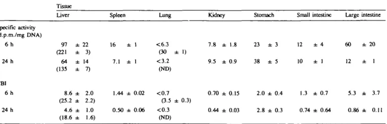
Liver. Radioactivity was associated with the DNA from livers
of mice treated with either compound (Table I). Since the specific
radioactivity of the DNA preparations did not appreciably change
on reisolation, it was assumed that the radioactivity represented
covalently bound or bioincorporated radiolabel. Analysis by
HPLC of DNA digests (Figure la and b) revealed that in every
case all recoverable radioactivity was only detected in fractions
having zero UV absorbance. This indicates that all of the
radio-activity associated with the DNA was due to covalent binding.
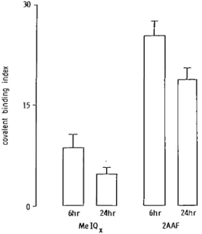
A conversion of the specific radioactivities of hepatic DNA to
covalent binding index values is shown in Figure 2. 2AAF
(CBI = 25 at 6 h) was about three times more potent than MelQx
(CBI = 8.6). The values at 24 h were lower (Table I; Figure 2),
Table I. DNA specific
Specific activity (d.p.m./mg DNA) 6 h 24 h CBI 6 h 24 h
activities and CBI Tissue liver 97 ± (221 ± 64 ± (135 ± 8.6 ± (25.2 ± 4.6 ± (18.6 ± 22 3) 14 7) 2.0 2.2) 1.0 1.6)
obtained from tissues
Spleen 16 7.1 1.44 0.50 ± 1 ± 1 ± 0.02 ± 0.06 of mice killed 6 Lung <6.3 (30 ± 1) <3.2 (ND) <0.7 (3.5 ± 0.3) <0.3 (ND) or 24 h after Kidney 7.8 ± 9.5 ± 0.70 ± 0.44 ± treatment 1.8 0.9 0.15 0.03 with MelQx Stomach 23 ± 3 38 ± 5 2.0 ± 0.4 2.8 ± 0.3
(data for 2AAF shown
Small 12 10 1.3 0.74 intestine ± 4 ± 1 ± 0.7 ± 0.64 in parentheses) Large intestine 60 ± 2 0 12 ± 1 5.3 ± 3.7 0.86 ± 0.11
DNA binding by MelQx
nominal half lives of the adducts were determined to be between
19 and 39 h.
Extrahepatic tissues. Treatment of mice with MelQx and killing
6 h later led to radioactivity being associated with DNA from
the spleen, kidney and sections of the gastrointestinal tract,
enabling the determination of CBI values (Table I). No
radio-activity was found to be associated with lung DNA (in contrast,
radioactivity was found with DNA from the Fungs of mice treated
with 2AAF and killed 6 h later). Although it was generally
a
l.O-i 0 . 5 -0J 10 .218
•I
L0 15 30 45 elutlon time (minutes)60 0 . 5 -0J V * > . 15
10 I
S.5
f
15 30elution time (minutes)
45 60
Fig. 1. HPLC analysis of nucleotides obtained from the enzymatic hydrolysis of hepatic DNA from mice treated with either MelQx (a) or 2AAF (b) and killed 6 h after treatment. The plot shows the UV absorbance of the eluate, while the bars show the radioactivity of the collected fractions fif significantly above control DNA). Letters indicate the order of nucleotide (deoxynucleoside-3'-phosphate) elution: A, adenine; C, cytosine; G, guanine; T, thymidine.
30 n 15-0J
1
T
I
T
6hr 24hr MeIQv 6hr 24hr 2AAFFig. 2. Covalent binding indices determined for hepatic DNA isolated from mice treated (per os) with either MelQx or 2AAF and killed 6 h or 24 h after administration. Data are presented as the means and standard deviations (bars) from two mouse liver pools (two livers per pool).
possible to purify DNA to a constant specific activity, it was not
possible to perform HPLC analysis of the nucleotides. This was
due to the small sample size and low specific activity. Although
it was not possible to show conclusively that all radioactivity was
due to covalent binding, it was assumed that the extrahepatic
DNA radioactivity was also a consequence of nucleotide—adduct
formation. In the case of the gastrointestinal tract there was,
additional, circumstantial, evidence to support this idea. Analysis
of DNA biosynthetic activity in the mouse (15) reveals the small
intestine to have the greatest activity followed by (in descending
order) large intestine, stomach and liver. The (descending) order
of CBI values at 6 h was: liver, large intestine, stomach and small
intestine (Table I). If radioactivity did represent biosynthesis it
would be expected that the order of CBI values would be similar
to that of biosynthesis. For all of the extrahepatic tissues the CBI
values were lower than those obtained for the corresponding liver
preparations. As in the case of the liver the CBI values obtained
for the spleen and kidneys were higher in mice killed 6 h after
dosing compared with the 24 h values. This result contrasted with
sections of the gastrointestinal tract which appeared to be
un-changed.
Control experiments. In order to ensure that the radioactivity
associated with DNA was not due to work-up techniques or
contaminating protein, other determinations were made. The
radioactivity determined for hepatic DNA isolated from either
solvent- or in vi/ro-control experiments were 13.2 and
13.5 c.p.m., respectively (MelQx, 24-h experiment). Succinate
buffer alone resulted in 13.6 c.p.m. total count. This indicates
that neither the work-up procedure nor the solvent contributed
to DNA radioactivity in treated mice.
Chromatin protein isolated from the liver showed specific
activities of between 400 and 900 d.p.m./mg, i.e. they were only
about 10-fold higher than for DNA. Similar results were obtained
for other tissues. It has previously been shown (12) that using
this method of DNA isolation, protein contamination accounts
for no more than 0.2% by weight. Thus contamination due to
protein cannot account for the radioactivity measured in the DNA
samples.
Discussion
We have observed that intragastric administration of radiolabelled
MelQx or 2AAF to mice leads to radioactivity being co-isolated
with DNA from a number of tissues 6 or 24 h after treatment.
In all tissues it was possible to purify DNA to a constant specific
activity. When hepatic DNA was digested to its component
nucleotides and subjected to HPLC analysis it was not possible
to detect radioactivity in the UV-absorbing regions of the eluates
(Figure 1). Thus it can be concluded that the radiolabel was
covalently bound in the form of adducts. The hepatic CBI values
obtained were compatible with both compounds being carcinogens
of medium potency (10).
Binding of MelQx to the DNA of extrahepatic tissues (with
the exception of the lung) was also observed. The results obtained
with the lung were surprising, since the lung was a target site
in the mouse carcinogenesis experiment (5). Four reasons can
be put forward to explain this observation, (i) Strain differences,
this study used BALB/c mice as opposed to CDF) used in the
carcinogenicity study, (ii) Only a small population of lung cells
(e.g. clara cells) are capable of performing the metabolic
processes necessary for the conversion of MelQx to an active
carcinogen. Since DNA is extracted from the complete tissue,
it is possible that adducted DNA is 'diluted' to such a level that
it cannot be detected by our methods, (iii) The limit of detection
of DNA binding (CBI ^ 0 . 3 at 24 h, averaged over all cells)
might not be low enough to exclude tumour formation in target
cells within the lung, (iv) MelQx induces lung tumours by an
adduct independent process. It has been demonstrated that 2AAF
can act as a promoter (16) and MelQx may have a similar
property.
Binding by MelQx to hepatic or lung DNA was not as great
as by 2AAF. This may in part be due to the different solvents
used and/or inherent differences in pharmacokinetics. Six hours
after administration, the radioactivity in the liver was
approxi-mately twice as high with 2AAF (see results). Nevertheless, the
adducts produced by these two compounds appeared to have
different properties. Up to 24 h after treatment adducts formed
by 2AAF persisted longer than by MelQx. Assuming first order
kinetics, the nominal half-lives for MelQx and 2AAF induced
adducts had a ratio of 1:2. The apparently faster rate of
disappearance of MelQx adducts may be due to one or a
combination of factors. Adducts produced by MelQx may be
repaired at a faster rate than those produced by 2AAF.
Alterna-tively MelQx-induced adducts may decay to form a second, more
toxic lesion. It is known that both compounds produce guanine
C-8 adducts (17,18). Furthermore it has been demonstrated that
some guanine C-8 adducts are labile, leading to the generation
of 8-hydroxyguanine (18). Since the half-life of MelQx appears
to be lower than that of 2AAF, it is possible that the MelQx
adduct unlike that of 2AAF has this property.
Acknowledgements
This work was partly supported by funds provided by the UK Ministry of Agriculture, Fisheries and Food and a Fellowship in Toxicology (PGT) provided by the European Science Foundation and European Medical Research Councils to A.J.A.
References
l.FeltonJ.S., Knize,M.G., Shcn.N.H., Anderson,BD., Bjcldanes.L.F. and Hatch.F.T. (1986) Identification of the mutagens in cooked foods. Env. Health
DNA binding by MelQx 2. Sugimura.T. and Sato.S. (1983) Mutagens and carcinogens in foods. Cancer
Res., 43 (Suppl.), 2415s-2421s.
3. Sugimura.T. (1985) Carcinogenicity of mutagenjc hetcrocylic amines formed during the cooking process. Mutat. Res., 150, 3 3 - 4 1 .
4. Kasai.H., Yamaizumi.S., Shiomi.T., Yokayama.S., Miyazawa.T., Wakabayashi.K., Nagao.M., Sugimura.T. and Nishimura.S. (1981) Structure of a potent mutagen isolated from fried beef. Chan. Lett., 485-488. 5.Ohgaki,H., Hasegawa,H., Suenaga.M,. Sato.S., Takayama.S. and
Sugimura.T. (1987) Carcinogenicity in mice of a mutagenic compound, 2-amino-3,8-dimemylimkiazo[4,5-/]quinoxaline (MelQx) from cooked foods.
Carcinogenesis, 8, 665—668.
6. Kato.T., Ohgaki.H., Hasegawa.H., Sato.S., Takayama.S. and Sugimura.T. (1988) Carcinogenicity in rats of a mutagenic compound: 2-amino-3,8- di-mcthybmidazo[4,5-/|quinoxaline. Carcinogenesis, 9, 71-74.
7.Turesky,R.J., Aeschbacher.H.U., Malnoe.A. and Wuerzner.H.P. (1988) Metabolism of the food-borne mutagen/carcinogen 2-amino-3,8-dimethyl-imidazo{4,5-/]quinoxaline in the rat: assessment of billiary metabolites for genotoxkity. Food Chem. Toxic, 26, 105-110.
8. Alldrick.A.J. and Rowland.I.R. (1988) Distribution of [2-14C]IQ and MelQx in the mouse. Toxicol. Lett., 44, 183-190.
9. Yamashita,K., Umenoto.A., Grivas.S., Kato,S., Sato.S. and Sugimura.T. (1988) Heterocyclic aminc-DNA adducts analysed by 32P-post labelling method. Nucleic Acid Res. Symp. Series, 19, 111-114.
10. Lutz.W.K. (1979) In vivo covalent binding of organic chemicals to DNA as a quantitative indicator in the process of chemical carcinogenesis. Mutat. Res., 65, 289-356.
11. Ditmer.D.S. (ed.) (1961) Blood and Other Body Fluids. Federation of American Societies of Experimental Biology, Washington, DC, USA. 12. Sagelsdorff.P., Lutz.W.K. and Schlatter.C. (1983) The relevance of covalent
binding to mouse liver DNA to the carcinogenic action of hexachlorocyclo-hexane isomers. Carcinogenesis, 4, 1267 — 1273.
13. Lowry,O.H., Rosebrough.N.J., Farr.A.L. and Randall.R.J. (1951) Protein measurements with the folin phenol reagent. J. Biol. Chem., 193, 265—275. 14. Sagelsdorff.P., Lutz.W.K. and Schlatter.C. (1988) DNA methylation by daminozide, 1,1-dimethylhydrazine and dimethylnitrosamine. Fund. Appl.
Toxicol, 11, 723-730.
15. Shephard.S.E. (1987) Towards an evaluation of the health risk posed by in
vivo nitrosation of food. PhD. Thesis, Swiss Federal Institute of Technology,
Zurich, Dissertation no. 8364.
16. Saeter.G., Schwarze.P.E., NestlandJ.M. and Seglen,P.O. (1988) 2-Acetyl-aminofluorene promotion of carcinogenesis by a noncytotoxic mechanism.
Carcinogenesis, 9, 581-587.
17. Kato.R. and Yamazoe.Y. (1987) Metabolic activation and covalent binding to nucleic acids of carcinogenic heterocyclic amines from cooked foods and amino acid pyrolysates. Gann, 78, 297—317.
18. KohdaJC., Tada.M., HalcuraA, Kasai.H. and Kawazoe.Y. (1987) Formation of 8-hydroxyguanine residues in DNA treated with 4-hydroxyamino-quinoline-1-oxide and its related compounds in the presence of seryl ATP.
Biochem, Biophys. Res. Commun., 149, 1141-1148.
Received on January 12, 1989; revised on May 16, 1989; accepted on May 24, 1989