Carciuogenesis vol.14 no.3 pp.355-36O, 1993
Covalent binding of styrene to DNA in rat and mouse
Sergio Cantoreggj and Werner K.Lutz
1Institute of Toxicology, Swiss Federal Institute of Technology and University of Zurich, CH-8603 Schwerzenbach, Switzerland 'To whom correspondence should be addressed
Covalent binding of ^ - ^ s t y r e n e (S) to DNA in vivo was
measured and evaluated in a quantitative manner in order
to investigate whether DNA adduct formation could form a
mechanistic basis for tumor induction in a carcinogenicity
bioassay. [7-
3H]S was administered by inhalation in a closed
chamber to male and female CD rats and B6C3F1 mice. After
4 . 5 - 6 h (rats) and 6 - 9 h (pools of four mice), S doses of
23—39 and 85-110 mg/kg respectively had been metabolized.
DNA was purified to constant specific radioactivity which was
measurable in all samples. DNA was enzymatically degraded
to the 3'-nucleotides which were separated by HPLC for the
detection of radiolabeled nucleotide-S adducts. The fractions
with the normal nucleotides contained most of the
radioactivity. In mouse liver DNA, a minute but significant
level of adduct radioactivity was also detected. In the units
of the Covalent Binding Index CBI = (/imol adduct/mol DNA
nucleotide)/(mmol chemical/kg body wt), values of 0.05-0.09
and 0.07-0.18 were calculated for males and females
respectively. In the rat, no DNA adducts were detectable in
the liver at a limit of detection of 0.1 CBI units. Two of the
four lung samples of the female rats showed adduct-related
radioactivity corresponding to 0.07 CBI units. The CBI values
are compatible with styrene 7,8-oxide as the reactive
intermediate. The data are compared with CBI values and
carcinogenic potencies of established genotoxic carcinogens.
It is concluded that the DNA-binding potency of S is so low
that significant tumor induction in a standard bioassay for
carcinogenicity is unlikely to be due to DNA adduct formation
alone. Consequences for a human risk estimation are
discussed.
Introduction
Styrene (S*; vinyl benzene; C6H5-CH=CH2) is widely used
for the manufacture of polymers and reinforced plastics.
High-level exposure of humans to S can occur by inhalation at the
workplace; low-level exposure is due to smoking and air pollution
(1). Exposure to S migrating from packaging into food appears
to be negligible (2).
Epidemiological studies investigated the carcinogenic potential
of S in humans (reviewed in 3). An insignificantly elevated level
of lymphatic and hematopoietic cancer was reported in some of
the older studies which were based on mixed chemical exposures,
including benzene. The two most recent studies, on the other hand,
showed a decrease in these tumor types. Carcinogenicity of S was
investigated in nine long-term animal studies. At very high dose
•Abbreviations: S, styrene; CBI, covalent binding index = DNA adduct level/dose = Otmol adduct/mol DNA nucleotide)/(mmol chemical/kg body wt); SO, styrene 7,8-oxide.levels, S appeared to affect the lung and the liver in the mouse
and the mammary gland in the rat, i.e. organs with high
spontaneous tumor incidence. However, all these studies had some
deficiencies or limitations (e.g. toxicity), so that the International
Agency for the Research on Cancer (IARC) considered the
evidence of carcinogenicity in animals to be limited (4).
Mutagenicity of S has been reviewed by Barale (5). In vitro,
S was occasionally found to be mutagenic, exclusively with
metabolic activation. In animals, S was very weakly genotoxic
(6). In humans, positive and negative cytogenetic studies were
reported (5). Detectable effects appeared to be limited to
workplace exposure levels >50 p.p.m. (7).
Covalent binding of S to DNA was investigated in the mouse
after i.p. injection (8). The DNA-binding potency expressed in
the units of the Covalent Binding Index (CBI) for liver DNA
was between 2 and 5; this is in the potency range of weakly
genotoxic carcinogens (9). It was therefore considered important
to reinvestigate the DNA-binding potency of S.
In this study, covalent binding of tritiated S to DNA was
investigated in rat and mouse by inhalation, and the data were
analyzed in terms of a DNA-binding potency. The results were
compared with other carcinogens (9) in order to answer the
question whether DNA adduct formation could form the
mechanistic basis for a putative tumor induction in a
carcinogenicity bioassay.
Materials and methods
Chemicals
Twice 20 mCi [7-3H]S (mol. wt 104.15), containing 10-15 p.p.m. 4-tert-butylcatechol as stabilizer, were purchased in 2 mCi ampoules from The Radiochemical Centre, Amersham, UK. The two batches had specific activities of 21 mCi/mmol (0.2 mCi/mg) and 20 mCi/mmol (0.19 mCi/mg) and were used for the experiments with the rats and mice respectively. The ampoules were stored at -20°C in the dark and used within 1 month after delivery. The radiochemKal purity determined by the suppher immediately prior to shipment was indicated to be 92.4 and 89.5% respectively. In order to check whether the [7-3H]S was in monomeric form when used, the specific activity was checked during the inhalation exposures by radio-GC, on a 20 mx0.3 mm column filled with PS 089 (95% dimethyl/5% diphenylpolysiloxan from HQls America, Inc.) and run with hydrogen at 50°C. The specific activity indicated by the supplier could be confirmed.
Animals
Six male and six female Crl:CD® BR rats and 24 male and 24 female B6C3Fl/CrlBR mice from Charles River Wiga, Sulzfeld, Germany were used. They were acclimatized in Macrolon cages for at least 1 week after delivery and were fed ad libitum with tap water and maintenance diet NAFAG 890, purchased from Nafag AG, Gossau SG, Switzerland.
Inhalation exposure
Single rats or groups of four mice (weights given in the tables) were exposed to tritiated S by inhalation in a closed chamber (10). The chamber consists of a 2 1 desiccator with an inlet for oxygen and a septum for air sampling. Expired carbon dioxide is adsorbed on soda lime placed into the chamber. The resulting atmospheric pressure reduction is compensated by an influx of oxygen with which tritiated S is carried along.
The ampoule containing 2 mCi tritiated S (10 mg for rats, 10.4 mg for the pools of four mice) was centrifuged at 2000 g for 5 min, cooled at the bottom with liquid nitrogen, cut open and immediately put into the oxygen flow system of the inhalation apparatus. Air samples taken from the chamber were analysed by GC for the time course of the S concentration. The exposure was terminated
S.Cantoreggi and W.K.Lutz
after at least four metabolism half-lives. The animals were killed with ether; liver and lung of rats and liver of mice were excised, pooled within the inhalation groups (mice) and kept frozen at -20°C. The mouse lung was not investigated because the yield of DNA would not have resulted in a satisfactory detection limit
Isolation of DNA
DNA was isolated via chromatin piecipilation according to Sagdsdorff a al. (11). Repurification to constant specific activity was carried out as previously reported (12).
Control experiments were performed using DNA isolated from untreated animals, (i) Radioactivity controls were compared with historical values to show that the work-up of the DNA samples had been performed without external contamination with radiolabd. (ii) An in vitro incubation of a chromatin supernatant from a treated animal with a chromatin pellet from a control (13) was done to investigate whether radioactive chemicals present in the tissue when the animals were killed could irreversibly associate with the DNA during work-up.
HPLC analysis of DNA nucleotides
DNA was digested to constituent nucleotides according to Sagelsdorff « al. (13). The resulting deoxynucleotides were separated by HPLC on an Eurosphere RP18 5jt column (250x4 mm) with 2 ml/min 30 mM ammonium formate buffer, pH 3.8/2—60% methanol gradient system according to Cantoreggi and Lutz (12). The fractions were dried down on a rotatory evaporator and dissolved in 1 ml water.
Scintillation counting
DNA samples and nucleotide fractions were counted in 10 ml Ultima Gold* (Packard) in plastic vials for 100 min in a liquid scintillation counter LS 6000 LL (Beckman, Fullerton CA) equipped with the LowLevel* option (rat liver DNA and nucleotide fractions). The significance level at two standard deviations, given by the Poisson distribution of the radioactive decay plus vial-to-vial
differences, had been determined to be 1.1 c.p.m. in an optimized tritium channel. The counting efficiency was between 47 and 52%.
The net radioactivity was calculated by subtracting the radioactivity of an equivalent sample isolated from animals treated with unlabeled S, both for DNA samples and for fractions of the nucleotide analyses.
Calculations
The concentration of a DNA solution was calculated from the optical density of a diluted aliquot at 260 nm using an absorbance of 20 for 1 mg DNA/ml. Depending on the amount of DNA available, an aliquot of 0.25-1 mg DNA was diluted with calcium succinate buffer to a standardized concentration (usually the concentration of the most dilute sample of the ongoing experiment).
The concentration of the nucleotide solutions was calculated from the optical density of a diluted aliquot at 260 nm assuming an absorbance of 35 for a nucleotide mixture equivalent to 1 mg digested DNA/ml.
The specific DNA radioactivity was expressed as DNA adduct level per dose administered and converted to the units of the CBI (14), CBI = Ounol adduct/mol DNA nucleotide)/(nimol chemical/kg body wt). Doses are given in the tables.
Results
Kinetics of S in the inhalation chamber
The concentration-time course of S in the chamber was
characterized by three phases. The first phase was dominated
by an influx of S into the chamber with maximum concentrations
of 274-300 and 385-464 p.p.m. registered after 1-2 h in the
experiments with rats and pools of mice respectively. Equilibrium
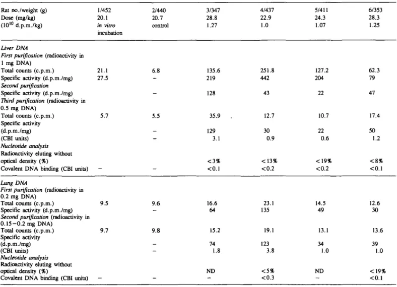
Table I. Investigation of the covalentRat no./weight (g) Dose (mg/kg) (1010 d.p.m./kg)
Liver DNA
First purification (radioactivity in
1 mg DNA) Total counts (c.p.m.) Specific activity (d.p.m./mg)
Second purification
Specific activity (d.p.m./mg)
Third purification (radioactivity in
0.5 mg DNA) Total counts (c.p.m.) Specific activity (d.p.m./mg) (CBI units) Nucleotide analysis
Radioactivity during without optical density (%)
Covalent DNA binding (CBI units)
binding of 1/452 20.1 in vitro incubation 21.1 27.5 5.7 —
[7-3H]styrene to DNA isolated 2/440 20.7 control 6.8 -5.5 -from liver 3/347 28.8 1.27 135.6 219 128 35.9 129 3.1 < 3 % <0.1
and lung of male CD 4/437 22.9 1.0 251.8 442 43 12.7 30 0.9 <13% <0.2
rats after inhalation exposure 5/411 24.3 1.07 127.2 204 22 10.7 22 0.6 <19% <0.2 6/353 28.3 1.25 62.3 79 47 17.4 50 1.2 < 8 % <0.1 Lung DNA
First purification (radioactivity in
0.2 mg DNA)
Total counts (c.p.m.) 9.5 Specific activity (d.p.m./mg)
Second purification (radioactivity in
0.15-0.2 mg DNA) Total counts (c.p.m.) 9.7 Specific activity (d.p.m./mg) (CBI units) Nucleotide analysis
Radioactivity eluting without optical density (%)
Covalent DNA binding (CBI units)
-9.6 9.8 16.6 64 15.2 74 1.8 ND 23.1 135 19.1 123 3.8 14.5 49 13.1 34 1.0 ND 12.6 30 13.6 39 1.0 <0.3
DNA binding of stymie
for S between the atmosphere and the animal was reached after
another 0 . 5 - 1 h. Thereafter, the concentration of S in the
chamber decreased as a result of metabolism. The experiment
was terminated when the concentration of S in the chamber
atmosphere had dropped to between 18 and < 5 p.p.m. (42
p.p.m. for the female mouse pool no. 3). This was after 4 . 5 - 6
h with the rats and 6.25-9 h with the mice. Taking into account
the metabolism half-life deduced from the third phase, >95%
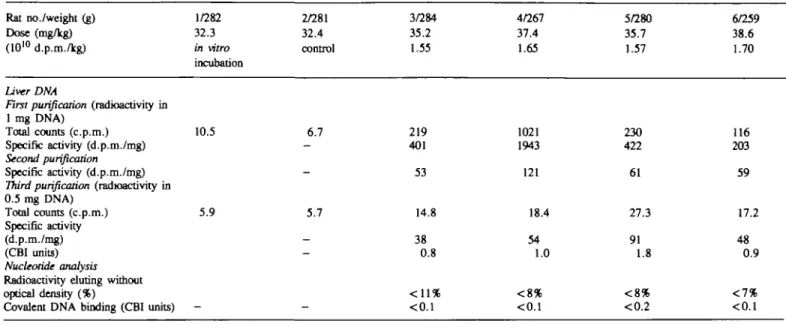
TaWe II. Investigation of the covalentRat no./weight (g) Dose (mg/kg) (1010 d.p.m./kg)
Liver DNA
First purification (radioactivity in
1 mg DNA) Total counts (c.p.m.) Specific activity (d.p.m./mg)
Second purification
Specific activity (d.p.m./mg)
Third purification (radioactivity in
0.5 mg DNA) Total counts (c.p.m.) Specific activity (d.p.m./mg) (CBI units) Nucleotide analysis
Radioactivity during without optical density (%)
Covalent DNA binding (CBI units)
binding of [7-1/282 32.3 in vitro incubation 10.5 5.9 -3H]styrene to DNA 2/281 32.4 control 6.7 -5.7 -_
-isolated from liver 3/284 35.2 1.55 219 401 53 14.8 38 0.8 < 1 1 % <0.1
and lung of female 4/267 37.4 1.65 1021 1943 121 18.4 54 1.0 < 8 % <0.1
CD rats after inhalation exposure 5/280 35.7 1.57 230 422 61 27.3 91 1.8 < 8 % <0.2 6/259 38.6 1.70 116 203 59 17.2 48 0.9 <1% <0.1 Lung DNA
First purification (radioactivity in
0.2 mg DNA)
Total counts (c.p.m.) 10.3 Specific activity (d.p.m./mg)
Second purification (radioactivity in
0.2 mg DNA) Total counts (c.p.m.) 9.5 Specific activity (d.p.m./mg) (CBI units) Nucleotide analysis
Radioactivity eluting without optical density (%) Specific activity (d.p.m./mg)
Covalent DNA binding (CBI units)
-10.6 10.0 18.8 84 18.3 85 1.7 3% 3.8 0.07 23.0 124 23.3 131 2.5 3% 3.6 0.07 20.9 104 19.5 96 1.9 <4.3 <0.08 36.0 251 37.1 272 4.9 <12.9 <0.23
A
20- M -
30 40 Time/min cpm 16- 14-12 ID-S' 6- 4-2-B
10 20 30 40 Time/minFig. 1. Reverse-phase HPLC profiles of optical density (solid line) and tritium radioactivity (bars; c.p.m.) of deoxyribonucleotides obtained by enzymatic hydrolysis of DNA. (A) From the liver of a male Crl:CD* BR rat (no. 4), 6 h after inhalation of l.OxlO10 d.p.m./kg (22.9 mg/kg) [7-3H]sryrene. (B) From the pooled livers of four male B6C3F1 mice (pool no. 4), 8 h after inhalation of 3.9x10'° d.p.m./kg (92.4 mg/kg) [7-3H]styrene.
S.Cantoreggl and W.K.Lutz
of the amount of S put into the system had been metabolized
within the exposure period.
DNA binding of [
3H]S in the rat
DNA isolated from the liver and the lung of the rats was analyzed
for radioactivity and nucleotide-S adducts. The results are
summarized in Tables I and II, for males and females
respectively.
In the liver, two rounds of DNA repurification were required
to remove all reversibly bound radioactivity and obtain constant
specific activities. All DNA samples were radiolabeled at levels
of 22-129 d.p.m./mg.
Radioactivity irreversibly associated with the DNA is not
necessarily due to nucleotide—carcinogen adduct formation, but
can be the result of biosynthetic incorporation of radiolabel via
DNA biosynthesis. Tritiated water is formed from the 7-3H
label of S during metabolism from mandelic acid to
phenylglyoxylic acid. Tritium from water can then be
incorporated into the 2'-position of the deoxyribose in the
ribonucleotide reductase step.
In order to distinguish adduct formation from radiolabel
incorporation, nucleotides were analyzed for radioactivity. HPLC
profiles with rat liver DNA showed that all radioactivity eluted
with normal nucleotides (Figure 1A). At elution times known
for S-nucleotide adducts (12), no radioactivity was measured at
a detection limit of 1.1 c.p.m. Conversion of this detection limit
to the units of the CBI showed a maximum possible DN A-binding
potency of CBI < 0.1 for both males (Table I) and females (Table
II).
In the rat lung, only one repurification round was necessary
to show constant specific DNA radioactivity for all samples. Not
all samples could be subjected to nucleotide analysis because of
low DNA yields. For the males (Table I), all detectable DNA
radioactivity eluted with the natural nucleotides. The limit of
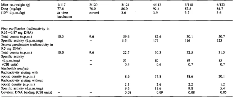
Table ID. Investigation of the covalent binding of Mice no./weight (g)
Dose (mg/kg) (1010 d.p.m./kg)
First purification (radioactivity in
0.35-0.87 mg DNA) Total counts (c.p.m.) Specific activity (d.p.m./mg)
Second purification (radioactivity in
0.5 mg DNA) Total counts (c.p.m.) Specific activity (d.p.m./mg) (CBI units) Nucleotide analysis
Radioactivity eluting with optical density (c.p.m.) Radioactivity eluting without optical density (c.p.m.) Specific activity (d.p.m./mg) Covalent DNA binding (CBI units)
1/117 77.6 in vitro incubation 10.3 10.0 — [7-3H]styrcne to 2/120 76.0 control 9.6 -9.6 -—
DNA isolated from the 3/121 86.0 3.6 59.6 115 22.7 51 0.4 8.6 2.1 9.6 0.08 liver of male B6C3F1 4/112 92.4 3.9 82.6 177 30.3 80 0.6 17.8 2.6 11.6 0.09
mice after inhalation exposure 5/118 87.8 3.7 30.1 116 32.5 89 0.7 18.6 2.2 9.8 0.08 6/123 84.7 3.6 50.7 123 31.5 85 0.7 20.1 1.2 5.4 0.05
Table IV. Investigation of the covalent binding of [7-3H]styrene to DNA isolated from the liver of female B6C3F1 mice after inhalation exposure Mice no./weight (g) 1/88 2/94 3/95 4/95 5/102 6/97 Dose (mg/kg) 103.3 96.9 109.9 109.0 102.1 107.7
(1010 d.p.m./kg) in vitro control 4.6 4.6 4.3 4.5
First purification (radioactivity in
0.40-0.58 mg DNA) Total counts (c.p.m.) Specific activity (d.p.m./mg)
Second purification (radioactivity in
0.5 mg DNA) Total counts (c.p.m.) Specific activity (d.p.m./mg) (CBI units) Nucleotide analysis
Radioactivity eluting with optical density (c.p.m.) Radioactivity eluting without optical density (c.p.m.) Specific activity (d.p.m./mg) Covalent DNA binding (CBI units)
9.3 9.6 9.0 9.4 68.0 295 60.6 206 1.4 22.9 2.5 26.6 0.18 84.3 377 49.4 156 1.0 33.3 4.0 18.1 0.12 29.7 101 36.8 107 0.8 23.4 2.1 9.6 0.07 75.3 332 49.6 157 1.1 40.8 3.0 13.6 0.09
DNA binding of styrene
detection of adducts was at CBI < 0.1 - < 0.3. With the females
(Table II), the yield of DNA was higher. In two samples, the
radioactivity measured in the fractions known to contain adducts
(12) was just detectable (CBI = 0.07).
DNA-binding of [
3H]S in the mouse
DNA was isolated from the pools of four livers. After the first
repurification, one sample already showed constant specific
activity and the remaining had only slightly been reduced (Tables
EH and IV). A second repurification was not necessary. HPLC
analysis of the nucleotides revealed, in all eight samples, a small
but significant amount of radioactivity in fractions eluting after
the natural nucleotides (Figure IB). The elution time was similar
to the early-eluting S-nucleotide adduct(s) generated from DNA
plus SO in vitro (12). Expressed in the units of the CBI, the
DNA-binding potency was at the extremely low level of 0.05-0.09
and 0.07-0.18 for males and females respectively. The average
was 0.10 ± 0.04 CBI units.
Discussion
S was shown to have a very low DNA-binding potency in vivo
after inhalation exposure of rodents. Using high levels of
radiolabeled S, the low limit of detection of DNA adducts allowed
to establish a DNA-binding potency of CBI ~ 0.1 for mouse
liver DNA. This very low genotoxic potency is in good
quantitative agreement with the borderline mutagenicity of S in
vivo.
What is the reactive intermediate? Metabolism of S involves,
to almost 100%, the intermediate formation of the 7,8-epoxide
(SO). The question is whether this epoxide could be responsible
for the DNA adduct formation seen with S. SO is a relatively
inert epoxide (15). SO is also weakly mutagenic in a number
of test systems without metabolic activation (5). DNA binding
by SO was not detectable in vivo but the limit of detection was
not as good as in the present experiments with S: a CBI <0.6
was found for mouse liver after i.p. injection (12). The CBI =
0.1 measured here for S is therefore not in contradiction to the
negative findings with SO. The data are consistent with the idea
that SO was responsible for the DNA adduct formation seen here
with S.
A minor pathway in S metabolism (<1%) results in the
formation of 4-vinylphenol (16,17), probably via the styrene
3,4-epoxide. The low CBI value of 0.1 does not allow for the
possibility that this or another metabolic pathway could result
in high levels of DNA adducts.
The present CBI values for S in mouse liver DNA are 2 0 - 5 0
times lower than the values that could be derived from another
report (8). A similar discrepancy was noted in the experiments
with SO: a CBI value of 2.2 was deduced from Byfalt Nordqvist
et al. (8), while our values were below a limit of detection of
CBI <0.6 under identical experimental conditions (12). Byfalt
Nordqvist et al. apparently did not purify the DNA to constant
specific activity. It cannot be excluded, therefore, that their DNA
had not completely been freed from non-covalently bound
radiolabeled S metabolites.
Chemical stability of DNA adducts is a prerequisite for a
correct interpretation of DNA-binding experiments. Evidence that
this requirement is met comes from Vodicka and Hemminki (18)
who showed a half-life of 10 days for SO-DNA adducts at pH
4.2 and room temperature in double-stranded DNA. Under our
experimental conditions for the DNA purification (neutral pH,
temperature mostly 4°C), the half-life could be even longer.
Furthermore, in the present experiments, DNA could be
repurified to constant specific activity. This would not be possible
with labile adducts. It can therefore be assumed that the adduct
concentrations measured after DNA purification reflected the
situation at the time the animals were killed.
No significant difference was seen between mice and rats or
between sexes. In the rat liver, the limit of detection was at CBI
< 0.1 in both males and females (Tables I and II), while the
highest value seen in the mouse liver was at CBI = 0.09 (male,
Table III) and CBI = 0.18 (female, Table IV). Therefore,
DNA-binding potency data would not help explain species or sex
differences tentatively seen in tumor induction (4).
It has been shown that potencies of genotoxic carcinogens
correlate quite well with CBI values for liver DNA [r = 0.81
for 29 activation-dependent mutagenic carcinogens (9)]. The range
of CBI values spanned from 2 to 10 000, carcinogenic potencies,
expressed as TD^ values, from 8 to 0.000001 mmol/kg/day.
Vinyl chloride, to mention one of the data points, had a CBI =
8 for mouse liver DNA (19) and a TD50 value for
hemangiosarcomas in the liver of 1 mmol/kg/day (20).
Using the above correlation of CBI versus TD50, the DNA
binding potency of S as determined here (CBI = 0.1) extrapolates
to a T D M value of — 1000 mmol S/kg/day. This is equivalent
to a dose of ~ 100 g S/kg/day, required theoretically to induce
a 50% tumor incidence in a standard lifespan if DNA adduct
formation is the mechanism of carcinogenic action. This
correlation could very tentatively be used to estimate a cancer
risk in humans, under the assumptions of a linear dose—response
relationship, the absence of species differences, and
proportionality with duration of exposure. Exposure of a worker
for 15 years to a daily dose of 10 mg/kg (resulting from an 8
h exposure at 20 p.p.m. for instance) would result in a theoretical
cancer risk of 1 in 105 lives (1 in 2 at 100 g/kg/day for life; 1
in 20 000 at 10 mg/kg for life; 1 in 100 000 if exposed only
for one-fifth of the standard human lifespan).
In conclusion, the level of DNA adduct formation by S is highly
unlikely to become responsible for a significant increase in tumor
incidence in a bioassay. Changes observed in the spontaneous
tumor pattern in standard bioassays with S are more likely due
to unspecific effects at maximum tolerated dose levels.
Acknowledgements
We thank Dr H.D.Hoffmann, BASF AG, Ludwigshafen, Germany, for valuable discussions. This work was supported by ECETOC, the European Chemical Industry Ecology and Toxicology Centre.
References
l.WHO (1983) Styrene. Environmental Health Criteria, 26, 1-123. 2. WQthrich.C, Muller.F., Blaser.O. and Marek,B. (1985) Die Belastung der
Bevdlkerung mit Pestiziden und anderen Fremdstoffen durch die Nahrung.
Mitt. Gebiete Lebensm. Hyg., 76, 260-276.
3.Bond,G.G., Bodner,K.M., Olsen.G.W. and Cook.R.R. (1992) Mortality among workers engaged in the development or manufacture of styrene-based products—an update. Scand. J. Work Environ. Health, 18, 145-154. 4. IARC (1987) 1ARC Monographs on the Evaluation of Carcinogenic Risks to
Humans, Suppl. 7. IARC, Lyon, pp. 345-347.
5. Barak,R. (1991) The genetic toxicology of styrene and styrene oxide. Mutat.
Res., 257, 107-126.
6. Simula^.P. and Priestly,B.G. (1992) Species differences in the genotoxicity of cyclophosphamide and styrene in three in vivo assays. Mutat. Res., 271, 4 9 - 5 8 .
7.Sorsa,M., Anttila.A., JSrventaus.H., Kubiak.R., Norppa.H., Nylander.L., Pekari.K., PfSffli.P. and Vainio.H. (1991) Styrene revisited—exposure assessment and risk estimation in reinforced plastics industry. Prog. din.
Biol. Res., 372, 187-195.
S.Cantoreggi and W.K.Lutz
Covalent binding of styrene and styrene-7,8-oxide to plasma proteins, hemoglobin and DNA in the mouse. Chan.- BioL Interactions, 55, 6 3 - 7 3 . 9. Lutz.W.K. (1986) Quantitative evaluation of DNA binding data for risk estimation and for classification of direct and indirect carcinogens. /. Cancer
Res. din. Oncol., 112, 8 5 - 9 1 .
10. Baertsch,A., Lutz.W.K. and Schlatter.C. (1991) Effect of inhalation exposure regimen on DNA binding potency of 1,2-dichloroethane in the rat. Arch.
Toxicol., 65, 169-176.
11. Sagelsdorff.P., Lutz.W.K. and Schlatter.C. (1983) The relevance of covalent binding to .mouse liver DNA to the carcinogenic action of hexachlorocyclohexane isomers. Carcinogenesis, 4, 1267-1273. 12. Cantoreggi.S. and Lutz.W.K. (1992) Investigation of the covalent binding
ofstyrene-7,8-oxidetoDNAinratandrnouse. Carcinogenesis, 13, 193-197. 13. Sagelsdorff.P., Lutz.W.K. and Schlatter.C. (1988) DNA methylation in rat
liver by daminozide, 1,1-dimethylhydrazine, and dimethyhiitrosamine.
Fundam. AppL Toxicol., 11, 723-730.
14. Lutz.W.K. (1979) In vivo covalent binding of organic chemicals to DNA as a quantitative indicator in the process of chemical carcinogenesis. Mutat. Res., 65, 289-356.
15.Betso,J.E.,Carreon,R.E. andMiner.V.M. (1991) The use of proton nuclear magnetic resonance spectroscopy ('H NMR) for monitoring the reaction of epoxides with butylamine and predictive capabilities of the relative alkyiatkm index (RAT) for skin sensitization by epoxides. Toxicol. AppL Pharmacol., 108, 483-488.
16.Pantarotto,C, Fanelli.R., Bidoli.F., Morazzoni.P., Salmona.M. and Szczawinska.K. (1978) Arene oxides in styrene metabolism, a new perspective in styrene toxicity? Scand. J. Work. Environ. Health, 4, 67—77. n.Pfaffli.P., Hesso.A., Vainio.H. and Hyvflnen.M. (1981) 4-Vinylphenol
excretion suggestive of arene oxide formation in workers occupationally exposed to styrene. Toxicol AppL Pharmacol., 60, 85-90.
18.Vodicka,P. and Hemminki,K. (1988) Identification of alkylation products of styrene oxide in single- and double-stranded DNA. Carcinogenesis, 9, 1657-1660.
19. Bergman.K. (1982) Reactions of vinyl chloride with RNA and DNA of various mouse tissues in vivo. Arch. Toxicol., 49, 117-129.
20.Gold,L.S., Sawyer.C.B., Magaw.R., Backman.G.M., deVeciana.M., Levinson.R., Hooper.N.K., Havender.W.R., Bemstein.L., Peto.R., Pike.M.C. and Ames.B.N. (1984) A carcinogenic potency data base of the standardized results of animal bioassays. Environ. Health Perspect., 58, 9 - 3 1 9 .
Received on October 6, 1992; revised on November 23, 1992; accepted on November 26, 1992