TECHNICAL REPORT
CT-guided cervical nerve root injections: comparing
the immediate post-injection anesthetic-related effects
of the transforaminal injection with a new indirect technique
Reto Sutter&Christian W. A. Pfirrmann&
Marco Zanetti&Juerg Hodler&Cynthia K. Peterson
Received: 10 August 2011 / Revised: 12 September 2011 / Accepted: 13 September 2011 / Published online: 1 October 2011 # ISS 2011
Abstract
Objective To describe an “indirect” cervical nerve root injection technique with a dorsal approach that should carry less inherent risk than the “direct” cervical transforaminal injection approach, and to compare the immediate post-injection results of the two procedures.
Materials and methods The indirect and direct cervical nerve root injection procedures are described in detail. Fifty-three consecutive patients receiving the indirect nerve root injections during 2009–2010 were age- and gender-matched to 53 patients who underwent direct transforaminal nerve root injections performed in 2006. Pain level data were collected immediately before and 20–30 min after each procedure. The percentages of pain change in the two
groups were compared using the unpaired Student’s t test. Results Fifty-two men (mean age 49) and 54 women (mean age 55) were included. The mean percentage of pain reduction for patients receiving indirect nerve root injec-tions was 38.4% and for those undergoing the direct nerve root injections approach it was 43.2%. This was not significantly different (P=0.455). No immediate or late adverse effects were reported after either injection procedure. Conclusions The indirect cervical nerve root injection procedure is a potentially safer alternative to direct cervical transforaminal nerve root injections. The short-term pain reduction is similar using the two injection methods.
Introduction
Compression or irritation of a nerve root in the cervical spine, either from a disc herniation or degenerative changes involving the intervertebral foramen, can be a source of considerable pain and disability, the most frequent incidence (203 out of 1,000) occurring between the ages of 50 and 54 [1–4]. When conservative treatments fail to improve the pain, many patients have increasingly been referred for cervical transforaminal nerve root injections as an alternative to surgery, injecting a combination of local anesthetic and corticosteroid [1–3,5–7]. A recent“best evidence synthesis” report states that there is some evidence supporting short-term symptomatic improvement of radicular symptoms when the treatment involves a short (< 3) course of selective root injections with corticosteroids [8], but there are no controlled trials investigating the efficacy of this procedure [1–3,5–9]. The literature to date supports a relatively low serious adverse events rate of less than 1% for cervical trans-foraminal nerve root injections [8]. However, increasing reports have surfaced describing very rare but extremely
R. Sutter
:
C. W. A. Pfirrmann:
C. K. Peterson (*)Department of Radiology,
Orthopaedic University Hospital of Balgrist, Forchstrasse 340, 8008 Zürich, Switzerland e-mail: [email protected] R. Sutter e-mail: [email protected] C. W. A. Pfirrmann e-mail: [email protected] M. Zanetti
Department of Radiology, Hirslanden Hospital, Seefeldstrasse 214,
8008 Zürich, Switzerland
e-mail: [email protected] J. Hodler
Department of Radiology, University Hospital, Rämistrasse 100,
8091 Zürich, Switzerland e-mail: [email protected]
serious complications, including several cases of either transient or permanent tetraplegia, brain infarctions leading to death, arterial dissection with resulting complications, or cortical blindness [2,3,9–19]. Recent investigations appear to link many of these serious complications to specific steroid preparations that consist of large particles or particles that coalesce into larger aggregates, forming emboli that infarct the spinal cord or brain when inadver-tently injected into an artery [2, 3, 10, 14–16]. Another suggested etiology is inadvertent arterial penetration leading to either dissection or vasospasm [15,17,18].
Owing to the very serious nature of these uncommon complications, protocols have been suggested to reduce the likelihood of such adverse events. These include: always performing the procedures under imaging guidance (real-time fluoroscopy or computed tomography); use of a non-particulate steroid; injection of iodinated contrast agent using digital subtraction imaging to detect vascular uptake before injecting medications; and a test dose of 1 ml of local anesthetic before injecting steroid [3,9,14,15]. While these guidelines are important to reduce the risks of serious complications from cervical transforaminal nerve root injections, they cannot guarantee that no adverse reaction will occur. Inadvertent vascular penetration during this procedure has been reported to be up to 19.4% using recommended imaging guidance procedures, as outlined above [19]. However, with the use of a contrast agent, some of these punctures are recognized immediately, and the needle repositioned, before the injection of potentially harmful medication.
Because of the risk of potentially serious adverse events with the use of cervical transforaminal nerve root injections, and as a direct result of two such cases in this specialized orthopedic/rheumatology hospital, the radiology department stopped carrying out this procedure in June 2008. However, patients with intractable nerve root pain, who wish to avoid surgery, need effective short-term treatment. The natural history of cervical disc herniations with radiculopathy is often an improvement in symptoms over time [20], and for many of these patients, cervical transforaminal nerve root injections helped them to cope, avoiding the risks and costs of surgery. A recent abstract (Drape JL et al., presented at the 2007 annual meeting of the Radiological Society of North America) suggested that patients with cervical radiculopathy might indeed respond favorably to the safer procedure of cervical facet joint injections, which may indirectly also provide medication to the related nerve root. This was a very small study consisting of only 17 patients; thus, further investigation is indicated.
Starting in 2009, our hospital began providing the new “indirect” approach, which uses a procedure similar to a posterolateral facet injection to indirectly allow the medi-cation to target the relevant cervical nerve root (henceforth called the indirect cervical nerve root injection). Shortly
thereafter, a prospective study investigating the outcomes in these patients was started, which is currently ongoing. Therefore, the purposes of this paper are:
1. To describe the procedure for CT-guided indirect cervical nerve root injections
2. To compare the immediate post-injection outcomes on patients receiving this new indirect cervical nerve root injection approach with patients receiving the more traditional cervical transforaminal “direct” nerve root injection
Materials and methods
The prospective indirect cervical nerve root injection part of this study was approved by the institutional ethics review board. The retrospective direct cervical transforaminal nerve root injection part of the study used data routinely collected in 2006 and patients signed informed consent before their procedures. Therefore, specific ethics approval for this aspect of the study was not required according to a waiver issued by the ethics committee.
Indirect cervical nerve root injection procedure
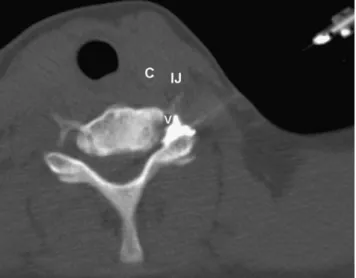
The indirect injection procedure and its benefits as well as the associated risks were discussed with the patient and informed consent was obtained. All cervical indirect nerve root injections were performed as outpatient procedures with CT fluoroscopic guidance (40-detector row CT, Philips Brilliance; Philips Medical Systems, Best, the Netherlands) by radiologists experienced in spinal inter-ventions. All radiologists proceeded according to a stan-dardized protocol to ensure the consistency of the indirect cervical nerve root injections. For the procedure, the patient was placed prone on the CT table with the head in a straight position and the forehead on a pillow for mechanical support. A lateral scout image was obtained to select the appropriate cervical level, then a CT acquisition was performed over 2–3 cervical levels. The entry site on the skin was chosen dorsal to the lateral aspect of the facet joint, which was directly adjacent to the targeted cervical foramen. Craniocaudal location information was transferred from the CT control station to the skin with the built-in laser navigation, and then the skin was disinfected. A second needle with the cap still on was placed flat on the skin surface to ascertain the correct mediolateral position under CT fluoroscopy before a 23-gauge needle was inserted into the skin. The needle position was aimed at the lateral aspect of the chosen facet joint and adjusted in several steps using CT fluoroscopy so that the needle tip directly touched the bone of the facet joint (Fig.1).
An injection of 0.5 mL iopamidol (Iopamiro 200, 200 mg of iodine per milliliter; Bracco, Milan, Italy) was performed to verify the correct position of the needle tip while manually holding the needle in position to avoid slight needle dislocation due to the injection pressure (Fig.2). The contrast material distribution was monitored with a single CT fluoroscopic scan (4 slices with a slice thickness of 3.75 mm; field of view, 15 cm; bone window setting): the observed contrast material distribution with this indirect cervical nerve root injection method was either dorsal to the facet joint at the level of the targeted nerve root or a portion of the injected contrast material was found lateral to the facet joint or even ventral to the joint with direct contact with the nerve root. Hence, in some patients who received an indirect cervical nerve root injection the drugs had immediate and direct contact with the targeted nerve root, as is usually the case with the direct cervical transforaminal nerve root injection approach. Following the injection of contrast material, 4 mg (1 mL) of the non-particulate corticosteroid preparation dexamethasone (Fortecortin Inject; Merck, Darmstadt, Germany) was slowly injected. This was fol-lowed by a slow injection of 1 mL of 0.2% ropivacaine (Naropin 0.2%; Astra-Zeneca, Södertälje, Sweden).
Direct cervical transforaminal nerve root injection approach
The direct anterolateral approach for CT-guided cervical transforaminal nerve root injections has been described in the literature [5, 21]. All direct cervical transforaminal nerve root injections were performed by radiologists with the same level of experience in spinal interventions as for the indirect cervical nerve root injections. While some elements are identical to the indirect approach described above, the following steps were performed differently. The patient was placed supine on the CT table with the head turned slightly to the side opposite to the injection site. The entry site on the skin was chosen at the same level or slightly ventral to the targeted cervical foramen on the CT
images correlating with a lateral or anterolateral approach in order to avoid the carotid and jugular vessels. A 23-gauge needle was inserted into the skin with the needle parallel to the table or angled slightly dorsally. The needle position was aimed at the posterior border of the cervical foramen and adjusted in several steps using CT fluoroscopy so that the needle tip was located at the outer edge of the posterior aspect of the foramen, dorsal to the nerve root (Fig.1).
An injection of 0.3 mL of iopamidol was performed to verify the correct position of the needle tip (Fig. 3). The intraforaminal or periradicular contrast distribution was monitored with a single CT fluoroscopic scan. A quantity of the crystalloid corticosteroid suspension triamcinolone (40 mg [1.0 mL]; Kenacort A 40; Dermapharm, Munich, Germany) and 1 mL of 0.2% ropivacaine were slowly injected.
Subjects
After a trial period of 3 months in 2009, during which the radiologists became familiar and comfortable with the indirect cervical nerve root injection procedure, 55 consecutive patients who received an indirect cervical nerve root injection were included in the study. Two patients had to be excluded because they did not present for the follow-up data collection, resulting in 53 patients. These 53 patients were matched for age and gender with 53 other patients who had previously received the traditional direct cervical transforaminal nerve root injection. Starting with January 2006, consecutive direct cervical transforaminal injection patients who were the exact age and gender matches for the indirect cervical nerve root injection patients were included in the study until all 53 indirect injection patients were matched. All of these patients were referred to the radiology department with clinical signs and symptoms of nerve root compression from disc hernia-tion, degenerative intervertebral foraminal stenosis or a combination of the two conditions.
As a routine part of all imaging-guided injection procedures at this hospital, pain data are collected before
Fig. 1 Schematic figure of cervical nerve root injections. a Indirect cervical nerve root injection. The needle is posi-tioned at the dorsal border of the facet joint and the injected drugs reach the target indirectly. b Direct cervical transforaminal nerve root injection. The needle is positioned at the ventral border of the facet joint, which forms the dorsal border of the intervertebral foramen. The injected drugs reach the target directly. Ccarotid artery, V vertebral artery, IJ internal jugular vein
and shortly after diagnostic or therapeutic injections. Using the numerical rating scale (NRS) where 0 is no pain and 10 is unbearable pain, the radiologists report any change in pain 20–30 min after the injection. This difference in pain before and after injection was converted to a percentage score. The age and gender matching of the patients was done before calculating their percentage pain change.
Statistical analysis
The 2006 pain change data were given by the radiologists as a direct percentage score, and were either decreased or
increased. In 2009–2010 these data was provided by the exact numbers on the 11-point visual analog pain scale (VAS) where 0 is no pain and 10 is unbearable pain. These values were converted to a percentage change score, and the mean percentage pain decrease or increase with the standard deviations for both the indirect and direct nerve root injection patients was compared using the unpaired Student’s t test. P<0.05 was set as indicating statistical significance. The computer software package SPSS (version 16; SPSS, Chicago, IL, USA) was used for statistical calculations.
Results
The mean patient age for the two groups was 52 years (SD= 11.4 for the indirect cervical nerve root injection group and SD=10.3 for the direct cervical transforaminal nerve injection group) with an age range of 29–74 years. There were 52 men and 54 women in the study, and the female patients were on average 6 years older than the male patients in both groups (mean age 55 years for women and 49 years for men). For the 53 patients receiving indirect cervical nerve root injections, C7 was the most common nerve root level targeted (29 patients) followed by the C6 level (22 patients). Similarly, C7 was the most common injection level for the direct nerve root injection patients (24 out of 53) and C6 was second with 20 patients receiving an injection at this level.
The mean percentage of pain decrease post-injection for the patients receiving indirect cervical nerve root injections was 38.4% (SD=34.5) and for patients undergoing the traditional direct cervical transforaminal nerve root
injec-Fig. 2 Indirect cervical nerve root injection. a The needle and contrast material are located behind the facet joint. b The needle is located behind the facet joint; contrast material can be seen next to the needle, but also lateral and in front of the facet joint next to the extraforaminal portion of the nerve root. Transverse CT image
Fig. 3 Direct cervical transforaminal nerve root injection. The needle and contrast material are located within the intervertebral foramen. Transverse CT image
tions it was 43.2% (SD=30.7). This was not statistically significant (P=0.455).
There were no adverse effects reported after either of the injection procedures.
Discussion
The purpose of this study was primarily to describe the technique for this indirect cervical nerve root injection method. Additionally, a comparison of the immediate post-injection patient responses between the direct cervical transforaminal nerve root injection and the indirect cervical nerve root injection approaches was made.
The two patient groups were purposefully age and gender matched before the immediate post-injection pain responses were analyzed so that the risk of bias was reduced. It is encouraging that although the direct nerve root injection procedure had a slightly higher mean percentage of pain reduction immediately after injection, this was not statistically significantly better than the indirect injection approach. The 43.2% pain reduction using the direct injection method is consistent with a previous study evaluating patient responses at this same time point after injection [5] where 46% pain reduction was obtained. It will be very interesting to follow these indirect nerve root block patients over time to determine whether or not their pain levels continue to improve beyond the immediate post-injection time point. It seems logical that it may take longer for the drugs to affect the involved nerve root using this procedure as the injections are not performed directly over the relevant anatomy, but at a slight distance. Even with the direct nerve root injection approaches it has been shown that the positive effects improve over time, at least up to 4 weeks using either liquid or crystalline steroid prepara-tions [2,22].
Another alternative to the traditional direct approach for cervical transforaminal CT-guided nerve root injections is a “modified direct” approach, the so-called dorsal approach as described by Wolter et al. [23,24]. With this method the patient lies prone on the table and the needle is inserted posterolateral to the nerve root foramen so that the tip of the needle can be placed directly between the extra-foraminal portion of the nerve root and the facet joint. This dorsal approach, based on extraforaminal rather than transforami-nal blocks, is suspected to be less prone to serious adverse effects because puncturing the nerve root artery, the vertebral artery but also the carotid and jugular vessels is less likely compared with the transforaminal nerve root injections for anatomical reasons [23, 24]. The technique used in our study is markedly different from the so-called dorsal approach described above because the tip of the needle is not placed in an extra-foraminal position, but
rather dorsal to the facet joint and the therapeutic effect originates from drugs that disperse to the extra-foraminal or intra-foraminal portion of the nerve root either initially or a few hours after the injection.
In the more commonly performed traditional cervical transforaminal nerve root injection procedure, the mecha-nism by which particulate steroids may enter the arterial system and travel to the spinal cord and brain causing infarction has been the subject of several recent papers [9– 19]. It is strongly suggested that the needle inadvertently penetrates one of the small arteries (sometimes called medullary feeding arteries, segmental arteries or radicular arteries) within the intervertebral foramen [9]. These small arteries are inconsistent in their anatomical course and thus their location cannot be predicted in order to direct needle placement away from them. They arise from the vertebral arteries and may contribute to the distribution of the anterior cervical artery [10, 11]. Indeed, many of the reported cases of tetraplegia have the symptoms of a cervical anterior spinal artery syndrome [10,11] consistent with accidental embolization to this vessel. The vertebral artery has also been implicated as the source of needle penetration and hence spinal cord or brain infarction [16– 18]. Based on the anatomy of the neck, puncture of the nerve root artery, the vertebral artery or the carotid and jugular vessels is extremely unlikely with our indirect cervical nerve root injection approach and we therefore expect this procedure to be safer than the cervical trans-foraminal approach and the risk of serious adverse effects to still be lower than with the dorsal approach used by Wolter et al. [23,24].
The indirect cervical nerve root injection is similar to the technique used for CT-guided cervical facet joint infiltration [25], except that there is no attempt to place the needle into the joint itself and the non-particulate corticosteroid preparation dexamethasone is used instead of a crystalloid corticosteroid. Serious adverse effects in image-guided cervical facet joint infiltration are rare [26].
A limitation to this study is the lack of longer term follow-up for patients receiving both of these injection procedures. Because the direct cervical transforaminal nerve root injection procedure is no longer performed at this hospital, it is not possible to obtain these data. Long-term outcomes using the indirect cervical nerve root injection procedure are currently being collected and will be reported in due course. Ideally, a randomized clinical trial would be carried out to compare outcomes from these two injection approaches. However, this is not possible as the direct procedure is no longer performed at this hospital owing to concerns over possible adverse events. Another limitation is that for the patient group who received a direct cervical transforaminal nerve root injection in 2006, there are no data recorded that indicate how many needle
placements required repositioning because of intra-vascular flow demonstrated by CT fluoroscopy. In the patient group with the indirect cervical nerve root injection, however, there were no cases where the needle had to be repositioned owing to intra-vascular flow.
Conclusions
The indirect cervical nerve root block injection procedure is described as a potentially safer alternative to the traditional cervical transforaminal nerve root injection. The short-term pain reduction is nearly as good using this indirect approach compared with the traditional direct injection method.
Acknowledgement The study was funded by the Vontobel-Stiftung,
Tödistrasse 17, 8002 Zürich, Switzerland.
References
1. Slipman CW, Lipetz JS, Jackson HB, Rogers DP, Vresilovic EJ. Therapeutic selective nerve root block in the nonsurgical treatment of atraumatic cervical spondylotic radicular pain: a retrospective analysis with independent clinical review. Arch Phys Med Rehabil. 2000;81:741–6.
2. Dreyfuss P, Baker R, Bogduk N. Comparative effectiveness of cervical transforaminal injections with particulate and nonparticu-late corticosteroid preparations for cervical radicular pain. Pain
Med. 2006;7:237–42.
3. Scanlon GC, Moeller-Bertram T, Romanowsky SM, Wallace MS. Cervical transforaminal epidural steroid injections. More
danger-ous than we think? Spine. 2007;32:1249–56.
4. Radhakrishnan K, Litchy WJ, O’Fallon WM, Kurland LT.
Epidemiology of cervical radiculopathy. A population-based study
from Rochester, Minnesota, 1976–1990. Brain. 1994;117:325–35.
5. Strobel K, Pfirrmann CWA, Schmid M, Hodler J, Boos N, Zanetti M. Cervical nerve root blocks: indications and role of MR imaging. Radiology. 2004;233:87–92.
6. Vallée JN, Feydy A, Carlier RY, Mutschler C, Mompoint D, Vallée CA. Chronic cervical radiculopathy: lateral-approach periradicular corticosteroid injection. Radiology. 2001;218:886–92.
7. Windsor RE, Storm S, Sugar R, Nagula D. Cervical trans-foraminal injection: review of the literature, complications, and a
suggested technique. Pain Physician. 2003;6:457–65.
8. Carragee EJ, Hurwitz EL, Cheng I, Carroll LJ, Nordin M, Guzman J, et al. Treatment of neck pain: injections and surgical
interventions: results of the bone and joint decade 2000–2010 task
force on neck pain and its associated disorders. Spine. 2008;33
(Suppl):S153–69.
9. Huntoon MA. Cervical spine: case presentation, complications,
and their prevention. Pain Med. 2008;9:S35–40.
10. Baker R, Dreyfuss P, Mercer S, Bogduk N. Cervical trans-foraminal injection of corticosteroids into a radicular artery: a possible mechanism for spinal cord injury. Pain. 2003;103:211–5. 11. Brouwers PJAM, Kottink EJBL, Simon MAM, Prevo RL. A cervical anterior spinal artery syndrome after diagnostic blockade of the right C6-nerve root. Pain. 2001;91:397–9.
12. Rosenkranz M, Grzyska U, Niesen W, Fuchs K, Schummer W, Weiller C, et al. Anterior spinal artery syndrome following
periradicular cervical nerve root therapy. J Neurol. 2004;251:229–31.
13. Karasek M, Bogduk N. Temporary neurologic deficit after cervical transforaminal injection of local anesthetic. Pain Med.
2004;5:202–5.
14. Tiso RL, Cutler T, Catania JA, Whalen BS, Pharm BCPS. Adverse central nervous system sequelae after selective transforaminal block:
the role of corticosteroids. Spine J. 2004;4:468–74.
15. Wallace MA, Fukui MB, Williams RL, Ku A, Baghai P. Complica-tions of cervical selective nerve root blocks performed with
fluoroscopic guidance. AJR Am J Roentgenol. 2007;188:1218–21.
16. Okubadejo GO, Talcott MR, Schmidt RE, Sharma A, Patel AA, Mackey RB, et al. Perils of intravascular methylprednisolone injection into the vertebral artery. An animal study. J Bone Joint Surg Am. 2008;90:1932–8.
17. Suresh S, Berman J, Connell DA. Cerebellar and brainstem infarction as a complication of CT-guided transforaminal cervical
nerve root block. Skeletal Radiol. 2007;36:449–52.
18. Rozin L, Rozin R, Koehler SA, Shakir A, Ladham S, Barmada M, et al. Death during transforaminal epidural steroid nerve root block (C7) due to perforation of the left vertebral artery. Am J
Forensic Med Pathol. 2003;24:351–5.
19. Furman MB, Giovanniello MT, O’Brien EM. Incidence of
intravascular penetration in transforaminal cervical epidural
steroid injections. Spine. 2003;28:21–5.
20. Rao R. Neck Pain, Cervical radiculopathy and cervical myelop-athy. Pathophysiology, natural history and clinical evaluation. J
Bone Joint Surg Am. 2002;84-A:1872–81.
21. Wagner AL. CT fluoroscopic-guided cervical nerve root blocks. Am J Neuroradiol. 2005;26:43–4.
22. Pfirrmann CW, Oberholzer PA, Zanetti M, Boos N, Trudell DJ, Resnick D, et al. Selective nerve root blocks for the treatment of sciatica: evaluation of injection site and effectiveness—a study with patients and cadavers. Radiology. 2001;221:704–11. 23. Wolter T, Mohadjer M, Berlis A, Knoeller S. Cervical CT-guided,
selective nerve root blocks: improved safety by dorsal approach.
Am J Neuroradiol. 2009;30:336–7.
24. Wolter T, Knoeller S, Berlin A, Hader C. CT-guided cervical selective nerve root block with a dorsal approach. Am J
Neuro-radiol. 2010;31:1831–6.
25. Hechelhammer L, Pfirrmann CW, Zanetti M, Hodler J, Boos N, Schmid MR. Imaging findings predicting the outcome of cervical
facet joint blocks. Eur Radiol. 2007;17:959–64.
26. Boswell MV, Colson JD, Sehgal N, Dunbar EE, Epter R. A systematic review of therapeutic facet joint interventions in