Psychopharmacology (2006) 185: 226–239 DOI 10.1007/s00213-005-0286-y
O R I G I N A L I N V E S T I G ATI O N
Daria Peleg-Raibstein . Esther Sydekum . Holger Russig . Joram Feldon
Withdrawal from continuous amphetamine administration
abolishes latent inhibition but leaves prepulse inhibition intact
Received: 30 September 2005 / Accepted: 2 December 2005 / Published online: 10 February 2006# Springer-Verlag 2006
Abstract Rationale: Schizophrenia has been associated with dysregulation of dopamine (DA) transmission and impairment in a number of experimental tasks, including sensorimotor gating assessed using prepulse inhibition (PPI) and selective attention assessed using latent inhibi-tion (LI). We have demonstrated in previous studies that after withdrawal from escalating (ESC) dosages of am-phetamine (AMPH), animals exhibited disruption of LI but no alteration of PPI. Moreover, these animals always showed behavioural sensitization to an AMPH challenge. Objective: In this study, we were interested in testing whether a different administration schedule would elicit disruption of both LI and PPI. Methods: Animals were treated with continuous AMPH release (via osmotic mini-pumps at a dosage of 10 mg kg−1day−1for 7 days) and tested for their performance in LI and PPI during withdrawal in a drug free state. Rats received AMPH treatment during the induction phase in their home cages or in the activity chambers. Following withdrawal, the ex-pression of behavioural sensitization to an AMPH chal-lenge was tested in both cases in the activity chambers. Results: Animals pretreated with AMPH from both groups did not exhibit behavioural sensitization. Withdrawal from continuous administration induced LI attenuation with no effect on PPI. Conclusions: These findings are similar to what was previously found with respect to an ESC AMPH regime. The only difference between the schedules was that the ESC AMPH schedule led to behavioural sensitization
whereas the continuous AMPH did not. It is suggested that the expression of sensitization may not be a prerequisite for observed LI disruption.
Keywords Latent inhibition . Prepulse inhibition . Withdrawal . Amphetamine . Sensitization . Rat . Schizophrenia
Introduction
One of the leading views on the pathophysiology of schizo-phrenia contends that the unmedicated schizophrenic brain is characterized by an endogenous sensitized dopaminergic system. Recent positron emission tomography (PET) and single-photon emission computerized tomography (SPECT) imaging techniques examining the displacement of a radio-active D2 receptor binding ligand by endogenously released dopamine (DA) have revealed that untreated schizophrenic patients show enhanced striatal DA release in response to an acute amphetamine (AMPH) challenge administration (i.e. a sensitized response) (Laruelle2000). Moreover, the DA release was positively correlated with the severity of their positive symptoms, supporting an endogenous sensi-tization hypothesis of at least the positive symptoms of schizophrenia (Abi-Dargham et al. 1998; Laruelle 2000; Laruelle et al.1999).
In experimental animals, injections of AMPH are known to induce a state of behavioural sensitization (Kalivas and Stewart 1991; Robinson and Becker 1986). Behavioural sensitization consists of two distinct phases, termed in-duction and expression. The inin-duction of behavioural sen-sitization to psychostimulants is defined as the transient sequence of cellular and molecular events precipitated by repeated psychostimulant administration that lead to the enduring changes in neuronal function responsible for the expression of behavioural sensitization (Kalivas and Stewart1991). The expression of behavioural sensitization is indicated by an augmentation of behaviours (e.g. loco-motion, stereotypes) (Kalivas et al. 1993; Robinson and Becker1986) combined with enhanced striatal DA release D. Peleg-Raibstein . E. Sydekum . J. Feldon (*)
Laboratory of Behavioural Neurobiology,
Swiss Federal Institute of Technology (ETH Zurich), Schorenstrasse 16, 8603 Schwerzenbach, Switzerland e-mail: feldon@behav.biol.ethz.ch Tel.: +41-44-6557448 Fax: +41-44-6557203 Present address: H. Russig
Brain Research Institute, University of Zurich/ETH Zurich, Winterthurerstrasse 190,
(Kalivas and Stewart1991) to subsequent drug challenges and can persist even after prolonged periods of abstinence. Over the last few years, our laboratory has attempted to develop an animal model of schizophrenia based on the endogenous sensitization hypothesis. To mimic symptoms of schizophrenia in rats, we exposed them repeatedly to AMPH and tested the sensitized animals following varying periods of withdrawal, without further exposing them to any challenge administration. We evaluated AMPH with-drawal following a variety of administration schedules (Murphy et al.2001a,b; Russig et al.2002,2003a,b,2005) and examined the animals in a number of behavioural paradigms relevant to schizophrenia, in particular latent inhibition (LI) and prepulse inhibition (PPI). LI refers to the observation that repeated exposure to a stimulus with-out consequence comes to impede the formation of sub-sequent associations with that stimulus (Lubow1973). PPI is a phenomenon whereby low-to-moderate intensity prepulse stimuli attenuate startle responses to a subsequent intense pulse (Graham 1975; Hoffman and Ison 1980; Hoffman and Searle1965). Reductions in both LI and PPI have been reported in schizophrenic patients (Baruch et al. 1988; Braff et al.1978,2001; Braff and Geyer1990; Gray et al. 1995; Swerdlow et al. 1996; Weiner and Feldon 1997). These deficits can be reversed by neuroleptic treat-ment in both humans (Baruch et al.1988; Braff et al.2001; Gray et al.1992,1995; Kumari and Sharma2002; Kumari et al. 1999, 2002; Leumann et al. 2002) and animals (Mansbach et al.1988; Swerdlow et al.1991; Warburton et al. 1994; Weiner et al. 1996). Since LI and PPI can be similarly tested in both humans and animals (Baruch et al. 1988; Braff et al.1978,2001), they can therefore be con-sidered as translational paradigms relevant for testing schizophrenia-like symptoms in animals and for detecting new antipsychotic drugs. Thus, pharmacological and phys-iological manipulations that induce LI and PPI deficits are used to model schizophrenic patients’ inability to ignore irrelevant stimuli and sensorimotor gating impairments, respectively (Geyer et al.1990; Weiner and Feldon1997). To develop an animal model of schizophrenia, we have previously characterized the behavioural consequences of withdrawal from an intermittent (five daily injections of 1.5 mg/kg) and an escalating (ESC; 6 days with three daily injections of 1.0–5.0 mg/kg) AMPH schedule. We found that the different schedules of AMPH administration pro-duce opposite effects on LI. Our findings indicated that withdrawal from intermittent administration resulted in enhanced LI (Murphy et al. 2001a) while ESC adminis-tration eliminated LI following various withdrawal periods (Murphy et al. 2001b; Russig et al. 2002, 2003b). Fur-thermore, we provided substantial support for the predict-ive validity of the ESC AMPH schedule model by showing that the disrupted LI can be reversed following treatment with either the typical antipsychotic haloperidol or the atypical antipsychotic clozapine (Russig et al.2002). Our studies further demonstrated that following long periods of withdrawal, these previously drug-free tested animals were indeed sensitized in their responses both to low (Russig et al.2003a,c,2005) and high (2003c) dosages of an AMPH
challenge administration. In contrast to the rather persistent and enduring effects on LI both AMPH schedules did not affect PPI (Murphy et al.2001b; Russig et al.2003b). Even when PPI was tested following a subthreshold dosage of a DA agonist challenge, such as apomorphine or AMPH, AMPH-withdrawn animals exhibited normal PPI (Russig et al.2003b).
Withdrawal from AMPH administration has also been used as an animal model of depression (Barr et al.1999; Barr and Phillips1999,2002; Lin et al.1999,2000; Russig et al.2003c). In this context, however, the animals dem-onstrate‘depressive-like’ symptoms only during the early drug withdrawal period (2–5 days). Recently, two studies (Cryan et al.2003; Paterson et al.2000) found that during short-term withdrawal from a continuous AMPH admin-istration delivered via osmotic mini-pumps, stronger anhe-donia (i.e. elevations in brain reward thresholds) was observed compared to withdrawal from repeated AMPH administration (Lin et al.1999). However, in contrast to the short-term effects of withdrawal from AMPH (that were already diminished on day 5 of withdrawal) (Lin et al. 1999; Paterson et al. 2000) studied in several depression-related paradigms, it was shown by us and others that there are withdrawal effects lasting for a much longer (up to 63 days subsequent to last injection) time in paradigms related to schizophrenia such as LI (Murphy et al.2001b; Tenn et al.2005) and PPI disruptions (Peleg-Raibstein et al. 2005a,b; Tenn et al. 2005). With these observations in mind, we sought to find a drug administration regime that would be most appropriate for producing an improved animal model of schizophrenia.
Accordingly, we decided to use a continuous AMPH administration method in experiment 1. This was done to evaluate the consequences of withdrawal on both PPI and LI. We chose a dosage of 10 mg kg−1day−1for seven con-secutive days. This election was made to enable a com-parison with the ESC AMPH schedule employed in our laboratory, which had a total dose of 75 mg AMPH leading to LI disruption but which left PPI intact. Since animals that were pretreated with AMPH exhibited LI attenuation, we examined a subgroup of animals in post-mortem neuro-chemistry in an attempt to characterize the underlying neurochemical mechanisms of disrupted LI. These animals participated in the LI and PPI parts of the study but were not exposed to AMPH challenge for evaluation of the expression of sensitization.
In experiment 1, animals received drug treatment in their home cage. The expression of behavioural sensitization was tested in a different environment (in an activity chamber). Under these conditions, AMPH-pretreated animals did not express behavioural sensitization. This result was expected since it is known that this method of drug administration does not produce sensitization to a subsequent AMPH challenge (Robinson and Becker1986).
In experiment 2, we were interested to ascertain whether drug administration, during the induction phase and in the same environment as the one used for testing, would facilitate the expression of behavioural sensitization tested in the same test environment. Hence, we had two main
reasons for carrying out the second experiment. First, we were interested in evaluating a dose-dependent response of AMPH on locomotor activity during the 7-day period of continuous drug release in the induction phase. In this experiment, animals were kept in activity chambers throughout the entire drug-release period. Locomotor activity was monitored continuously during the course of the treatment. Second, we sought to evaluate whether this environment, previously paired with drug treatment, had an influence on the expression of behavioural sensitization. Therefore, animals were tested on withdrawal day 77 for sensitization of locomotor activity in response to an AMPH challenge in the same activity chambers.
Materials and methods
Subjects
Male Wistar rats (bred at the animal facilities of the Behavioural Neurobiology Laboratory, Schwerzenbach, Switzerland) weighing 300–350 g at the time of surgery (experiment 1, 26 rats; experiment 2, 24 rats) were used. The rats were housed under reversed light–dark cycle (lights on between 1900 and 0700 hours) in temperature-controlled (21±1°C) and humidity-temperature-controlled (55±5%) animal facilities. Free access to food and water was pro-vided throughout the experiment. All experimental proce-dures were carried out in the dark phase of the cycle. Before surgery, the rats were housed in groups of four per cage; after surgery, the rats were individually housed. Rats were handled daily for 3 days prior to surgery. All ex-periments were in agreement with the Principles of Lab-oratory Animal Care (NIH publication no. 86-23, revised 1985) and Swiss regulations for animal experiments.
Osmotic pump implantation and removal
Subjects were anesthetized with Nembutal (sodium pento-barbital, 50 mg/kg, Abbott Labs, North Chicago, IL, USA) and implanted with subcutaneous osmotic mini-pumps (Alzet model 2 ML1 for AMPH at a dosage of 10 mg kg−1 day−1 or model 2001 for a dosage of 1 mg kg−1 day−1 AMPH; Alza Corporation, Palo Alto, CA, USA) along the back, parallel to the spine, with the flow meter directed posteriorly. Pumps were filled with either physiological saline (SAL) or AMPH solution (Sigma Chemical Com-pany, St. Louis, MO, USA). The concentration of the latter was adjusted according to animal weight and pumping rate to deliver a dosage of 1 or 10 mg kg−1day−1. To ensure that the pumps started dispensing immediately upon implant, they were “primed” according to instructions from Alzet. The pumps were filled the afternoon prior to surgery and placed in a SAL-filled tube. This tube was placed in a 37°C water-bath overnight. The wound was closed with stainless
steel wound clips and cleaned with an aqueous polyvinyl-pyrrolidone (PVP)–iodine solution (Braunol 2000, Braun B, Braun Medical AG, Switzerland). Pumps were surgi-cally removed after 7 days using the same procedure.
For experiment 1, the groups were as follows: SAL (n=10) and 10 mg kg−1day−1AMPH (n=16). In the second experiment, rats were divided into three groups as follows: SAL (n=12), 1 mg kg−1day−1AMPH (n=6) and 10 mg kg−1 day−1AMPH (n=6).
Behavioural testing procedures and apparatus Experiment 1
Two-way active avoidance apparatus Testing was con-ducted in four identical shuttle boxes (Coulbourn Instru-ments, Allentown, PA, USA; model E10-16TC). Each box was set in a ventilated, sound- and light-attenuating shell (model E10-20). The internal dimensions of each chamber were 35×17×21.5 cm. The grid floor of each chamber was divided into two identical compartments by an aluminium hurdle (17 cm long, 4 cm high). The barrier was very thin to prevent animals from balancing on it, thereby avoiding shock. Foot shocks were applied to the grid floor by a constant direct current source (model E13-14) and a scan-ner (model E13-13) set at 0.5 mA intensity. During the experimental session, each chamber was illuminated by a diffuse light source (house light), mounted 19 cm above the grid floor in the centre of the side walls. The con-ditioned stimulus (CS) was a tone of 85 dB produced by a speaker (model E12-02) placed behind the shuttle box on the floor of the shell.
Active avoidance procedure The LI procedure in the two-way active avoidance paradigm was conducted over 3 days: two consecutive daily sessions of pre-exposure to either the tone and the apparatus or to the apparatus only and a conditioning session on the third day.
– Withdrawal days 2 and 3
– Exposure to the tone CS and the apparatus
– The pre-exposed (PE) rats received 50 presenta-tions of the tone with a duration of 10 s and a mean variable inter-stimulus interval of 50 s (range 10–90 s).
– Exposure to apparatus only
– Each non-pre-exposed (NPE) animal was con-fined to the box with the house light on for an equivalent period of time without any stimulus presentation. A general evaluation of each animal’ s activity level (PE and NPE groups) was obtained by recording the total number of crossings during the sessions.
– Withdrawal day 4
– Conditioning to the CS
– Each animal was placed into the shuttle box and received 100 avoidance trials according to a variable interval schedule of 50 s (ranging from 10 to 90 s). Each avoidance trial began with a 10-s tone followed by a 2-s, 0.5-mA shock, with the tone remaining on with each shock. If the rat crossed the barrier to the opposite compartment during the tone, the stimulus was terminated and no shock was delivered (avoidance response). A crossing response during the shock terminated both the tone and the shock (escape response). If the rat failed to cross during the entire tone–shock trial, the tone and the shock terminated after 12 s (unfinished trial). The total number of inter-trial crossings was recorded as an additional measure of locomotor activity.
Acoustic startle and PPI apparatus Testing was conducted in four ventilated startle chambers (SR-LAB, San Diego Instruments, San Diego, CA, USA), each containing a transparent Plexiglas tube (diameter 8.2 cm, length 20 cm) mounted on a Plexiglas frame. Noise bursts were presented via a speaker mounted 24 cm above the tube. Motion inside the tube was detected by a piezoelectric accelerometer below the frame. The amplitude of the whole body startle to an acoustic pulse was defined as the average of one hundred 1-ms accelerometer readings collected from pulse onset.
Acoustic startle response and PPI procedure After 6 days of withdrawal from AMPH administration, the rats were tested for the acoustic startle response and PPI. At the beginning of the PPI testing session, the rats were placed into the PPI chamber for a 2-min acclimatization period with a 68-dB background white noise level that continued throughout the session. After the acclimatization period, six pulse-alone trials (two trials per pulse intensity) were presented, comprising a 30-ms broadband burst of the following intensities: 100, 110 and 120 dB (P100, P110 and P120, respectively). These pulse-alone trials served to stabilize the rats’ startle response. Subsequently, 10 blocks of 16 discrete trials were presented to assess PPI. Each block included four different trial types presented pseudorandomly: pulse-alone (one trial for each pulse in-tensity), prepulse-alone (one trial for each prepulse inten-sity), prepulse followed by a pulse 100 ms after prepulse onset (one trial for each pulse–prepulse combination) and a single no-stimulus trial, i.e. the background noise level (one trial). The prepulses (20 ms of broadband burst) had in-tensities of 74, 80 and 86 dB (which correspond to +6, +12 and +18 dB above background). The session was concluded with a final block of six consecutive pulse-alone trials (two trials for each of the three pulse intensities). A variable interval between the trials was used with a mean of 15 s (ranging from 10 to 20 s).
Post-mortem monoamine measurements Animals (SAL, n=6; AMPH, n=6) were decapitated in a drug-free state 8 days after their last drug treatment. The brains were extracted from the skull within 1 min and placed ventral side up in a rat brain matrix (Harvard Apparatus, South Natick, MA, USA) on an ice-chilled plate. Double-edged ice-cooled blades were used to prepare 2-mm-thick cor-onal sections. The slices were placed on an ice-cold dis-section plate for the removal of discrete brain regions, using a 2-mm micropunch for the caudate–putamen (CPu), medial prefrontal cortex (mPFC), amygdala, dorsal hip-pocampus (dHippo) and ventral hiphip-pocampus (vHippo). To dissociate nucleus accumbens (NAC) shell from core, tissue containing the core and a small part of the anterior commissure were first extracted using a 1-mm punch needle, and then the tissue of the shell, surrounding the remaining hole, was removed with a 2-mm needle. The mPFC punch consisted primarily of its prelimbic and dorsal infralimbic parts, whereas the amygdalar punch included the central and basal regions of the nucleus. The CPu punch was obtained from the dorsolateral region of the striatum. Punch tips were pushed into the region of interest and then withdrawn. Tissue punches from the left and right hemispheres of each area of interest were weighed, placed in 1.5-ml polypropylene microcentrifuge tubes containing 300 μl ice-cold 0.4 M HClO4 and homogenized using
ultrasound. After centrifugation at 10,000g for 20 min at 4°C, the clear supernatant layers were removed into a 1-ml syringe and filtered through a 0.2-μm nylon filter to separate the insoluble residue. This solution was immedi-ately frozen and stored at −80°C until injection into the high-performance liquid chromatography (HPLC) system for the assessment of monoamines and their metabolites. For all brain regions, with the exception of CPu and NAC shell, an aliquot of 50μl was injected in the HPLC system. Due to the much higher concentration of DA in the CPu and NAC shell, only 10μl was injected into the column. Chromatographic conditions An HPLC system coupled with an amperometric electrochemical detector (Decade II; Antec, Leyden, The Netherlands) was used to determine concentrations of DA, dihydroxyphenylacetic acid (DO PAC), homovanillic acid (HVA), 5-hydroxytryptamine (5-HT) and 5-hydroxyindoleacetic acid (5-HIAA). The samples were injected via a refrigerated autoinjector (ASI-100, Dionex, USA) equipped with a 250μl injection loop. The samples were separated on a reversed-phase column (125×3 mm glass column, Nucleosil 120-3 C 18, Knauer, Berlin, Germany) maintained at 30°C by a column oven as part of the electrochemical detector. An HPLC pump (P680, Dionex, USA) connected to a pulse damper and a degasser was used to pump the mobile phase (see below) through the system. The working potential of the electrochemical glassy carbon flow cell (VT-03; Antec) was +0.70 V vs an ISAAC reference electrode. A chromatography workstation (Chromeleon, Dionex, Olten, Switzerland) was used for data acquisition and calculations. The mobile phase used consisted of 250 ml of HPLC-grade acetonitrile, which was added to 5 litres of aqueous solution containing 0.27 mM
sodium ethylendiammoniumtetraacetate (C10H14N2O8Na2·
2H2O), 0.43 mM triethylamine (C6H15N), 8 mM potassium
chloride and 0.925 mM octanesulphonic acid (C8H17O3SNa)
that acted as an ion pairing reagent and for which pH was adjusted to 2.95 by adding concentrated phosphoric acid. The mobile phase was pumped through the system at a flow rate of 0.4 ml/min. DA, 5-HT and their metabolites could be separated in a single run of about 14 min. The positions and heights of the peaks of the endogenous components were compared with samples of an external calibrating standard solution containing 1, 5, 10, 50, 100 and 500 nM of DA, DOPAC, HVA, 5-HT and 5-HIAA.
Sensitization apparatus The apparatus for the assessment of locomotor activity has been previously described (Russig et al. 2003b,c). Sixteen stations (25×40×40 cm) were used; each was equipped with a monochrome mini video camera with a wide-angle (100°), 2.5-mm lens which contained a sound-attenuating wooden cabin. Reversed light–dark cycle (lights on between 1900 and 0700 hours) was kept under the same conditions as in the animal rooms and animals had free access to food and water throughout the experiment. Video images were recorded by a video recorder and later transferred to a PC with a dedicated analysis program (Image,http://rsb.info.nih.gov/nih-image) and macroprogram (P. Schmid, Laboratory of Behavioural Neurobiology, Swiss Federal Institute of Technology, Zurich, Switzerland). The percentage of changed pixels between two adjacent 1-s images was used as measure of activity (Richmond et al. 1998).
Sensitization procedure Animals (SAL, n=4; AMPH, n=8) were tested for AMPH-induced locomotion. Locomotion was assessed in 12 of the 16 test boxes and consisted of three stages. In the first stage, each rat was weighed and habituated to the apparatus on withdrawal day 39 at 1600 hours for a period of 18 h. On withdrawal day 40, the animals were removed from the apparatus at 1000 hours and injected with SAL and placed back into the apparatus for the second stage, which consisted of 1 h of free exploration. At 1100 hours, the animals were injected with 1.0 mg/kg AMPH and returned to the apparatus for 4 h of free exploration.
Experiment 2
Locomotor activity and sensitization apparatus As des-cribed above for experiment 1.
Locomotor activity procedure All animals were housed in the animal room prior to surgery. Three hours after im-plantation of the osmotic mini-pumps, all rats were housed in the activity chambers for the duration of the experiment (7 days), and locomotor activity was continuously re-corded for 24 h a day for the entire 7-day period of SAL or AMPH release. Animal activity was analysed following
approximately 18 h of recovery. The analysis started from 0700 hours the following morning with a 12-h dark period (0700–1900 hours) and, thereafter, in blocks of 12 h for five 24-h periods. We analysed 5 days of dark–light cycle from the second day to the sixth day of drug release. On the seventh day, the rats were taken out of the activity chambers, and the mini-pumps were surgically removed. After a 3-h recovery period from surgery, all animals were transported back to the animal room.
Sensitization procedure Animals (SAL, n=12; 1 mg kg−1 day−1 AMPH, n=6; and 10 mg kg−1day−1 AMPH, n=6) were tested for AMPH-induced locomotion. The experi-ment was run in two replications according to a counter-balanced schedule, which included the factor of test boxes. All animals (n=24) were weighed and placed in the apparatus at 1600 hours for an 18-h habituation phase (day 74 or 76 of withdrawal). On withdrawal day 75 or 77, at 1000 hours, the animals were removed from the paratus, injected with SAL and placed back into the ap-paratus for the second stage, which consisted of 1 h of free exploration. At 1100 hours, the animals were injected with 1.0 mg/kg AMPH and returned to the apparatus for 4 h of free exploration.
Data collection and analysis Experiment 1
LI in an active avoidance
– Pre-exposure The total number of spontaneous crosses was subjected to a 2×2×2 split-plot analysis of variance (ANOVA) with the between-subjects factors of treat-ment (SAL and AMPH) and pre-exposure (PE, NPE) and repeated measures factor of pre-exposure day (day 1 and day 2).
– Conditioning The 100 trials were divided into 10 blocks of 10 trials and the average number of avoidance responses was calculated per block. These averaged numbers of avoidance responses were submitted to a 2×2×10 split-plot ANOVA with the between-subjects factors of treatment and pre-exposure and the repeated measures factor of 10 blocks of 10 trial blocks.
Acoustic startle response and PPI
– Startle habituation The mean reactivity obtained in the first block was compared against that measured in the last block of six pulse-alone trials across the three pulse intensities (P100, P110 and P120). For this purpose, the average reactivity of the two pulse presentations during the first block and the last block of six pulse-alone trials was calculated for each pulse intensity and submitted to a 2×3×2 (treatment×pulse intensity× block) split-plot ANOVA consisting of a
between-subjects factor of treatment (SAL and AMPH) and repeated measures factors of block (first and last) and pulse intensities (P100, P110 and P120).
– Startle reactivity The mean reactivity obtained on the pulse-alone trials in the 10 test blocks across the three pulse intensities was submitted to a 2×3×10 (treatment× pulse intensity×trials) split-plot ANOVA consisting of a between-subjects factor of treatment and repeated measures factors of pulse intensity (P100, P110, and P120) and pulse-alone trials (10).
– Prepulse inhibition (%PPI) The startle reactivity data obtained from the 10 test blocks to measure PPI were used to calculate the percent PPI (%PPI) per each pulse intensity induced by each prepulse intensity using the following formula: 100−[100×(startle amplitude on prepulse plus pulse trial/startle amplitude on pulse-alone trial)]. PPI was analysed using a 2×3×3 (treat-ment×pulse intensity×prepulse intensity) split-plot ANOVA consisting of a between-subjects factor of treatment and repeated measures factors of pulse intensity and of prepulse intensity.
– Prepulse-elicited reactivity The data of the startle reactivity elicited by the three types of prepulse-alone presentations plus the no-stimulus trials was first sub-jected to natural logarithmic transformation to conform to the homogeneity and normality assumptions of parametric ANOVA. The data were then submitted to a 2×4 (treatment × prepulse intensity) split-plot ANOVA with a between-subject factor of treatment (2) and a repeated measures factor of prepulse intensity (4). Post-mortem monoamine measurements For the analysis of neurochemical data, the tissue concentrations of trans-mitters and metabolites were calculated in nanograms per milligram of wet tissue weight. Since there was no sig-nificant difference between the right and left hemispheres, an average of wet tissue weight was calculated from both hemispheres of each brain region. Animals were taken according to a counterbalanced schedule, which included the factor of PE/NPE treatment from the LI experiment. There was no significant pre-exposure effect. From these values, metabolite/DA ratios (DOPAC/DA, HVA/DA) and 5-HIAA/5-HT were calculated for each individual rat. Neurochemical data were analysed by separate one-way ANOVAs of tissue concentrations and ratios for each region.
Sensitization of locomotor activity Locomotor activity data for the baseline period (last hour prior to the SAL injection) on withdrawal day 40 and SAL challenge injection were analysed by two separate 2×6 ANOVA with a between-subjects factor of treatment (SAL, AMPH) and a repeated measures factor of six 10-min blocks. The period following the AMPH challenge injection was analysed by a 2×24 (treatment×10-min blocks) split-plot ANOVA.
Experiment 2
Locomotor activity during drug release Locomotor activity during the 5 days of drug release was analysed by a 3×5×2×12 split-plot ANOVA. The between-subject factor of treatment (SAL, 1 mg kg−1day−1AMPH and 10 mg kg−1 day−1 AMPH) and repeated measures factors of days (5), cycle (dark, light) and hours (12).
Sensitization of locomotor activity Locomotor activity data on withdrawal day 75 or 77 baseline period and SAL chal-lenge injection were analysed by two separate 3×6 ANOVA with a between-subjects factor of treatment (SAL, 1 mg kg−1 day−1 AMPH and 10 mg kg−1 day−1 AMPH) and a re-peated measures factor of six 10-min blocks. The period following the AMPH challenge injection was analysed by a 3×24 (treatment×10-min blocks) split-plot ANOVA.
All statistical analyses were performed using the sta-tistical software StatView 5.01 (SAS Institute Inc., Cary, NC, USA) implemented on a PC using the Microsoft Windows XP operating system. Statistical significance was set at a probability level of p<0.05 for all tests. When main effects or interactions were found to be significant, post hoc comparisons were conducted using Fisher’s protected least significant test.
Results
Experiment 1
Effects of drug treatment on body weight
One-way ANOVA examining the effect of drug treatment on body weight, expressed as percentage change over the 7 days of drug administration, revealed that AMPH-treated animals exhibited weight loss (−6.4±1.1%) as compared with SAL-treated animals that gained weight slightly (+2.6±0.4%). This was supported by a significant main effect of treatment [F(1,24)=31.12, p<0.0001, data not
shown].
LI active avoidance
Activity during the pre-exposure session on withdrawal days 2 and 3 AMPH-treated animals were significantly less active compared with the SAL-treated animals, as was supported by a significant main effect of treatment [F(1,22)=9.23, p<0.007]. A comparison of the total number
of crossings during the two pre-exposure sessions revealed that AMPH-treated animals crossed less frequently com-pared with SAL-treated animals on the first day of pre-expossure (SAL 41.1±4.7 and AMPH 23.4±2.3; p<0.001) but not on the second day of pre-exposure (SAL 21.6±2.7
and AMPH 15.2±2.1; p>0.07). This observation was sup-ported by a significant treatment×pre-exposure day in-teraction [F(1,22)=8.88, p<0.007].
Conditioning session on withdrawal day 4 As can be seen in Fig. 1, all animals improved in their avoidance re-sponses over the 10 blocks of testing, which was supported by a highly significant main effect of blocks [F(9,198)=19.30,
p<0.0001]. AMPH-pretreated animals showed retarded avoidance acquisition as compared with the SAL-pretreat-ed animals. This was reflectSAL-pretreat-ed by a significant main effect of treatment [F(1,22)=4.85, p<0.04]. PE animals made
fewer avoidance responses than NPE animals, as reflected by a significant main effect of pre-exposure [F(1,22)=19.63,
p<0.0003]. As seen in Fig 1a, LI was clearly evident in SAL-pretreated animals (restricted post hoc tests compar-ing SAL-NPE and SAL-PE revealed, p<0.001) and was absent in AMPH-pretreated animals (p<0.1) (Fig.1b). The latter was exclusively due to reduced learning demonstrat-ed by the AMPH-NPE-treatdemonstrat-ed group (post hoc tests comparing SAL-NPE and AMPH-NPE, p<0.0001).
Acoustic startle response and PPI on withdrawal day 6
Startle habituation Comparing the startle response in the first block and the last block revealed a clear habituation effect (first block>last block), as was supported by a significant main effect of block [F(1,24)=6.98, p<0.02]. As
expected, the startle reactivity increased as a function of
the pulse intensity, reflected by a significant main effect of pulse intensity [F(2,48)=62.53, p<0.0001]. The degree
of startle habituation was lower in the P100 condition as compared with the P110 and P120, which was support-ed by a significant pulse intensity×block interaction [F(2,48)=12.60, p<0.0001]. The analysis yielded neither a
significant treatment effect nor any of its interactions (data not shown).
Startle reactivity The mean startle reactivity during the middle 10 pulse-alone trials increased as a function of the pulse intensity, supported by a significant main effect of pulse intensity [F(2,48)=88.12, p<0.0001]. With respect to
the measure of startle habituation, this reduction in startle appeared to be more pronounced under the higher pulse intensities, which was supported by a significant pulse intensity×trials interaction [F(18,432)=1.79, p<0.03]. The
analysis yielded neither a significant treatment effect nor any of its interactions (data not shown).
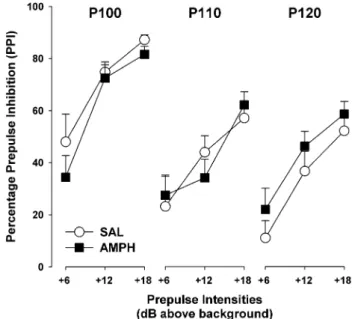
Prepulse inhibition (%PPI) A gradual increase in the amount of inhibition was observed as a function of the increasing intensity of the prepulse stimulus, which con-stitutes the PPI effect (Koch 1999). This was support-ed by a significant main effect of prepulse intensity [F(2,48)=86.28, p<0.0001]. Furthermore, an inverse
rela-tionship between mean %PPI and pulse intensity was found: an increase of the pulse intensity led to a reduction of %PPI, as supported by the significant main effect of pulse intensity [F(2,48)=23.67, p<0.0001] (Fig. 2). The analysis
Fig. 1 Percentage of avoidance responses made during a 100-trial test of conditioned two-way active avoidance acquisition in animals previously treated with either saline (SAL, a) or amphetamine (AMPH, b). Animals were tested on AMPH withdrawal day 4. Values are means (±SEM) from 10 blocks of 10 consecutive trials each. NPE Non-pre-exposed, PE pre-exposed. SAL-NPE, n=6; SAL-PE, n=4; AMPH-NPE, n=8; AMPH-PE, n=8
Fig. 2 Prepulse inhibition (PPI) expressed in terms of percentage inhibition, calculated with reference to the respective pulse-alone trials of a given pulse intensity and illustrated separately for the two treatment groups. All values are means (±SEM). SAL, n=10; AMPH, n=16
yielded neither a significant treatment effect nor any of its interactions.
Prepulse-elicited reactivity The 2×4 split-plot ANOVA of the ln-transformed reactivity scores on the no-stimulus and prepulse-alone trials revealed a significant prepulse inten-sity effect [F(3,72)=3.30, p<0.03]. This supports the
ob-servation that the prepulse-induced reactivity increased as a function of increasing prepulse intensities (Yee et al. 2004a,b,2005). The analysis yielded neither a significant treatment effect nor any of its interactions (data not shown).
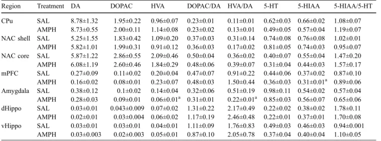
Post-mortem monoamine levels
The seven brain regions (CPu, NAC core, NAC shell, mPFC, dHippo, vHippo and amygdala)×the eight measures (DA, DOPAC, HVA, 5-HT, 5-HIAA, DOPAC/DA, HVA/ DA and 5-HIAA/5-HT) resulted in a total of 56 parameters, of which only 3 parameters were significantly different between the two treatment groups (see Table1).
In the amygdala, the AMPH-treated animals exhibited lower HVA levels as compared with SAL-treated animals, which was supported by a significant main effect of treatment [F(1,10)=10.14, p<0.01]. Moreover, the
AMPH-treated animals exhibited lower HVA/DA ratio (lower turnover) in the amygdala as compared with the SAL-treated animals. This observation was supported by a main effect of treatment [F(1,10)=5.09, p<0.05]. In the mPFC,
AMPH-treated animals exhibited lower 5-HIAA levels as compared with SAL-treated animals, as was supported by a significant main effect of treatment [F(1,10)=6.12, p<0.04].
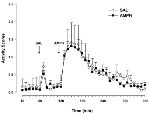
Expression of behavioural sensitization in response to a 1 mg/kg AMPH challenge injection on withdrawal day 40
Baseline stage Analysis of the last hour baseline test did not reveal any significant difference between the AMPH and the SAL groups (Fig.3).
Saline stage Following an acute SAL administration, ac-tivity increased and returned rapidly to baseline levels. This result was supported by a main effect of 10-min blocks [Fig. 3: F(5,50)=11.04, p<0.0001]. The analysis yielded
neither a significant treatment effect nor any interaction involving this factor.
AMPH stage In response to the 1 mg/kg AMPH challenge administration, all animals increased their activity levels, followed by a steady decline in activity after the challenge administration. This observation was supported by a main effect of 10-min blocks [Fig. 3: F(23,230)=11.52,
p<0.0001]. The analysis revealed no influence of treat-ment on AMPH-stimulated activity.
Experiment 2
Effects of drug treatment on body weight
One-way ANOVA examining the effect of drug treatment on body weight expressed as percentage of change over the 7 days of drug administration revealed that rats treated with 1 and 10 mg kg−1day−1AMPH exhibited weight loss (−2.3± 0.5 and −7.0±0.8%, respectively) as compared with the
Table 1 DA, DOPAC, HVA, 5-HT, 5-HIAA and metabolite/neurotransmitter ratios in the caudate–putamen (CPu), nucleus accumbens (NAC) core and shell, medial prefrontal cortex (mPFC), amygdala, dorsal hippocampus (dHippo) and ventral hippocampus (vHippo) after 8 days of withdrawal from continuous saline (SAL) or amphetamine (AMPH) administration of a dosage of 10 mg kg−1day−1for 7 days (SAL, n=4; AMPH, n=8)
Region Treatment DA DOPAC HVA DOPAC/DA HVA/DA 5-HT 5-HIAA 5-HIAA/5-HT
CPu SAL 8.78±1.32 1.95±0.22 0.96±0.07 0.23±0.01 0.11±0.01 0.62±0.03 0.66±0.02 1.08±0.07 AMPH 8.73±0.55 2.00±0.11 1.14±0.08 0.23±0.02 0.13±0.01 0.49±0.05 0.57±0.04 1.19±0.07 NAC shell SAL 5.25±1.55 1.83±0.42 1.09±0.20 0.37±0.03 0.31±0.14 0.74±0.08 0.76±0.08 1.02±0.01 AMPH 5.82±1.01 1.99±0.31 0.91±0.12 0.36±0.03 0.17±0.02 0.81±0.05 0.74±0.03 0.95±0.07 NAC core SAL 5.87±1.22 2.86±0.55 2.09±0.46 0.50±0.04 0.36±0.02 0.40±0.07 0.55±0.04 1.47±0.20 AMPH 6.08±1.19 2.60±0.46 1.84±0.29 0.48±0.06 0.39±0.07 0.31±0.04 0.44±0.03 1.57±0.17 mPFC SAL 0.27±0.09 0.11±0.02 0.20±0.04 0.47±0.07 0.91±0.22 0.44±0.06 0.37±0.02 0.87±0.10 AMPH 0.16±0.02 0.08±0.01 0.23±0.07 0.48±0.03 1.50±0.44 0.36±0.03 0.31±0.01a 0.89±0.06 Amygdala SAL 0.38±0.12 0.1±0.02 0.14±0.04 0.32±0.06 0.51±0.19 0.98±0.11 0.54±0.02 0.57±0.04 AMPH 0.28±0.03 0.09±0.01 0.06±0.01a 0.31±0.01 0.22±0.01a 0.85±0.03 0.56±0.07 0.65±0.06 dHippo SAL 0.03±0.01 0.043±0.009 0.07±0.02 1.31±0.22 2.17±0.49 0.22±0.02 0.38±0.02 1.78±0.11 AMPH 0.02±0.01 0.03±0.004 0.06±0.02 1.17±0.19 2.46±0.48 0.22±0.01 0.37±0.01 1.70±0.08 vHippo SAL 0.03±0.01 0.03±0.01 0.04±0.01 1.11±0.09 1.76±0.83 0.49±0.03 0.46±0.03 0.94±0.001 AMPH 0.03±0.003 0.02±0.003 0.05±0.01 0.87±0.10 2.05±0.78 0.37±0.04 0.40±0.04 1.10±0.05 Values are expressed as nanograms per milligram of tissue weight (±SEM)
a
SAL-treated control group which gained weight slightly (+1.0±0.3%). This was supported by a main effect of treat-ment [F(2,21)=78.94, p<0.0001, data not shown]. Post hoc
tests revealed that 1 and 10 mg kg−1day−1AMPH-treated animals exhibited significant weight loss compared with SAL-treated animals (both p<0.0001). Moreover, the 10 mg kg−1day−1AMPH-treated animals lost significantly more weight compared with the 1 mg kg−1 day−1 AMPH-treated animals (p<0.0001).
Locomotor activity
Days 2–6 of drug release The 3×5×2×12 (treatment×days× cycle×12 h) split-plot ANOVA revealed a significant main effect of treatment [Fig.4a: F(2,21)=47.07, p<0.0001]. The
10 mg kg−1day−1AMPH-treated animals exhibited higher locomotor activity compared with the SAL and 1 mg kg−1 day−1AMPH groups. The 10 mg kg−1day−1AMPH group exhibited higher activity levels during the 12 h averaged Fig. 3 Locomotor activity
measured during an initial 18-h habituation period (only the last hour shown), a 1-h period fol-lowing an injection of SAL and a 4-h period following a chal-lenge injection of 1.0 mg/kg AMPH. Testing was on with-drawal day 40 in animals that had been pretreated with a con-tinuous AMPH, at a dosage of 10 mg kg−1day−1for 7 days, or SAL. Values are means (±SEM). SAL, n=4; 10 mg kg−1day−1 AMPH, n=8
Fig. 4 a Mean activity during the entire drug administration period. b Locomotor activity during 5 days of drug administration. c Loco-motor activity during a 12:12-h dark–light cycle. Values are means
(±SEM). SAL, n=12; 1 mg kg−1day−1AMPH, n=6; 10 mg kg−1day−1 AMPH, n=6. *p<0.0001, #p<0.05
over the dark–light periods as compared with SAL and 1 mg kg−1 day−1 AMPH groups. All groups exhibited higher activity levels during the first hour of the cycle and then activity levels declined. These observations were supported by a significant treatment×12-h interaction [F(22,231)=2.73, p<0.0001] and by a significant main effect
of 12 h [F(11,231)=9.58, p<0.0001]. All treatment groups
exhibited higher locomotor activity during the dark cycle as compared with the light cycle. However, animals treated with 10 mg kg−1 day−1 AMPH exhibited higher locomotor activity during both the light and dark cycles as compared with animals treated with SAL and 1 mg kg−1 day−1 AMPH (p<0.0001). Furthermore, animals treated with 1 mg kg−1day−1AMPH exhibited significantly lower activity levels during the dark cycle as compared with the SAL-treated group (p<0.004). However, both groups exhibited similar activity levels during the light cycle (p>0.6). These observations were supported by a treatment× cycle×12-h interaction [Fig.4b: F(33,308)=2.95, p<0.02], a
significant treatment×cycle interaction [F(2,21)=5.44,
p<0.0001] and a main effect of cycle [F(1,28)=150.49,
p<0.0001].
Expression of behavioural sensitization in response to a 1 mg/kg AMPH challenge injection on withdrawal day 77
Baseline stage Analysis of the last hour baseline test did not reveal any significant difference between the AMPH and the SAL groups (Fig. 5).
Saline stage Following an acute SAL administration, ac-tivity increased and returned rapidly to baseline levels. This result was supported by a main effect of 10-min blocks [Fig.5: F(5,105)=63.68, p<0.0001].
AMPH stage In response to the 1 mg/kg AMPH challenge administration, all animals increased locomotor activity, followed by a steady decline in activity after the challenge administration, as was supported by a significant main effect of 10-min blocks [Fig.5: F(23,483)=39.40, p<0.0001].
There was no difference between the three treatment groups.
Discussion
This study investigated the behavioural and neurochemical consequences of withdrawal from continuous release of AMPH administration via subcutaneously implanted os-motic mini-pumps. The continuous low-level administra-tion of AMPH to animals might uniquely mimic some of the alterations in brain chemistry that occur during psy-chotic onset in schizophrenia. Two behavioural paradigms that are widely employed in schizophrenia research were examined: LI in an active avoidance paradigm and PPI (both tested in drug-free conditions). The major findings were that withdrawal from continuous AMPH administra-tion disrupted LI while leaving startle response and PPI unaffected. Behavioural sensitization in response to an AMPH challenge was evaluated in two separate experi-ments to test whether the expression of behavioural sen-sitization to AMPH is strengthened by the association of drug release with environmental cues. Expression of behavioural sensitization was not evident in AMPH-sensitized animals—neither when treatment was not paired with the test environment (first experiment) nor when treatment was paired with the test environment (second experiment). In the post-mortem monoamine measure-ments, seven brain regions were examined, and in each brain region, eight parameters were measured. Significant differences were detected only in the amygdala (reduced Fig. 5 Animals received
con-tinuous AMPH from two differ-ent dosages (1 and 10 mg kg−1 day−1for 7 days) or SAL in the activity boxes and tested for locomotor sensitization on with-drawal day 77 in the same boxes. Locomotor activity measured during an initial 18-h habituation period (only the last hour shown), a 1-h period following an injection of SAL and a 4-h period following a challenge injection of 1.0 mg/kg AMPH. Values are means (±SEM). SAL, n=12; 1 mg kg−1day−1 AMPH, n=6; 10 mg kg−1day−1 AMPH, n=6
HVA levels and reduced HVA/DA ratio) and in the mPFC (reduced 5-HIAA) in the AMPH-pretreated group as compared with the SAL-pretreated group.
We employed a continuous AMPH administration sched-ule that was recently used in the context of an animal model of depression (Cryan et al. 2003; Paterson et al. 2000). These studies have demonstrated that this schedule induced more pronounced anhedonia, as expressed by higher re-ward thresholds during the first few days of drug with-drawal, as compared with withdrawal from repeated AMPH administration (Lin et al. 1999). Our hypothesis was that like the situation within the framework of an animal model of depression, continuous AMPH treatment will induce a more pronounced withdrawal effect which might attenuate both LI and PPI. However, while observing a clear dis-ruption of LI, we obtained an intact PPI phenomenon. It could be argued that if the animals had been tested for PPI earlier, we would have seen a disruption. However, un-published results from animals that were exposed to an identical AMPH administration schedule have shown no such trend (Peleg-Raibstein et al., unpublished results). Disrupted LI and intact PPI are similar to what we observed previously with an ESC AMPH schedule (Murphy et al. 2001b; Russig et al.2002,2003b). It should be pointed out that both AMPH administration schedules were almost equivalent in the total AMPH dose (70 in the continuous schedule vs 75 mg/kg in the ESC schedule). Where the two AMPH schedules do differ is in their ability to induce expression of behavioural sensitization in response to a low dose of AMPH. While the ESC AMPH schedule clearly leads to behavioural sensitization in AMPH-treated ani-mals (Russig et al.2003a,c,2005), the continuous AMPH administration does not (Post1980; Robinson and Becker 1986). Behavioural sensitization was not expressed in response to a challenge injection of AMPH on day 40 of withdrawal (experiment 1) in AMPH continuously-treated animals. It is known that longer withdrawal periods lead to more augmented sensitization effect (Robinson and Becker 1986) and that the expression of sensitization may be context-dependent (Badiani et al. 1997; Browman et al. 1998; Robinson et al. 1998). Therefore, in the second experiment, animals were tested for the expression of sensitization on withdrawal day 77 using the same test environment as the one used for drug delivery during the induction phase. Even though it appeared graphically (Fig.5) that the 10 mg kg−1day−1AMPH group exhibited some behavioural sensitization as compared with the control and 1 mg kg−1day−1AMPH groups, these changes were far from statistically significant. The current results support and extend previous findings that continuous release, as opposed to repeated discrete AMPH injections, does not lead to the expression of behavioural sensitization (Post1980; Robinson and Becker1986).
In addition to our failure (in line with previous reports in the literature) to reveal expression of sensitization follow-ing continuous AMPH administration, the present study is the first to examine locomotor activity during the entire drug-release period of AMPH delivered by osmotic
mini-pumps. We employed a highly sensitive method of detec-tion of locomotor activity, continuously recording each second 24 h a day (Russig et al.2003c). The augmentation of locomotor activity was dose dependent, as animals treated with 10 mg kg−1day−1AMPH exhibited marked day and night hyperactivity compared with the SAL and 1 mg kg−1 day−1 AMPH-treated animals. The mean locomotor activity across days and cycles of the 1 and 10 mg kg−1day−1 AMPH-treated animals gradually declined from the second day to the last day of treatment. In contrast, activity levels of the SAL-treated animals remained unchanged for the du-ration of the experiment. This decreased activity observed in the AMPH-treated animals may be attributed to tolerance to the motor stimulant effect of AMPH with continuous exposure (Nielsen et al.1980).
LI was clearly demonstrated in the SAL group as the NPE animals made more avoidance responses than the PE animals. Conversely, in the withdrawn AMPH group, an attenuation of LI was observed that was largely due to impaired avoidance learning exhibited by the AMPH-NPE as compared with the SAL-NPE group. We have seen before that LI disruption is not necessarily due to improved learning in the AMPH-PE group only but can also be due to impaired learning in the AMPH-NPE group (Murphy et al. 2001b). Furthermore, this phenomenon was also demon-strated in acute schizophrenic patients where the PE sub-jects learned faster than the NPE subsub-jects (Baruch et al. 1988). Hence, the observed LI attenuation, due to impaired learning in the AMPH-NPE group, might partially mimic these (perhaps cognitive) deficits observed in schizo-phrenia patients since poor learning has been suggested to contribute to the LI deficits of schizophrenia patients (Green et al.2000; Swerdlow et al.1996). Alternatively, it can be suggested that the poor avoidance performance in the AMPH-NPE group can reflect a phenomenon called “learned helplessness” since similar behavioural learning deficits have been described in the literature after exposure to inescapable shocks (Anisman and Zacharko1990). The reduced avoidance of the AMPH-NPE group may indicate a depressive state similar to that seen in reductions of reward-motivated behaviours reported following short pe-riods of withdrawal from AMPH (Barr et al.1999; Barr and Phillips 1999; Lin et al. 1999). One measure clearly re-flecting learned helplessness is an enhanced number of escape failures (sometimes referred to as unfinished trials) (Overmier and Seligman1967; Vollmayr and Henn2001). However, the SAL-NPE (2.0±0.9) animals did not differ from the AMPH-NPE (4.1±1.2) animals in terms of unfinished trials. Since our data did not reveal an effect of AMPH withdrawal on unfinished trials, we can safely conclude that learned helplessness cannot account for the observed results. This conclusion is in line with previous findings from our laboratory which did not find an effect of withdrawal from an ESC AMPH schedule on learned helplessness (Russig et al.2003c).
In the current study, we employed a recently developed PPI protocol to enhance the detection sensitivity of PPI disrupting treatments (Yee et al. 2005). To achieve this
goal, this PPI paradigm manipulates the pulse intensity by using three levels of intensities (i.e. 100, 110 or 120 dB), as compared with the conventional PPI paradigm that em-ploys a single pulse intensity (Murphy et al.2001b; Russig et al.2003b). In line with previous observations (Pothuizen et al.2006; Yee et al.2005), our results clearly demonstrate that the magnitude of %PPI is not only a function of increasing prepulse intensities but is also affected by changes in intensity of the pulse stimulus. The weakest pulse stimulus of 100 dB was associated with the highest level of mean %PPI, which constitutes a confirmation of previous observations using this paradigm. We expected that this new PPI procedure would reveal a PPI disruption that would not have been observed using the conventional PPI procedure. Despite employing a more sensitive PPI program, we observed no difference between treatment groups (SAL and AMPH), in %PPI, or in startle reactivity. As the AMPH schedule had showed LI attenuation (in experiment 1), we examined one subgroup of animals in post-mortem neurochemistry with the aim of unravelling the underlying neurochemical mechanisms of disrupted LI by comparing it to the SAL-treated animals. On withdrawal day 8, we did not find DA depletion in either the CPu or in the NAC. Our results are in contrast to neurochemical studies that have shown that continuous release, albeit of higher AMPH doses, as compared with repeated intermit-tent or ESC AMPH administration, has a selective neu-rotoxic effect on DA terminals in the CPu (Ellison et al. 1996; Gately et al.1987; Ricaurte et al.1984; Ryan et al. 1990). However, the current results are in line with our previous findings that withdrawal from intermittent and ESC AMPH schedules did not lead to any significant neu-rochemical changes as compared with SAL treatment (Murphy et al. 2003). It seems that the dosage of 10 mg kg−1 day−1 over 7 days did not lead to an enduring depletion or neural damage in the striatum of Wistar rats. There are a number of factors that determine the extent to which the neurotoxic effects of continuous AMPH treat-ment are regionally and neurochemically specific, which include dose used, duration of treatment, age of the or-ganism, prior drug history and species (Robinson and Becker 1986). The only significant effects that emerged from the neurochemistry post-mortem experiment were reduced levels of 5-HIAA in the mPFC in the pretreated AMPH animals. In the amygdala, AMPH-pretreated ani-mals exhibited reduced HVA levels and reduced turnover indicated by reduced HVA/DA ratio. Perhaps one can relate the findings in the amygdala to the well-established retardation of active avoidance learning acquisition in-duced by amygdala lesions (Coover et al.1973; Werka and Zielinski 1998). Furthermore, clinical evidence suggested the mPFC abnormalities are involved in the pathopysiol-ogy of depression (Drevets et al.1997; Mann et al.1996; Williams et al. 2004). This may explain our findings of reduced 5-HIAA, the serotonin metabolite, in the mPFC.
Conclusion
The present study represents the first report investigating the effects of withdrawal from continuous AMPH release on LI and PPI. Continuous AMPH release, unlike ESC AMPH administration, has not led to any signs of sen-sitization either in the induction or in the expression eval-uation. We have found that the LI deficits observed following continuous AMPH administration are similar to those following ESC administration. Furthermore, both continuous and ESC AMPH regimes did not affect PPI. Thus, based on the results of the current study, sensitization cannot be seen as a prerequisite to LI disruption, a con-clusion which may have been drawn from the ESC AMPH studies. Future studies should further explore the neuro-biological mechanisms by which the different AMPH treaments induce disruption of LI and the underlying mechanisms that are responsible for these deficits.
Acknowledgements This study was supported by the Swiss Federal Institute of Technology (ETH Zurich, Switzerland). We would like to thank the animal care technicians for the excellent care of the animals used in these experiments. We also thank Mr. Peter Schmidt for his technical support and Mrs. Natalie Aeschbach-Jones for her editorial support.
References
Abi-Dargham A, Gil R, Krystal J, Baldwin RM, Seibyl JP, Bowers M, van Dyck CH, Charney DS, Innis RB, Laruelle M (1998) Increased striatal dopamine transmission in schizophrenia: confirmation in a second cohort. Am J Psychiatry 155:761–767 Anisman H, Zacharko RM (1990) Multiple neurochemical and behavioral consequences of stressors: implications for depres-sion. Pharmacol Ther 119–136
Badiani A, Camp DM, Robinson TE (1997) Enduring enhancement of amphetamine sensitization by drug-associated environmental stimuli. J Pharmacol Exp Ther 282:787–794
Barr AM, Phillips AG (1999) Withdrawal following repeated exposure toD-amphetamine decreases responding for a sucrose solution as measured by a progressive ratio schedule of re-inforcement. Psychopharmacology (Berl) 141:99–106 Barr AM, Phillips AG (2002) Increased successive negative contrast
in rats withdrawn from an escalating-dose schedule ofD
-amphet-amine. Pharmacol Biochem Behav 71:293–299
Barr AM, Fiorino DF, Phillips AG (1999) Effects of withdrawal from an escalating dose schedule ofD-amphetamine on sexual
behavior in the male rat. Pharmacol Biochem Behav 64: 597–604
Baruch I, Hemsley DR, Gray JA (1988) Differential performance of acute and chronic schizophrenics in a latent inhibition task. J Nerv Ment Dis 176:598–606
Braff DL, Geyer MA (1990) Sensorimotor gating and schizophrenia. Human and animal model studies. Arch Gen Psychiatry 47:181–188
Braff D, Stone C, Callaway E, Geyer M, Glick I, Bali L (1978) Prestimulus effects on human startle reflex in normals and schizophrenics. Psychophysiology 15:339–343
Braff DL, Geyer MA, Swerdlow NR (2001) Human studies of prepulse inhibition of startle: normal subjects, patient groups, and pharmacological studies. Psychopharmacology (Berl) 156:234–258
Browman KE, Badiani A, Robinson TE (1998) Modulatory effect of environmental stimuli on the susceptibility to amphetamine sensitization: a dose–effect study in rats. J Pharmacol Exp Ther 287:1007–1014
Coover G, Ursin H, Levine S (1973) Corticosterone and avoidance in rats with basolateral amygdala lesions. J Comp Physiol Psychol 85:111–122
Cryan JF, Hoyer D, Markou A (2003) Withdrawal from chronic amphetamine induces depressive-like behavioral effects in rodents. Biol Psychiatry 54:49–58
Drevets WC, Price JL, Simpson JR Jr, Todd RD, Reich T, Vannier M, Raichle ME (1997) Subgenual prefrontal cortex abnormal-ities in mood disorders. Nature 386:824–827
Ellison G, Irwin S, Keys A, Noguchi K, Sulur G (1996) The neurotoxic effects of continuous cocaine and amphetamine in habenula: implications for the substrates of psychosis. NIDA Res Monogr 163:117–145
Gately PF, Segal DS, Geyer MA (1987) Sequential changes in behavior induced by continuous infusions of amphetamine in rats. Psychopharmacology (Berl) 91:217–220
Geyer MA, Swerdlow NR, Mansbach RS, Braff DL (1990) Startle response models of sensorimotor gating and habituation deficits in schizophrenia. Brain Res Bull 25:485–498
Graham FK (1975) Presidential address, 1974. The more or less startling effects of weak prestimulation. Psychophysiology 12:238–248
Gray NS, Hemsley DR, Gray JA (1992) Abolition of latent inhibition in acute, but not chronic, schizophrenics. Neurol Psychiatry Brain Res 1:83–89
Gray NS, Pilowsky LS, Gray JA, Kerwin RW (1995) Latent inhibition in drug naive schizophrenics: relationship to duration of illness and dopamine D2 binding using SPET. Schizophr Res 17:95–107
Green MF, Kern RS, Braff DL, Mintz J (2000) Neurocognitive deficits and functional outcome in schizophrenia: are we measuring the“right stuff”? Schizophr Bull 26:119–136 Hoffman HS, Ison JR (1980) Reflex modification in the domain of
startle: I. Some empirical findings and their implications for how the nervous system processes sensory input. Psychol Rev 87:175–189
Hoffman HS, Searle JL (1965) Acoustic variables in the modifica-tion of startle reacmodifica-tion in the rat. J Comp Physiol Psychol 60:53–58
Kalivas PW, Stewart J (1991) Dopamine transmission in the initiation and expression of drug- and stress-induced sensitiza-tion of motor activity. Brain Res Brain Res Rev 16:223–244 Kalivas PW, Sorg BA, Hooks MS (1993) The pharmacology and
neural circuitry of sensitization to psychostimulants. Behav Pharmacol 4:315–334
Koch M (1999) The neurobiology of startle. Prog Neurobiol 59: 107–128
Kumari V, Sharma T (2002) Effects of typical and atypical antipsychotics on prepulse inhibition in schizophrenia: a critical evaluation of current evidence and directions for future research. Psychopharmacology (Berl) 162:97–101
Kumari V, Soni W, Sharma T (1999) Normalization of information processing deficits in schizophrenia with clozapine. Am J Psychiatry 156:1046–1051
Kumari V, Soni W, Sharma T (2002) Prepulse inhibition of the startle response in risperidone-treated patients: comparison with typical antipsychotics. Schizophr Res 55:139–146
Laruelle M (2000) The role of endogenous sensitization in the pathophysiology of schizophrenia: implications from recent brain imaging studies. Brain Res Brain Res Rev 31:371–384 Laruelle M, Abi-Dargham A, Gil R, Kegeles L, Innis R (1999)
Increased dopamine transmission in schizophrenia: relationship to illness phases. Biol Psychiatry 46:56–72
Leumann L, Feldon J, Vollenweider FX, Ludewig K (2002) Effects of typical and atypical antipsychotics on prepulse inhibition and latent inhibition in chronic schizophrenia. Biol Psychiatry 52: 729–739
Lin D, Koob GF, Markou A (1999) Differential effects of withdrawal from chronic amphetamine or fluoxetine adminis-tration on brain stimulation reward in the rat–interactions be-tween the two drugs. Psychopharmacology (Berl) 145:283–294 Lin D, Koob GF, Markou A (2000) Time-dependent alterations in
ICSS thresholds associated with repeated amphetamine admin-istrations. Pharmacol Biochem Behav 65:407–417
Lubow RE (1973) Latent inhibition. Psychol Bull 79:398–407 Mann JJ, Malone KM, Diehl DJ, Perel J, Cooper TB, Mintun MA
(1996) Demonstration in vivo of reduced serotonin responsivity in the brain of untreated depressed patients. Am J Psychiatry 153:174–182
Mansbach RS, Geyer MA, Braff DL (1988) Dopaminergic stimu-lation disrupts sensorimotor gating in the rat. Psychopharma-cology (Berl) 94:507–514
Murphy CA, Di Iorio L, Feldon J (2001a) Effects of psychostim-ulant withdrawal on latent inhibition of conditioned active avoidance and prepulse inhibition of the acoustic startle re-sponse. Psychopharmacology (Berl) 156:155–164
Murphy CA, Fend M, Russig H, Feldon J (2001b) Latent inhibition, but not prepulse inhibition, is reduced during withdrawal from an escalating dosage schedule of amphetamine. Behav Neurosci 115:1247–1256
Murphy CA, Russig H, Pezze MA, Ferger B, Feldon J (2003) Amphetamine withdrawal modulates FosB expression in mesolimbic dopaminergic target nuclei: effects of different schedules of administration. Neuropharmacology 44:926–939 Nielsen EB, Lee TH, Ellison G (1980) Following several days of
continuous administration D-amphetamine acquires
hallucino-genlike properties. Psychopharmacology (Berl) 68:197–200 Overmier JB, Seligman ME (1967) Effects of inescapable shock
upon subsequent escape and avoidance responding. J Comp Physiol Psychol 63:28–33
Paterson NE, Myers C, Markou A (2000) Effects of repeated withdrawal from continuous amphetamine administration on brain reward function in rats. Psychopharmacology (Berl) 152:440–446
Peleg-Raibstein D, Sydekum E, Feldon J (2005a) Differential effects on prepulse inhibition of withdrawal from two different repeated administration schedules of amphetamine. Int J Neuropsychopharmacol (in press)
Peleg-Raibstein D, Sydekum E, Russig H, Feldon J (2005b) Withdrawal from repeated amphetamine administration leads to disruption of prepulse inhibition but not to disruption of latent inhibition. J Neural Transm (in press)
Post RM (1980) Intermittent versus continuous stimulation: effect of time interval on the development of sensitization or tolerance. Life Sci 26:1275–1282
Pothuizen HHJ, Neumann KR, Feldon J, Yee BK (2006) Selective nucleus accumbens core lesions enhance dizocilpine-but not apomorphine-induced disruption of prepulse inhibition in rats. Behav Pharmacol (in press)
Ricaurte GA, Seiden LS, Schuster CR (1984) Further evidence that amphetamines produce long-lasting dopamine neurochemical deficits by destroying dopamine nerve fibers. Brain Res 303:359–364
Richmond MA, Murphy CA, Pouzet B, Schmid P, Rawlins JN, Feldon J (1998) A computer controlled analysis of freezing behaviour. J Neurosci Methods 86:91–99
Robinson TE, Becker JB (1986) Enduring changes in brain and behavior produced by chronic amphetamine administration: a review and evaluation of animal models of amphetamine psychosis. Brain Res 396:157–198
Robinson TE, Browman KE, Crombag HS, Badiani A (1998) Modulation of the induction or expression of psychostimulant sensitization by the circumstances surrounding drug adminis-tration. Neurosci Biobehav Rev 22:347–354
Russig H, Murphy CA, Feldon J (2002) Clozapine and haloperidol reinstate latent inhibition following its disruption during amphetamine withdrawal. Neuropsychopharmacology 26: 765–777
Russig H, Durrer A, Yee BK, Murphy CA, Feldon J (2003a) The acquisition, retention and reversal of spatial learning in the morris water maze task following withdrawal from an escalating dosage schedule of amphetamine in Wistar rats. Neuroscience 119:167–179
Russig H, Murphy CA, Feldon J (2003b) Prepulse inhibition during withdrawal from an escalating dosage schedule of amphet-amine. Psychopharmacology (Berl) 169:340–353
Russig H, Pezze MA, Nanz-Bahr NI, Pryce CR, Feldon J, Murphy CA (2003c) Amphetamine withdrawal does not produce a depressive-like state in rats as measured by three behavioral tests. Behav Pharmacol 14:1–18
Russig H, Murphy CA, Feldon J (2005) Behavioural consequences of withdrawal from three different administration schedules of amphetamine. Behav Brain Res 165:26–35
Ryan LJ, Linder JC, Martone ME, Groves PM (1990) Histological and ultrastructural evidence thatD-amphetamine causes
degen-eration in neostriatum and frontal cortex of rats. Brain Res 518:67–77
Swerdlow NR, Keith VA, Braff DL, Geyer MA (1991) Effects of spiperone, raclopride, SCH 23390 and clozapine on apomor-phine inhibition of sensorimotor gating of the startle response in the rat. J Pharmacol Exp Ther 256:530–536
Swerdlow NR, Braff DL, Hartston H, Perry W, Geyer MA (1996) Latent inhibition in schizophrenia. Schizophr Res 20:91–103 Tenn CC, Fletcher PJ, Kapur S (2005) A putative animal model of
the “prodromal” state of schizophrenia. Biol Psychiatry 57: 586–593
Vollmayr B, Henn FA (2001) Learned helplessness in the rat: improvements in validity and reliability. Brain Res Brain Res Protoc 8:1–7
Warburton EC, Joseph MH, Feldon J, Weiner I, Gray JA (1994) Antagonism of amphetamine-induced disruption of latent inhibition in rats by haloperidol and ondansetron: implications for a possible antipsychotic action of ondansetron. Psycho-pharmacology (Berl) 114:657–664
Weiner I, Feldon J (1997) The switching model of latent inhibition: an update of neural substrates. Behav Brain Res 88:11–25 Weiner I, Shadach E, Tarrasch R, Kidron R, Feldon J (1996) The
latent inhibition model of schizophrenia: further validation using the atypical neuroleptic, clozapine. Biol Psychiatry 40:834–843
Werka T, Zielinski K (1998) CS modality transfer of two-way avoidance in rats with central and basolateral amygdala lesions. Behav Brain Res 93:11–24
Williams W, Reimold M, Kerich M, Hommer D, Bauer M, Heinz A (2004) Glucose utilization in the medial prefrontal cortex correlates with serotonin turnover rate and clinical depression in alcoholics. Psychiatry Res 132:219–224
Yee BK, Chang DL, Feldon J (2004a) The Effects of dizocilpine and phencyclidine on prepulse inhibition of the acoustic startle reflex and on prepulse-elicited reactivity in C57BL6 mice. Neuropsychopharmacology 29:1865–1877
Yee BK, Russig H, Feldon J (2004b) Apomorphine-induced prepulse inhibition disruption is associated with a paradoxical enhancement of prepulse stimulus reactivity. Neuropsycho-pharmacology 29:240–248
Yee BK, Chang T, Pietropaolo S, Feldon J (2005) The expression of prepulse inhibition of the acoustic startle reflex as a function of three pulse stimulus intensities, three prepulse stimulus intensities, and three levels of startle responsiveness in C57BL6/J mice. Behav Brain Res 163:265–276