PHYSIOLOGY
CA-125 Concentrations in the Serum and Pregnancy Outcome
in IVF Cycles
ALFRED W. BRANDENBERGER,1,3 NICOLAS A. BERSINGER,1
PETER R. HUBER,2 ELISABETH BERGER,1 PATRICK GLANZMANN,1 and MARTIN H. BIRKHAEUSER1
Submitted: September 29, 1997 Accepted: December 19, 1997
Purpose: CA-125 has been proposed as a potential marker
for endometrial receptivity in assisted reproduction. This study was designed to evaluate whether the levels of CA-125 in the serum of patients undergoing IVF-embryo trans-fer (ET) is correlated with the outcome.
Methods: Levels of serum CA-125 were measured on the
day before and on the day of human chorionic gonadotropin (hCG) administration, ovum pickup (OPU), and ET in 74 patients undergoing 100 IVF cycles between January 1994 and March 1995. Patients were treated with a midluteal-phase gonadotropin-releasing hormone (GnRH) agonist pro-tocol and follicular-phase human menopausal gonadotropin.
Results: One hundred oocyte retrievals resulted in 91 ETs,
and 22 clinical pregnancies (22%|OPU and 24.2%/ET). The live-born rate was 21%/OPU and 23.1%/ET. Neither the CA-125 serum levels nor their increase from the day of hCG until the day of ET showed any prognostic significance to the outcome of IVF, and they were not correlated with the endometrium thickness or the number of oocytes retrieved or fertilized.
Conclusions: The CA-125 serum levels in conventional IVF
cycles were not correlated with the IVF outcome and yielded no prognostic information in a GnRH agonist down-regula-tion protocol.
KEY WORDS: CA-125; in vitro fertilization; endometrium; receptivity.
INTRODUCTION
Pregnancy outcome after in vitro fertilization (IVF) depends on many factors, but mainly on the quality of the embryos and the endometrial receptivity.
CA-125, well-known as a tumor marker for epithe-lial ovarian cancer (1), is a high molecular weight glycoprotein that is produced not only by ovarian can-cer, but also by nonovarian tumors, normal epithelia of the peritoneum, the endometrium, the fallopian tube, and the ovary. In some patients undergoing ovarian hyperstimulation, increased serum levels are detected. The exact source and amount of production are not entirely known. Because CA-125 is also produced by the endometrium, some authors suggest that CA-125 can be used as a marker for endometrial receptivity in patients undergoing IVF. Its role as a predictor for pregnancy outcome is controversial (2-4). Therefore, we sought to evaluate whether CA-125 serum levels have any predictive prognostic value regarding the outcome of conventional IVF.
1 Division of Endocrinology, Department of Obstetrics and
Gyne-cology, University of Bern, Bern, Switzerland.
2 Hormone and Tumormarker Laboratory, Department of Central Laboratories, University Hospital, Basel, Switzerland.
3 To whom correspondence should be addressed at Division of
Endocrinology, Department of Obstetrics and Gynecology, Schan-zeneckstrasse 1, 3012 Bern, Switzerland.
MATERIALS AND METHODS
Patients and IVF Protocols
Sera from 74 patients who underwent 100 conven-tional IVF cycles between January 1994 and March
1995 were analyzed. Fifty-five patients underwent one cycle, twelve underwent two cycles, and seven underwent three cycles during that period. Stimula-tion protocols were identical in all patients: D-Trp6 -gonadotropin-releasing hormone agonist (0.1 mg; Decapeptyl; Ferring, Duebendorf, Switzerland) was administered subcutaneously daily from the luteal phase of the preceding cycle. After the onset of menses, if transvaginal ultrasound showed no ovarian cyst and the estradiol (E2) levels were <40 pg/ml, stimulation was initiated with human menopausal gonadotropin (hMG) (Pergonal, Serono, Zug, Swit-zerland, or Humegon, Organon, Switzerland). The daily doses ranged from 150 to 300 1U per day. D-Trp was continued until the day of human chorionic gonadotropin (hCG) administration. On the basis of E2 levels and the presence of at least two follicles 17 mm or greater in diameter, as monitored by transvaginal ultrasound, 10,000 IU of hCG (Profasi, Serono, or Pregnyl, Organon) was given. Thirty-five to 38 hours later, oocyte pickup (OPU) was performed under i.v. sedation. Oocytes were aspirated into hepa-rin-containing flushing medium at 37 °C (Medi-Cult; Copenhagen, Denmark), cultured in IVF medium (Medi-Cult) and inseminated with 50,000 to 100,000 motile spermatozoa prepared by Percoll gradient in the same medium (1 ml). On the following day, fertilization was assessed microscopically and the culture medium was replaced; supernumerary pronu-clear-stage zygotes were frozen. Embryos were replaced 2 days after oocyte retrieval. One thousand international units of hCG was given intramuscularly every other day until the first measurement of hCG 14 to 15 days after embryo transfer (ET). Clinical pregnancy was defined as a positive heart action in transvaginal ultrasound 4 to 6 weeks after ET.
Blood was collected 2 and/or 3 days before OPU (day hCG/hCG-1), on the day of OPU, and on the day of ET. Care was taken that on the day of OPU, the blood samples were collected by venipuncture prior to insertion of the oocyte aspiration needle, to avoid any possible spilling of follicular CA-125 into the circulation. Serum was obtained by immediate centrif-ugation after coagulation and stored at — 30°C until CA-125 and hormone analyses were performed in batches. Blood was not taken on all 4 days for all the cycles.
Measurement of Serum Levels
Serum concentrations of CA-125 were determined by CA-125-11 enzyme immunoassay (ElA), based on
a double-antibody method using both the OC 125 and the M 11 antibody. The tests were run in batches on an automatic Cobas Core immunoanalyzer (F. Hoff-man-La Roche, Switzerland). The sensitivity of the test was 1.0 lU/ml. Intra- and inter-assay coefficients of variance at the low end of the standard assay range had been determined in a trial involving four centers and were 4.4% (number of repetitions = 21) and 5.1%, respectively, for a CA-125 concentration of 17.2 lU/ml. Serum levels of E2 were determined by dissociation-enhanced lanthanide immunofluorometric assay (Del-fia; Wallac, Finland) according to the manufacturer's instructions.
Progesterone was measured by radioimmunoassay using a coated tube kit (Coat-a-count; DPC, Los Angeles, CA). Due to the high serum progesterone levels encountered after ovarian hyperstimulation, all samples obtained on the day of ET were diluted 1:10 with zero diluent (DPC) prior to assay.
Statistical Analysis
Comparisons of the different parameters between the pregnant and the nonpregnant groups were made using the nonparametric Mann-Whitney test for two-tailed P values, because the data were not normally distributed. Only cycles with ET were included in that analysis. A P value < 0.05 was considered significant. Regression was used to detect any correla-tion between CA-125 levels and the number of oocytes retrieved, fertilization, the endometrial thick-ness, the embryo score, and the E2 and progesterone levels, including all cycles. Statistical calculations were performed using the StatView SE & Graphics package for Macintosh.
RESULTS
This study included 74 patients undergoing a total of 100 oocyte retrievals, resulting in 91 ETs and 22 clinical pregnancies (22% per OPU and 24.2% per ET). Twenty-one patients delivered live-born infants, and one patient had a pregnancy loss at 8 weeks of gestation. The take-home baby rate was 21% per OPU and 23.1% per ET.
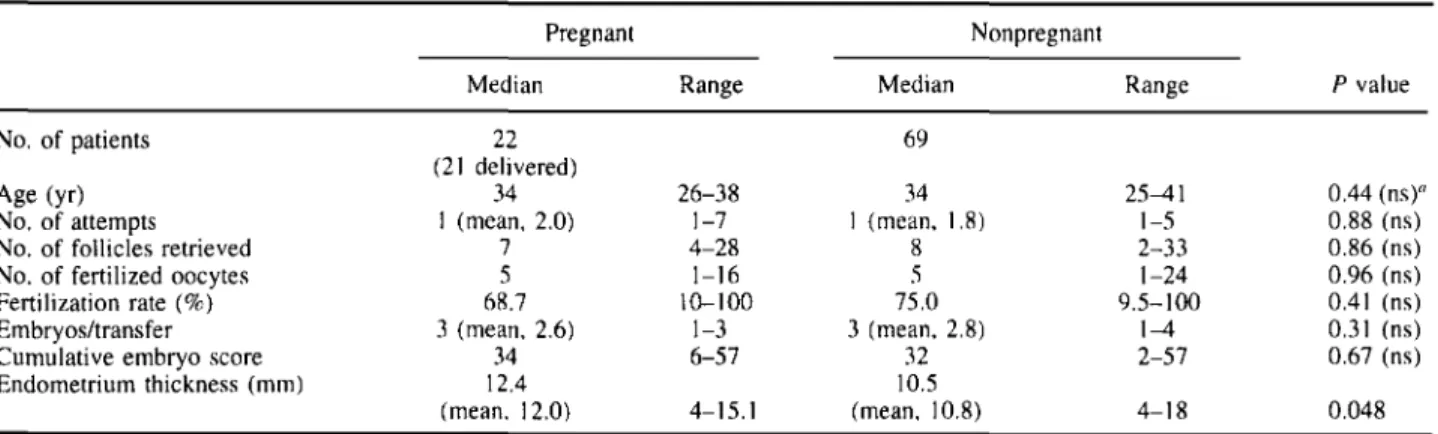
The patient characteristics are summarized in Table I. The patients' ages ranged from 25 to 41 years, with a median of 34 years. There were no statistically signi-ficant differences in the patient age or attempt number, number of follicles retrieved, fertilization rate, cumula-tive embryo score (sum of blastomere numbers times
the quality factor, ranging from 1 = least to 4 = best, totaled for all transferred embryos), or number of embryos transferred. The endometrium thickness was higher in the pregnancy group (P = 0.048). Endo-metriosis was observed in 19 cycles (18 patients); 3 of them resulted in ongoing pregnancies.
Table II summarizes the levels of progesterone and E2, which were not of prognostic value.
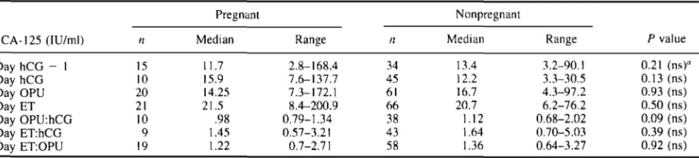
Table III shows the CA-125 serum levels on the day before and at hCG administration, oocyte retrieval, and ET, respectively. The differences in CA-125 levels between the pregnant and the nonpregnant groups were found to be nonsignificant. No correlation existed between serum CA-125 levels and endometrial thick-ness, number of retrieved and number of fertilized oocytes, cumulative embryo score, and serum estradiol and progesterone concentrations, respectively. The presence or absence of endometriosis did not have a
statistically significant effect on the observed CA-125 levels on any of the 4 days investigated, whether the whole study group was taken into account or the con-ceptual and nonconcon-ceptual cycles were analyzed sepa-rately (data not shown).
CA-125 increased significantly from the day of hCG administration until the day of ET, but the increase did not differ significantly between the pregnant (increase,
1.59-fold) and the nonpregnant (1.83-fold) groups.
DISCUSSION
CA-125 has been detected in the follicular fluid of patients undergoing IVF, as well as in their serum. Several groups, however, have postulated that the determination of CA-125 in the follicular fluid has no prognostic value with respect to the outcome (2, 4-7).
Table II. Serum Steroid Hormone Levels on Days hCG-1, hCG, OPU, and ET Pregnant E2 (pg/ml)a Day hCG Day hCG Day OPU Day ET Progesterone Day hCG Day hCG Day OPU Day ET - 1 n 15 10 21 20 Median 2,138.3 2,358.9 953.4 889.4 Range 563.8-5,066.1 487.6-7,518.2 264.2-2,495.2 315.9-2,132.6 n 34 45 58 66 Nonpregnant Median 1,648.0 2,299.1 1,084.1 727.3 Range 359.6-4,528.8 795.8-6,619.3 171.6-3,050.8 73.5-2,590.5 P value 0.17(ns)c 0.83 (ns) 0.85 (ns) 0. 1 8 (ns) (pg/ml)b - 1 15 10 20 21 911.9 880.5 8,569.0 78,709.3 471.7-1,446.5 566.0-4,591.8 4,150.8-45,942.6 5,377.5-186,663.45 37 42 58 66 723.3 1,037.7 7,688.5 58,914.1 251.5-2,484.2 408.8-3,836.4 2,955.9-40,282.2 5,477.2-343,799.1 0.13 (ns) 0.47 (ns) 0.26 (ns) 0.34 (ns) " Conversion factor to SI 3.671. b Conversion factor to SI 3. 1 80. c ns = not signicant.
Table I. Patient Synopsis Pregnant
No. of patients Age (yr) No. of attempts No. of follicles retrieved No. of fertilized oocytes Fertilization rate (%) Embryos/transfer Cumulative embryo score Endometrium thickness (mm) Median 22 (21 delivered) 34 1 (mean, 2.0) 7 5 68.7 3 (mean, 2.6) 34 12.4 (mean, 12.0) Range 26-38 1-7 4-28 1-16 10-100 1-3 6-57 4-15.1 Nonpregnant Median 69 34 1 (mean, 1.8) 8 5 75.0 3 (mean, 2.8) 32 10.5 (mean, 10.8) Range 25-41 1-5 2-33 1-24 9.5-100 1-4 2-57 4-18 P value 0.44 (ns)a 0.88 (ns) 0.86 (ns) 0.96 (ns) 0.41 (ns) 0.31 (ns) 0.67 (ns) 0.048 a ns = not significant.
On the other hand, C A-125 is produced by the endome-trial epithelium, and therefore, an endocrine or a para-crine function could be possible. The role of CA-125 as a marker for uterine receptivity has been controversial (2-4). These two aspects prompted us to investigate the usefulness of secreted (i.e., present in the serum) CA-125 as a potential indicator for the possible occur-rence of a pregnancy in IVF treatment.
Our study shows that there was no correlation between the CA-125 levels on the 4 days on which blood samples were taken and the pregnancy outcome. Serum concentrations of CA-125 showed great vari-ability within each patient group, both pregnancy and nonpregnancy outcomes. Although the endometrium was slightly thicker in the pregnancy group, there was no correlation between endometrial thickness and CA-125 levels—this would have been expected to some extent in the case of a significant contribution of the endometrium to CA-125 production and release. The lack of correlation argues against a quantitative rela-tionship, however, not against a qualitative (physio-logic) one. There was no correlation between CA-125 levels and ovarian steroid hormone concentrations found in the serum. These results may be explained by the multiple-site production of CA-125, which is not restricted to the endometrium. In patients without cancer the fallopian tube, endocervix, peritoneum, pleura, and pericardium have been found to be sources of production.
The exact location and amount of CA-125 produced in the female genital tract during ovarian hyperstimula-tion remain unclear. Sources of produchyperstimula-tion are the ovary (7), endometrium (8), and peritoneum (9). In women undergoing ovarian superovulation before and after vaginal hysterectomy, the differences in luteal CA-125 levels were similar (10), suggesting major extraendometrial production of CA-125, mainly in the ovary and/or peritoneum. Ozaksit et al. (5), studying
patients either with a normal cycle, stimulated for intra-uterine insemination, or undergoing IVF, found increased CA-125 levels only in the luteal phase of patients with ovarian hyperstimulation syndrome, sug-gesting peritoneal rather than significant endometrial or corpus luteum production of CA-125. Levels of CA-125 can be 15 to 50 times higher in pregnant patients with ovarian hyperstimulation syndrome and in patients with ectopic pregnancy with peritoneal irri-tation (9,11), suggesting that high CA-125 levels in stimulated patients might be due to not only an ovarian and endometrial, but also a peritoneal, contribution. We believe that our results are due to the fact that CA-125 production is not restricted to the endome-trium. Our results are comparable to those of Phocas
et al. (2), who found no correlation between serum
or follicular fluid CA-125 and estradiol, testosterone, oocyte quality, fertilization, and pregnancy rate. On the other hand, a recent study showed elevated serum C A-125 concentrations on the day of hCG administra-tion in conceptual cycles (3). The sensitivity was 72%, the specificity 97%, and the positive predictive value for pregnancy 95% in patients with CA-125 levels greater than 16 IU/ml on the day of hCG administra-tion. The positive significance of CA-125 was present, but less strong, on the day before hCG. In that particu-lar study, CA-125 was not measured on the day of oocyte retrieval or ET.
In contrast, Chryssikopoulos et al. (4) found increased CA-125 serum concentrations in the preg-nancy group only on the day of retrieval, and not on the day of hCG administration and ET. The increase in CA-125 from the day of hCG until the day of oocyte retrieval was higher in the pregnancy group.
The different results of the four reports are difficult to explain. The stimulation protocols were different: three groups (Refs. 2 and 3 and this study) used a gonadotropin releasing hormone (GnRH) agonist Table III. Serum CA-125 Concentrations and Rates of Increase
Pregnant CA-125(IU/ml) Day hCG - 1 Day hCG Day OPU Day ET Day OPU:hCG Day ET:hCG Day ET:OPU n 15 10 20 21 10 9 19 Median 11.7 15.9 14.25 21.5 .98 1.45 1.22 Range 2.8-168.4 7.6-137.7 7.3-172.1 8.4-200.9 0.79-1.34 0.57-3.21 0.7-2.71 n 34 45 61 66 38 43 58 Nonpregnant Median 13.4 12.2 16.7 20.7 1.12 1.64 1.36 Range 3.2-90.1 3.3-30.5 4.3-97.2 6.2-76.2 0.68-2.02 0.70-5.03 0.64-3.27 P value 0.21 (ns)a 0.13 (ns) 0.93 (ns) 0.50 (ns) 0.09 (ns) 0.39 (ns) 0.92 (ns) a ns = not significant.
down-regulation protocol, while Chryssikopoulos et
al. (4) used a short GnRH protocol. Because the
half-life of CA-125 in the serum is 4.5 days (12), ovarian down-regulation will have a different effect than the flare protocol. Such stimulation-induced differences in the control of CA-125 production and release might be stronger than the variation in endometrial glandular function, which in turn has an effect on the outcome (pregnancy or not).
Other factors, such as the number of embryos trans-ferred and the different CA-125 assay protocols used by various groups, might also provide an explanation for the conflicting results. Our group used the Cobas Core (Roche) method, Chryssikopoulos et al. (4) a RIA kit from Abbott Laboratories, and Miller et al. (3) an immunoradiometric assay from Centocor (Malvern, PA). For this reason, the different studies in the litera-ture cannot be directly compared, because the afore-mentioned methods show considerable differences in the antigen determinant specificity and the type of standard used in the respective kits. The assay used in our study used CA-125 antibodies OC 125 and Ml 1 and has shown a good reproducibility in the low ranges(<20 lU/ml) (13).
The findings presented in this study, however, relate to the CA-125 assay we used and may not be applicable to other assays. The sensitivity, specificity, and positive predictive value will also depend on the prevalence of the condition, in this case pregnancies, for the days sampled, which was different among the studies. It might also be argued that within a larger population, the difference might become statistically significant. However, CA-125 showes a wide range even in healthy women, with values ranging from less than 5 to greater than 60 U/L (13). Because of the great variance and overlapping values in the pregnancy and nonpregnancy groups, in our population, as well as the controversial reports in the literature, determination of CA-125 is currently not performed routinely as an IVF outcome prognosticator at our clinic. The study of a very large patient population, treated with identical ovarian stim-ulation protocols and using identical CA-125 assays, might, however, be warranted.
ACKNOWLEDGMENTS
We are indebted to Andy Maurer (Roche, Switzer-land) for providing the immunoassay kits and to Collin
B. Smikle for carefully reviewing the manuscript. Our thanks go to the staff of the Assisted Reproduction Unit for their technical assistance in this study.
REFERENCES
1. Bast RC Jr., Feeney M, Lazarus H, Nadler LM, Colvin RB, Knapp RC: Reactivity of a monoclonal antibody with human ovarian carcinoma. J Clin Invest 1981;68:1331-I337 2. Phocas I, Sarandakou A, Rizos D, Dimitriadou F, Mantzavinos
T, Zourlas PA: Tumour-associated antigens, CEA, CA 125 and SCC in serum and follicular fluid of stimulated and unstimu-lated cycles. Eur J Obstet Gynecol Reprod Biol 1994; 54:131-136
3. Miller KA, Deaton JL, Pittaway DE: Evaluation of serum CA 125 concentrations as predictors of pregnancy with human in vitro fertilization. Fertil Steril 1996;65: 1184-1189
4. Chryssikopoulos A, Mantzavinos T, Kanakas N, Karagouni E, Dotsika E, Zourlas PA: Correlation of serum and follicular fluid concentrations of placental protein 14 and CA-125 in in vitro fertilization-embryo transfer patients. Fertil Steril
1996:66:599-603
5. Ozaksit G, Turhan NO, Oral H, Dogu N, Gokmen O: Relation-ship between serum CA 125 levels, endometrial thickness and corpus luteum function in different stages of ovarian activity. J Endocr Invest 1993;16(3):175-179
6. Jimena P, Castilla JA, Ramirey JP, Gil T, Acebal M, Molina R. Herruzo AJ: Follicular fluid alpha-fetoprotein, carcinoem-bryonic antigen, and CA-125 levels in relation to in vitro fertil-ization and gonadotropin and steroid hormone concentrations. Fertil Steril 1993;59:1257-1260
7. Mordel N, Anteby SO, Zajicek G, Roisman I, Treves A, Barak V: CA-125 is present in significant concentrations in periovula-tory follicles of in vitro fertilization patients. Fertil Steril
1992:57:377-380
8. Bishop P, Tseng L, Brioschi PA, Herrmann WL: Cancer antigen 125 is produced by human endometrial cells. Hum Reprod 1986:1:423-426
9. Jager W, Diedrich K, Wildt L: Elevated of CA-125 in serum of patients suffering from ovarian hyperstimulation syndrome. Fertil Steril 1987:48:675-678
10. Gurgan T.Urman B, Kisnisel HA: Circulating CA-125 levels in superovulated women are mainly derived from the ovaries. Fertil Steril 1993;59:928-930
11. Bischof P, Mignot TM, Cedard L: Are pregnancy-associated plasmaprotein-A (PAPP-A) and CA-125 measurements after IVF-ET predictors of early pregnancy wastage? Hum Reprod 1989:4:843-847
12. Olt B, Berchuck A, Bast RC Jr: The role of tumor markers in gynecologic oncology. Obstet Gynecol Surv 1990:45:570-577 13. Filella X, Ballesta AM, Fox M, Mitchell H, Molina R, Purstner P, Thome H: Multicentre clinical evaluation of the COBAS CORE CEA, CA 125 II and PSA tumor marker assays. Int J Biol Markers 1996:11:40-45