DOI 10.1007/s00276-010-0626-4
O R I G I N A L A R T I C L E
Liver segments: an anatomical rationale for explaining
inconsistencies with Couinaud’s eight-segment concept
Jean H. D. Fasel · Pietro E. Majno · Heinz-Otto Peitgen
Received: 18 August 2009 / Accepted: 11 January 2010 / Published online: 29 January 2010 © Springer-Verlag 2010
Abstract
Background and purpose An increasing number of surgi-cal and radiologisurgi-cal observations surgi-call Couinaud’s concept of eight liver segments into question and such inconsisten-cies are commonly explained with anatomical variations. This paper was intended to demonstrate that, beyond vari-ability, another anatomical principle may allow to under-stand supposedly diVering concepts on liver segmentation. Materials and methods The study was performed on 25 portal vein casts scanned by helical CT. The branches of the right and left portal vein and their corresponding territo-ries were determined both anatomically and mathematically (MEVIS LiverAnalyzer, MEVISLab).
Results The number of branches coming-oV the right and left portal vein was never 8, but many more (mean number 20, range 9–44). DiVerent combinations of these branches and their respective territories, carried out in this study, yielded larger entities and supposedly contradictory subdi-visions (including Couinaud’s eight segments), without calling upon anatomical variability.
Conclusions We suggest the human liver to be considered as corresponding to 1 portal venous territory at the level of the portal vein, to 2 territories at the level of the right and left branch of the portal vein, and to 20 at the level of the rami of the right and left branch. This “1-2-20-concept” is a rationale for reconciling apparent discrepancies with the eight-segment concept. On a pragmatic level, in cases in which imaging or surgical observations do not Wt with Couinaud’s scheme, we propose clinicians not to autonom-ically conclude to the presence of an anatomical variation, but to become aware of the presence of an average of 20 (and not 8) second-order portal venous territories within the human liver.
Keywords Liver · Human · Segments · Couinaud · 1-2-20 Concept
Introduction
Subdivision of the liver into functional vascular and biliary entities is routinely performed by radiologists in view of localising focal intrahepatic lesions and during preoperative investigations for liver surgery, in particular living donor liver transplantation. The commonly applied concept of hepatic segmentation in our days divides the organ into eight segments, delimited by three vertical and one trans-verse plane. This concept is credited to the French surgeons Couinaud [5] and Bismuth [2].
An increasing number of observations, however, call the concept into question. Surgeons report intraoperative Wndings inconsistent with those expected on the basis of Couinaud’s scheme [3, 17, 19], and several have made proposals for reclassiWcations of liver territories [4, 18]. Radiological investigations describe vascular entities that J. H. D. Fasel (&)
Clinical Anatomy Research Group,
Department of Cellular Physiology and Metabolism, University Medical Centre, Rue M. Servet 1, 1211 Geneva 4, Switzerland
e-mail: [email protected]
P. E. Majno
Department of Visceral Surgery, University Hospital, Geneva, Switzerland
H.-O. Peitgen
Fraunhofer MEVIS (Medical Visualisation Center), Bremen, Germany
do not correspond to those predicted by the eight-segment concept. Several conclude to limitations and pitfalls of Couinaud’s classiWcation [9, 12, 20, 24]. In the anatomi-cal literature also, Couinaud’s subdivision turns out to be disputed, some authors considering the liver to be built-up by four, others by Wve or nine territories [8, 13, 15, 21]. Even Couinaud himself suggested successive modiWca-tions. In 1998 and 2000, he advocated the existence of nine segments [6, 11], and later revised this view [1].
At Wrst glance, such inconsistencies are explained by anatomical variability. There is obviously a large number of variants in the portal venous branching pattern, and they have been extensively described. The purpose of the present study was not to add further descriptions, but to submit a working hypothesis, according to which discrepancies are not necessarily due to variations, but can be explained by another, neglected anatomical principle concerning portal venous territories in the human liver. To achieve this goal, the study design had to exclude any anatomical variation.
Materials and methods
Livers
The study was performed on 25 human livers of deceased individuals who had donated their body to the Anatomy Department (16 women, 9 men). The age of the donors ranged from 59 to 97, with an average of 79 years. Donors with known liver pathology or organs with lesions discov-ered during the anatomical examination were excluded. Anatomical preparation
The portal vein was injected with acrylic resin using man-ual pressure. After hardening of the resin, the livers were macerated in 20% KOH for 4 days, at a temperature of 55°C. The vascular casts were rinsed and air-dried (Fig.1). CT imaging
Imaging of the corrosion casts was performed with a 64-row-multidetector helical CT scanner (Somatom Sensation 64; Siemens, Erlangen, Germany) using the following acquisition protocol: A peak kV of 120, 17 mAS dose, 5.0-mm section thickness, 0.6-mm slice collimation, a table feed of 0.4-mm per rotation, 0.5-s rotation time, and without gantry tilt. Image reconstruction was performed with 0.4-mm reconstruction interval and a 285-mm Weld of view. A high contrast of 1,130 HounsWeld units (HU) could be achieved between the hardened resin (130 HU) and the surrounding air (¡1,000 HU), thus allowing identiWcation and segmentation of small vascular branches. Image noise was
low with a standard deviation of 9.80 HU measured in the surrounding air.
Image analysis and reconstructions
The CT images were analysed with the software-assistant MEVIS LiverAnalyzer, and with the research and develop-ment platform MEVISLab (Frauenhofer MEVIS GmbH, Bremen, Germany). This software is based on a fractal approach [14] and allows to reconstruct the vascular trees on CT or MRI, and to calculate the patient’s individual vascular territories for diVerent vessel orders. In the present study, the territories of the second-order portal venous branches were determined. Image analysis consisted of the following steps: (1) extraction of vessels from the CT data by means of a modiWed region growing algorithm; (2) auto-matic calculation of the centrelines of the extracted vessels; (3) interactive labelling of the vascular branches according to the diVerent classiWcations mentioned in the literature; and (4) calculation of the portal venous territories for each of these classiWcations.
Results
Out of the 25 livers investigated, a conventional right–left branching pattern of the main portal vein was seen in 22 cases. In two casts, a large branch for the right posterior sec-tor originated from the portal vein. In one case, a portal vein trifurcation was observed. At the second-order level, the total number of branches originating from the left and the right portal vein was always higher than 8. A mean of 20 branches (range 9–44) was counted. An example of a liver with 22 second-order branches is illustrated in Fig.1. Pool-ing these branches and their territories to obtain a smaller number of secondary units yielded to seemingly contradic-tory segmentations in the same livers (thus excluding any variation)—such as four sectors (Fig.2c), three (and not 2 or 4) segments in the right hemiliver (Fig.2d), three (and not 1 or 2) territories in the left anterior sector (Fig.2e), and eight segments for the liver as a whole (Fig.2f).
Discussion
Several anatomical, radiological and surgical observations challenge Couinaud’s concept of eight liver segments [1, 3, 4, 6, 8, 9, 11–13, 15, 17–22, 24]. Discrepancies are commonly explained with the frequency of anatomical variations. The present paper shows that beyond variations, inconsistencies can be explained by the understandable need to Wt the high number of secondary territories into a simpliWed scheme.
This interpretation is based on the undisputable anatomi-cal observation that the branches of the right and left portal vein (and thus their corresponding territories) are much more numerous than generally admitted in clinical practice. The high number of secondary branches can be conWrmed by an unprejudiced look at corrosion casts (Fig.1). Natu-rally enough and despite remarkable advances in imaging and surgical techniques, such a detailed analysis of the intrahepatic vascular tree remains the anatomist’s privilege yet. In other words, a systematic, hierarchically graded description of the detailed portal venous branching pattern suggests the following (Fig.3): at the level of the portal vein (which can be seen as the order zero vessel) the liver consists of one territory—the whole organ. At the Wrst-order level (generally the right and left branches of the
portal vein), the same liver consists of two territories—the right and left liver of the French nomenclature, the right and left hemiliver in the English terminology. On the second-order level, the branches are not eight and, therefore, the liver not made of eight segments, but of many more. An average of 20 second-order branches (and thus territories) was observed [10].
It is this high number of second-order branches that explains presumed inconsistencies with Couinaud’s eight segments. The view, for instance, that Goldsmith’s and Woodburne’s [13] left medial sector (Fig.2c) can be subdi-vided into two segments according to Couinaud (Fig.2f), but into three as stated by Platzer and Maurer [21] (Fig.2e) is neither an antinomy nor necessarily due to anatomical variability. It is the result of gathering various branches of
Fig. 1 Portal venous branching
pattern in a corrosion cast, with emphasis on the second-order branches. In the liver illustrated, the number of those branches was 22, 18 of which can be seen in the inferior view (1–18). LPV left branch of the portal vein, RPV right branch of portal vein
same order, considered individually by Platzer and Maurer [21], to one territory for the sake of obtaining eight seg-ments. Another example concerns the right hemiliver, which has been considered to be built-up by two [21], three [10, 15], or four [5] territories. Attributing diVerent vessels to one territory is observer dependent and yields diVerent results by diVerent authors, whereas the underlying reality does not diVer. The fact that such seemingly inconsistencies can neither be automatically reduced to anatomical varia-tions is illustrated in Fig.2, which is from one and the same liver—thus, excluding any variability of the portal venous branching pattern.
The existence of an average of 20 (and not 8) second-order portal vein territories explains the common surgical observation that intraoperative dye injections into second-order branches stain small areas of hepatic parenchyma [19]. The high number of secondary branches is also in good accordance with the territories computed from CT data sets [12, 22] and those identiWed by selective angiography [20].
We would like to emphasise that Couinaud’s eight segments are not in contradiction to the “1-2-20 concept”. On the contrary, they can be deduced from it. The latter is the underlying principle, from which eight segments, as an example amongst others, can be obtained by putting together the smaller basic entities. All due distance respected, the theory of relativity did not contradict
Newton’s classical mechanics, but acknowledges it as one possible phenotype of a more general principle.
When compared with other organs, the fractal approach used in this and previous studies [14] is in good agreement with an asymmetric branching model described for the human airway tree [16]. The portal venous system corresponds to a well ordered, but highly asymmetric tree and, including the many side branches of various size, cannot be explained by a symmetric top-down branching pattern.
On a pragmatic clinical level, we suggest to continue using Couinaud’s eight-segment scheme for what it is: a brilliantly founded and extremely useful referential frame-work for communication between gastroenterologists, radi-ologists, and surgeons. In cases, however, in which the patient’s anatomy does not Wt with the eight-segment scheme, we suggest not to automatically conclude to the presence of an anatomical variation, but to become aware of the presence of an average of 20 (and not 8) second-order portal territories within the human liver.
In conclusion, we would like to quote again the remark-able statement made by Skandalakis et al. [23]: “Despite its multiple vital functions and its regenerative abilities, the liver has been misunderstood at nearly all levels of organi-sation and in almost every period of time since Galen. The most paradoxical aspect of the understanding of hepatic
Fig. 2 Arbitrary combination of the second-order territories in the
same liver—i.e. excluding any anatomical variability—yields seem-ingly contradictory segmentations. a The anterior view of the native specimen, b the second-order territories coloured. c Goldsmith’s and Woodburne’s [13] sectors that were generally used by American
radi-ologists before the intervention of Dodd [7], d three territories in the right liver, as postulated by Hjorstö [15], e the left medial sector built-up by three (and not 1 nor 2) territories [21], f Couinaud’s eight seg-ments, including the left medial sector being subdivided into two parts [2, 5]
anatomy has not been lack of knowledge, but questions of interpretation; there is a tendency to ignore details that do not Wt preconceived ideas”.
References
1. Abdalla EK, Vauthey JN, Couinaud C (2002) The caudate lobe of the liver: implications of embryology and anatomy for surgery. Surg Oncol Clin N Am 11:835–848
2. Bismuth H (1982) Anatomical surgery and surgical anatomy of the liver. World J Surg 6:3–9
3. Cervone A, Sardi A, Conaway GL (2000) Intraoperative ultra-sound (IOUS) is essential in the management of metastatic colo-rectal liver lesions. Am Surg 66:611–615
4. Cho A, Okazumi S, Makino H et al (2004) Relation between hepatic and portal veins in the right paramedian sector: proposal for anatomical reclassiWcation of the liver. World J Surg 28:8–12 5. Couinaud C (1957) Le foie. Etudes anatomiques et chirurgicales,
Masson, Paris
6. Couinaud C (1998) Secteur dorsal du foie. Chirurgie 123:8–15 7. Dodd GD III (1993) An American guide to Couinaud’s numbering
system. Am J Roentgenol 161:574–575
8. Elias H, Petty D (1952) Gross anatomy of the blood vessels and ducts within the human liver. Am J Anat 90:59–111
9. Fasel JHD, Selle D, Evertz CJG, Terrier F, Peitgen HO, Gailloud P (1998) Segmental anatomy of the liver: poor correlation with CT. Radiology 206:151–156
10. Fasel JHD (2008) Portal venous territories within the human liver: an anatomical reappraisal. Anat Rec 291:636–642
11. Filipponi F, Romagnoli P, Mosca F, Couinaud C (2000) The dorsal sector of human liver: embryological, anatomical and clinical relevance. Hepatogastroenterology 47:1726–1731
12. Fischer L, Cardenas C, Thorn M, Benner A, Grenacher L, Vetter M, Lehnert T, Klar E, Meinzer HP, Lamade W (2002) Limits of Couinaud’s liver segment classiWcation: a quantitative computer-based three-dimensional analysis. J Comput Assist Tomogr 26:962–967
13. Goldsmith NA, Woodburne RT (1957) The surgical anatomy per-taining to liver resection. Surg Gynecol Obstet 105:310–318 14. Hahn HK, Evertsz CJG, Peitgen HO, Fasel JHD (2003) Fractal
properties, segmental anatomy and interdependence of the human portal and hepatic vein in 3D. Fractals 11:53–62
15. Hjortsjö CH (1951) The topography of the intrahepatic duct sys-tems. Acta Anat 11:599–615
16. HorsWeld K (1990) Diameters, generations, and orders of branches in the bronchial tree. J Appl Physiol 68:457–461
17. Ko S, Murakami G, Kanamura T, Sato TJ, Nakajima Y (2004) Cantlie’s plane in major variations of the primary portal vein ram-iWcation at the porta hepatis: cutting experiment using cadaveric livers. World J Surg 28:13–18
18. Koqure K, Kuwano H, Fujimaki N, Ishikawa H, Takada K (2002) Reproposal for Hjortsjo’s segmental anatomy of the anterior seg-ment in human liver. Arch Surg 137:1118–1124
19. Makuuchi M, Hasegawa H, Yamazaki S (1985) Ultrasonically guided subsegmentectomy. Surg Gynecol Obstet 161:346–350 20. Ohashi I, Ina H, Okada Y, Yoshida T, Gomi N, Himeno Y,
Hanafusa K, Shibuya H (1996) Segmental anatomy of the liver under the right diaphragmatic dome: evaluation with axial CT. Radiology 200:779–783
21. Platzer W, Maurer H (1966) Zur Segmenteinteilung der Leber. Acta Anat 63:8–31
22. Rieker O, Mildenberger P, Hintze C, Schunk K, Otto G, Thelen M (2000) Segmentanatomie der Leber in der Computertomographie: Lokalisieren wir die Läsionen richtig? (Segmental anatomy of the liver evaluated with CT: do we localize focal lesions as we should?). Fortschr Roentgenstr 172:147–152
23. Skandalakis JE, Skandalakis LJ, Skandalakis PN, Mirilas P (2004) Hepatic surgical anatomy. Surg Clin North Am 84:413–435 24. Strunk H, Stuckmann G, Textor J, Willinek W (2003) Limitations
and pitfalls of Couinaud’s segmentation of the liver in transaxial imaging. Eur Radiol 13:2472–2482
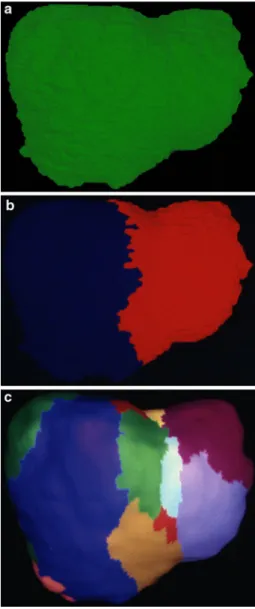
Fig. 3 Portal venous segmentation of the liver, in function of the level
of the portal venous branching pattern considered. a At the level of the portal vein (which can be addressed as the order 0 vessel), the liver cor-responds to 1 territory. b At the Wrst-generation level, the liver (in the usual case of portal vein bifurcation) consists of two territories—the right and left liver. c On the next, second-order level, the same liver has 20 segments (11 of which can be seen in this anterior view