HAL Id: hal-01632250
https://hal.archives-ouvertes.fr/hal-01632250
Submitted on 29 May 2019
HAL is a multi-disciplinary open access
archive for the deposit and dissemination of
sci-entific research documents, whether they are
pub-lished or not. The documents may come from
teaching and research institutions in France or
abroad, or from public or private research centers.
L’archive ouverte pluridisciplinaire HAL, est
destinée au dépôt et à la diffusion de documents
scientifiques de niveau recherche, publiés ou non,
émanant des établissements d’enseignement et de
recherche français ou étrangers, des laboratoires
publics ou privés.
4
Xavier Fabrèges, E. Ressouche, F. Duc, S. de Brion, Mehdi Amara, C. Detlefs,
L. Paolasini, E. Suard, L.-P. Regnault, B. Canals, et al.
To cite this version:
Xavier Fabrèges, E. Ressouche, F. Duc, S. de Brion, Mehdi Amara, et al.. Field-driven
magne-tostructural transitions in GeCo 2 O 4. Physical Review B, American Physical Society, 2017, 95 (1),
pp.014428. �10.1103/PhysRevB.95.014428�. �hal-01632250�
X. Fabr`eges,1, 2 E. Ressouche,3 F. Duc,2 S. de Brion,4, 5 M. Amara,4, 5 C. Detlefs,6 L. Paolasini,7 E. Suard,8 L.-P. Regnault,3 B. Canals,4 P. Strobel,4, 5 and V. Simonet4, 5,∗
1Laboratoire L´eon Brillouin, CEA, CNRS, Universit´e Paris-Saclay,
CEA Saclay, F-91191 Gif-sur-Yvette Cedex France
2
Laboratoire National des Champs Magn´etiques Intenses, CNRS-INSA-UJF-UPS, 31400 Toulouse, France
3CEA/Grenoble, INAC/SPSMS-MDN, 17 rue des Martyrs, 38054 Grenoble Cedex 9, France 4
CNRS, Institut N´eel, 38042 Grenoble, France
5
Univ. Grenoble Alpes, Institut N´eel, 38042 Grenoble, France
6European Synchrotron Radiation Facility, 71 Avenue des Martyrs, 38000 Grenoble, France 7
European Synchrotron Radiation Facility, Grenoble, France
8
Institut Laue Langevin, BP 156, 38042 Grenoble Cedex 9, France (Dated: November 5, 2018)
In the spinel compound GeCo2O4, the Co2+ pyrochlore sublattice presents remarkable magnetic
field-induced behaviors that we unveil through neutron and X-ray single-crystal diffraction. The N´eel ordered magnetic phase is entered through a structural lowering of the cubic symmetry. In this phase, when a magnetic field is applied along a 2-fold cubic direction, a spin-flop transition of one fourth of the magnetic moments releases the magnetic frustration and triggers magnetostructural effects. At high field, these ultimately lead to an unusual spin reorientation associated with structural changes.
PACS numbers: 75.25.-j,75.50.Ee,75.30.Gw,75.80.+q
I. INTRODUCTION
Spinel compounds with the generic formula AB2O4
crystallize in the cubic space group F d¯3m. The B sites can accomodate a magnetic ion, in this case Co2+. They form a pyrochlore lattice, a network of corner-sharing tetrahedra, that is the archetype of geometrical frustra-tion. In addition, they combine magnetic and lattice degrees of freedom, which confer them a magnetostruc-tural flexibility in zero and finite magnetic field. The interplay between magnetic frustration and magnetoelas-tic coupling has been intensively studied in spinels with, for instance, either V3+ or Cr3+ on the B site and
var-ious ions on the A site (Hg, Mg, Cd and Zn) [1–6]. In these systems, at the N´eel temperature (TN), a transition
to an antiferromagnetic (AFM) ordering is accompanied by a cubic to tetragonal or orthorhombic structural dis-tortion. It is interpreted as a 3-dimensional spin-Peierls transition acting to reduce the frustration [1, 3]. In the Cr compounds, when applying a magnetic field, a magne-tization plateau at half of the saturation magnemagne-tization is stabilized by the spin-lattice coupling on a wide range of fields [2,3,7,8]. Moreover, the beginning of the plateau coincides with a structural distortion. It corresponds to a recovery of the cubic structure as a consequence of the release of the frustration by the magnetic field [4,5].
Ge spinels, with Ni2+ or Co2+ on the pyrochlore
lat-tice, have comparable rich phase diagrams but have been less studied because the frustration effects are not as straightforward, in particular not solely driven by the
∗Corresponding author: [email protected]
first neighbor interactions. They were both shown, by powder neutron diffraction, to order in a magnetic struc-ture characterized by a propagation vector k=(1/2, 1/2, 1/2) and stabilized through competing interactions be-yond the third neighbors [9,10]. The magnetic arrange-ment was described assuming a single propagation vec-tor (single-k structure) as follows: alternating kagome (KGM) and triangular (TRI) ferromagnetic (FM) planes, perpendicular to the <111> directions associated with the propagation vector, and antiferromagnetically cou-pled to the nearest planes of the same kind (see figure
1a). In GeNi2O4, which retains the cubic structure in
the magnetic phase, both KGM and TRI sites are mag-netically decoupled, i.e. the molecular field created by the magnetization of one type of site on the other one is zero. It leads to an independent ordering of the two kinds of planes with distinct transition temperatures [11–
13]. This is at variance with GeCo2O4 in which a unique
magnetic transition is observed at TN=23.5 K. This is
as-cribed to a structural distortion that couples the two sites and allows to reduce the frustration in the absence of any external magnetic field. This distortion was proposed to be mainly cubic-to-tetragonal from powder neutron and X-ray diffraction [1,15].
Besides this zero-field magnetostructural behavior, a complex H-T phase diagram has been observed in GeCo2O4 by magnetization, ultrasound and electron
spin resonance measurements, evidencing several field-induced anomalies [1, 9, 16–22]. Their microscopic ori-gin was investigated by powder neutron diffraction under magnetic field up to 10 T [10]. A transition, observed at ≈ 4 T, was attributed to an antiferromagnetic-to-ferromagnetic spin rearrangement between the triangular planes while retaining the antiferromagnetic arrangement
(a)$
(c)$
b$
c$
a$
(b)$
H
c1<H<H
c2$k
1#
c$
a$
b$
k
1#
KGM#
[11*2]$
TRI#
[11*2]$
k
3#
KGM#
[110]$
b$
c$
a$
H<H
c1$[1*10]$
[111]$
[1*10]$
[1*10]$
KGM#
[11*2]$
[111]$
[1*1*1]$
H>H
c2$FIG. 1: AFM ordered components of the KGM (red) and TRI (green) moments determined from low temperature single-crystal neutron diffraction refinements in zero-field and under a magnetic field applied along the [1-10] direction (black arrow): at µ0H=0 T for the k1domain (one of the 3 equipopulated S-domains represented) (a), at µ0H=5.5 T for the k1 domain (b)
and at µ0H=12 T for the k3 domain after the high-field spin-reorientation (c). Note that the ferromagnetic component that
develops under magnetic field at the expense of the antiferromagnetic component is not shown. The AFM component of the TRI magnetic moments is only present in (a) since they get polarized by the magnetic field above Hc1. From (b) to (c), the
KGM planes, in which lie the AFM component of the magnetic moments, are reoriented from (111) to (1-1-1), consistent with the change of propagation vector.
between the kagome planes. The final ferromagnetic or-der of the kagome planes was proposed to occur at the second transition around µ0H=10 T.
We hereafter present the results of single-crystal neu-tron and synchroneu-tron X-ray diffraction under zero and finite magnetic fields applied along a 2-fold axis of the high temperature cubic structure. We show in particular that the high field transition, for this orientation of the field, is much more subtle than a plain antiferromagnetic-to-ferromagnetic one. It implies a change of the magnetic anisotropy, a switch of the magnetostructural domains, and the stabilization of a new canted magnetic structure. We discuss the interplay between magnetostructural ef-fects and frustration in triggering these field-induced un-conventional behaviors.
II. EXPERIMENTAL DETAILS
Single-crystals of GeCo2O4 with an octahedral shape
of approximately 2 mm size were synthesized by chemical vapor transport in 0.1 atm of HCl gas. A heat treatment was performed with a heating plateau at 950◦C followed by a slow cooling at 1◦/mn.
Magnetization measurements on the oriented single-crystal were performed up to 10 T in the temperature range 2K -300 K using an extraction magnetometer. The magnetic susceptibility data are in agreement with those published in the literature with a N´eel temperature of
23.5 K (see Supplemental Material).
Single crystal neutron diffraction was carried out at the Institut Laue Langevin (ILL). Single-crystal diffraction in zero magnetic field was obtained on the CEA-CRG D15 diffractometer operated in the 4-circle mode, with the sample mounted in a displex refrigerator, and with an incident wavelength of 1.173 ˚A. Measurements under magnetic field up to 12 T was performed on the CEA-CRG D23 two-axis diffractometer with a lifting arm de-tector and an incident wavelength of 1.280 ˚A. The single-crystal was mounted in the cryomagnet with the [1-10] axis set vertical, parallel to the applied field. An ad-ditional diffraction experiment using polarized neutrons with spherical polarization analysis was performed on the single-crystal installed inside the CRYOPAD device on the CEA-CRG IN22 spectrometer at the ILL using a wavelength of 2.36 ˚A. This set-up allows measuring the three orthogonal components of the polarization vector of the neutron beam after scattering by the sample what-ever the direction of the incident neutron beam polariza-tion. It gives for a reflection (Qh, Qk, Ql) a polarization
matrix Pij, with i,j=X,Y,Z (X k scattering vector, Z ⊥
the scattering plane) [23,24].
High magnetic field X-ray single-crystal diffraction experiments were performed at the ID06 beamline of the European Synchrotron Radiation Facility using a monochromatic beam selected by a Si(111) double monochromator. Magnetic field was provided by a 10 T split-coil superconducting magnet [25]. A 2x2x1 mm
H>Hc2# k3=#(½,#,(½,(½)# [110]'''[1(12]# [01(1]##[211]# [101]###[12(1]# Hdom<H<Hc2# k2=(½,½,(½)# [1(10]##[112]' [011]###[2(11]# [101]###[1(2(1]# Hdom<H<Hc2# k1=(½,#½,#½)# [1(10]###[11)2]# [01(1]###[2(1(1]# [10(1]###[1(21]# H>Hc2# k4=(½,(½,#½)# [110]'''[1(1(2]# [011]###[21(1]# [10(1]##[121]# a' b' c' H//[1(10]#
FIG. 2: Magnetic domains in cubic symmetry: the direction of the propagation vectors is shown by red lines for the pair (k1, k2) selected between Hdomand Hc2, and by blue lines for
the other pair (k3, k4) selected above Hc2. For each k-domain,
the 3 S-domains are listed in the boxes for two possible ori-entation of the magnetic moments, either along the <110> directions (left) or along the <112> directions (right). The domains in bold and the double arrows give the orientation of the AFM component on the KGM sites for Hdom< H <
Hc2 and for H > Hc2.
single-crystal was aligned with the [110] and [001] axes within the horizontal plane giving access to the (h,h,l ) Bragg reflections. Magnetic field was applied along the [1-10] direction. The measurements were performed with E = 32 keV (λ = 0.3876 ˚A) and the diffracted intensities were collected with a photodiode.
III. RESULTS
A. Magnetic structure in zero and finite magnetic fields
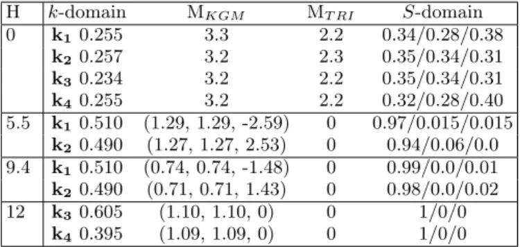
To analyze the single-crystal neutron diffraction results of GeCo2O4, we first assume a cubic structure and a
single-k magnetic arrangement. Both hypotheses will be discussed later on. We take into account the presence of several magnetic domains, that appear at the phase tran-sition (see figure2): we consider four k-domains that cor-respond to the symmetry-equivalent directions of propa-gation of the AFM order given by the propapropa-gation vec-tors k1=(1/2, 1/2, 1/2), k2=(1/2, 1/2, -1/2), k3=(1/2,
-1/2, -1/2), k4=(1/2, -1/2, 1/2). Moreover, if the
mag-netic moments lie in the {111} planes, there are three S-domains per k-domain with moment directions rotated by 120◦ between S-domains [26].
Note that the magnetic refinements reported below are based on the analysis of the magnetic Bragg reflections indexed by the four propagation vectors with half-integer indices mentioned above. Therefore, it concerns only the antiferromagnetic component of the magnetic arrange-ment. While it describes the whole magnetic structure in zero-field, it is accompanied by a rising ferromagnetic
component when a magnetic field is applied, which is not refined in the present work.
In zero field, our refinement of the magnetic intensities collected on D15 at T =10 K (see figure3) confirms the antiferromagnetic structure deduced from powder neu-tron diffraction [9, 10]. It additionally proves that the TRI and KGM magnetic moments lie in the plane per-pendicular to the <111> directions given by the propa-gation vectors. Due to the presence of 12 domains that we found approximately equipopulated (see TableI), the moment orientation could not be further determined us-ing unpolarized neutrons. Polarized neutrons and spheri-cal polarization analysis allowed us to determine the rela-tive orientation of the magnetic moments on the TRI and KGM sites (but not their absolute direction that was only inferred from neutron scattering under magnetic field, as presented below). The best fit of the measured polariza-tion matrix at 1.5 K for 23 reflecpolariza-tions is shown with blue dots in figure4. It is obtained for a parallel orientation of TRI and KGM magnetic moments in the {111} planes (see figure1a) and a population of the three S-domains equal to 26/40/34 %. 0 100 200 300 400 500 600 0 100 200 300 400 500 600 0 100 200 300 400 500 0 100 200 300 400 500
Calculated Intensity (a. u.)
k4 domain wR2=22% k2 domain wR2=22% O b se rv e d In te n si ty ( a . u .) k1 domain wR2=22% O b se rv e d In te n si ty ( a . u .)
Calculated Intensity (a. u.)
k3 domain
wR2=18%
FIG. 3: Observed versus calculated intensity of the magnetic Bragg reflections measured for the four k-domains on D15 at H=0 T and T =10 K. The wR2 factors give the goodness of the fit.
Under a magnetic field, the populations of the mag-netic domains are expected to vary, the domains with the AFM component perpendicular to the magnetic field being favored by the exchange and Zeeman terms. We followed four AFM reflections while applying a magnetic field along the 2-fold axis [1-10] direction: (0.5, 0.5, 0.5), (0.5, 0.5, -0.5), (0.5, -0.5, -0.5), (0.5, -0.5, 0.5). They are characteristic of the four k-domain populations and have roughly the same intensity at H=0. As shown in figure
FIG. 4: Spherical polarization analysis at T =1.5 K using CRYOPAD on IN22: observed versus calculated components of the final polarization for 23 magnetic Bragg peaks belong-ing to the k1domain. The magnetic moments on the TRI and
KGM sites lie in the plane perpendicular to the [111] direc-tion. The best fit is obtained when they are parallel to each other (blue filled circles, goodness of the fit wR2=21.4%), and it deteriorates as soon as they depart from collinearity. For comparison, an example is given for perpendicular TRI and KGM spins (red empty circles, wR2=63.0 %). Note that 4 points out of 207 are still not well described by the best model with collinear TRI and KGM magnetic moments. They corresponds to the PY Y and PZZ components of the
polar-ization matrix for the (0.5, 0.5, 2.5) and (1.5, 1.5, -0.5) Bragg reflections. The origin of this discrepancy is unclear at the moment.
5a, when increasing the magnetic field, the 4 equipop-ulated k-domains start to split into two pairs of reflec-tions with different intensities, one pair being selected (k1, k2) whereas the other pair (k3, k4) is suppressed
with increasing field. This selection occurs irreversibly mostly below µ0Hdom ≈ 2 T. At µ0Hc1=4.3 T, there is
a slight change of curvature of the field dependence of the (0.5, 0.5, 0.5) and (0.5, 0.5, -0.5) signal. This field corresponds to the first anomaly observed in the mag-netization curve measured on a single-crystal shown in figure6, in agreement with the results of Hoshi et al. [1]. The second magnetization anomaly occurs at µ0Hc2=9.5
T, when the populations of the four k-domains change again abruptly in a remarkable way: the two selected do-mains (k1, k2) vanish and the two k-domains that were
absent (k3, k4) are populated (see figure 5a).
In addition to the AFM components probed with the previous reflections, a gradual rise of the intensity of the (1, 1, 1) nuclear reflection is observed (see figure 5b). It is associated with the FM component (M ) developing along the field on the TRI and KGM sites. It leads to the integrated magnetic intensity on the (1, 1, 1) reflec-tion, given by the square of the magnetic structure fac-tor, ∝ (MT RI− 3MKGM)2 [10]. Two step-like increases
of this signal are observed, a small one at Hc1and a
pro-nounced one at Hc2. Above Hc2, both a FM and an AFM
TABLE I: Results of the magnetic structure refinements in zero field (D15) and under applied magnetic fields (D23). In the successive columns are reported the value of the magnetic field in T, the k-domains population, the antiferromagnetic component of the magnetic moments in Bohr magneton on the KGM and TRI sites (only the amplitude at 0 T and the magnetic moment vector components under finite fields), and the population of the corresponding S-domains, listed as in figure2. H k-domain MKGM MT RI S-domain 0 k1 0.255 3.3 2.2 0.34/0.28/0.38 k2 0.257 3.2 2.3 0.35/0.34/0.31 k3 0.234 3.2 2.2 0.35/0.34/0.31 k4 0.255 3.2 2.2 0.32/0.28/0.40 5.5 k1 0.510 (1.29, 1.29, -2.59) 0 0.97/0.015/0.015 k2 0.490 (1.27, 1.27, 2.53) 0 0.94/0.06/0.0 9.4 k1 0.510 (0.74, 0.74, -1.48) 0 0.99/0.0/0.01 k2 0.490 (0.71, 0.71, 1.43) 0 0.98/0.0/0.02 12 k3 0.605 (1.10, 1.10, 0) 0 1/0/0 k4 0.395 (1.09, 1.09, 0) 0 1/0/0
contributions are thus present indicating a canted mag-netic structure, consistent with the magnetization still increasing in higher fields [9].
The field-induced antiferromagnetic component of the magnetic orders could be determined from refinements of the half integer magnetic reflections collected on D23 at T =4.5 K under the selected fields of 5.5, 9.4 and 12 T as shown in figure7. At µ0H=5.5 and 9.4 T, i.e. for
Hc1<H<Hc2, the KGM AFM component of the moments
is found aligned along the [11-2] direction for the k1
se-lected domain (see figure1b) and along the [112] direction for the k2 selected domain. The amplitude of this
com-ponent decreases from 3.1 to 1.8 µB between 5.5 and 9.4
T while the FM component increases. On the TRI site, the AFM component is found equal to zero above Hc1,
as already proposed [10]. At µ0H=12 T, i.e. for H>Hc2,
the KGM AFM component of the two new k3and k4
se-lected domains is found aligned along a unique direction [110], also perpendicular to the applied magnetic field (see figure1c). At this field, the AFM and FM compo-nents of the KGM magnetic moment are found ≈ 1.5 and 2.4 µB respectively from neutron diffraction refinement
and magnetization measurements.
The field-induced magnetic behavior is summarized in Table II and leads to the following scenario: at zero-field, we assume that both the TRI and KGM moments are parallel to each other and along the 12 equivalent <112> directions. Increasing the field applied along [1-10] selects, above Hdom, the two [11-2] and [112]
direc-tions perpendicular to it, and thus the two associated k1
and k2domains. Hc1marks an AFM to FM transition of
the TRI magnetic moments that become oriented along the field. At Hc2, a new magnetic order is stabilized with
the AFM component on the KGM sites aligned along the [110] direction. This orientation is compatible with the
0 50 100 150 30 60 90 1300 1400 1500 1600 (d) (b) (c) (0.5,0.5,0.5) (0.5,0.5,-0.5) (0.5,-0.5,-0.5) (0.5,-0.5,0.5) (a) (1,1,1) 0 2 4 6 8 10 12 0 5 10 15 !0
H (T)
(0,0,2) N eu tr on c ou nt s (a . u. ) (-4,-4,0)H
dom$H
c1$H
c2$FIG. 5: Neutron counts at the peak maximum versus mag-netic field applied along the [1-10] direction at 4.5 K for: (a) four magnetic Bragg reflections representative of the AFM k-domains, (b) the (1, 1, 1) weak nuclear reflection on top of which grows a field-induced FM component, (c) the (-4, -4, 0) strong nuclear reflection sensitive to the extinction, (d) the (0, 0, 2) forbidden nuclear reflection. The Hdom, Hc1and
Hc2 characteristic fields are indicated by vertical lines. The
filled (open) symbols correspond to increasing (decreasing) field measurements.
selection of two new domains, k3 and k4, and implies a
redefinition of the KGM and TRI planes. A canted mag-netic structure is induced by the field on the former while the latter are ferromagnetic.
B. Structural changes
Correlated to the complex magnetic behavior of GeCo2O4, there is a remarkable variation of the
extinc-tion. It corresponds to a decrease of the intensity of strong Bragg reflections when the structural crystal qual-ity is improved possibly due to magnetostructural effects [27]. An irreversible decrease of the intensity is observed
0 2 4 6 8 10 12 0 1 2 !0H (T) D e riv a tiv e d M /d H H // [1-10] M ( µB /C o) Hc1 Hc2
FIG. 6: Single-crystal magnetization M and its derivative (red dashed line) measured as a function of the magnetic field oriented along the [1-10] direction at 2 K.
TABLE II: Selection of the antiferromagnetic domains under a magnetic field applied along [1-10]. For the three field ranges given in the Table, the columns indicate the selected propa-gation vectors and the orientation of the antiferromagnetic component of the KGM and TRI magnetic moments. Note that in zero field the four magnetic domains are equipopulated and that they get selected for H > Hdom. Above Hc1, the TRI
moments are ferromagnetically aligned along the field. Hdom< H < Hc1 Hc1< H < Hc2 Hc2< H k KGM TRI k1 [11-2] [11-2] k2 [112] [112] k KGM TRI k1 [11-2] none k2 [112] none k KGM TRI k3 [110] none k4 [110] none
on the intense nuclear reflection (-4, -4, 0) up to Hdom
(see figure5c), which shows that the structural quality is improved when the magnetic domains are selected. The intensity is then constant up to Hc2 where a very narrow
peak of extra intensity is visible, indicating a transient crystal deterioration at the transition toward the new magnetic structure.
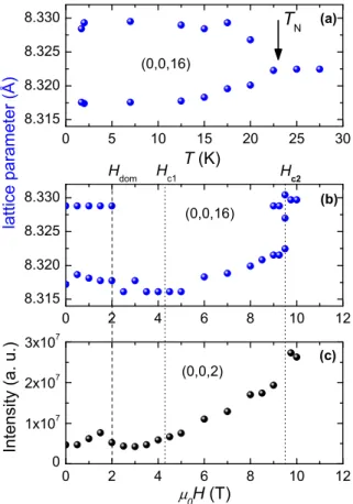
In the present paper, these magnetostructural effects were further investigated by single-crystal X-ray diffrac-tion on ID06. Back to the temperature dependence in zero field, the (0, 0, 16) structural reflection splits into two peaks below TN (see figure 8a). This agrees with a
main cubic-to-tetragonal structural distortion implying a 0.12 % elongation of one of the cubic axes [1], hence yielding three tetragonal domains (see TableIIIand the Supplemental Material for complementary powder neu-tron diffraction results).
It should be noted however that, in a single-k picture, a (1/2, 1/2, 1/2) propagation vector for the magnetic structure cannot result in a tetragonal strain. An im-portant issue is therefore whether the magnetic order is described by only one or several members of the star of the k-vector. A similar puzzling behavior was reported in CoO: this oxide was known to undergo a cubic-to-tetragonal structural transition at the temperature of the collinear magnetic ordering, itself described by an AFM
0 20 40 60 80 100 120 0 20 40 60 80 100 120 0 10 20 30 40 50 60 0 10 20 30 40 50 60 0 20 40 60 80 100 120 0 20 40 60 80 100 120 0 10 20 30 40 50 60 0 10 20 30 40 50 60 0 5 10 15 20 25 30 0 5 10 15 20 25 30 0 5 10 15 20 25 30 0 5 10 15 20 25 30 O b se rv e d I n te n si ty ( a . u .) !0H=5.5 T k1 domain wR2=13% O b se rv e d I n te n si ty ( a . u .) !0H=9.4 T k1 domain wR2=15% !0H=5.5 T k2 domain wR2=14% !0H=9.4 T k2 domain wR2=15%
Calculated Intensity (a. u.)
!0H=12 T k4 domain wR2=15% O b se rv e d I n te n si ty ( a . u .)
Calculated Intensity (a. u.)
!0H=12 T
k3 domain
wR2=16%
FIG. 7: Observed versus calculated intensity of the magnetic Bragg reflections measured on D23 at T=4.5 K, and refined at 5.5 T, at 9.4 T (Hc1<H<Hc2), and at 12 T (H>Hc2). The
wR2 factors give the goodness of the fit.
stacking of ferromagnetic planes with a (1/2, 1/2, 1/2) propagation vector and the magnetic moment along the <112> directions [28]. Symmetry arguments [29] were in-voked showing that, if the tetragonal distortion is driven by the magnetic ordering, the magnetic structure has to be described by 4 propagation vectors, leading to a non-collinear magnetic arrangement. On the other hand, if the main tetragonal distortion is not caused by the mag-netic structure, a single-k magmag-netic structure will induce an additional small trigonal distortion leading to a mono-clinic space group. In CoO, the evolution of the magnetic domains under applied stress allowed to discriminate be-tween the two scenarios, in favor of a single-k structure [29]. Direct evidence of a small but finite trigonal lat-tice distortion was finally found through X-ray diffraction [30], which agreed with the magnetic structure symme-try.
In GeCo2O4, as in CoO, a tetragonal distortion
result-ing from the magnetic orderresult-ing would imply 4 propaga-tion vectors and a non-collinear magnetic structure. This is hardly compatible with the field-induced magnetic do-main selection that we observe and that we can explain straightforwardly in a single-k magnetic structure.
Simi-0 5 10 15 20 25 30 8.315 8.320 8.325 8.330 0 2 4 6 8 10 12 8.315 8.320 8.325 8.330 0 2 4 6 8 10 12 0 1x10⁷ 2x10⁷ 3x10⁷ T (K) (a) (0,0,16) !" ##$ %& '( ") "* &# &) '+,
-T
N ' (b) (0,0,16) Hc1 Hdom !0H (T) (c) (0,0,2) ./ #& /0 $#1 '+" 2'3 2-Hc2FIG. 8: Single-crystal X-ray diffraction: temperature depen-dence in zero field of the lattice parameter determined from the (0, 0, 16) reflection (a). Field dependence at 2 K under a magnetic field along the [1-10] direction of the lattice pa-rameter determined from the (0, 0, 16) reflection (b), of the intensity of the (0, 0, 2) forbidden reflection (c). The Hdom,
Hc1and Hc2characteristic fields are indicated by vertical lines.
larly to CoO [29,30], we therefore suggest that the single-k magnetic structure emerges in a primarily tetragonal context, inducing an additional small trigonal distortion (see figure 9). This should lead to a monoclinic space group, that has still to be evidenced [31]. This scenario is consistent with the reported first-order transition at TN
in GeCo2O4 [32, 33]. Also supporting this assumption
is the orientation of the Co2+ magnetic moments that
we determined along the same direction as in CoO [28]. The monoclinic distortion is characterized by a unique 2-fold axis along the < 110 > direction perpendicular to the selected cubic trigonal < 111 > direction and to the tetragonal axis. In this case, each tetragonal domain is further split into four monoclinic domains, yielding 12 structural domains (see TableIII).
It is interesting at this stage to correlate the struc-tural and the magnetic domains. For this purpose, we have studied the field evolution of the (0, 0, 16) nuclear Bragg peak that got split below TN in zero field. Under
[1#10]& [111]& β& c& a& b&
FIG. 9: Structural distortion: example of monoclinic cell (in blue), with a unique 2-fold axis [1-10], derived from the tetrag-onally distorted cubic cell (black) with the elongated c axis. The monoclinic distortion is achieved through an additional trigonal distortion (here along the [111] direction) leading to a deviation of the β angle from 125.264◦.
TABLE III: Description of the magnetostructural domains. The successive columns indicate the tetragonal domains char-acterized by their elongated axis, the 12 monoclinic domains characterized by the trigonal axis of the cubic cell and the monoclinic 2-fold axis, and the associated 24 magnetic do-mains for the magnetic moments along the < 112 > direction, with the additional requirement that the magnetization must be the closest to the plane perpendicular to the tetragonal axis. The domains selected by a magnetic field applied along the [1-10] direction are in bold.
Tetragonal Monoclinic Magnetic a [111] [01-1] [11-2] [1-21] [11-1] [011] [112] [1-2-1] [1-1-1] [01-1] [1-12] [12-1] [1-11] [011] [1-1-2] [121] b [111] [10-1] [2-1-1] [11-2] [11-1] [101] [2-11] [112] [1-1-1] [101] [211] [1-12] [1-11] [10-1] [21-1] [1-1-2] c [111] [1-10] [2-1-1] [1-21] [11-1] [1-10] [2-11] [1-2-1] [1-1-1] [110] [211] [12-1] [1-11] [110] [21-1] [121]
are still visible up to µ0Hdom=2 T, but only one peak
remains above Hdom associated with the smaller lattice
parameter (see figure 8b). It means that the field se-lects two tetragonal domains out of three, with an a or b elongated axis. To understand the relation between this field-induced structural domain selection and the mag-netic domain selection, we refer to the behavior of the magnetic moment of Co2+ in CoO films submitted to
different strains that compress or elongate the tetragonal axis. It has been shown in particular that for an elon-gated tetragonal axis, the magnetic moment should lie in the plane perpendicular to this axis [34]. This condi-tion in GeCo2O4allows to identify 24 magnetostructural
domains listed in TableIII. The trigonal cubic axis char-acterizing the distortion coincides with the direction of the magnetic propagation vector. Our single-crystal neu-tron diffraction results indicate that H k [1-10] selects, below Hc2, the magnetostructural domains with the [112]
and [11-2] magnetic moments directions. These are in-deed closer to the (100) and (010) planes than to the (001) one, hence compatible with the a and b elongated axis, as observed.
From Hc1 to Hc2, the position of the remaining peak
progressively changes signaling an increase in the c lat-tice parameter. Above Hc2, a unique peak finally appears
abruptly shifted at a value corresponding to a larger c lattice parameter. In addition to the lattice parame-ter variation, other structural changes start to occur for H > Hc1. For instance, the (0, 0, 2) structural reflection,
forbidden by the 41/d symmetry element of the Fd-3m
space group, rises in intensity from Hc1 to Hc2 where it
presents a marked step, as observed both with neutron and X-ray diffraction (see figures5d and 8c) [35].
IV. DISCUSSION
These results provide a strong indication that the field-induced evolution of the magnetic structure of GeCo2O4
is not merely due to an energy balance between the Zee-man term, the magnetic exchange interactions and the single ion magnetocristalline anisotropy. The key ingredi-ent is actually the magnetostructural coupling. The TRI and KGM sites, which were coupled in zero-field through a structural distortion at TN, get decoupled above Hc1
when the field strength is enough to polarize the TRI planes. This renders ineffective the zero-field structural distortion in reducing the magnetic frustration and low-ering the energy of the system. It thus triggers new structural changes evidenced by the rise of the (0, 0, 2) structural reflection and the increase of the c lattice pa-rameter. These magnetoelastic effects ultimately lead at Hc2 to an abrupt structural change and to a novel
mag-netic configuration. The latter consists in a stacking of FM planes of KGM moments. The canted plane to plane arrangement exhibits a ferromagnetic and an antiferro-magnetic components, both perpendicular to the <111> directions. The AFM component is now orientated along the [110] direction, which implies the magnetic domain switching from k1 and k2 to k3 and k4 at Hc2, and a
change in the KGM and TRI sites distribution.
This remarkable spin reorientation can be ascribed to a change of the magnetocrystalline anisotropy of the Co2+ ions induced by the structural deformation acting on the Crystal Field parameters. The exact nature of the structural changes occurring at Hc2 cannot be
un-ambiguously established. From an analogy with the Cr spinels, the first possibility is that this is a transition towards a new structure of cubic symmetry with a lat-tice parameter enlarged with respect to the paramagnetic state. The second option rather implies a change of the
selected structural domains, triggered by the magnetism. The weakening of the structural distortion tilts the mag-netic moments from the [112] and [11-2] directions to the (001) plane. The magnetic moments are then per-pendicular to the c-axis thus favoring, above Hc2, the
tetragonal structural domain with the elongated c-axis instead of those with elongated a and b axes selected for Hdom< H < Hc2. This is supported by the fact that the
c lattice parameter above Hc2is identical to the one of the
zero-field domain that disappears above Hdom. Finally,
complementary studies are crucially needed, first to con-firm the single-k nature of the magnetic structure and the monoclinic distortion, and then to decide between the two above scenarios.
V. CONCLUSION
In conclusion, the spinel compound GeCo2O4exhibits
common features to both Cr/V spinels and binary oxides
such as CoO, in spite of different interaction schemes or different topology of the lattice. Our neutron and X-ray single-crystal diffraction study has shown that unconven-tional behaviors are generated by the strong interplay between the structural and frustrated magnetic degrees of freedom under the influence of an external magnetic field. The high field transition is particularly rich, involv-ing deep structural changes and a reshufflinvolv-ing of the inho-mogeneous magnetization distribution, associated with a rare field-induced change of magnetic anisotropy.
We thank J. Debray for the single-crystal orientation and polishing necessary for the X-ray experiment, E. Lhotel, R. Ballou and J. Robert for fruitful discussions, and L. Chaix, S. Diaz and A. deMuer for their help dur-ing the sdur-ingle-crystal neutron diffraction experiments.
[1] S.-H. Lee, C. Broholm, T. H. Kim, W. Ratcliff, S-W. Cheong, Phys. Rev. Lett. 84, 3718 (2000).
[2] H. Ueda, H. A. Katori, H. Mitamura, T. Goto, H. Takagi, Phys. Rev. Lett. 94, 047202, (2005).
[3] H. Ueda, H. Mitamura, T. Goto, Y. Ueda, Phys. Rev. B 73, 094415 (2006).
[4] M. Matsuda, H. Ueda, A. Kikkawa, Y. Tanaka, K. Kat-sumata, Y. Narumi, T. Inami, Y. Ueda, S.-H. Lee, Nature Physics 3, 397 (2007).
[5] M. Matsuda, K. Ohoyama, S. Yoshii, H. Nojiri, P. Frings, F. Duc, B. Vignolle, G. L. J. A. Rikken, L.-P. Regnault, S.-H. Lee, H. Ueda, Y. Ueda, Phys. Rev. Lett. 104, 047201 (2010).
[6] E. D. Mun, G.-W. Chern, V. Pardo, F. Rivadulla, R. Sin-clair, H. D. Zhou, V. S. Zapf, and C. D. Batista, Phys. Rev. lett. 112, 017207 (2014).
[7] K. Penc, N. Shannon, H. Shiba, Phys. Rev. Lett. 93, 197203 (2004).
[8] D. L. Bergman, R. Shindou, G. A. Fiete, L. Balents, Phys. Rev. Lett. 96 097207 (2006).
[9] S. Diaz, S. deBrion, G. Chouteau, B. Canals, V. Simonet, P. Strobel, Phys. Rev. B 74, 092404 (2006).
[10] M. Matsuda, T. Hoshi, H. Aruga Katori, M. Kosaka, H. Takagi, Journal of the Physical Society of Japan 80, 034708 (2011).
[11] E. F. Bertaut, Vu Van Qui, R. Pauthenet, A. Murasik, J. Phys. 25, 516 (1964).
[12] M. K. Crawford, R. L. Harlow, P. L. Lee, Y. Zhang, J. Hormadaly, R. Flippen, Q. Huang, J. W. Lynn, R. Stevens, B. F. Woodfield, J. Boerio-Goates, R. A. Fisher, Phys. Rev. B 68, 220408 (2003).
[13] M. Matsuda, J.-H. Chung, S. Park, T. J. Sato, K. Mat-suno, H. Aruga Katori, H. Takagi, K. Kakurai, K. Ka-mazawa, Y. Tsunoda, I. Kagomiya, C. L. Henley, S.-H. Lee, Europhysics Letters, 82, 37006 (2008).
[14] T. Hoshi, H. Aruga Katori, M. Kosaka, H. Takagi, Jour-nal of Magnetism and magnetic Materials, 310, E448
(2007).
[15] P. T. Barton, M. C. Kemei, M. W. Gaultois, S. L. Moffitt, L. E. Darago, R. Seshadri, M. R. Suchomel, B. C. Melot, Phys. Rev. B 90, 064105 (2014).
[16] R. Plumier, C. R. Acad. Sc. Paris, s´erie B, 264, 278 (1967).
[17] S. Diaz, S. deBrion, M. Holzapfel, G. Chouteau, P. Stro-bel, Physica B 346, 146 (2004).
[18] S. Diaz, S. deBrion, G. Chouteau, P. Strobel, B. Canals, J. Rodriguez-Carjaval, H. Rakoto, J.-M. Broto, J. App. Phys. 97, 10A512 (2005).
[19] H. Sasame, H. Yoshimoto, Y. Takahashi, T. Watanabe, K. Takase, Y. Takano, S. Hara, SI. Ikeda, J. Phys.: Conf. Series 92, 012154 (2007).
[20] T. Watanabe, S. Hara, S.-I. Ikeda, K. Tomiyasu, Phys. Rev. B 84, 020409(R) (2011).
[21] T. Yamasaki, S. Okubo, H. Ohta, T. Sakurai, S. Ikeda, H. Oshima, M. Takahashi, S. Hara, K. Tomiyasu, T. Watan-abe, J. Phys.: Conf. Series 400, 032119 (2013).
[22] K. Tomiyasu, A. Tominaga, S. Hara, H. Sato, T. Watan-abe, S.-I. Ikeda, H. Hiraka, K. Iwasa, and K. Yamada, J. Phys.: Conf. Series 320 012038 (2011).
[23] F. Tasset, Physica B 156 & 157 627 (1989).
[24] P.J. Brown, J.B. Forsyth and F. Tasset, Proc. R. Soc. London A 442 147 (1993).
[25] L. Paolasini, C. Detlefs, C. Mazzoli, S. Wilkins, P. P. Deen, A. Bombardi, N. Kernavanois, F. De Bergevin, F. Yakhou, J. P. Valade et al., Journal of Synchrotron Radi-ation 14, 301 (2007).
[26] 3 S-domains per k-domains are expected for parallel KGM and TRI magnetic moments, which is validated by the refinement. Our measurements are not sensitive to the 180◦domains, which are thus ignored.
[27] P. J. Becker and P. Coppens, Acta Crystallogr. Sect. A 30, 129 (1974).
[28] E. Ressouche, N. Kernavanois, L.-P. Regnault, J. Y. Henry, Physica B 385 (2006) 394.
[29] D. Herrmann-Ronzaud, P. Burlet, and J. Rossat-Mignod, J. Phys. C: Solid State Phys. 11 2123 (1978).
[30] W. Jauch, M. Reehuis, H. J. Bleif, F. Kubanek, and P. Pattison, Phys. Rev. B 64, 052102 (2001).
[31] The Bragg reflections of the type (0, 0, `), measured dur-ing the X-ray diffraction experiment, do not allow to esti-mate any additional trigonal distortion superimposing the main tetragonal one.
[32] J. C. Lashley, R. Stevens, M. K. Crawford, J.
Boerio-Goates, B. F. Woodfield, Y. Qiu, J. W. Lynn, P. A. God-dard, and R. A. Fisher, Phys. Rev. B 78, 104406 (2008). [33] J. Hubsch, G. Gavoille, J. Mag. Mag. Mat. 66, 17 (1987). [34] S. I. Csiszar, M. W. Haverkort, Z. Hu, A. Tanaka, H. H. Hsieh, H.-J. Lin, C. T. Chen, T. Hibma, L. H. Tjeng, Phys. Rev. Lett. 95, 187205 (2005)
[35] The (0, 0, 2) reflection measured in neutron diffraction has both a nuclear and a FM contribution.
Supplemental material for ”Field driven magnetostructural transitions in GeCo
2O
4”
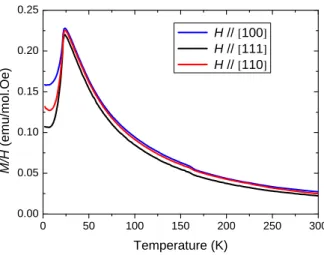
VI. MAGNETIZATION VERSUS TEMPERATURE
0 5 0 1 0 0 1 5 0 2 0 0 2 5 0 3 0 0 0 . 0 0 0 . 0 5 0 . 1 0 0 . 1 5 0 . 2 0 0 . 2 5 H / / [1 0 0 ] H / / [1 1 1 ] H / / [1 1 0 ] M /H ( e m u /m o l. O e ) T e m p e r a t u r e ( K )
FIG. 10: Temperature dependence of the magnetic susceptibility (magnetization divided by the magnetic field, M/H, in the linear regime) measured on the GeCo2O4 single-crystal used in the neutron diffraction experiments, for three orientations of
the magnetic field µ0H=1 T with respect to the main crystallographic directions. The cusp in the three curves indicates the
N´eel temperature, TN=23.5 K, at which the antiferromagnetic ordering occurs.
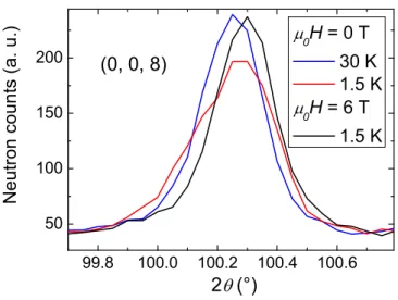
VII. POWDER NEUTRON DIFFRACTION
In addition to single-crystal X-ray diffraction, we performed powder neutron diffraction under magnetic field to investigate the structural distortion. Powder patterns were recorded on the D2B high-flux diffractometer of the Institut Laue Langevin (ILL) under a magnetic field up to 6 T on a pressed pellet of GeCo2O4at the wavelength λ=1.594 ˚A.
Our powder neutron measurements are in agreement with the proposed cubic-to-tetragonal main distortion with an elongated tetragonal axis occurring at TN [1]: The large 2θ angle (0, 0, 8) nuclear reflection splits into 2 below TN
with the lower angle reflection being twice smaller in intensity than the higher angle reflection (see figure11). Under a 6 T magnetic field, only the higher 2θ angle peak is still visible which corresponds to the selection of tetragonal domains with an elongated a or b axis.
99.8 100.0 100.2 100.4 100.6 50 100 150 200 ! !
![FIG. 1: AFM ordered components of the KGM (red) and TRI (green) moments determined from low temperature single- single-crystal neutron diffraction refinements in zero-field and under a magnetic field applied along the [1-10] direction (black arrow):](https://thumb-eu.123doks.com/thumbv2/123doknet/13141468.388646/3.918.111.814.89.376/ordered-components-determined-temperature-diffraction-refinements-magnetic-direction.webp)

![FIG. 6: Single-crystal magnetization M and its derivative (red dashed line) measured as a function of the magnetic field oriented along the [1-10] direction at 2 K.](https://thumb-eu.123doks.com/thumbv2/123doknet/13141468.388646/6.918.100.427.85.603/single-magnetization-derivative-measured-function-magnetic-oriented-direction.webp)
![FIG. 9: Structural distortion: example of monoclinic cell (in blue), with a unique 2-fold axis [1-10], derived from the tetrag-onally distorted cubic cell (black) with the elongated c axis.](https://thumb-eu.123doks.com/thumbv2/123doknet/13141468.388646/8.918.178.363.81.281/structural-distortion-example-monoclinic-unique-derived-distorted-elongated.webp)