HAL Id: hal-03020053
https://hal.archives-ouvertes.fr/hal-03020053
Submitted on 23 Nov 2020
HAL is a multi-disciplinary open access
archive for the deposit and dissemination of
sci-entific research documents, whether they are
pub-lished or not. The documents may come from
teaching and research institutions in France or
abroad, or from public or private research centers.
L’archive ouverte pluridisciplinaire HAL, est
destinée au dépôt et à la diffusion de documents
scientifiques de niveau recherche, publiés ou non,
émanant des établissements d’enseignement et de
recherche français ou étrangers, des laboratoires
publics ou privés.
Learning from BMPs and their biophysical extracellular
matrix microenvironment for biomaterial design
Elisa Migliorini, Amaris Guevara-Garcia, Corinne Albiges-Rizo, Catherine
Picart
To cite this version:
Elisa Migliorini, Amaris Guevara-Garcia, Corinne Albiges-Rizo, Catherine Picart. Learning from
BMPs and their biophysical extracellular matrix microenvironment for biomaterial design. BONE,
Elsevier, 2020, 141, �10.1016/j.bone.2020.115540�. �hal-03020053�
Learning from BMPs and their biophysical extracellular matrix microenvironment for biomaterial design
Elisa Migliorini 1,2*, Amaris Guevara-Garcia1,2,3, Corinne Albiges-Rizo3 and Catherine Picart1,2*
1 CNRS, Grenoble Institute of Technology, LMGP, UMR 5628, 3 Parvis Louis Néel, 38016 Grenoble, France
2 CEA, Institute of Interdisciplinary Research of Grenoble (IRIG), Biomimetism and Regenerative Medicine Lab, Université
Grenoble-Alpes/CEA/CNRS
3 INSERM U1209, CNRS 5309, Université de Grenoble Alpes, Institut for Advanced Biosciences, Institut Albert Bonniot, Allée des
Alpes, Site Santé, 38700 La Tronche, France
*corresponding authors: [email protected] ; [email protected]
KEYWORDS: BMPs, Extracellular matrix, bioinspiration, biomaterials, mechanotransduction
ABSTRACT: It is nowadays well-accepted that the extracellular matrix (ECM) is not a simple reservoir for growth factors but is an
organization center of their biological activity. In this review, we focus on the ability of the ECM to regulate the biological activity of BMPs. In particular, we survey the role of the ECM components, notably the glycosaminoglycans and fibrillary ECM proteins, which can be promoters or repressors of the biological activities mediated by the BMPs. We examine how a process called mechano-transduction induced by the ECM can affect BMP signaling, including BMP internalization by the cells. We also focus on the spatio-temporal regulation of the BMPs, including their release from the ECM, which enables to modulate their spatial localization as well as their local concentration. We highlight how biomaterials can recapitulate some aspects of the BMPs/ECM interactions and help to answer fundamental questions to reveal previously unknown molecular mechanisms. Finally, the design of new biomaterials inspired by the ECM to better present BMPs is discussed, and their use for a more efficient bone regeneration in vivo is also highlighted.
1.
Introduction
Bone morphogenetic proteins (BMPs) are secreted cytokines, part of the transforming growth factors- superfamily, with pivotal roles during embryogenesis [1, 2], tissue homeostasis [3], cellular proliferation [4, 5], migration [6, 7] and differentiation [8, 9]. BMPs signaling is widely investigated for vertebral skeletal development and homeostasis [10] but it has fundamental roles in the development of the majority of tissues [11]. The main BMPs involved in the skeletal development are BMP-2,-4,-5,-6,-7 as well as their antagonist Noggin and Gremlin (reviewed in [10]). The conditional ablation of these BMP genes selectively in the limb bud or the total deficiency of these genes generated defects at the cell level (osteoblasts, chondrocytes) and at the tissue level (joint defect) [10]. BMPs have different physiological and pathological roles. BMP-2 plays an important role in the development of the musculoskeletal system [12], notably cartilage and bone formation. BMP-2 and BMP-4 have an important role in the regulation of normal and cancer stem cells, in particular in the hematopoietic system [13], in brain cancer and neurological diseases [11], BMP-5 in nephrogenesis [14] and lung carcinoma [1BMP-5], BMP-6 in the regulation of ion metabolism [16] and BMP-7 in inflammation, macrophage polarization and differentiation of fat cells [17]. BMP-9 and BMP-10 have a key role in vascular development and angiogenesis [18, 19]. Their role in bone formation in vivo has emerged very recently [20]
BMPs bind to BMP receptors (BMPR) which are composed by a complex of type-1 (BMPR1) also called Activin receptor-like kinase (ALK)- 1, 2, 3, or 6 and type-2 (BMPR2), Activin A receptor, type-2 A (ACVR2A) and ACVR2B) [21, 22]. They are serine/threonine kinase receptors which trigger the phosphorylation of SMAD 1/5/8 and together with SMAD 4 translocate to the nucleus to activate the transcription of target genes [23]. BMPs signaling induces also a SMAD-independent (non-canonical) pathway via the phosphorylation of MAPK which also leads to osteogenic differentiation [24].
The extracellular matrix (ECM) is composed of several fibrillar proteins (fibronectin, collagen, fibrillins) and proteoglycans. The fibrillar ECM proteins have not only the structural role of supporting organs and tissues allowing cellular adhesion via integrins but they have the role of binding and delivery signaling molecules like growth factors [25]. Finally, the physical properties such as the stiffness are also important to guide cellular fate [26]. The fibrillar proteins described in this review can bind BMPs and/or their prodomains to regulate BMP-mediated signaling. Fibronectin, collagen type IIA and IV and fibrillin are known to have specific BMPs and/or BMP prodomains binding sites, as reviewed elsewhere [25] and, close to them, interaction sites for other cell membrane receptors such as integrins or heparin binding sites [27, 28].
Proteoglycans (PGa) are glycosylated proteins composed by core-protein on which one or more glycosaminoglycans (GAGs) chain are covalently attached to a serine residue of the core protein. GAGs are polysaccharides chains of repeated disaccharides
2
anchored via their reducing end to the core proteins [29]. In particular, a tetrasaccharide primer is elongated by adding disaccharides repetitions in the Golgi apparatus where other enzymes as sulfotransferases and N-deacetylases modify the GAG chain. They are then expressed at the cell membrane. There are four families of GAGs: heparin/heparan sulfate (HS), chondroitin/ dermatan sulfate (DS), keratan sulfate and hyaluronic acid (HA). For the first three families, the GAGs are synthetized in the Golgi apparatus while hyaluronic acid is synthetized directly at the plasma membrane by transmembrane enzymes and immediately secreted [30]. The ectodomains of cell-surface proteoglycans (PGs) can be released by the action of sheddases generating extracellular PGs.
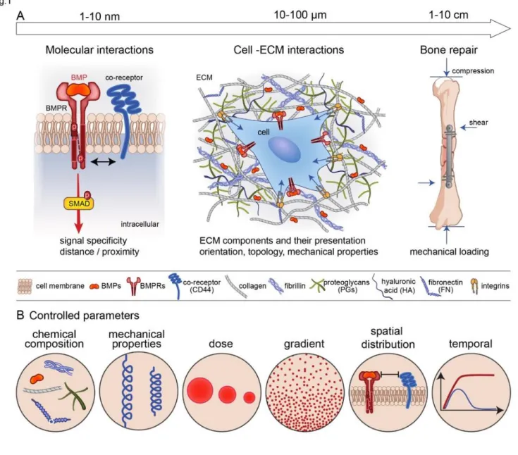
It is now widely accepted that the ECM is not simply the reservoir for these BMPs but is a direct regulator of the biological activities of BMPs [31]: by providing the binding site for BMPs, regulating their dose and availability, and providing a mechanical signal to the cells. In terms of length scale, the BMPs may act at different length scales (FIG. 1A). At the nanoscale, which is the level of the BMP receptors, there are different types of co-receptors engaged by the ECM proteins and GAGs. At the cellular level, the cell is embedded in the ECM proteins and GAGs that present and orient the BMPs to the cells and provide mechanical forces on the receptors. At the bone tissue level, mechanical forces are playing a key role on bone repair induced by the bone progenitors and environmental factors. Whatever the length scale considered (nanoscale, cellular and tissue scale), the chemical composition and mechanical properties of the ECM, the dose and the gradient of BMPs, their spatial and temporal availability will impact the responses of the receptors, cells and tissue (FIG. 1B).
In this review, we aim to show the effect of the ECM as an important regulator of the activity of BMPs. We first summarize the role of the ECM components, notably the glycosaminoglycans (part 2) and fibrillary ECM (part 3), which are important for the binding and presentation of BMPs to the BMPRs. ECM components can be either promoters or inhibitors of BMP bioactivity. We then present how the forces transmitted from the ECM and converted in biochemical signals into the cell, the so-called mechano-transduction, affects BMP signaling (part 4) and internalization (part 5). We also show the effect of the spatio-temporal regulation of BMP release from the ECM (part 6). For each aspect, we report how biomaterial approaches can be used in vitro to answer fundamental biological questions and help to reveal previously unknown mechanisms. Finally, we show how biomaterials inspired from ECM/BMP interactions can enable a better presesentation of BMPs for a more efficient bone regeneration in vivo.
2. Glycosaminoglycans and BMP signaling
After their synthesis and secretion, BMPs interact with proteoglycans, which are present both at the cell surface and within the ECM. Indeed, proteoglycans decorate both ECM proteins such as collagen, fibronectin, fibrillin and other receptors present at the cell membrane, including MuSK (muscle-specific kinase) and BAMBI (BMP and activin membrane-bound inhibitor), which are reviewed somewhere else [32]. By interacting with the cell surface and ECM, cell-secreted BMPs have a constrained range of action in the pericellular space.
GAGs binding sites and BMPs recognition motifs
BMP-2 and 7 can bind to GAGs [33, 34], mainly to heparin and heparan sulfate-GAG family [35, 36]. However, their binding to other GAGs with a lower affinity than to heparin has also been demonstrated [37]. For example, BMP-2 binding affinity to heparin has been measured to be in the range of 2 and 20 nM [35, 37] while the binding affinity to dermatan sulfate was 10 fold lower with a slower association and a faster dissociation rate [37]. BMP-9 and 10 do not present GAG binding sites. However in the pericellular space, they are not free to circulate since they can bind to cell-surface co-receptors such as endoglin [19].
The heparin binding site on BMP-2 has been revealed by Ruppert et al. in 1996 [35]. This sequence of alternated basic amino acids is located at the N-terminal of the protein is called Cardin-Weintraub motif (FIG. 2A). This motif is generally composed by a sequence like XBBXBX or XBBBXXBX where B are the basic residues and X corresponds to hydropathic residues [38]. Molecular studies presented Cardin-Weintraub motifs located at the N-terminus of BMP-2 and BMP-4 (FIG. 2A) and at the C-terminus of BMP-5/6/7 [34] (FIG. 2A) while no heparin-binding site is presented on BMP-9 and BMP-10 [39]. The BMP homolog in Drosophila decapentaplegic (Dpp) also presents the heparin binding site at the N-terminal domain [40]. It is not clear whether the Cardin-Weintraub motif mediates the binding of BMPs to other GAGs such as dermatan sulfate (CS) [41] and to chemically sulfated hyaluronic acid (HA) [42].
BMPs bind to HS specifically at the sulfated regions of the polysaccharides (FIG. 2B). In these regions, HS presents specific sulfation motifs (the number and distribution of N- or O-sulfate groups along the chain) that work as molecular recognition elements [43, 44]. An extensive analysis of HS structure and function has shown that the cell type-specific pattern of sulfation determines the binding affinity of HS to growth factors, cytokines, and other extracellular proteins [43, 45-47], thereby modeling their distribution and activity in the extracellular space [29]. The basis of the biochemical interaction and the importance of a specific sulfation pattern on BMPs bioactivity, have not been deciphered at the molecular level. Indeed, characterization of BMP-2 binding to specific HS-sulfation motifs has so far relied on molecular indirect studies on enzymatically de-sulfated heparin on specific positions, chemically sulfated HA or sulfated chitosan [42, 48, 49]. More recently, it has been proposed, by enzymatical removal of specific sulfations of HS decasaccharides, that the N-sulfations have an important role on the binding of BMP-2 followed by the 6-O-sulfation while 2-O-sulfation has a secondary role in binding BMP-2 [48]. By these indirect methods, the sequence and sulfation pattern of HS motifs recognized by growth factors cannot be revealed and the binding affinity of growth factors to the different HS motifs cannot be directly quantified. The saccharides analysis of long HS fragments is difficult to achieve due to the
complexity of the HS polymer while progress on the synthetic heparin and HS are expected to be the solution to determine the structure-function relationship between HS sulfation pattern and BMP-2 bioactivity [50].
Computational data with highly sulfated GAGs derivatives as sulfated hyaluronic acid and chondroitin sulfate proved a conformational change of the N-terminal region of 2 upon binding. This conformational change reduces the affinity of BMP-2 to BMPR1A in a sulfation-dependent manner [4BMP-2]. This SPR measurement is in contradiction with previous results showing that BMP-2–HS interaction helped the BMP-2/BMPR1A recognition, [51] leaving this finding still under debate.
Regarding HA, since it has no sulfated group, it might not be expected to interact with BMPs at neutral pH. However, a study showed that HA-based matrices could retain and then deliver BMP-2 over 4 weeks [52]. Using the Van der Waals forces between the BMPs and HA, an innovative approach was used to improve the retention of BMP-2 on HA-based hydrogel by changing the protonation state of the carboxylic acid residues of the GAG [53]. Future studies may use HA to retain more efficiently BMP and may also aim to understand the underlying mechanism of BMP/HA molecular interactions.
Role of GAGs on BMPs signaling
After the discovery of the heparin binding-site on BMP-2, the majority of studies regarding the roles of GAGs on BMPs bioactivity were concentrated on heparin and HS. The importance of HS in BMPs signaling has been demonstrated in Drosophila where the abrogation of heparin synthesis was found to disrupt the Dpp signaling pathway [54, 55] and on a rare paediatric disease called hereditary multiple exostoses which causes bone masses at the side of long-bones [56, 57]. This disease is due to mutations of Ext1 glycosyltransferase responsible for the elongation of the HS chain, in the Golgi apparatus by adding to the initial tetrasaccharide primer alternating units of N-acetylglucosamine (GlcNAc) and glucuronic acid (GlcA). It has been reported that Ext1-deficient cells in the cartilaginous region of endochondral bones, present an increased BMP signaling at early tumour stages [58]. This implies that the deficiency of HS at the cellular surface enhances BMP-2 bioactivity. In line with that other studies have supported the inhibitory effect of Heparin/HS on BMP-SMAD signaling. The mutated form of BMP-2 for the heparin binding site presented 15–20 fold increased SMAD 1/5/9 phosphorylation as compared to the wild type BMP-2 [35]. However, this variant leads to a reduction in osteoinductivity of BMP-2 in vivo [59]. On the other hand, addition of basic amino acids at the N-terminal region of BMP-2 induces an increase in the binding to heparin, leading to an enhancement in the osteoinductive properties and resulting in higher and faster bone formation [59].
Another study has shown that the injection of soluble heparin inhibits the ALP activity mediated by BMP-6 in vitro on C2C12 cells and consequently in vivo bone and cartilage formation in rat [60].
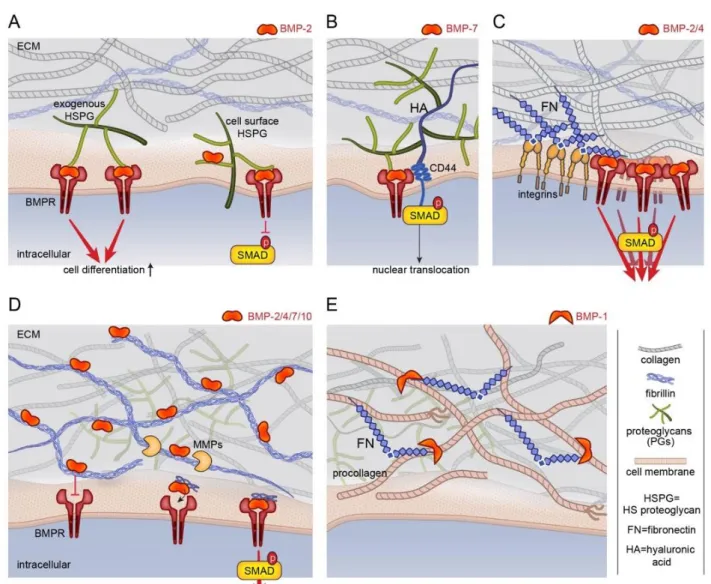
Exogenous HS injected in solution was also found to enhance BMP-2-mediated SMAD 1/5/9 phosphorylation [51] in C2C12 cells. In vivo soluble HS seems to improve bone regeneration, suggesting the use of this GAG in future regenerative medicine applications [61, 62]. This controversial result is likely due to a different but unknown function of cell-surface and extracellular HS-proteoglycans (FIG. 3A). It has been shown that the exogenous soluble HS improves BMP-2-mediated chondrogenic differentiation as well as heparitinase treatment of cell-surface HSPGs, while the up-regulation of syndecan-3 (cell-surface HSPGs) suppresses BMP-2-mediated SMAD phosphorylation [63]. Studies in Drosophila showed that Dally, a cell-surface HSPG, potentiates Dpp signaling and acts as a co-receptor [64].
The studies on exogenous HS used a soluble form of it. However, in the natural environment, HS is not in solution but rather grafted on core proteins forming HSPGs. To mimic this presentation mode, novel bioinspired 2D surfaces have been developed to graft HS as it is in nature by its reducing end [65-68]. They consist in streptavidin platforms on which biotinylated HS is grafted and oriented (iHS) thanks to the site-specific biotinylation [68]. Once BMP-2 is adsorbed iHS and presented to cells the p-SMAD 1/5/9 signaling is sustained over time and up-regulated with respect to the soluble form [65]. It has been suggested that the antagonist Noggin is not able to inhibit BMP-2 once it is adsorbed to iHS, which is not the case for soluble HS [65]. To clarify the mechanism of action of cell-surface and extracellular HS on BMPs signaling, it would be important to compare the effect of these biomimetic platforms on HS-deficient cells.
Although less studied, some evidence has shown that other GAGs including chondroitin and dermatan sulfate (CS and DS) impact BMP-SMAD signaling [69] through their binding to different BMPs [37, 41] . CS is structurally closely related to HS but contains N-acetylgalactosamine (GalNAc) instead of GlcNAc. It is sulfated at specific positions generating different type o CS called CS-A (sulfation on carbon 4 of GalNAc) B (dermatan sulfate DS composed by sulfation on carbon 4 of GalNAc and iduronic acid) CS-C (sulfation on carbon 6 of the GalNAc), CS-CS-D (sulfation on carbon 2 of GlcNAc) and CS-CS-E (sulfation on carbon 4 and 6 of GalNAc). Studies using soluble of E bound to BMP-2 and BMP-4 provided opposite results on BMPs bioactivity. In one study soluble CS-E inhibited p-SMAD 1/5/9 in presence of BMP-2 [70] while in the second study soluble CS-CS-E enhanced BMP-4-mediated osteogenic differentiation [71]. The treatment of primary chondrocytes with chondroitinase reduced the bioactivity of BMP-2 showing that cell-surface CS enhances BMP-2 bioactivity [69].
Another type of ECM PGs, the small leucine-rich proteoglycans (SLRP) consists of a protein core with leucine rich-repeat motifs that is covalently linked to GAG side chains of CS, dermatan and keratan sulfate [72]. Generally, SLRP presents an inhibition effect on BMPs bioactivity [72].
Sulfated GAGs were used to form biomaterials based on a multilayer assembly of chitosan as polycation and oxidized HS and CS as polyanion, together with adsorbed BMP-2. These biomaterials with oxidized GAGs act as a reservoir for the sustained release of BMP-2 and promote cell adhesion, migration and osteogenic differentiation [73].
The non-sulfated GAG HA has also an important function on bone growth. Indeed, it is a main component of the bone ECM and it has been isolated on archeological human skeleton as well as CS [74]. It has been reported that in the chondrocyte response to BMP-7, SMAD1 might immunoprecipitate with the HA receptor CD44 (FIG. 3B) and that this complex is needed for SMAD1 and 5 nuclear translocation [75]. Soluble HA enhances BMP-2-mediated SMAD 1/5/9 phosphorylation and ALP expression [76]. In this
4
study, the authors proposed that HA enhances BMP-2 signaling by blocking the cellular expression of Noggin and Follistatin, another BMP antagonist [76]. A HA-based hydrogel with modified protonation state of the carboxylic acid residues enables the binding of BMP-2 and promotes bone formation in vivo, in comparison to a non-modified HA hydrogel [53]. This result is in line with our studies on Poly(L-lysine) (PLL)/HA polyelectrolyte multilayer films showing that the incorporation of BMP-2 was potentiated at pH 3 in the absence of salt, in comparison to a neutral salt-containing solution (pH 7.4 at 150 mM NaCl) [77]. Such (PLL/HA) films present 2 to cells in a matrix-bound manner, due to the physico-chemical (non-covalent) interactions of BMP-2 with the film. Matrix-bound BMP-BMP-2 was shown to increase p-SMAD 1/5/9 activity and osteogenic differentiation [77] in comparison to soluble BMP-2 [78]. In addition, soft films enabled to reveal a so-far hidden role of BMP-2 on cell adhesion and migration [7]. To investigate the possibility of enhanced BMP-2 binding using sulfated GAGs, heparin was added to HA to form mixed PLL/HA-heparin films [79]. The thickness of the LbL was significantly lower for (PLL/heparin) in comparison to the (PLL/HA) films. Moreover, the presence of heparin was not improving the amount of incorporated BMP-2 nor the ALP activity on C2C12 cells [79], which was solely BMP-2-dose dependent and not dependent on the presence of sulfated groups. Thus, HA by itself may also be a good way to retain BMP-2.
3. Fibrillar ECM and BMP signaling
Fibrillar ECM as a dynamic modulator of BMP signaling
Fibrillar proteins from ECM include multiple, independently folded domains. Some of these domains bind adhesion receptors such as integrins that mediate cell-matrix adhesion. Other domains found in fibronectin, collagen and fibrillin are capable of binding directly to BMP and/or BMP signaling molecules [31, 80]. They act as dynamic modulators of BMP bioavailability and activity [25, 81-83]. This property inspired the field of bioengineering. Indeed, different engineered material surfaces have been designed to mimic the BMP-2 microenvironment and tune cellular as already described in our previous review [84]. It is known that Fibronectin is able to bind BMP-2/4 through the FN 12-14 domain which is localized very close to the FN 7-11 domain known to interact with integrins [83], providing functional proximity between integrins and BMPRs (FIG. 3C). Structural analyses have also revealed that the N-terminal prodomain of collagen type IIA binds 2 [85] (FIG. 2C), whereas the C-terminal part of collagen IV binds BMP-4 [86]. A mechanism based on type IIA procollagen mRNA splicing provides an interesting model for BMP reservoir establishment and induction and differentiation of bone. At its N terminus, this major collagen of cartilage, contains a chordin-like the von Willebrand factor type C (VWC) domain (FIG. 2C) that binds BMP-2 and TGF-1 two chondrogenic growth factors. The VWC domain is alternatively spliced, included in prechondrogenic mesoderm and early developing cartilage but excluded in mature cartilage when the cells differentiate into chondrocytes [85]. This VWC or chordin domain is found in many ECM proteins acting as a negative regulator of BMP functions [87]. Collagen IV, the universal constituent of basement membranes, restricts Dpp/BMP signaling range in Drosophila model [86]. The Dpp-binding motif identified in the C-terminal domains of the two Drosophila collagen IV subunits is highly conserved across phyla, suggesting that this interaction may be important in other contexts [86]. Fibrillins are other major components of the ECM that bind via their N-terminal part to BMP prodomain in the case of BMP-2, -4, -7, and -10 [81, 88, 89] (FIG. 3D). Fibrillins 1, 2 and 3 are self-assembling glycoproteins. They are critically involved in determining tissue elasticity and stiffness [90], which can eventually impact BMP diffusion and gradient. Findings from Nistala et al. [91] identify fibrillins as critical regulators of bone formation through the modulation of endogenous BMP signaling. BMP/fibrillin interactions have been also described as crucial for limb digit formation patterning [92]. However, sequestration of BMPs by fibrillin can inhibit binding to BMPR. The binding of the BMP-7 prodomain to fibrillin-1 causes a conformational change that prevents BMP-7 from binding to BMP receptors (FIG. 3D) [91]. However, BMPs can later be released through matrix remodeling, so that BMP sequestration functions to provide a reservoir [91]. BMPs assemble non-covalently with fibrillins [88], but BMP-propeptide association does not prevent necessarily the ligand from interacting with its receptors [93]. It has been suggested that the elevation of osteoinductive BMP signals in Fibrillin1-/- osteoblast cultures overrides the inhibitory action of improper TGF- activity.
As such, fibrillin-1 microfibrils might act as negative regulators of TGF- and BMP bioavailability in the forming bone. Interestingly, BMPR2-deficient cells express genes indicative of altered biophysical properties, including up-regulation of ECM such as Fibrillin1 and of the adhesion receptors integrins [94]. Finally, dynamic properties of ECM are important not only for BMP sequestration but also for BMP protection from neutralization by soluble inhibitors.
BMP signaling as regulator of matrix remodeling
BMP and BMP-related molecules participate in matrix remodeling. BMPs promote the production of ECM proteins in vitro [7, 95-97]. Analysis of BMPR1aCKO and BMPR1b –/– embryos reveals an essential role of BMP signaling in the production and remodeling
of cartilage ECM proteins in vivo which likely impact the cartilage-to-bone transition through a defect of chondrocyte proliferation and/or survival [95]. Furthermore, the mammalian tolloid (TLD) family of metalloproteinases (BMP-1/TLD) is essential for tissue patterning and ECM assembly. The BMP-1/TLD family is conserved in species ranging from Drosophila to humans and their importance is highlighted by the embryonic lethal phenotype of BMP-1/Tll1 homozygous null mice, which display heart malformations and abnormal procollagen processing. In vertebrates, BMP-1/TLD proteinases are involved in the biosynthetic processing of a wide range of ECM precursors including collagens, the crosslinking enzymes lysyl oxidase (LOX) and LOX-like, laminin basement membrane proteins and SLRPG osteoglycin and probiglycan [98, 99]. As an example, assembly of collagen requires proteolytic processing of procollagen by distinct proteinases ADAMTS-2 and of BMP-1 that cleave the N- and C-terminal
propeptides, respectively [100-103]. However, it is worth to note that cleavage of collagen by BMP-1 induces the production of endotrophin known to be highly inflammatory and to be present in fibrosis and tumoral tissue [104]. BMP-1 and its enhancer PCPE-1 have been shown to bind to FN [105, 106]. More recent results indicate that collagen fibrillogenesis depends on the FN matrix at least in part since FN fibrils provide binding sites for BMP-1 to facilitate procollagen processing [107]. An increased rate of processing is also observed in presence of heparin. This supports that the activity, bioavailability and diffusion of extracellular BMP signaling agonists and antagonists are further regulated by ECM components [82]. More investigations would be helpful to discriminate whether, how and in what circumstances ECM-bound BMPs need to be released to soluble form or can act as solid-phase ligands given that fragments of ECM proteins can be released by proteolysis (FIG. 3E).
4. BMPs signaling in a biomechanical contest
Several reviews have already been dedicated to growth factor signaling and its relation to the biomechanical environment. Kopf et al. [96] have reviewed more specifically the synergy between the integrin adhesion receptors and BMPs-mediated signaling. Several other recent reviews highlighted the fact that growth factors signaling and mechano-transduction mediated via integrin receptors can be combined by engineering cellular microenvironments [108-110], including a very recent review published in bone [111]. At the tissue scale, it is already known that mechanical loading is important for bone growth and regeneration [112] and that BMP signaling and mechano-transduction pathways are tightly interconnected and represent an elaborate signaling network that is active during development [96]. At the cellular scale, cells interpret the biophysical properties of their microenvironment via cell surface mechanoreceptors that are coupled to the contractile cytoskeleton. Signalling is achieved via direct and indirect pathways that control cell fate via nuclear organization and gene expression [113, 114].
Involvement of BMPR in mechanosensing
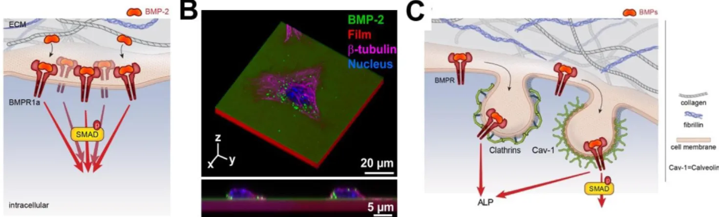
The proximity between FN 12-14 and FN 7-11 domains of FN is crucial to favor the cross-talk between BMP receptors and integrins. Using the (PLL/HA) biopolymeric films presenting bound BMP-2 (bBMP-2), we have shown that the level of SMAD phosphorylation in the presence of BMP-2 on stiff films was similar to that of soluble BMP-2 (sBMP-2), but higher phosphorylation was observed for 2 on soft films [78] in comparison to sBMP-2. An unexpected finding was that, besides BMP-2-mediated signaling, bBMP-2 is able to induce cell adhesion and spreading of soft films (FIG. 4A) and to potentialize cell migration on both soft and stiff films, the effects being stronger for soft films. This BMP-2 induced cell spreading was found to be mostly mediated by 3 integrins [7].
and to depend on the FN that is already present in the pericellular coat of C2C12 skeletal myoblasts. BMPR and 3 integrins work
together to control SMAD signaling and cell adhesion/migration through a LIM kinase pathway involving Cdc42–Src–FAK–ILK, independently of ROCK. β3 integrin was found to regulate a multistep process to control first the BMP receptor activity and second
the inhibitory role of GSK3 on SMAD signaling (FIG. 4A).
Interestingly, BMP-2 can also be printed onto the layer-by-layer biopolymeric films and remain bioactive, which can be used to spatially control cell differentiation [115]. Mixed fibronectin/BMP-2 single-cell micropatterns on soft films were used to study the mechanism underlying BMP-2-mediated mechano-transduction [97]. Cell spreading itself potentiated BMP-2-dependent phosphorylation and nuclear localization of pSMAD 1/5/8 (FIG. 4B). This SMAD signaling was found to depend on LIM kinase 2 and ROCK, rather than myosin II activation. This result was different from the effect of cell spreading induced by sBMP-2 on mesenchymal stem cells cultured on stiff substrates, which was found to be regulated solely by RhoA/ROCK [116].
The same type of collaboration is illustrated in the case of Drosophila collagen IV which binds Dpp (a BMP homolog) and enhances its interactions with BMPR to regulate the dorsoventral axis and the numbers of germinal stem cells in the ovary through integrin activation [117, 118]. However, integrin can also act as an inhibitor of BMP signaling. Indeed, in a specific cellular context and experimental conditions, integrins can act as inhibitors. It has been shown that upon interaction with collagen type II, 1 integrin
compete with BMPR to bind with SMAD1 and then inhibit SMAD1 activation and nuclear import [119]. This helps to suppress helping articular chondrocytes hypertrophy and osteoarthritis progression. The diversity of BMP signaling may also result from the combination of BMP ligands. Recently, a BMP-2/BMP-6/activin A chimera with increased binding affinity to BMP receptors was developed, notably to interact with the ALK2 receptor involved in bone formation[120]. These examples illustrate the capacity of conserved elements of ECM proteins to regulate, either positively or negatively, the functions of various BMPs. However, to date, the ability of ECM to bind a combination of BMP ligand has not been systematically investigated yet. It would be important to better characterized and understand ECM/BMP ligand interactions.
Conversely, the ECM may be affected by BMPR mutations. Shore et al [121] showed that fibroblasts mimicking the Fibrodysplasia Ossificans Progressiva (FOP) pathology, a mutation in ALK2 (R206H/+), have an altered collagen content and organization within the fibroproliferative tissue. Basal activation of BMP signaling pathways, even in the absence of ligand, has also been shown to regulate cell contractility in mesenchymal stem cells [122]. When cultured on tissue culture plastic (a stiff substrate), Rho activation was more robustly increased in Acvr1R206H/+ cells compared with controls and increased levels of phospho-cofilin, of phospho-myosin light chain 2 and nuclear stiffness, and a sensitivity to the ROCK inhibitor Y27, indicating the over-activation of the mechanotransduction pathway Rho/ROCK in mutant cells cultured on stiff substrates. When cultured on polyacrylamide gels of different stiffness, the Acvr1R206H/+ cells exhibited significantly greater cell areas and spreading compared with control cells, especially on the soft gels. In addition, the nuclear localization of the transcription factor RUNX2 was also higher in Acvr1R206H/+ compared with Acvr1+/+ control cells on soft gels. All these data suggested that Acvr1R206H/+ cells misinterpret their biophysical
6
microenvironment, responding to soft substrates similarly to stiff ones, which leads to important changes in key factors that direct cellular differentiation and extracellular matrix production.
More recently, the same team further studied the interplay between Rho/ROCK and YAP pathways in the Acvr1R206H/+ cells [123]. The YAP/TAP complex is a key intracellular regulator of cell differentiation [124] that is known to be regulated by ECM stiffness and cell geometry. YAP/TAZ proteins shuttle between the cytoplasm and the nucleus in response to substrate stiffness and other environmental cues [125]. Cytoplasmic localization of YAP/TAZ is associated with a soft ECM and adipogenic condition, whereas YAP translocation to the nucleus occurs with a stiffer ECM, proliferation and osteogenic conditions [124]. YAP1 was recently found to influence the osteogenic differentiation of MC3T3-EA cells through the regulation of ID1 [126]. A recent study showed that YAP1 governs the initiation of fracture repair by inhibition of cartilaginous callus tissue formation and that it can interact with RunX2 [127]. Interestingly, Shore et al. [123] found that YAP1 has a nuclear localization in Acvr1R206H/+ cells on soft substrates, which was inhibited by the LDN-193189 drug targeting Acvr1 receptors. RhoA expression was increased in mutant cells and this was associated with an increased cellular adhesion and focal adhesion formation. The mutant cells also exerted a higher force compared to normal cells. The ACVR1R206H/+ cells were also poised to chondro/osteogenic differentiation even in the absence of inductive factors. Their results established RhoA and YAP 1 signaling as modulators of mechanotransduction in FOP and showed that the increased BMP pathway signaling through mutant ACVR leads to a higher sensitivity to mechanical stimuli and to increased chondro/osteo differentiation. This synergistic increase in BMP signaling and mechanical signaling promotes heterotopic ossification formation.
An additional step to understand the interaction between cells and the ECM would be to recapitulate the viscoelasticity of the native microenvironment. Viscoelastic matrices enable cell-mediated reorganization of the matrix nanoarchitecture, which in turn modifies local mechanics. Interestingly, in these 3D matrices, stiffness appears to be less important than the stress relaxation rate and fiber topography and remodeling [128-130]. The use of newly-developed viscoelastic materials, possibly as surface coatings, may enable to decouple the effects of stiffness, stress relaxation, fiber remodeling, and ligand density. Indeed, the spatio-temporal control of mechanical properties and the presentation of adhesive ligands might direct cell attachment and migration, or even modulate the mechano-active state of the cell through mechanical priming.
Contribution of microgravity in BMP-2-induced cytoskeleton organization
Recent interesting findings were obtained from experiments performed under simulated microgravity, since bone loss is the most prominent and well recognized phenomenon occurring during spaceflight [131]. One of the consequences of the microgravity is to disrupt the organization of the actin cytoskeleton. Dai et al. [132] showed using drug disruption of the actin cytoskeleton, that the cytoskeleton is mediating the Cbfa1 (RunX2) responsiveness to BMP-2. On the contrary, stabilization of the cytoskeleton by Jasplakinolide can reverse the responsiveness of RunX2 to BMP-2. The same team later demonstrated that integrin v3
participated in the synergetic regulation of RunX2 activity by insulin growth factor 1 under simulated microgravity [133] via PI3K signaling. The role of the actin cytoskeleton in the BMP-2 mediated SMAD signaling studied under microgravity was recently shown to involve calponin 1 [134], an actin-binding protein that promotes and sustain actin polymerization and is crucial for cell contractility. In fact, calponin 1 appears to decrease BMP-2 induced p-SMAD signaling and to block SMAD translocation to the nucleus. Calponin 1 can interact directly with SMAD and p-SMAD. These studies provide a new piece of information on the mechanisms of mechanotransduction under simulated microgravity conditions, which probably contributes to bone formation decrease induced by this experimental condition.
The primary cilium has recently emerged as a sensory organelle in signal transduction in microgravity and electromagnetic field sensing involved in bone disease and cancers [135]. Indeed, Xie et al. [136] have proposed that BMP-2-SMAD 1/5/8 signaling plays a key role in primary cilium-dependent electromagnetic field sensation. They found that pulsed electromagnetic fields stimulated the osteogenic differentiation and maturation of osteoblasts by upregulating the expression of BMPR2, which is localized at the base of the primary cilium.
All these studies highlight the importance of mechanical stimuli in BMP-mediated cell response and the mechanical sensitivity of BMP receptors.
5. BMP internalization to modulate BMP signaling
While BMP receptor and BMP ligand endocytosis has been proposed to promote receptor turnover and regulate BMP signal transduction [137], it is important to consider the ability of BMP to be internalized or not. Previous studies have indicated that clathrin/dynamin-dependent endocytosis of BMP type I receptors is conserved in various models, including C. Elegans, Drosophila, mouse, and human fibroblasts [138]. It is worth to note that the level of BMPR expression impacts BMP internalization. Indeed overexpression of BMPR1A induces an increase of BMP-2 uptake [139] and higher expression of BMPR2 short form at the plasma membrane results in enhanced activation of SMAD signaling (FIG. 5A), stressing the potential importance of the multilayered regulation of BMPR2 expression at the plasma membrane [140]. The endocytic machinery can drive specific BMP signaling or attenuate retrograde BMP signaling [141-144]. The (PLL/HA) films of controlled stiffness were used to study the internalization of matrix-bound BMP-2 (i.e. non-covalently bound BMP-2) and its dependence on film stiffness [141]. Interestingly, bBMP-2 internalization was found to be strongly stiffness-dependent: the amount of internalized BMP-2 is 8 fold higher and the internalization kinetics 5 times faster when BMP-2 is internalized from a soft film in comparison to a stiff film. BMP-2
internalization was independent of the presentation mode (soluble BMP-2 versus bound BMP-2) for low crosslinked films (soft) in striking contrast with high crosslinked (stiff) films where internalization was much lower and slower for bBMP-2. BMPR1A (ALK3) and BMPR-II receptor receptors are both involved in the BMP-2 internalization process in C2C12 cells. Caveolin-mediated internalization was found to be related to both SMAD and ALP signaling while clathrin-mediated internalization was only related to ALP signaling (FIG. 5B). To note, the scission of the endocytic vesicles was not required for SMAD or ALP signaling, highlighting the fact that signaling can still occur at the plasma membrane even in the absence of BMP-2 internalization.
BMP-2 internalization on soft ECM might be considered as an induction mechanism of stem cell differentiation by ECM elasticity [7, 141, 145]. More recently, it has been shown that retrograde BMP signaling including micropinocytosis, a mode of clathrin- and dynamin-independent endocytosis, might be also a key instructive regulator of BMP-dependent bone development [146]. This mode of endocytosis is indispensable for signaling-dependent downregulation of BMPRs at the developing neuro-muscular junction synapse. However, BMP internalization might be affected by BMP antagonists in different ways. While gremlin and Noggin strongly increased BMP-2 internalization, chordin blocked BMP-2 uptake explaining some of the selective biological functions of BMP antagonists [139, 147]. Since BMP-2 is not significantly degraded upon internalization, sorting to late endosomal particles could also provide a reservoir for later use or may be associated with other cellular functions.
In line with the fact that BMP internalization is not required to trigger downstream signaling, several studies used BMPs covalently immobilized to the surface [148-152]. BMP-2 and BMP-6 were covalently coupled to gold nanoparticles with the heterobifunctional linker MU-(N-hydroxysuccimide) NHS [148-150]. Both BMPs were still active after covalent coupling and enabled SMAD 1/5/9 phosphorylation. Moreover, the effect of immobilized BMPs vs soluble BMPs on SMAD 1/5/9 phosphorylation was significant at low BMP-6 doses (1 ng) [148]. Site-specific immobilization of BMP-2 ensures a good orientation of the BMP toward BMPRs that permits a reduction of the BMP dose in comparison to non-specific immobilization. Site-directed immobilization of BMP-2 by click chemistry has been applied on beads and it promotes ALP expression of C2C12 cells in vitro [151, 152]. Nevertheless, BMP2 internalization should be considered in all biomaterials since BMP endocytosis is highly regulated. Besides, BMP signal transduction may depend on the context-specific endocytic routes taken by the receptors.
6. Spatio-temporal control of BMP signaling
The development and homeostasis of multicellular organisms require the coordinated interaction between different biomolecules and receptors. These mechanisms allowing communication between cells within and between tissues are required to confirm the proper assignment of cell types during development and repair. By nature, the signaling by morphogens, like BMPs, is closely related to their spatial position and dose, so that signaling varies across a tissue depending on the amount of ligand, the presence and concetnration of antagonists, and the receptors that are present in the cells of this tissue. The function and efficiency of cell signaling pathways are highly dependent on their coordinated organization both in space and time.
Spatial regulation of BMP signaling
Signaling components in cells are organized to transmit information across cells in nanomter/micrometer size-compartments. This spatial organization of signaling pathways depends on molecular interactions that occur at the nanoscale between the cellular components.
BMP interactions with ECM components facilitate their localization and spatially-regulated signaling, thanks to the high affinity and promiscuous ECM protein-binding, as described in detail in the previous parts. BMPs are recognized to have numerous interactions with GAGs [35, 36] and fibrillar ECM proteins [25, 28, 83, 85, 86, 88, 91, 97, 104, 105, 153-157]. Thus, the ECM components contribute to control the availability of BMPs, since they act as a localized reservoir [91]. ECM proteins regulate cell signaling via their interactions with the adhesive cellular receptors (integrins and others). The adhesive receptors engaged by the ECM act in close proximity to BMP receptors and have an important role as BMP co-receptors. Via their interactions, ECM proteins can thus have a role of enhancers or inhibitors of BMP signaling, depending on the context. For instance, the presence of metalloproteases (BMP-1/TLD) can establish an antagonist sink regulating the bioavailability of BMPs, which is considered as the primary mechanism to drive the formation of BMP gradients [97, 158].
In vivo, the spatial organization of BMP signaling has fundamental roles in both embryonic development and postnatal bone homeostasis [3]. Much of our understanding of spatially-distinct signaling comes from studies on murine and avian limb buds where BMP-2 and BMP-7 were localized to the distal tip of the limb bud [159]. The growth factor gradient appears to be crucial for cell fate decisions. A gradient in genes related to BMP-2 expression has been observed across the cartilage growth plate, likely playing a role in zonal differentiation [160], the gradients for BMP-2 and 6 signaling being mainly present within the hypertrophic zones. This suggests that BMP signaling gradients exist across both growth plate and articular cartilage. These spatial gradients favor the differentiation of chondrocytes in the endochondral skeleton. Furthermore, BMP signaling is one of the key pathways regulating craniofacial development and the formation of mineralized structures. It is involved in the early patterning of the head, development of cranial neural crest cells, and facial patterning [161].
The presence of BMP gradients demonstrates that BMP can be confined to a specific volume, due to a spatially restricted pattern of expression. A recent study elaborated on a model to propose different mechanisms to explain how components work together to create and maintain BMP signaling gradient in the embryo [162]. This model relies on a restricted diffusion BMP antagonists acting as a sink that drives BMP signaling through a dorsal gradient. This data suggests that the spatio-temporal patterns of chordin and BMPs gene expression are dominant drivers of shape in 3D [162]. Another mathematical model [163] demonstrated the
8
importance of BMPR localization in human embryonic stem cells in vitro and mouse embryos in vivo, to shape morphogen signaling during embryogenesis. It shows that the receptor localization facilitates a robust BMP signaling gradient in the mouse embryo. The BMP receptor signaling during embryogenesis depends on the restricted, basolateral localization of BMPR, since the BMP receptor localization at the apical side (mislocalization) results in ectopic BMP signaling in the mouse epiblast in vivo.
Several steps of embryonic development are also found during bone healing [164], indicating that there is a spatio-temporal pattern of BMP-2 expression through the bone fracture. Indeed, the spatially and temporally restricted patterns of BMP expression during bone regeneration appear to be necessary for determining the sites of coupled angiogenesis and osteogenesis that underlie bone regeneration.
Temporal regulation of BMP signaling
Fracture healing involves a synchronized discrete temporal association of cell types and many local and systematic regulatory factors. In the case of bone fracture, bone healing presents five temporal phases that are in chronological order: i) hematoma formation, ii) inflammation, iii) angiogenesis, iv) cartilage formation with subsequent calcification and cartilage removal, and finally v) bone formation and bone remodeling [165]. Furthermore, the expression of BMPs is temporally distinct during fracture healing [160], BMP-2 being expressed at all stages of bone repair.
Besides, the intensity, duration, and fluctuation of signaling pathway stimulation are important in determining the output from the downstream transduction network. Improving our understanding of how these differences in the input are converted into distinct outputs will be essential to unravel the mechanisms of cell fate decisions [166]
A recent review recapitulates the different strategies for targeted and control bone formation in vivo [167]. The ideal carrier should protect the BMPs from degradation, maintain its bioactivity whilst releasing the protein in a spatially and timely-controlled manner to promote the formation of new bone at the treatment site, avoiding secondary and undesirable effects. Traditionally, the immobilization of BMPs on material surfaces is achieved using various methods: via adsorption, entrapment or by covalent binding [168]. While spatial control has been developed by engineering gradients [115] and surface patterning [97], developing a temporal control is more challenging.
A few strategies imply temporal control over BMPs presentation on the substrate or degradation of the links to release BMPs in a time-controlled manner. This strategy also protects BMPs from rapid degradation. Controlled degradation can be achieved using degradable substrates or degradable chemical tethers. These approaches allow researchers to tightly control temporal release kinetics simply by increasing or decreasing the concentration of the cleavable element. The release of BMPs in vitro may be via active degradation, such as a macrophage-mediated release system coupled with the bone repair inflammatory stage [169]. Other examples of active delivery include the release of the BMPs through rupture hydrolysis[170] or proteolytic release [171] induced by the cells, or by external stimuli, such as heat in the case of thermosensitive hydrogels [172]. BMP-2 delivery via the matrix-metallo protease-degradable linker is used to induce osteogenic differentiation in C2C12 cells and mesenchymal stem cells [173]. As a perspective, the control of BMPs release over time is a promising approach to mimic the time-pattern of BMP expression during development and bone repair [174]. Biomaterials may be combined with genetic manipulation as already illustrated in the case of mesenchymal stem cells [175]. Indeed, genetically engineered BMP expressing cells can release the growth factors in situ at the site of injury over extended periods of time at a physiological concentration [176]. To deliver the BMP-plasmids, the coupling with the material-based strategies allows generating BMP signaling over a specific period [177]. This can be accomplished by assembling functional genetic modules to control intrinsic gene expression patterns and similarly, assemble functional ECM domains to create custom-made microenvironments for directing cell behavior. The use of degradable matrices containing the expression plasmids [178] and particularly the use of alginate has been successfully used for the transfer of BMP plasmid [179]. The BMP-genes may also be activated by a pharmacological approach, heat or light to offer a robust and time-tunable protein expression [180]. Optogenetic systems may enable a more precise bone regeneration, inducing the gene BMP-2 on mesenchymal stem cells in a specific location of the bone defect area [181]. It remains to demonstrate whether such regulatory issues raised by too complex systems are not counterproductive.
7. Bone regeneration in vivo using biomaterials inspired from ECM /BMP interactions
As we have seen above that BMP naturally exert their morphogenetic functions within the context of the ECM microenvironment, the interactions between BMPs and other ECM components may be used to optimize the delivery of growth factors for human clinics [182-186]. Overall, biomaterial engineering explores the concept that ECM proteins bind and present growth factors as solid-phase ligands. Cellular responses to nanometer scale signals provided by biomaterial may be exploited to ensure effective osteointegration and osteoinduction of orthopedic devices. Three major interrelated-parameters may be optimized thanks to ECM-inspired biomaterials: i) the dose of BMP-2 that is delivered, which should be as low as possible to avoid side effects (like inflammation and osteolysis) while being as efficient as possible [187, 188]; ii) the temporal control of BMP-2 release, which should as controlled as possible to avoid the so-called burst release since a very fast release at the beginning often leading to an overdose and side effect [189]; iii) the spatial control of BMP-2 to deliver BMP-2 at the right place and avoid its diffusion in fluids or other tissues [187]. Some strategies use surface coatings or nanoparticles to trap or encapsulate BMP-2 and deliver it in a spatially-controlled manner [190, 191].
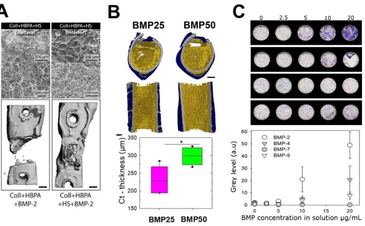
In their recent review, Hachim et al. [192] described how GAGs-based biomaterials can also be used to reduce the dose of BMPs, given their ability to bind and slowly release BMPs, to their natural composition recognized by remodeling ECM-enzymes and to their anti-inflammatory effect [193]. Heparin has been proposed as carriers for BMPs in several types of biomaterials: microparticles [194-196], hydrogels [197] and scaffolds [198, 199]. The release of BMP-2 from heparin-based biomaterials is in the range between 13 days [200] and 28 days [201], depending on the type of biomaterial used: fibrin hydrogel or microparticles, respectively. Due to its anticoagulant effect other GAGs as CS and HS have been proposed as an alternative candidate for BMPs delivery [51, 61, 71, 202, 203]. Stupp’s group proposed to enhance bone regeneration by developing peptide amphiphile nanofibers able to bind HS and a low dose of BMP-2 [204]. These hybrid materials released only the ~ 34% of BMP-2 after 8 days and enhanced bone repair in a rat critical-size femoral defect (FIG. 6A). It has been shown that CS scaffolds can release BMP-2 for two weeks [205]. GAGs-mimetics has also been developed to promote osteogenic differentiation. Some examples are RGTA© (ReGeneraTing Agents) molecules [206], which consist of a saccharides backbone chemically modified with specific sulfate and carboxyl groups [207]. On the contrary of HS they are not recognized by glycanases and once complexed with collagen sponges they promote bone repair in vivo [206]. 6-O and 2-N-sulfated chitosan has also been proposed as an alternative to HS to promote osteogenic differentiation and angiogenesis in vitro and in vivo [208].
Other GAGs or ECM proteins can also be exploited. Recently, Salmeron-Sanchez and coworkers [209] proposed a controlled material-based approach to enhance the activity of BMP-2 during tissue healing using fibrillary fibronectin driven by adsorption onto the polymer poly(ethyl acrylate) (PEA). This fibrillar fibronectin adsorbed onto PEA, but not a fibronectin globular conformation obtained on control polymers, promoted a synergistic presentation of integrin-binding sites and bound BMP-2, which enhanced mesenchymal stem cell osteogenesis in vitro and enabled full regeneration of a non-healing bone defect in vivo at low BMP-2 concentrations. Using this approach, it was possible to reduce the BMP-2 dose by adsorbing it at 50 µg/mL in comparison to 1.5 mg/mL for the concentration used with the collagen sponge using currently in clinics. In their latest study [210], the same team translated this technology and used plasma-polymerized PEA on 2D and 3D substrates to enhance osteogenic differentiation in vitro, proving that this combined fibrillar FN /BMP-2 presentation via the PEA is able to heal a critical-sized bone injury in mice as well as a non-healing humerus fracture in dogs.
A surface coating of implantable materials made of the (PLL/HA) films of controlled stiffness, for which an integrin/ BMPR receptor crosstalk was otherwise revealed [7], was used as osteoinductive coatings of orthopedic and maxillo-facial implants: the (PLL/HA coatings was deposited on ceramics [211], titanium alloy [185], or on the biodegradable polymer polylactic-co-glycolic acid (PLGA) [184] (FIG. 6B). The film-coated implants were tested in rats for their capacity to be osteoinductive and to trigger bone formation in muscle [185, 211]. When coated at the surface of a 3D polymeric hollow tube, they could regenerate a femoral bone defect in rats [184]. The amount of BMP-2 loaded in the film was controlled by tuning the initial concentration of BMP-2 in solution (used for BMP-2 loading in the film), while the BMP-2 released from the film was controlled via film crosslinking level: the low crosslinked film releasing the highest amount of BMP-2. By tuning these parameters, the amount of newly formed bone and the bone repair kinetics could be modulated: an effective and fast repair was obtained in 1 to 2 weeks in the best conditions, including complete defect bridging, the formation of vascularized and mineralized bone tissue. Histological staining and high-resolution computed tomography revealed the presence of bone regeneration inside and around the tube with a spatially distinct organization for trabecular-like and cortical bones. Notably, the amount of cortical bone and its thickness increased with the BMP-2 dose delivered via the polyelectrolyte film [184].
Altogether, these results show that ECM protein and GAG can be used to potentiate and optimized BMP-2 induced bone regeneration.
8. Conclusions and Perspectives
Clearly, the ECM appears to play a significant role in the presentation of BMP to cells to control BMP-mediated signaling. BMP signaling is ECM-dependent and the cellular context depends on the BMPR repertoire and the co-receptor involvement to potentiate or inhibit the BMP signaling. The role of BMP inhibitors, which are also tissue and fluid specific should not be overlooked. In addition, BMP signaling also depends on the BMP dose, spatial presentation time scale and pattern (duration, presence of gradients…). The contribution of biophysicists is and will be useful to design simplified experimental approaches to control the accurate parameters to decorticate the complex biological phenomena that occur in vivo during development and tissue regeneration. The future biomaterials for in vivo applications are also likely to be inspired by the natural ECM. The new paradigm of these future engineered biomaterials is to reduce the BMP dose and to spatially and temporally control the BMP delivery to repair bones in an optimized manner.
Last but not least, technological developments regarding the automated fabrication of biomaterials to enable high content screening of cell behaviors in vitro could boost the time needed to design and test the biomaterials in a parallelized manner. In this context, our team recently developed a process to deposit the biomimetic layer-by-layer films directly in cell culture microplates using a commercial liquid handling robot to study BMP-mediated cellular processes, including cell adhesion and differentiation in 96-well plates in [212]. Being optical transparent, the biomimetic coatings are compatible with microplate readers and common optical microscopes. A proof-of-concept was made to assess the differentiation of skeletal progenitors into bone cells at high content using the coated microplates with different BMP proteins (FIG. 6C). Using such BMPs loaded film-coated plates, it could also be envisioned to study the combined effects of film stiffness and bioactive molecules on stem cell differentiation, to unravel signaling pathways associated with cell fate and thus to propose new strategies to further optimize bone regeneration.
10
The future research for skeletal regeneration will need to be strongly interdisciplinary at the frontier of biology, synthetic biology, genetics, and material engineering to offer the tools to recapitulate the in vivo ECM patterns that are the most appropriate for bone repair in a sophisticated but not too complex way.
ACKNOWLEDGEMENTS
C. Picart is a senior member of the Institut Universitaire de France, whose financial support is acknowledged. A Guevara is supported by a fellowship from the mexican gouvernment CONACYT - (CVU: 532484)). This work was funded by the Fondation Recherche Médicale (DEQ20170336746 to CP and DEQ20170336702 to CAR), by the ANR (CO-DECIDE, grant ANR-17-CE13-022 and GlyCON, grant ANR-19-CE13-0031-01 PRCI), by the European Research Council (ERC Biomim, GA 259370, POC OSCODI 334966 and POC BioactiveCoatings, GA692924), and by Initiative de Recherche Stratégique, University Grenoble Alps (IDEX-IRS 2018-2021).
Bibliographic references
[1] S.V. Hegarty, G.W. O'Keeffe, A.M. Sullivan, BMP-Smad 1/5/8 signalling in the development of the nervous system, Progress in neurobiology 109 (2013) 28-41.
[2] M. Hashiguchi, M.C. Mullins, Anteroposterior and dorsoventral patterning are coordinated by an identical patterning clock, Development (Cambridge, England) 140(9) (2013) 1970-1980.
[3] M. Chau, J.C. Lui, E.B.M. Landman, S.-S. Späth, A. Vortkamp, J. Baron, O. Nilsson, Gene Expression Profiling Reveals
Similarities between the Spatial Architectures of Postnatal Articular and Growth Plate Cartilage, PloS one 9(7) (2014) e103061. [4] J. Ning, Y. Zhao, Y. Ye, J. Yu, Opposing roles and potential antagonistic mechanism between TGF-β and BMP pathways: Implications for cancer progression, EBioMedicine 41 (2019) 702-710.
[5] S.E. Joppé, L.K. Hamilton, L.M. Cochard, L.-C. Levros, A. Aumont, F. Barnabé-Heider, K.J.L. Fernandes, Bone morphogenetic protein dominantly suppresses epidermal growth factor-induced proliferative expansion of adult forebrain neural precursors, Front Neurosci 9 (2015) 407-407.
[6] J. Fiedler, G. Röderer, K.P. Günther, R.E. Brenner, BMP-2, BMP-4, and PDGF-bb stimulate chemotactic migration of primary human mesenchymal progenitor cells, Journal of cellular biochemistry 87(3) (2002) 305-12.
[7] L. Fourel, A. Valat, E. Faurobert, R. Guillot, I. Bourrin-Reynard, K. Ren, L. Lafanechere, E. Planus, C. Picart, C. Albiges-Rizo, beta3 integrin-mediated spreading induced by matrix-bound BMP-2 controls Smad signaling in a stiffness-independent manner, The Journal of cell biology 212(6) (2016) 693-706.
[8] M. Morikawa, R. Derynck, K. Miyazono, TGF-β and the TGF-β Family: Context-Dependent Roles in Cell and Tissue Physiology, Cold Spring Harbor Perspectives in Biology 8(5) (2016).
[9] A.H. Reddi, BMPs: from bone morphogenetic proteins to body morphogenetic proteins, Cytokine & growth factor reviews 16(3) (2005) 249-50.
[10] V.S. Salazar, L.W. Gamer, V. Rosen, BMP signalling in skeletal development, disease and repair, Nature reviews. Endocrinology 12(4) (2016) 203-21.
[11] D. Obradovic Wagner, C. Sieber, R. Bhushan, J.H. Börgermann, D. Graf, P. Knaus, BMPs: From bone to body morphogenetic proteins, Science Signaling 3(107) (2010) mr1-mr1.
[12] A. Bandyopadhyay, K. Tsuji, K. Cox, B.D. Harfe, V. Rosen, C.J. Tabin, Genetic analysis of the roles of BMP2, BMP4, and BMP7 in limb patterning and skeletogenesis, PLoS genetics 2(12) (2006) e216.
[13] P. Toofan, H. Wheadon, Role of the bone morphogenic protein pathway in developmental haemopoiesis and leukaemogenesis, Biochemical Society transactions 44(5) (2016) 1455-1463.
[14] C.P. Bramlage, G.A. Müller, B. Tampe, J. Bevanda, I. Maatouk, M. Koziolek, K. Lange, K. Ahrens, H. Schmid, C.D. Cohen, P. Bramlage, M. Kretzler, F. Strutz, The role of bone morphogenetic protein-5 (BMP-5) in human nephrosclerosis, Journal of nephrology 24(5) (2011) 647-55.
[15] T. Deng, D. Lin, M. Zhang, Q. Zhao, W. Li, B. Zhong, Y. Deng, X. Fu, Differential expression of bone morphogenetic protein 5 in human lung squamous cell carcinoma and adenocarcinoma, Acta biochimica et biophysica Sinica 47(7) (2015) 557-63. [16] B. Andriopoulos, Jr., E. Corradini, Y. Xia, S.A. Faasse, S. Chen, L. Grgurevic, M.D. Knutson, A. Pietrangelo, S. Vukicevic, H.Y. Lin, J.L. Babitt, BMP6 is a key endogenous regulator of hepcidin expression and iron metabolism, Nature genetics 41(4) (2009) 482-7.
[17] L. Grgurevic, G.L. Christensen, T.J. Schulz, S. Vukicevic, Bone morphogenetic proteins in inflammation, glucose homeostasis and adipose tissue energy metabolism, Cytokine & growth factor reviews 27 (2016) 105-18.
[18] H. Chen, J. Brady Ridgway, T. Sai, J. Lai, S. Warming, H. Chen, M. Roose-Girma, G. Zhang, W. Shou, M. Yan, Context-dependent signaling defines roles of BMP9 and BMP10 in embryonic and postnatal development, Proceedings of the National Academy of Sciences of the United States of America 110(29) (2013) 11887-92.
12
[19] L. David, C. Mallet, S. Mazerbourg, J.J. Feige, S. Bailly, Identification of BMP9 and BMP10 as functional activators of the orphan activin receptor-like kinase 1 (ALK1) in endothelial cells, Blood 109(5) (2007) 1953-61.
[20] R.M. Salmon, J. Guo, J.H. Wood, Z. Tong, J.S. Beech, A. Lawera, M. Yu, D.J. Grainger, J. Reckless, N.W. Morrell, W. Li, Molecular basis of ALK1-mediated signalling by BMP9/BMP10 and their prodomain-bound forms, Nature communications 11(1) (2020) 1621.
[21] B.B. Koenig, J.S. Cook, D.H. Wolsing, J. Ting, J.P. Tiesman, P.E. Correa, C.A. Olson, A.L. Pecquet, F. Ventura, R.A. Grant, et al., Characterization and cloning of a receptor for BMP-2 and BMP-4 from NIH 3T3 cells, Molecular and cellular biology 14(9) (1994) 5961-74.
[22] D. Chen, M. Zhao, G.R. Mundy, Bone morphogenetic proteins, Growth factors (Chur, Switzerland) 22(4) (2004) 233-41. [23] F. Liu, A. Hata, J.C. Baker, J. Doody, J. Carcamo, R.M. Harland, J. Massague, A human Mad protein acting as a BMP-regulated transcriptional activator, Nature 381(6583) (1996) 620-3.
[24] C. Sieber, J. Kopf, C. Hiepen, P. Knaus, Recent advances in BMP receptor signaling, Cytokine & growth factor reviews 20(5-6) (2009) 343-55.
[25] R.O. Hynes, The extracellular matrix: not just pretty fibrils, Science (New York, N.Y.) 326(5957) (2009) 1216-9.
[26] A.J. Engler, S. Sen, H.L. Sweeney, D.E. Discher, Matrix elasticity directs stem cell lineage specification, Cell 126(4) (2006) 677-89.
[27] M.M. Martino, F. Tortelli, M. Mochizuki, S. Traub, D. Ben-David, G.A. Kuhn, R. Muller, E. Livne, S.A. Eming, J.A. Hubbell, Engineering the growth factor microenvironment with fibronectin domains to promote wound and bone tissue healing, Science translational medicine 3(100) (2011) 100ra89.
[28] M.M. Martino, P.S. Briquez, A. Ranga, M.P. Lutolf, J.A. Hubbell, Heparin-binding domain of fibrin(ogen) binds growth factors and promotes tissue repair when incorporated within a synthetic matrix, Proceedings of the National Academy of Sciences of the United States of America 110(12) (2013) 4563-8.
[29] S. Sarrazin, W.C. Lamanna, J.D. Esko, Heparan Sulfate Proteoglycans, Cold Spring Harbor Perspectives in Biology 3(7) (2011). [30] N. Itano, T. Sawai, M. Yoshida, P. Lenas, Y. Yamada, M. Imagawa, T. Shinomura, M. Hamaguchi, Y. Yoshida, Y. Ohnuki, S. Miyauchi, A.P. Spicer, J.A. McDonald, K. Kimata, Three isoforms of mammalian hyaluronan synthases have distinct enzymatic properties, The Journal of biological chemistry 274(35) (1999) 25085-92.
[31] G. Sedlmeier, J.P. Sleeman, Extracellular regulation of BMP signaling: welcome to the matrix, Biochemical Society transactions 45(1) (2017) 173-181.
[32] J. Nickel, P. Ten Dijke, T.D. Mueller, TGF-beta family co-receptor function and signaling, Acta biochimica et biophysica Sinica 50(1) (2018) 12-36.
[33] P.C. Billings, M. Pacifici, Interactions of signaling proteins, growth factors and other proteins with heparan sulfate: mechanisms and mysteries, Connect Tissue Res 56(4) (2015) 272-80.
[34] P.C. Billings, E. Yang, C. Mundy, M. Pacifici, Domains with highest heparan sulfate-binding affinity reside at opposite ends in BMP2/4 versus BMP5/6/7: Implications for function, The Journal of biological chemistry 293(37) (2018) 14371-14383.
[35] R. Ruppert, E. Hoffmann, W. Sebald, Human bone morphogenetic protein 2 contains a heparin-binding site which modifies its biological activity, European journal of biochemistry / FEBS 237(1) (1996) 295-302.
[36] B. Ohkawara, S. Iemura, P. ten Dijke, N. Ueno, Action range of BMP is defined by its N-terminal basic amino acid core, Current biology : CB 12(3) (2002) 205-9.
[37] M. Kisiel, A.S. Klar, M. Ventura, J. Buijs, M.K. Mafina, S.M. Cool, J. Hilborn, Complexation and sequestration of BMP-2 from an ECM mimetic hyaluronan gel for improved bone formation, PloS one 8(10) (2013) e78551.
[38] A.D. Cardin, H.J. Weintraub, Molecular modeling of protein-glycosaminoglycan interactions, Arteriosclerosis (Dallas, Tex.) 9(1) (1989) 21-32.
[39] A. Lawera, Z. Tong, M. Thorikay, R.E. Redgrave, J. Cai, M. van Dinther, N.W. Morrell, G.B. Afink, D.S. Charnock-Jones, H.M. Arthur, P. Ten Dijke, W. Li, Role of soluble endoglin in BMP9 signaling, Proceedings of the National Academy of Sciences of the United States of America 116(36) (2019) 17800-17808.
[40] J. Groppe, K. Rumpel, A.N. Economides, N. Stahl, W. Sebald, M. Affolter, Biochemical and biophysical characterization of refolded Drosophila DPP, a homolog of bone morphogenetic proteins 2 and 4, The Journal of biological chemistry 273(44) (1998) 29052-65.
[41] N. Sasaki, T. Hirano, T. Ichimiya, M. Wakao, K. Hirano, A. Kinoshita-Toyoda, H. Toyoda, Y. Suda, S. Nishihara, The 3'-phosphoadenosine 5'-phosphosulfate transporters, PAPST1 and 2, contribute to the maintenance and differentiation of mouse embryonic stem cells, PloS one 4(12) (2009) e8262-e8262.
[42] V. Hintze, S.A. Samsonov, M. Anselmi, S. Moeller, J. Becher, M. Schnabelrauch, D. Scharnweber, M.T. Pisabarro, Sulfated glycosaminoglycans exploit the conformational plasticity of bone morphogenetic protein-2 (BMP-2) and alter the interaction profile with its receptor, Biomacromolecules 15(8) (2014) 3083-92.
[43] X. Zhang, F. Wang, J. Sheng, "Coding" and "Decoding": hypothesis for the regulatory mechanism involved in heparan sulfate biosynthesis, Carbohydrate research 428 (2016) 1-7.
[44] R. El Masri, A. Seffouh, H. Lortat-Jacob, R.R. Vives, The "in and out" of glucosamine 6-O-sulfation: the 6th sense of heparan sulfate, Glycoconjugate journal 34(3) (2017) 285-298.
[45] B.J. Connell, H. Lortat-Jacob, Human immunodeficiency virus and heparan sulfate: from attachment to entry inhibition, Frontiers in immunology 4 (2013) 385.
[46] K. Grobe, J. Ledin, M. Ringvall, K. Holmborn, E. Forsberg, J.D. Esko, L. Kjellen, Heparan sulfate and development: differential roles of the N-acetylglucosamine N-deacetylase/N-sulfotransferase isozymes, Biochimica et biophysica acta 1573(3) (2002) 209-15.
[47] S. Ashikari-Hada, H. Habuchi, Y. Kariya, N. Itoh, A.H. Reddi, K. Kimata, Characterization of growth factor-binding structures in heparin/heparan sulfate using an octasaccharide library, The Journal of biological chemistry 279(13) (2004) 12346-54.
[48] R.A.A. Smith, S. Murali, B. Rai, X. Lu, Z.X.H. Lim, J.J.L. Lee, V. Nurcombe, S.M. Cool, Minimum structural requirements for BMP-2-binding of heparin oligosaccharides, Biomaterials 184 (2018) 41-55.
[49] F. Zhang, J.S. McLellan, A.M. Ayala, D.J. Leahy, R.J. Linhardt, Kinetic and structural studies on interactions between heparin or heparan sulfate and proteins of the hedgehog signaling pathway, Biochemistry 46(13) (2007) 3933-41.
[50] C.T. Tsai, M.M.L. Zulueta, S.C. Hung, Synthetic heparin and heparan sulfate: probes in defining biological functions, Current opinion in chemical biology 40 (2017) 152-159.
[51] D.S. Bramono, S. Murali, B. Rai, L. Ling, W.T. Poh, Z.X. Lim, G.S. Stein, V. Nurcombe, A.J. van Wijnen, S.M. Cool, Bone marrow-derived heparan sulfate potentiates the osteogenic activity of bone morphogenetic protein-2 (BMP-2), Bone 50(4) (2012) 954-64.
[52] H.D. Kim, R.F. Valentini, Retention and activity of BMP-2 in hyaluronic acid-based scaffolds in vitro, Journal of biomedical materials research 59(3) (2002) 573-84.
[53] H.J. Yan, T. Casalini, G. Hulsart-Billstrom, S. Wang, O.P. Oommen, M. Salvalaglio, S. Larsson, J. Hilborn, O.P. Varghese, Synthetic design of growth factor sequestering extracellular matrix mimetic hydrogel for promoting in vivo bone formation, Biomaterials 161 (2018) 190-202.
[54] D.J. Bornemann, J.E. Duncan, W. Staatz, S. Selleck, R. Warrior, Abrogation of heparan sulfate synthesis in Drosophila disrupts the Wingless, Hedgehog and Decapentaplegic signaling pathways, Development (Cambridge, England) 131(9) (2004) 1927-38.
[55] S. Nishihara, Chapter Fifteen - Glycosyltransferases and Transporters that Contribute to Proteoglycan Synthesis in
Drosophila: Identification and Functional Analyses Using the Heritable and Inducible RNAi System, in: M. Fukuda (Ed.), Methods in Enzymology, Academic Press2010, pp. 323-351.
[56] M. Pacifici, Hereditary Multiple Exostoses: New Insights into Pathogenesis, Clinical Complications, and Potential Treatments, Current osteoporosis reports 15(3) (2017) 142-152.
![FIG. 2. Structures of selected BMPs and GAGs and ECM proteins. (A) Structure of BMP-2 (PDB 6OMN) and BMP-6 (PDB 6OMO) [120] as example of BMPs presenting the Heparin/Heparan sulfate-binding site (Hp/HS) at the N and C-terminal domains, re](https://thumb-eu.123doks.com/thumbv2/123doknet/13302508.397962/25.892.55.837.212.576/structures-selected-proteins-structure-presenting-heparin-heparan-terminal.webp)