HAL Id: hal-00656858
https://hal.archives-ouvertes.fr/hal-00656858
Submitted on 5 Jan 2012HAL is a multi-disciplinary open access archive for the deposit and dissemination of sci-entific research documents, whether they are pub-lished or not. The documents may come from teaching and research institutions in France or abroad, or from public or private research centers.
L’archive ouverte pluridisciplinaire HAL, est destinée au dépôt et à la diffusion de documents scientifiques de niveau recherche, publiés ou non, émanant des établissements d’enseignement et de recherche français ou étrangers, des laboratoires publics ou privés.
Photon-in-Photon-out Spectroscopy
Marcin Sikora, Amélie Juhin, Tsu-Chien Weng, Philippe Sainctavit, Carsten
Detlefs, Frank de Groot, Pieter Glatzel
To cite this version:
Marcin Sikora, Amélie Juhin, Tsu-Chien Weng, Philippe Sainctavit, Carsten Detlefs, et al.. Strong K-edge Magnetic Circular Dichroism Observed in Photon-in-Photon-out Spectroscopy. Physical Review Letters, American Physical Society, 2010, 105 (3), pp.037202. �10.1103/PhysRevLett.105.037202�. �hal-00656858�
Strong K-edge Magnetic Circular Dichroism Observed in Photon-in–Photon-out Spectroscopy
Marcin Sikora,1,*Ame´lie Juhin,2,†Tsu-Chien Weng,3Philippe Sainctavit,4Carsten Detlefs,3 Frank de Groot,2and Pieter Glatzel3,‡
1Faculty of Physics and Applied Computer Science, AGH University of Science and Technology, 30-059 Krako´w, Poland 2Department of Inorganic Chemistry and Catalysis, Utrecht University, 3584 CA Utrecht, The Netherlands
3European Synchrotron Radiation Facility, BP 220, 38043 Grenoble Cedex, France
4Institut de Mine´ralogie et de Physique des Milieux Condense´s (IMPMC), UMR CNRS 7590, Universite´ Pierre et Marie Curie, IPGP,
75052 Paris Cedex 05, France
(Received 27 February 2010; published 13 July 2010)
A large enhancement of the x-ray magnetic circular dichroism is observed at the iron K absorption preedge of magnetite. This is achieved by performing resonant inelastic x-ray scattering (RIXS) experi-ments with a 2p hole in the final state of the second-order optical process. We measured and calculated the full 1s2p RIXS planes for opposite helicities of the incoming circularly polarized x rays. The crystal field multiplet calculations show that the enhancement arises from 2p-3d Coulomb repulsions and 2p and 3d spin-orbit coupling. The observed magnitude of the RIXS magnetic circular dichroism effect is!16%. This opens up new opportunities for a broad range of research fields allowing for truly bulk-sensitive, element-, and site-selective measurements of 3d transition metal magnetic moments and their ordering using hard x-ray photons.
DOI:10.1103/PhysRevLett.105.037202 PACS numbers: 75.25."j, 71.70.Ch, 75.47.Lx, 78.70.En
X-ray magnetic circular dichroism (XMCD) is a power-ful tool for the element-specific study of the magnetic structure of complex systems [1–3]. It enables at spin-orbit split absorption edges to determine spin and orbital mag-netic moments by means of sum rules [4,5]. It has been almost 25 years since the effect of magnetic dichroism was anticipated for the x-ray absorption spectra [6] and the first experimental observations of the linear [7] and circular [8] x-ray magnetic dichroism spectra were reported. Over the years the techniques became a routine probe of element-specific magnetization in antiferro- and ferro(ferri)-magnetic systems [9,10].
The magnetic moments of 3d transition metals (TM) are generally studied at the L absorption edge, i.e., dipole-allowed 2p! 3d transitions using soft x rays. Soft x-ray XMCD measurements are typically performed using total electron yield, because significant self-absorption effects that distort the spectral shape are observed when using total fluorescence yield detection. Thus, L-edge XMCD is mainly sensitive to the sample surface and, in addition, it is not compatible with demanding sample environments such as high-pressure cells, due to short penetration depth. Therefore, the 3d TM element-specific studies of bulk magnetic properties under extreme conditions, that require the penetrating properties of hard x rays, have been using K-edge XMCD [11,12] and K! emission spectroscopy [12–15]. The latter is sensitive only to the spin and orbital momenta (S and L) of individual ions, but not to the magnetic moments (ms and ml). As such, it does not
provide quantitative information on interatomic magnetic interactions. The magnetic signal in K-edge XMCD is very weak and the absence of spin-orbit split edges prohibits a
detailed quantitative interpretation. There is hence a need for a magnetic spectroscopy in the hard x-ray range that can provide information on the ordering and the value of magnetic moments. We show in this Letter that this goal can be achieved by combining XMCD with 1s2p resonant inelastic x-ray scattering (RIXS) at the K absorption preedge.
A RIXS signal was observed for the first time in the early 1970s [16], but the technique only developed its full po-tential with the advent of x-ray synchrotron radiation sources. With improved instrumentation [17] and a deeper theoretical understanding [18] this photon-in–photon-out spectroscopy has turned into an important analytical tool to study the electronic structure of TM compounds [19]. RIXS is a second-order optical process where the deep core hole in the intermediate state is replaced by a shal-lower hole in the final state. This results in sharp spectral features and often a rich multiplet structure that reveals electron-electron and spin-orbit interactions. Magnetic di-chroism measurements have been combined with RIXS previously, but these studies were mainly performed at the L2;3 absorption edges, probing low energy transfer
excitations in 3d TM [20,21] or the quadrupole transitions (2p! 4f) in lanthanides [22–26].
The 1s2p RIXS probes the evolution of K" emission (2p! 1s) following resonant excitation of a 1s electron. The K-edge absorption spectra of most 3d TM compounds show weak preedge features that arise from quadrupole (1s! 3d) excitations and possibly additional dipole (1s! 4p) transitions. The intensity of the latter depends on the degree of 3d-4p orbital hybridization and thus the local symmetry around the TM ion [27]. The K preedge is
sensitive to the valence orbitals that are predominantly composed of the 3d density of unoccupied states. The 2p53dnþ1 final state electron configuration in 1s2p RIXS
is identical to the 2p spin-orbit split L2;3absorption edges
[28]. The idea of the present work is to combine 1s2p RIXS in 3d transition metals with XMCD.
The measurements were performed at beam line ID26 of the European Synchrotron Radiation Facility using incom-ing radiation from the undulator fundamental. The incident energy was selected by means of a pair of Si(311) crystals. The circular polarization of the incident x rays was achieved using a 500 #m thick diamond (111) quarter wave plate (QWP). The inelastically scattered photons were analyzed using a set of four spherically bent Ge(440) crystals that were arranged with sample and pho-ton detector (Avalanche Photo Diode) in a vertical Rowland geometry (R$ 1 m). The scattering angle was set to 90%with sample surface at 45%to the incident beam. The combined resolution of the setup was determined using the full width at half maximum of the elastically scattered peak to !E¼ 1:4ð1Þ eV. The 1s2p RIXS-MCD spectrum was acquired at room temperature from a poly-crystalline sample of magnetite ½FeIII*
tetra½FeIIFeIII*octaO4
as a set of constant emission energy scans over the energies of the K"1 and K"2 fluorescence lines. The sample was
kept in magnetic saturation with the magnetization vector parallel to the incident beam using a Nd2Fe14B permanent
magnet, while the photon helicity was reversed after every two constant emission energy spectra. The constant degree of circular polarization, PC! 75ð5Þ%, was maintained by
simultaneous scanning of the incident photon energy and $QWP. The spectra are normalized to perfect circular po-larization degree (i.e., divided by PC), but they are not
corrected for self-absorption effects. The latter may con-siderably affect the shape and dichroism of the main K edge. In the preedge range, however, they are weak and do not influence the analysis presented here.
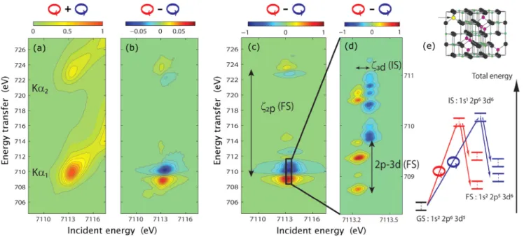
Figure 1 presents the RIXS planes, i.e., the observed fluorescence intensity as a function of energy transfer and incident photon energy. The energy transfer is the differ-ence between incident and emitted energy. The RIXS signal shown in Fig. 1is averaged over both photon hel-icities, whereas the RIXS-MCD signal [Fig. 1(b)] is ob-tained by taking their difference. Their comparison shows that only the resonant features give rise to the XMCD, while the features due to nonresonant fluorescence, that are visible as diagonal structures in the RIXS plane, do not show any detectable XMCD effect. The experimental RIXS-MCD plane reveals two groups of final states, which correspond, respectively, to the K"1 and K"2 emission lines. They are each composed of two strong features with different spin polarization, and the sign of their XMCD is opposite. The characteristic dispersion of the resonant features is due to the lifetime of the 1s hole along the incident energy and of the 2p hole along the energy trans-fer. The experimental data are compared with the theoreti-cal spectrum theoreti-calculated within the crystal field multiplet approach [Fig.1(c)and1(d)].
In a first step towards a quantitative simulation of the 1s2p RIXS-MCD signal, we considered only the
contri-FIG. 1 (color online). (a) The 1s2p RIXS plane measured at the Fe K edge in magnetite. (b) The RIXS-MCD plane, plotted as the difference between the RIXS planes measured for opposite helicities of circularly polarized light. (c) The theoretical RIXS-MCD plane calculated for tetrahedral FeIII. (d) The theoretical RIXS-MCD plotted for the 2p5
3=23d6final state with a small broadening reveals the
origin of the energy shifts that give rise to the dichroism. The arrows with labels indicate the interactions to which the splittings are proportional. (e) Energy level diagram that was used in the crystal field multiplet calculations, involving only a tetrahedral FeIIIion of magnetite [arrow marked (yellow) atom in structure]. See text for details.
bution from tetrahedral FeIII. The lack of inversion sym-metry enables 3d-4p hybridization resulting in strong 1s! 4p dipole contributions to the preedge, while only weak 1s! 3d quadrupole transitions are present for the octahedral Fe sites [19]. The spectrum was simulated using the crystal field multiplet approach developed by Thole et al. [29]. We considered an electric quadrupole excitation from 1s22p63d5 (ground state, GS) to 1s12p63d6
(inter-mediate state, IS), followed by an electric dipole emission to 1s22p53d6 (final state, FS), as shown schematically in
Fig. 1(e) [30]. The radial integrals for interelectronic re-pulsion and spin-orbit coupling were calculated using the atomic Hartree-Fock code of Cowan [31], where the Slater integrals were scaled down to 80% in agreement with previously published experimental works. The effects of the crystal field and of the magnetic exchange field were considered using the O3" Td" S4branching [32]. Values
of 10Dq¼ 0:6 eV and g#BB¼ "10 meV were taken for
the crystal field and the exchange field, respectively. The RIXS cross section was calculated using the Kramers-Heisenberg formula for a right-circular and a left-circular polarization vector. The transition lines were convoluted using a Lorentzian and a Gaussian function to account for the core-hole lifetime and instrumental resolution, respec-tively. For the Lorentzian, the half width at half maximum was chosen as 0.75 eV (IS) and 0.15 eV (FS). The half width of the Gaussian is 0.25 eV.
The experimental and theoretical RIXS-MCD planes [Figs.1(b)and1(c)] are in good agreement, in particular, with respect to the energy splittings and relative transition strengths of the left and right polarized channels. The theoretical plane shows additional features along the en-ergy transfer direction, in particular, at higher energies of the 2ðp3=2Þ53dnþ1 (K"1) final states. These discrepancies
are likely smoothed in experiment due to convolution with the energy-dependent lifetime broadening and experimen-tal resolution function. The experimenexperimen-tal data show a weak feature at 7112 eV incident energy and 707 eV energy transfer that is not observed in the calculations. This fea-ture can be ascribed to octahedral FeII, considering that
(i) its K preedge [27] and L edges [33] are shifted by "1:5 eV with respect to FeIII, and (ii) this feature is absent
from the RIXS-MCD measured on a Mn½FeIII*
2O4 (not
shown). The low intensity of the FeII-induced XMCD
signal and the good agreement between the experimental and calculated RIXS-MCD planes justify a posteriori the assumptions made in the calculation.
The overall agreement of the experimental results with a simple ionic crystal field model provides the possibility for a fundamental understanding of the RIXS-MCD effect. We show the theoretical RIXS-MCD plane at the K"1
reso-nance in Fig.1(d). The line broadenings were reduced in order to reveal the underlying interactions. The states that give rise to the XMCD in the 1s13d6intermediate state are
split by the 3d spin-orbit coupling and the exchange field. They are reached by left and right circular polarized x rays
with 100% selectivity and decay into final states with different orbital and spin momenta that are split by 2p-3d Coulomb repulsion. In the RIXS-MCD plane, the energy shift between left and right polarization along incident energy and energy transfer is thus mainly given by the 3d spin-orbit coupling in the intermediate state and the 2p-3d Coulomb repulsions combined with the 2p spin-orbit interaction in the final state, respectively. As a con-sequence, one can expect strong XMCD enhancement in RIXS spectra of those systems that reveal well-defined preedge features as it is the case in, e.g., semiconductors and molecular complexes. Itinerant and metallic systems generally do not show distinct spectral features that can be assigned to dominantly 3d orbitals possibly limiting the enhancement of the XMCD effect. However, this prelimi-nary assertion has to be tested in future experiments.
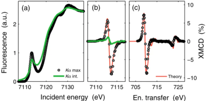
A striking consequence of combining RIXS with XMCD is a dramatic enhancement of the dichroism as compared to conventional K-edge absorption measurements. Fig-ure 2(a) compares the Fe K-edge spectra of magnetite measured at the maximum of the K"1 emission to the
K"1;2-integrated spectra, i.e., the RIXS spectral intensity summed over the energy transfer. The latter is equivalent to conventional fluorescence-detected absorption spectros-copy, while the K"1-detected spectrum corresponds to a
diagonal cut through the RIXS plane. Both spectra reveal similar features with a broad preedge peak at!7113:5 eV. The K"1-detected spectrum shows sharper spectral
fea-tures due to the reduced lifetime broadening [34,35]. The K"1;2-integrated XMCD spectrum [Fig. 2(b)] shows a
shape that is similar to what was observed in previous
7110 7115 (b) 7110 7120 7130 0 1 2 Fluorescence (a.u.) (a)
Incident energy (eV)
705 715 725 −10 −5 0 5 10 XMCD (%)
En. transfer (eV) (c)
Kα max
Kα int. Theory
FIG. 2 (color online). Line scans extracted from the RIXS-MCD plane. Comparison of the Fe K edge spectra (a) and their magnetic circular dichroism (b) acquired at the maximum of K"1 fluorescence line (dots) and using the K"1;2-integrated
spectrum [thick (green) line]. The K"1-detected XMCD spec-trum shows almost eightfold enhancement of the dichroism as compared to K"1;2-integrated and is in good agreement with the theoretical spectrum [thin (red) line]. (c) Comparison between theoretical [thin (red) line] and experimental (dots) XMCD spectra acquired at constant incident photon energy, which corresponds to a vertical line through the RIXS-MCD plane at the incident energy of 7113.6 eV. The measured XMCD effect shown in (b) and (c) is relative to the maximum preedge intensity, while the intensity of the theoretical XMCD is scaled down by factor of 5.67 to obtain best match.
experiments [36] and band structure calculations [37]. However, the intensity of the K"1 detected XMCD is
strongly enhanced and shows a peak-to-peak amplitude as large as 16% of the preedge maximum [Fig.2(b)]. The RIXS-MCD plotted as a function of the energy transfer at constant incident energy [Fig.2(c)] corresponds in the final states to L-edge absorption spectroscopy. We observe the same order of magnitude in the XMCD signal as what has been reported at the Fe L3 edge in magnetite [38,39]. The
crystal field multiplet calculations show that the enhance-ment of the XMCD signal in RIXS is a result of reduced lifetime broadening and increased splitting of the XMCD spectral features in the 2p53dnþ1final state.
An interesting aspect of RIXS-MCD is the possibility for site-selective measurements in mixed valence and mul-tisite compounds. The incident and emission energies de-pend on the oxidation state of the absorber and it is possible to increase the selectivity by tuning either of these, for example, on the weak feature at 7012 eV incident and 707 eV transfer energy in Fig.1(b), in order to study the magnetic behavior of octahedrally coordinated FeII in
magnetite. Therefore, 1s2p RIXS-MCD can be adopted for element- and site-selective magnetometry and mag-netic microscopy of 3d TM with hard x rays, by monitoring the changes in the XMCD amplitude as a function of space, pressure, temperature, or time, either in line scans or, if time allows, by measuring the full RIXS plane. Supported by crystal field multiplet calculations and using RIXS sum rules [40,41], this could be used to extract the values of many GS moments by using angle-dependent experiments. Although the applicability of sum rules has to be con-firmed, the significant progress achieved in synchrotron radiation sources and beam lines equipped with high reso-lution fluorescence spectrometers will allow the technique to become widely accessible.
We acknowledge ESRF for the provision of beam time. We thank C. Brouder for discussion of the theoretical results, A. Rogalev and F. Wilhem for discussion on the correct sign of XMCD, and the ID26 staff for help in setting up the experiment. M. S. acknowledges financial support from the Polish Ministry of Science and Higher Education. A. J. and F. d. G. acknowledge financial support from the Netherlands National Science Foundation (NWO/ VICI program).
*marcin.sikora@agh.edu.pl
†A.F.Juhin@uu.nl ‡glatzel@esrf.fr
[1] H. A. Du¨rr et al.,Science 277, 213 (1997).
[2] P. Gambardella et al.,Nature (London) 416, 301 (2002). [3] M. Mannini et al.,Nature Mater. 8, 194 (2009).
[4] B. T. Thole, P. Carra, F. Sette, and G. van der Laan,Phys. Rev. Lett. 68, 1943 (1992).
[5] P. Carra, B. T. Thole, M. Altarelli, and X. Wang, Phys. Rev. Lett. 70, 694 (1993).
[6] B. T. Thole, G. van der Laan, and G. A. Sawatzky,Phys. Rev. Lett. 55, 2086 (1985).
[7] G. van der Laan et al.,Phys. Rev. B 34, 6529 (1986). [8] G. Schu¨tz et al.,Phys. Rev. Lett. 58, 737 (1987). [9] C. T. Chen et al.,Phys. Rev. B 48, 642 (1993). [10] J. Sto¨hr,J. Magn. Magn. Mater. 200, 470 (1999). [11] O. Mathon et al.,Phys. Rev. Lett. 93, 255503 (2004). [12] Y. Ding et al.,Phys. Rev. Lett. 100, 045508 (2008). [13] J. Badro et al.,Science 300, 789 (2003).
[14] J. Badro et al.,Science 305, 383 (2004). [15] J.-F. Lin et al.,Nat. Geosci. 1, 688 (2008). [16] C. J. Sparks,Phys. Rev. Lett. 33, 262 (1974).
[17] K. Ha¨ma¨la¨inen, D. P. Siddons, J. B. Hastings, and L. E. Berman,Phys. Rev. Lett. 67, 2850 (1991).
[18] P. Carra, M. Fabrizio, and B. T. Thole,Phys. Rev. Lett. 74, 3700 (1995).
[19] F. de Groot and A. Kotani, Core Level Spectroscopy of Solids (CRC Press, Boca Raton, 2008).
[20] L. Braicovich et al.,Phys. Rev. Lett. 82, 1566 (1999). [21] L. Braicovich et al.,Phys. Rev. Lett. 95, 267402 (2005). [22] T. Iwazumi et al.,Phys. Rev. B 56, R14267 (1997). [23] F. M. F. de Groot, M. Nakazawa, A. Kotani, M. H. Krisch,
and F. Sette,Phys. Rev. B 56, 7285 (1997).
[24] F. M. F. de Groot, M. H. Krisch, F. Sette, and J. Vogel, Phys. Rev. B 62, 379 (2000).
[25] K. Fukui, H. Ogasawara, I. Harada, and A. Kotani, J. Synchrotron Radiat. 8, 407 (2001).
[26] T. Nakamura et al.,J. Synchrotron Radiat. 8, 428 (2001). [27] T. E. Westre et al.,J. Am. Chem. Soc. 119, 6297 (1997). [28] W. A. Caliebe et al.,Phys. Rev. B 58, 13452 (1998). [29] B. T. Thole et al.,Phys. Rev. B 32, 5107 (1985). [30] For the tetrahedral FeIIIsite (6A
1ground state) the electric
dipole (E1) operator has T2 symmetry and the electric
quadrupole (E2) operator has T2and E symmetries. In the present case, the quadrupole operators for left and right polarized light (2~111 and 2~11–1) only select T2transitions
and not E transitions. Then the only symmetry reached in the intermediate state is T2 symmetry: T2 (hybridized 4p-3d) for E1 transitions and T2 (pure 3d) for E2 tran-sitions. Since all the intermediates states have T2
symme-try with a dominant 3d character, we restricted the calculation to E2 transitions in order to limit the number of free parameters.
[31] R. D. Cowan, The Theory of Atomic Structure and Spectra (University of California Press, Berkeley, 1981).
[32] P. H. Butler, Point Symmetry Group Applications (Plenum Press, New York, 1981).
[33] P. Kuiper, B. G. Searle, L.-C. Duda, R. M. Wolf, and P. J. van der Zaag, J. Electron Spectrosc. Relat. Phenom. 86, 107 (1997).
[34] K. Ha¨ma¨la¨inen et al.,Phys. Rev. B 46, 14274 (1992). [35] P. Glatzel and U. Bergmann,Coord. Chem. Rev. 249, 65
(2005).
[36] K. Matsumoto et al.,Jpn. J. Appl. Phys. 39, 6089 (2000). [37] V. N. Antonov, B. N. Harmon, and A. N. Yaresko, Phys.
Rev. B 67, 024417 (2003).
[38] D. J. Huang et al.,Phys. Rev. Lett. 93, 077204 (2004). [39] C. Carvallo et al.,Am. Mineral. 93, 880 (2008).
[40] M. van Veenendaal, P. Carra, and B. T. Thole,Phys. Rev. B 54, 16010 (1996).
[41] F. Borgatti et al.,Phys. Rev. B 69, 134420 (2004).