HAL Id: hal-01534412
https://hal-amu.archives-ouvertes.fr/hal-01534412
Submitted on 3 Jul 2018
HAL is a multi-disciplinary open access
archive for the deposit and dissemination of
sci-entific research documents, whether they are
pub-lished or not. The documents may come from
teaching and research institutions in France or
abroad, or from public or private research centers.
L’archive ouverte pluridisciplinaire HAL, est
destinée au dépôt et à la diffusion de documents
scientifiques de niveau recherche, publiés ou non,
émanant des établissements d’enseignement et de
recherche français ou étrangers, des laboratoires
publics ou privés.
component of Mimivirus fibers.
Francesco Piacente, Cristina de Castro, Sandra Jeudy, Matteo Gaglianone,
Maria Elena Laugieri, Anna Notaro, Annalisa Salis, Gianluca Damonte,
Chantal Abergel, Michela G Tonetti
To cite this version:
Francesco Piacente, Cristina de Castro, Sandra Jeudy, Matteo Gaglianone, Maria Elena Laugieri,
et al.. The rare sugar N-acetylated viosamine is a major component of Mimivirus fibers.. Journal
of Biological Chemistry, American Society for Biochemistry and Molecular Biology, 2017, 292 (18),
pp.7385-7394. �10.1074/jbc.M117.783217�. �hal-01534412�
The rare sugar N-acetylated viosamine is a major component
of Mimivirus fibers
Received for publication, February 27, 2017, and in revised form, March 16, 2017 Published, Papers in Press, March 17, 2017, DOI 10.1074/jbc.M117.783217 Francesco Piacente‡, Cristina De Castro§, Sandra Jeudy¶, Matteo Gaglianone‡, Maria Elena Laugieri‡, Anna Notaro¶储1, Annalisa Salis‡, Gianluca Damonte‡, Chantal Abergel¶, and Michela G. Tonetti‡2
From the‡Department of Experimental Medicine and Center of Excellence for Biomedical Research, University of Genova, 16126 Genova, Italy, the Departments of§Agricultural Sciences and储Chemical Sciences, University of Napoli, 80138 Napoli, Italy, and the
¶Aix-Marseille Université, Centre National de la Recherche Scientifique, Information Génomique et Structurale, UMR 7256,
IMM FR3479, 13288 Marseille Cedex 9, France
Edited by Gerald W. Hart
The giant virus Mimivirus encodes an autonomous glycosyla-tion system that is thought to be responsible for the formaglycosyla-tion of complex and unusual glycans composing the fibers surrounding its icosahedral capsid, including the dideoxyhexose viosamine. Previous studies have identified a gene cluster in the virus genome, encoding enzymes involved in nucleotide-sugar pro-duction and glycan formation, but the functional characteriza-tion of these enzymes and the full identificacharacteriza-tion of the glycans found in viral fibers remain incomplete. Because viosamine is typically found in acylated forms, we suspected that one of the genes might encode an acyltransferase, providing directions to our functional annotations. Bioinformatic analyses indicated that the L142 protein contains an N-terminal acyltransferase domain and a predicted C-terminal glycosyltransferase. Se-quence analysis of the structural model of the L142 N-terminal domain indicated significant homology with some character-ized sugar acetyltransferases that modify the C-4 amino group in the bacillosamine or perosamine biosynthetic pathways. Using mass spectrometry and NMR analyses, we confirmed that the L142 N-terminal domain is a sugar acetyltransferase, cata-lyzing the transfer of an acetyl moiety from acetyl-CoA to the C-4 amino group of UDP-D-viosamine. The presence of
acetylated viosamine in vivo has also been confirmed on the glycosylated viral fibers, using GC-MS and NMR. This study represents the first report of a virally encoded sugar acetyltransferase.
Previous reports have provided evidence that the genome of some giant and large DNA viruses encodes autonomous glyco-sylation systems (1, 2). These machineries include glycosyl-transferases and the enzymes required to produce their nucle-otide-sugar substrates. Interestingly, for the few cases reported so far (3–5), novel and atypical glycans have been described. Pathways for monosaccharide synthesis and complex glycan
structures have been characterized in Chlorella viruses (Phy-codnaviridae family) and in some members of the Mimiviridae family (6 –11). In this frame, analysis of the genomes of newly identified giant viruses suggests that Chlorella viruses and Mimiviridae are not isolated cases, because genes associated with glycosylation pathways are found in other members of the nucleocytoplasmic large DNA viruses (12).
Mimivirus is the first identified member of the growing fam-ily of Mimiviridae (13). Its 1.2-Mbp genome encodes ⬃1000 proteins, and the pseudoicosahedral virions of 400-nm diame-ter are covered by a dense array of 150-nm-long, highly glyco-sylated fibers (14). The micrometer size of the viral particles together with the glycosylated fibers mimics the bacteria on which the Mimivirus natural host, Acanthamoeba castellanii, feeds. In a previous study, data from compositional analysis of Mimivirus glycans preparations revealed the presence of vari-ous monosaccharides, including N-acetylglucosamine, glucose, rhamnose, and the very rare 4-amino-4,6-dideoxyhexose viosamine along with its methylated derivative (6). The genes encoding the putative enzymes of the UDP-D-viosamine
bio-synthesis pathway have been identified in a 9-gene cluster local-ized at the 5⬘ end of Mimivirus genome, possibly devoted to glycan production (6). Specifically, the first enzyme, R141, is an UDP-D-glucose 4,6-dehydratase that catalyzes the formation of
a UDP-4-keto-6-deoxy intermediate, also common to the UDP-L-rhamnose pathway (8). A pyridoxal phosphate-depen-dent aminotransferase, L136, then transfers an amino group from glutamate to the 4-keto group, leading to UDP-D-Vio3
production (6).
Viosamine has only been identified in some bacterial species. In Pseudomonas syringae it is a component of the flagellin-as-sociated trisaccharide required for virulence, and in this case the 4-amino group is acylated, and a 2-O-methyl group is also present (15, 16). This unusual sugar, 2-O-methyl-4-(3-hy-droxy-3-methylbutanamido)-4,6-dideoxy-D-glucose, is also
termed anthrose, because of its occurrence in Bacillus
anthra-This work was supported in part by the Fondo per la Ricerca di Ateneo, Uni-versity of Genova, and French National Research Agency Grant ANR-14-CE14-0023-01. The authors declare that they have no conflicts of interest with the contents of this article.
1Recipient of a UIF Vinci program 2015 Ph.D. Fellowship C3_90.
2To whom correspondence should be addressed: Dept. of Experimental
Medicine, University of Genova, Viale Benedetto XV, 1-16132 Genova, Italy. Tel.: 39-010-3538151; Fax: 39-010-35338162; E-mail: tonetti@unige.it.
3The abbreviations used are: Vio, viosamine; VioNAc, N-acetylviosamine; ESI,
electrospray ionization; HSQC, heteronuclear single-quantum correlation spectroscopy; HMBC, heteronuclear multiple-bond correlation spectros-copy; PMAA, partially methylated alditol acetate; LH, left-handed -helix; GT, glycosyltransferase; N-L142, N-terminal domain of L142; TOCSY, total correlation spectroscopy; CPK, Corey-Pauling-Koltun.
cros
ARTICLE
J. Biol. Chem. (2017) 292(18) 7385–7394
7385
cisexosporium (17). Modified Vio has also been reported in few other bacterial organisms (18 –25).
Because Vio is always acylated on the amino group, we undertook a study to verify its status in Mimivirus by adopting a dual strategy. First, we scrutinized the cluster in search of a candidate for Vio acetylation and identified the N-terminal domain of L142 protein as a possible acyltransferase. Second, we performed GC-MS and NMR analyses of Mimivirus glycans. We now report the characterization of the N-terminal domain of L142 (N-L142) as an acetyl-CoA-dependent enzyme able to modify the 4-amino group of UDP-D-Vio. GC-MS and NMR
analyses of Mimivirus glycans then confirmed that Vio is com-pletely acetylated in vivo. To our knowledge, this represents the first report of a virally encoded sugar-N-acetyltransferase.
Results
Sequence and structure analysis
L142gene product is a bifunctional protein with an N-termi-nal domain with a predicted left-handed-helix (LH) super-family fold and a C-terminal one, predicted as glycosyltrans-ferase because of sequence homology. L142 is part of the 9-gene cluster (6) encoding two enzymes involved in UDP-D-Vio pro-duction (L136 and R141), the R135 protein already identified as a component of the outer fibers (26, 27) and several putative glycosyltransferases, some of them displaying two domains possibly derived from gene duplication events, as shown in Fig. 1. In particular, the C-terminal domain of L142 has strong ho-mology with the N-terminal regions of L137 and L138 (Fig. 1), both predicted to be type 2 glycosyltransferases (GT-2) (26). The C-terminal domain of L142 is also predicted to belong to GT-2 family in the CAZy database (28).
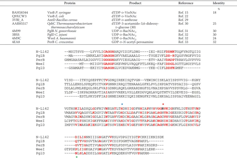
Bioinformatic analysis of the N-L142 revealed the presence of a hexapeptide repeat motif, typical of the LH domain, ini-tially described in UDP-N-acetylglucosamine 3-O-acyltrans-ferase (LpxA) (29). The LH domain is found in many acyltransferases and contributes to the subunit interface, pro-moting trimerization, as well as active site formation. As already observed for L136, the enzyme that transfers the C-4 amino group, no close homologue of N-L142 was found in other viral genomes, excluding Mimivirus close relatives. In the Mimiviridae family, BLAST best hits corresponded to unchar-acterized proteins from different bacterial species (with 30 –37% identity over 196 amino acids) and from environmen-tal sequences. The sequence conservation of N-L142 with known sugar acetyltransferases is reported in Table 1.
Interestingly, N-L142 did not show homology with enzymes known to modify Vio, such as VioB and AntD (15, 22, 30). Low homology was found with QdtC, which modifies the 3-amino group in the synthesis of dTDP-3-acetamido-3,6-dideoxy-␣-D
-glucose (31). On the other hand, a significant homology was observed with the well-characterized PglD from
Campylobac-ter jejuni, as well as with the C-terminal ATD domains of
Neis-seria gonorrhoeaePglB and Acinetobacter baumannii WeeI, all catalyzing an acetyl transfer to the C-4 amino group for
UDP-D-BacNAc2 (32, 33). Among the characterized sugar
acetyl-transferases, the best hit was with Caulobacter crescentus PerB catalyzing the last step of GDP-D-N-acetyl-perosamine
synthe-sis (34). The structural alignment of N-L142 with PglD, PglB, WeeI, and PerB is reported in Fig. 2, which also highlights the residues involved in catalysis and substrate recognition (32–34).
Structural comparison of the N-L142 Phyre2 model with
N. gonorrhoeaePglB confirmed that it possesses all the neces-sary determinants for acetyl-CoA and UDP-D-Vio recognition,
as well as the catalytic residues involved in acetyl transfer and those involved in trimer formation. Interestingly, the structural comparison of the N-L142 model (Fig. 3), based on PglB (4M99), with the PerB (4EA8) structure in complex with acetyl-CoA and GDP-D-N-acetylperosamine highlighted a steric
hindrance caused by an aspartate residue (Asp-232 in 4M99, Asp-40 in L142, and Asp-39 in 4EA8) in the nucleotide-binding site, preventing the accommodation of GDP but allowing a UDP to be properly positioned in the cavity. Asp-55 in PerB is also replaced by Thr-247 in PglB and Ile-55 in N-L142. The overall loop is in a closer state than in the GDP bound structure. Moreover, whereas in the PglB structure an asparagine residue is known to interact with the acetyl bound to the C-2 amino group in bacillosamine (Asn-162), in the N-L142 model, it is another strand of the LH that provides a specific residue (Arg-155), able to engage a H-bond with the hydroxyl group in C-2 of the viosamine. This suggests that viosamine methylation in C-2 is the last step of the sugar modification, probably after the transfer of VioNAc on its acceptor.
Purification of recombinant N-L142 and enzymatic activity WT N-142 and H136A and H145A mutants, expressed as GST-fusion proteins, were purified to homogeneity using affin-ity purification. WT and mutant proteins were soluble after proteolytic release from GST; comparable amounts, 2– 4 mg/li-Figure 1. Organization of the L142 gene cluster. A, in addition to L142, the 9-gene cluster contains the two enzymes for UDP-D-Vio production (R141 and L136), a putative pyruvoyltransferase (L143), a structural protein of the outer fibers (R135), and putative glycosyltransferases (L137, L138, R139, and L140). B, organization of the glycosyltransferase domains. AT, acetyltransferase domain; GT2, glycosyltransferase domain.
ter of bacterial culture, were obtained for WT and mutant proteins.
UDP-D-Vio was produced as described (6) and used as
sub-strate in the enzymatic activity assays. UDP-D-Vio (Fig. 4, peak
A) was incubated with purified WT N-L142 and acetyl-CoA; product formation was monitored by anion-exchange HPLC (Fig. 4, peak B). Enzymatic activity of H136A mutant incubated in identical condition resulted in a decrease by 3 orders of
mag-Table 1
Sequence identity of N-L142 with characterized sugar acetyltransferases
Protein Product Reference Identity
%
BAH58344 VioB P. syringae dTDP-D-VioNAc Ref. 15 18
Q9XCW3 VioB E. coli dTDP-D-VioNAc Ref. 22
3VBJ_A AntD Bacillus cereus dTDP-D-anthrose Ref. 29
AAR85517 QdtC Thermoanaerobacterium thermosaccharolyticum
dTDP-3-acetamido-3,6-dideoxy-D-glucose (30)
Ref. 30 25
4M99 PglB N. gonorrhoeae UDP-D-BacNAc2 Ref. 31 30
3BSS PglD C. jejuni UDP-D-BacNAc2 Ref. 32 30
4M9C WeeI A. baumannii UDP-D-BacNAc2 Ref. 32 24
4EA8 PerB C. crescentus GDP-D-N-acetyl-perosamine Ref. 33 32
Figure 2. Structural alignment of N-L142. Shown are the C-terminal ATD domains of N. gonorrhoeae PglB (4M99), C. crescentus PerB (4EA8), A. baumannii
WeeI (4M9C), and C. jejuni PglD (3BSS). Residues involved in trimer interface and in acetyl-coA binding site are marked in red (based on cd03360, Ref. 43, and our analyses of the model). The conserved catalytic histidine is marked by a blue star, and the position corresponding to viosamine C-2-O binding in L142 is marked by a red star (Arg-155 in bold), whereas the PglD Asn-162 residue involved in C-2 acetylbacillosamine binding is marked by a green star.
Figure 3. Ribbon representation of three perpendicular orientations (A, B, and C) of the N-L142 homotrimer model. Each monomer has a different color
for clarity (chain A in red, chain B in orange, chain C in ice blue). Panel B corresponds to a 90° rotation of the A orientation around the horizontal axis and C to a 90° rotation around the vertical axis. The acetyl-CoA (ACO) and UDP-D-N-acetylviosamine (UDV) are represented as CPKs colored by atom types. The Arg-155 residue making H-bonds with the viosamine C-2O in each monomer is represented as yellow CPK, and the catalytic His-136 is represented as green CPK. The figure was produced using VMD - Visual Molecular Dynamics.
A Mimivirus sugar acetyltransferase
nitude of the enzymatic activity (3992⫾ 554 mol/min/mg protein for the WT versus 2.1⫾ 1.5mol/min/mg protein for the H136A mutant tested within 48 h from purification). In addition, the WT protein activity remained stable over a week of storage, whereas the H136A mutant displayed⬃20–30% decrease. An increase in absorbance at 332 nm, suggestive of protein aggregation, or the presence of a precipitate were not observed for the WT or H136A proteins. Indeed, the reduced enzymatic activity found for this mutant is likely due to the role that His-136 has in the catalytic mechanism rather than to the perturbation of the protein fold. This conclusion is also in agreement with the proposed role of the active site His in the catalytic mechanism for the PglD and PerB enzymes (33). No significant changes in the enzymatic activity were observed for the H145A mutant, in agreement with previous reports for PglD and PerB, where mutation of the corresponding His caused only minor effects in the catalytic activity (33).
Structural characterization of L142 product
The L142 product was purified using anion-exchange HPLC and solid-phase extraction, as described (6), for further ESI-MS and NMR analyses. ESI-MS revealed the presence of a main ion at 590 m/z, consistent with the expected mass of UDP-D
-Vi-oNAc (Fig. 5). A minor peak of 611.9 m/z, was consistent with the sodium adduct of UDP-D-VioNAc.
The purified product was also analyzed by NMR. Identifica-tion of the UDP-D-VioNAc signals was possible by using both 2D homo- and heteronuclear NMR sequences (Table 1). The area at 6.0 –5.5 ppm (Fig. 6A) contained H-5 of the uracil moi-ety and two anomeric signals; the one at 5.99 ppm belonged to the expected ribofuranose unit of the UDP. Our analysis focused on that second one, which had to be related to the Vio residue. The proton at 5.57 ppm correlated with a carbon at 96.5 ppm (HSQC spectrum in Fig. 6B), a chemical shift similar to that previously reported for the non N-acetylated form of UDP-D-Vio (6). Similarly to that, H-1 of N-acetyl-Vio appeared
as a double doublet because of the coupling with two NMR active nuclei, phosphorous (3J
H1,P6.7 Hz) and H-2 ( 3J
H1,H23.3
ppm). Identification of all the ring proton resonances was accomplished by COSY spectrum interpretation (Fig. 6C). H-2 was at 3.61 ppm, partially overlapped to H-4. Nevertheless it was possible to evaluate the multiplicity of both proton signals:
H-2 was a double triplet (Fig. 6, inset) caused by the coupling to H-1 (3J
H2,H13.3 Hz), to phosphorous ( 4J
H2,P3.3 Hz) and H-3
(3JH2,H3 9.8 Hz). Both H-3 (3.76 ppm) and H-4 (3.62 ppm)
appeared as triplet (3J10.0 Hz) meaning that, like H-2 and H-5,
they were axial substituents of the pyranose ring of the sugar. This information confirmed the gluco stereochemistry of the sugar, whereas the carbon chemical shift of C-4 (57.6 ppm) together with H-6/C-6 (1.17/18.2 ppm) values confirmed that it was Vio. Importantly, compared with the non-acetylated Vio, H-4 was at lower field (3.62 versus 3.02 ppm), indicating that the amino function was acetylated, as confirmed by the occurrence of an acetyl group in the proton/carbon spectrum at1H/13C
2.03/23.3 ppm, in a 1:1 ratio with the methyl group of the 6-de-oxy position. The HMBC spectrum (Fig. 6B) disclosed that the methyl of this acetyl and H-4 correlated with the same carbonyl group (175.6 ppm), demonstrating that Vio was N-acetylated. VioNAc from Mimivirus surface glycans
The sugar composition of Mimivirus glycans was already investigated by GC-MS in a previous study (6). After the alditol acetate derivatization, the major components of viral glycans were rhamnose, glucose, N-acetylglucosamine, and Vio. Anal-ysis of the fragmentation spectrum of an unknown peak revealed the presence of methyl-Vio. However, the presence of acetylation on the C-4 amino group could not be detected using this method. Thus, Vio acylation on the surface of purified Mimivirus particles was analyzed by partially methylated aldi-tol acetate (PMAA) method, followed by GC-MS analysis. The presence of a single peak containing one methyl group on Vio C-4 amino group, as evidenced by GC-MS fragmentation spec-trum, is consistent with complete N-acylation of this monosac-charide in Mimivirus glycan (Fig. 7). In addition, PMAA analy-sis indicated that Vio is terminal and not further elongated by other sugars (Fig. 7).
The presence of Vio and the identity of the N-linked substit-uent were confirmed by NMR analyses (Fig. 8 and Table 1) of the glycans extracted from Mimivirus. Inspection of the HSQC spectrum (Fig. 8C) disclosed a complex pattern of anomeric signals (1H range 5.1– 4.5 ppm), a crowded carbinolic area (1H
range 4.4 –3.1 ppm), two main acetyl signals (⬃ 2.0 ppm), and a group of methyl signals (not shown) characteristic of 6-deoxy-sugars at⬃1.3 ppm. All these signals suffered of low resolution, and recording the spectra at high temperature did not improve their overall quality. However, identification of the Vio unit was achieved, even though it was not possible to determine at which residue it was further connected.
Vio anomeric signal was at (1H/13C) 4.59/105.3 ppm, values
diagnostic of a residue configured at the anomeric center. The TOCSY spectrum (Fig. 8B) disclosed that this signal correlated with other five protons (3.64, 3.56, 3.54, 3.22, and 1.24 ppm): this correlation pattern along with the presence of a methyl group at 1.24 ppm, identified this unit as Vio, labeled with V. Combination of COSY and TOCSY spectra established the sequence of the different protons, whereas HSQC identified the corresponding carbon chemical shifts (Table 2). H-2 (3.22 ppm) correlated with a carbon at low field (84.3 ppm) because it was methylated at the corresponding hydroxyl function (O-CH3at
1H/13C 3.63/60.9 ppm), as confirmed by the V
2,2OMe and
Figure 4. Anion-exchange HPLC analysis of nucleotide sugars. UDP-D-Vio (peak A) was incubated with the recombinant L142 N-terminal domain in the presence of acetyl-CoA at 25 °C. The progressive formation of a new com-pound (peak B) was observed.
V2OMe,2 cross-peaks in the HMBC spectrum (Fig. 8C). H-2 enabled H-3 identification, which in turn led to H-4; H-5 was found almost coincident with H-3, as suggested from NOESY spectrum (Fig. 8A), which had one intense cross-peak at⬃3.55 ppm embracing both H-3 and H-5; H-5 correlated further with H-6. Inspection of long-range correlations from H-6 (1.24 ppm) identified both C-4 and C-5 (57.8 and 72.0 ppm, respectively) so that C-3 value was finally selected and confirmed, and its value was indicated by the H-4/C-3 correlation in the HMBC spec-trum (Fig. 8C). Of note, H-4 chemical shift (3.64 ppm) was similar to that of UDP-D-VioNAc (3.62 ppm) and not to that of
UDP-D-Vio (3.02 ppm), in agreement with the amino function
acylation. Accordingly, both H-4 and the methyl of the acetyl at 2.07 ppm had a long-range correlation with a carbonyl at 175.1 ppm, disclosing that the acyl of the amino group was an acetyl. Indeed, in the polysaccharide fibers, Vio has a-anomeric linkage, is methylated at position 2, is acetylated at the amino function, and has no other substituent; it occupies a termi-nal position in agreement with PMAA atermi-nalysis. HMBC and NOESY spectra identified the density of a monosaccharide linked with Vio (see at the cross of the dotted lines in Fig. 8), but poor spectra resolution hampered the elucidation of the nature of this residue.
Discussion
Previous studies showed that the unusual sugar Vio is a com-ponent of Mimivirus glycans and that it is mainly contained in the long fibers that surround the capsid (5, 6). Here we demon-strate that the 4-amino group of UDP-D-Vio is acetylated by the
N-terminal domain of the L142 gene product. The in vitro data obtained with the recombinant enzyme are consistent with the finding that Vio is also N-acetylated in vivo in Mimivirus gly-cans. Indeed, GC-MS and NMR data clearly indicate that, in fibers, Vio is terminal and is both N-acetylated on C-4 and methylated on C-2.
Viosamine, acetylated on the C-4 amino group and often also methylated on C-2, is restricted to some bacterial species, path-ogenic to both vertebrates and plants. In P. syringae, dTDP-VioNAc is produced by a set of enzymes contained in a gene cluster named “Vio island,” which includes VioA, which trans-fers the amino group to the 4-keto group of dTDP-4-keto-6-deoxy-D-glucose and VioB that acetylates the 4-amino group
(15). Other enzymes in the cluster further convert
dTDP-D-VioNAc to dTDP-D-N-(3-hydroxy-1-oxobutyl)Vio and its 2-methylated derivative. Modified Vio was found in the flagel-lin-associated glycans, and disruption of its biosynthetic path-way impairs motility and virulence on host tobacco leaves (16). Similar “Vio islands” were also identified in P. aeruginosa PAK (15, 35) and in B. anthracis (17). In this latter organism, modi-fied Vio is a component of the exosporium pentasaccharide (17, 29). The biosynthetic genes for dTDP-VioNAc were also iden-tified in Escherichia coli O7 and Shigella dysenteriae type 7 (22). However, in most cases the acetyl group is further modified into more complex moieties, and simple acetylation has been rarely reported.
Bioinformatic analysis of N-L142 sequence showed that, sur-prisingly, it has no homology to known enzymes involved in Vio
N-acetylation or acylation. Similarly, the first enzyme of the Mimivirus Vio pathway, L136, has very low homology with the corresponding VioA of P. syringae or E. coli. On the other hand, a significant homology was found with enzymes that catalyze the acetylation of the 4-amino group of UDP-D-BacNAc (31,
33). BacNAc2is an essential component of bacterial N-linked
and O-linked glycans, where it represents the first sugar attached to the protein. Moreover, good homology was found with C. crescentus PerB, responsible for GDP-D-perosamine acetylation (34). This finding was also confirmed by compari-son of N-L142 structural model with N. gonorrhoeae PglB and
C. crescentusPerB (Fig. 3). N-L142 shows a typical LH super-Figure 5. ESI-MS spectrum of the purified N-L142 product. The purified product of N-L142 was analyzed by ESI-MS. The main ion of 590 m/z corresponds to
the calculated UDP-D-VioNAc molecular mass. Ion 612 m/z represents the sodium adduct. Other peaks are consistent with residual Acetyl-CoA and CoA-SH and their sodium adducts, copurified with the UDP-D-VioNAc.
A Mimivirus sugar acetyltransferase
family fold, typical of this type of acyltransferases. Chantigian
et al. (36), starting from X-ray structures and site-directed mutagenesis analyses, have proposed the presence of two
dif-ferent classes of LH enzymes able to acylate nucleotide sugars, based on substrate binding orientations and reaction mecha-nisms. N. gonorrhoeae PglB, C. jejuni PglD, and C. crescentus Figure 6. NMR analysis of purified UDP-D-VioNAc. A, full proton spectrum recorded for UDP-D-VioNAc (600 MHz, 25 °C, D2O). The inset details H-2 and H-4
signals, and the gray dotted lines dissect the contribution given by each proton to the complex signal. Only signals belonging to ribose (R) and Vio (V) are labeled; other signals belong to residual NADPH or impurities. B, expansion of COSY spectrum of UDP-D-VioNAc (600 MHz, 25 °C, D2O). Only densities belonging
to ribose (R) and viosamine (V) cross-peak densities are attributed; other signals belong to residual NADPH or impurities. C, expansion of COSY spectrum of UDP-D-VioNAc (600 MHz, 25 °C, D2O). Only cross-peak densities belonging to ribose (R) and viosamine (V) are attributed; other signals belong to residual
NADPH.
Figure 7. Identification of N-acylated Vio in Mimivirus by PMAA GC-MS analysis. The viral particles were derivatized by PMAA technique and then analyzed
by GC-MS. The chromatogram of VioNAc was obtained by extraction of m/z 117 ion current. Peak identification was performed by comparing UDP-D-VioNAc standard analysis and fragmentation spectra. A, zoomed-in view of the m/z 117 extracted chromatogram of Mimivirus. B, fragmentation spectrum of VioNAc
PerB belongs to class I, because they use a conserved histidine in the active site as a catalytic base. On the other hand, for class II enzymes, comprising QdtC and AntD, a substrate-assisted cat-alytic mechanism has been proposed (36). To confirm that N-L142 also belongs to class I acetyltransferases, we have per-formed site-directed mutagenesis of the proposed catalytic His residue. Indeed, the H136A mutant activity, assayed immedi-ately after purification, was 3 orders of magnitude lower when
compared with the WT. This finding matches what is described for PglB and PglD His mutants, although the decrease of the catalytic efficiency of these other enzymes ranged from 4 to 6 orders of magnitude. Upon storage, the N-L142 H136A mutant activity decreased faster than the WT, being 70 – 80% of the original value after 1 week of storage. On the other hand, pro-tein aggregation or precipitation could not be detected, sug-gesting that His-136 has a minor role in perturbing the struc-Figure 8. NMR of crude polysaccharide from Mimivirus fibrils. Shown are 2D spectra recorded for the crude polysaccharide from Mimivirus fibrils (600 MHz,
56 °C, D2O). A, expansion of NOESY spectrum showing correlation from Vio (V) anomeric proton. B, expansion of TOCSY spectrum showing correlation from Vio
anomeric proton. C, overlap of HSQC (black/dark gray) and HMBC (pale gray) spectra. D, expansion of HMBC spectrum detailing the long-range correlations with the carbonyl group. Vio densities are labeled with V, and dotted lines point to the HSQC density of the residue that has Vio linked. *, inter-residual NOE density between Vio and the other monosaccharide; **, NOESY artifact caused by minimal spin diffusion.
Table 2
1H and13C chemical shifts of UDP-␣-VioNAc as UDP precursor and in the polysaccharide
Spectra calibration used the methyl group of acetone (1
H/13
C 2.225/31.45 ppm) added as internal standard.
Nucleus 1 2 3 4 5 6 UDP-VioNAc VioNAc 1 H 5.57 3.61 3.76 3.62 4.04 1.18 13 C 96.4 73.3 71.8 57.6 69.3 18.2 Ac 1 H 2.03 13 C 175.6 23.3 Ribose 1 H 5.99 4.38 4.37 4.29 4.24–4.20 13 C 89.5 74.9 70.8 84.5 66.2 Uracil 1 H 5.97 7.96 13 C 152.9 167.5 103.7 142.7 VioNAc (polysaccharide) -VioNAc 1H 4.59 3.22 3.56 3.64 3.54 1.24 13C 105.3 84.3 73.9 57.8 72.0 18.3 OMe 1H 3.63 13C 60.9 Ac 1H 2.07 13C 175.1 23.5
A Mimivirus sugar acetyltransferase
ture of the protein. Accordingly, our data and the comparison of the N-L142 structural model with the published homologous structures clearly pinpointed a catalytic role for this His, as well as the other molecular determinants responsible for substrate specificity.
The C-terminal part of the L142 protein displays a glycosyl-trasferase GT-2 fold, suggesting that this domain can be responsible for the attachment of the acetylated Vio on the Mimivirus fibers. Interestingly, NMR analysis has revealed that VioNAc is bound via a-anomeric linkage, thus indicating that the involved enzyme behaves as an inverting transferase. Sev-eral GT-2 enzymes are contained in the same gene cluster as L142 and are probably derived from gene duplication and fusion events. The origin of this cluster is not clear, because it contains genes related to both prokaryotes (i.e. L136 and L142) and eukaryotes (R141). However, because these putative GTs are only found in Mimiviridae members of group A, it is likely that they are involved in the transfer of the monosaccharides that are uniquely produced by these viruses, i.e. rhamnose and Vio (6, 7).
The mechanisms of production of the complex carbohy-drates of Mimivirus and other large DNA viruses are largely unknown. In addition to Chlorella viruses, which revealed the presence of novel and unique structures (3), glycans from other viral families still await characterization. Several evidences have already suggested that glycosylation occurs in the cytosol, in the so-called “viral factories,” but information about the organiza-tion of the glycosylaorganiza-tion machinery in these factories is still lacking, as well as on the origin of the enzymes involved in these processes. Identification and characterization of the enzymes encoded by the viral genomes will help shed light on these issues.
Experimental procedures
L142 sequence and structural analyses
The most similar homologues of the N- and C-terminal domains of L142 were identified using the BLAST tool on the NCBI server, using the “nr” and the “env-nr” databases. The CAZy database was also used for glycosyltransferase identifica-tion (29). The N-L142 sequence was submitted to the Phyre server (37), which returned a model of the L142 N-terminal domain (Leu-8 to Ile-206) based on the PglB structure from
N. gonorrhoeae (100% confidence, 4M99) (32). We used the 4M99 structure to model the acetyl-CoA cofactor in the N-ter-minal domain of the L142 binding site and the PerB structure in complex with acetyl-CoA and GDP-D-N-acetylperosamine
(4EA8) to define the nucleotide acetylated sugar-binding site (34). A model of UDP-D-VioNAc manually built from the GDP-D-N-acetylperosamine and UDP-D-BacNAc2(3BSS, 31) was
fit-ted in the nucleotide sugar-binding pocket of N-L142. We used the molecular visualization program VMD (38) to compare the four structures (PglB, PerB, PglD, and WeeI) and define the amino acids involved in ligand binding.
Expression and purification of recombinant L142 proteins The N-L142 (amino acids 1–213) was expressed as a recom-binant GST-fusion protein in E. coli strain BL21 (DE3) (New England Biolabs) using the pGEX-6-P1 vector (GE Healthcare).
The PCR-amplified sequence corresponding to bases 1– 639 of
L142ORF was digested with BamHI-HF威 and XhoI restriction enzymes (New England Biolabs) and ligated in the plasmid vector. Site-directed mutagenesis was performed using QuikChange (Agilent), following the recommended protocols. Primers were designed using QuikChange primer design pro-gram. Sequencing of WT and mutants was performed by Tib-Molbiol (Genova, Italy). Protein expression, purification, and proteolytic cleavage were performed as described previously (8). Proteins were concentrated to⬃1 mg/ml using Amicon Ultra-4 10K (Millipore) and stored in PBS at 4 °C. They were analyzed by UV absorbance from 210 to 340 nm, and concen-tration was determined using⑀280⫽ 15,930M⫺1 cm⫺1(39).
Purity was determined by SDS-PAGE. Enzymatic assays
Mimivirus N-L142 enzymatic activity was assayed on the UDP-D-Vio, produced using recombinant Mimivirus R141 and
L136 (6, 8). UDP-D-Vio was incubated with acetyl-CoA in pres-ence of N-L142 in PBS, pH 7.3, at 25 °C. Reactions with differ-ent concdiffer-entrations of substrates were performed. The reactions were stopped by heat inactivation for 3 min at 80 °C at different time points of incubation, and the solutions were clarified by microfiltration (Millipore). After clarification, the reaction mixtures were analyzed by HPLC, as previously described (7). Specific activity was determined using the reduction of UDP-D
-Vio peak area and expressed as means ⫾ S.D. ofmol con-verted per min/mg protein, using 0.2 mMUDP-D-Vio and 0.4
mM acetyl-CoA as substrates. Analyses were performed in
duplicate at different time points after enzyme purification from two independent protein preparations.
Structural characterization of L142 product
To confirm the acyltransferase activity of Mimivirus N-L142, its product was purified as previously described (6) and ana-lyzed by ESI-MS and by NMR. ESI-MS analysis was performed in direct infusion analysis at 5l/min on an Agilent 1100 series LC/MSD ion trap XCT instrument (Agilent Technologies, Palo Alto, CA). Nucleotide sugars were diluted up to 10 pmol/ml in a water:acetonitrile (50:50) solution containing 0.1% formic acid. The spectra were acquired in negative ion mode in the mass range of the expected m/z ratios, as described (6).
For NMR analysis, 1D and 2D NMR spectra were recorded on a Bruker 600 DRX equipped with a CryoProbeTMon a
solu-tion of 500l of D2O, at 25 °C. Double quantum filtered
phase-sensitive homonuclear COSY experiment was performed using data sets of 2048⫻ 512 points (40, 41); the data matrix was zero-filled in both dimensions to give a matrix of 4K⫻ 2K points and was resolution-enhanced in both dimensions by a cosine-bell function before Fourier transformation. Coupling constants were determined on a first order basis from high-resolution 1D spectra. HSQC and HMBC spectra were mea-sured in the1H-detected mode via single quantum
coher-ence with proton decoupling in the13C domain, using data
sets of 2048 ⫻ 512 points. Experiments were carried out in the phase-sensitive mode (40), and the data matrix was extended to 4096⫻ 2048 points using forward linear prediction extrapolation.
Analysis of VioNAc presence on Mimivirus glycan
The presence of acylation on the C-4 amino group of Vio in Mimivirus glycans was verified using the PMAA derivatization technique (42). Approximately 0.5–1⫻ 1011whole Mimivirus
particles were lyophilized overnight to remove any trace of water that could inhibit the following permethylation reaction. The dry viral particles were suspended in DMSO (1 ml) with powdered NaOH. Then methyl iodide (0.5 ml) was added, and the sample was stirred for 45 min at room temperature. The reaction was quenched by dropwise addition of 5% acetic acid aqueous solution. To purify the permethylated particles, 2 ml of chloroform was added, and the reaction mixture was made up to 5 ml with ultrapure water. The sample was thoroughly mixed and allowed to settle into two layers. The upper aqueous layer was removed and discarded. The chloroform layer was washed several times with ultrapure water and dried under a gentle stream of nitrogen.
The dry permethylated particles were hydrolyzed with TFA and further processed to obtain the alditol acetate derivatives, as previously described (7). The PMAA were extracted from the solid crust at the bottom of the vial four times with 0.5 ml of dichloromethane and collected in a new vial. The samples, evaporated and suspended in a small volume of dichlorometh-ane, were analyzed by GC-MS as previously described (7). Isolation of Mimivirus polysaccharides and NMR analysis
Mimivirus suspension (2 ml, ⬃ 2.4 ⫻ 1011 particles) were
stirred with 6 ml of 0.5MDTT solution at 100 °C for 1 h to promote the complete removal of the fibrils. The slurry was centrifuged (8000 rpm, 10 min), and the solid was washed twice with ultrapure water. Supernatants were pooled and dialyzed, yielding to a crude polysaccharide preparation (32 mg), and an aliquot (5 mg) was analyzed via NMR without further purifica-tion. NMR conditions were the same as described for UDP-D
-VioNAc, except temperature that was set to 56 °C, and TOCSY and NOESY spectra were recorded, by using 100- and 200-ms mixing time, respectively. The spectra were calibrated using the methyl group of acetone (1H/13C 2.225/31.45 ppm) added as
internal standard.
Author contributions—F. P., M. G., and M. E. L. performed protein cloning and expression, mutagenesis, enzyme analysis, sample prep-aration, and purification for further characterization. F. P. and C. A. performed bioinformatic analyses. A. S. and G. D. analyzed samples by ESI-MS and GC-MS. C. D. C. and A. N. performed NMR analysis. C. A. and S. J. developed and analyzed the L142 structural model. M. G. T. conceived and coordinated the study and wrote the paper. All authors analyzed the results and approved the final version of the manuscript.
References
1. Piacente, F., Gaglianone, M., Laugieri, M. E., and Tonetti, M. G. (2015) The autonomous glycosylation of large DNA viruses. Int. J. Mol. Sci. 16, 29315–29328
2. Van Etten, J. L., Gurnon, J. R., Yanai-Balser, G. M., Dunigan, D. D., and Graves, M. V. (2010) Chlorella viruses encode most, if not all, of the ma-chinery to glycosylate their glycoproteins independent of the endoplasmic reticulum and Golgi. Biochim. Biophys. Acta 1800, 152–159
3. De Castro, C., Molinaro, A., Piacente, F., Gurnon, J. R., Sturiale, L., Palmi-giano, A., Lanzetta, R., Parrilli, M., Garozzo, D., Tonetti, M. G., and Van Etten, J. L. (2013) Structure of N-linked oligosaccharides attached to chlo-rovirus PBCV-1 major capsid protein reveals unusual class of complex
N-glycans. Proc. Natl. Acad. Sci. U.S.A. 110, 13956 –13960
4. De Castro, C., Speciale, I., Duncan, G., Dunigan, D. D., Agarkova, I., Lan-zetta, R., Sturiale, L., Palmigiano, A., Garozzo, D., Molinaro, A., Tonetti, M., and Van Etten, J. L. (2016) N-Linked glycans of chloroviruses sharing a core architecture without precedent. Angew. Chem. Int. Ed. Engl. 55, 654 – 658
5. Hülsmeier, A. J., and Hennet, T. (2014) O-Linked glycosylation in
Acan-thamoeba polyphagaMimivirus. Glycobiology 24, 703–714
6. Piacente, F., Marin, M., Molinaro, A., De Castro, C., Seltzer, V., Salis, A., Damonte, G., Bernardi, C., Claverie, J. M., Abergel, C., and Tonetti, M. (2012) Giant DNA virus Mimivirus encodes pathway for biosynthesis of unusual sugar 4-amino-4,6-dideoxy-D-glucose (Viosamine). J. Biol. Chem.
287,3009 –3018
7. Piacente, F., De Castro, C., Jeudy, S., Molinaro, A., Salis, A., Damonte, G., Bernardi, C., Abergel, C., and Tonetti, M. G. (2014) Giant virus megavirus chilensis encodes the biosynthetic pathway for uncommon acetamido sugars. J. Biol. Chem. 289, 24428 –24439
8. Parakkottil Chothi, M., Duncan, G. A., Armirotti, A., Abergel, C., Gurnon, J. R., Van Etten, J. L., Bernardi, C., Damonte, G., and Tonetti, M. (2010) Identification of anL-rhamnose synthetic pathway in two nucleocytoplas-mic large DNA viruses. J. Virol. 84, 8829 – 8838
9. Piacente, F., Bernardi, C., Marin, M., Blanc, G., Abergel, C., and Tonetti, M. G. (2014) Characterization of a UDP-N-acetylglucosamine biosyn-thetic pathway encoded by the giant DNA virus Mimivirus. Glycobiology
24,51– 61
10. Luther, K. B., Hülsmeier, A. J., Schegg, B., Deuber, S. A., Raoult, D., and Hennet, T. (2011) Mimivirus collagen is modified by bifunctional lysyl hydroxylase and glycosyltransferase enzyme. J. Biol. Chem. 286, 43701– 43709
11. Rommel, A. J., Hülsmeier, A. J., Jurt, S., and Hennet, T. (2016) Giant Mimivirus R707 encodes a glycogenin paralog polymerizing glucose through␣- and -glycosidic linkages. Biochem. J. 473, 3451–3462 12. Iyer, L. M., Balaji, S., Koonin, E. V., and Aravind, L. (2006) Evolutionary
genomics of nucleo-cytoplasmic large DNA viruses. Virus Res. 117, 156 –184
13. Raoult, D., Audic, S., Robert, C., Abergel, C., Renesto, P., Ogata, H., La Scola, B., Suzan, M., and Claverie, J. M. (2004) The 1.2-megabase genome sequence of Mimivirus. Science 306, 1344 –1350
14. Legendre, M., Santini, S., Rico, A., Abergel, C., and Claverie, J. M. (2011) Breaking the 1000-gene barrier for Mimivirus using ultra-deep genome and transcriptome sequencing. Virol. J. 8, 99
15. Yamamoto, M., Ohnishi-Kameyama, M., Nguyen, C. L., Taguchi, F., Chiku, K., Ishii, T., Ono, H., Yoshida, M., and Ichinose, Y. (2011) Identifi-cation of genes involved in the glycosylation of modified viosamine of flagellins in Pseudomonas syringae by mass spectrometry. Genes 2, 788 – 803
16. Taguchi, F., Yamamoto, M., Ohnishi-Kameyama, M., Iwaki, M., Yoshida, M., Ishii, T., Konishi, T., and Ichinose, Y. (2010) Defects in flagellin glyco-sylation affect the virulence of Pseudomonas syringae pv. tabaci 6605.
Microbiology 156,72– 80
17. Dong, S., McPherson, S. A., Wang, Y., Li, M., Wang, P., Turnbough, C. L., Jr., and Pritchard, D. G. (2010) Characterization of the enzymes encoded by the anthrose biosynthetic operon of Bacillus anthracis. J. Bacteriol.
192,5053–5062
18. Arbatsky, N. P., Kondakova, A. N., Shashkov, A. S., Drutskaya, M. S., Belousov, P. V., Nedospasov, S. A., Petrova, M. A., and Knirel, Y. A. (2010) Structure of the O-antigen of Acinetobacter lwoffii EK30A: identification of d-homoserine, a novel non-sugar component of bacterial polysaccha-rides. Org. Biomol. Chem. 8, 3571–3577
19. Kondakova, A. N., Linder, B., Fudala, R., Senchenkova, S. N., Moll, H., Shashkov, A. S., Kaca, W., Zähringer, U., and Knirel, Y. A. (2004) New structures of the O-specific polysaccharides of proteus: 4. polysaccharides containing unusual acidic N-acyl derivatives of 4-amino-4,6-dideoxy-D -glucose. Biochemistry 69, 1034 –1043
A Mimivirus sugar acetyltransferase
20. Liu, B., Chen, M., Perepelov, A. V., Liu, J., Ovchinnikova, O. G., Zhou, D., Feng, L., Rozalski, A., Knirel, Y. A., and Wang, L. (2012) Genetic analysis of the O-antigen of Providencia alcalifaciens O30 and biochemical charac-terization of a formyltransferase involved in the synthesis of a Qui4N derivative. Glycobiology 22, 1236 –1244
21. Perepelov, A. V., Wang, Q., Senchenkova, S. N., Shevelev, S. D., Shashkov, A. S., Feng, L., Knirel, Y. A., and Wang, L. (2008) Structure and char-acterization of the gene cluster of the O-antigen of Escherichia coli O49 containing 4,6-dideoxy-4-[(S)-3-hydroxybutanoylamino]-D- glucose.
Biochemistry 73,406 – 410
22. Wang, Y., Xu, Y., Perepelov, A. V., Qi, Y., Knirel, Y. A., Wang, L., and Feng, L. (2007) Biochemical characterization of dTDP-D-Qui4N and dTDP-D -Qui4NAc biosynthetic pathways in Shigella dysenteriae type 7 and
Esch-erichia coliO7. J. Bacteriol. 189, 8626 – 8635
23. Ovchinnikova, O. G., Valueva, O. A., Kocharova, N. A., Arbatsky, N. P., Maszewska, A., Zablotni, A., Shashkov, A. S., Rozalski, A., and Knirel, Y. A. (2013) Structure of the O-polysaccharide of Providencia alcalifaciens O35 containing an N-[(S)-1-carboxyethyl]-L-alanine (alanopine) derivative of 4-amino-4,6-dideoxyglucose. Carbohydr. Res. 375, 73–78
24. Kondakova, A. N., Kirsheva, N. A., Shashkov, A. S., Shaikhutdinova, R. Z., Arabtsky, N. P., Ivanov, S. A., Anisimov, A. P., and Knirel, Y. A. (2011) Low structural diversity of the O-polysaccharides of Photorhabdus
asymbi-otica subspp.asymbioticaandaustralisandtheirsimilaritytotheO-polysac-charides of taxonomically remote bacteria including Francisella
tularen-sis. Carbohydr. Res. 346, 1951–1955
25. Vinogradov, E., Nossova, L., Korenevsky, A., and Beveridge, T. J. (2005) The structure of the capsular polysaccharide of Shewanella oneidensis strain MR-4. Carbohydr. Res. 340, 1750 –1753
26. Boyer, M., Azza, S., Barrassi, L., Klose, T., Campocasso, A., Pagnier, I., Fournous, G., Borg, A., Robert, C., Zhang, X., Desnues, C., Henrissat, B., Rossmann, M. G., La Scola, B., and Raoult, D. (2011) Mimivirus shows dramatic genome reduction after intraamoebal culture. Proc. Natl. Acad.
Sci. U.S.A. 108,10296 –10301
27. Klose, T., Herbst, D. A., Zhu, H., Max, J. P., Kenttämaa, H. I., and Ross-mann, M. G. (2015) A Mimivirus enzyme that participates in viral entry.
Structure 23,1058 –1065
28. Lombard, V., Golaconda Ramulu, H., Drula, E., Coutinho, P. M., and Hen-rissat, B. (2014) The carbohydrate-active enzymes database (CAZy) in 2013. Nucleic Acids Res. 42, D490 –D495
29. Raetz, C. R., and Roderick, S. L. (1995) A left-handed parallel helix in the structure of UDP-N-acetylglucosamine acyltransferase. Science
270,997–1000
30. Kubiak, R. L., and Holden, H. M. (2012) Structural studies of AntD: an
N-acyltransferase involved in the biosynthesis ofD-anthrose. Biochemistry
51,867– 878
31. Thoden, J. B., Cook, P. D., Schäffer, C., Messner, P., and Holden, H. M. (2009) Structural and functional studies of QdtC: an N-acetyltransferase required for the biosynthesis of dTDP-3-acetamido-3,6-dideoxy-␣-D -glu-cose. Biochemistry 48, 2699 –2709
32. Morrison, M. J., and Imperiali, B. (2013) Biochemical analysis and structure determination of bacterial acetyltransferases responsible for the biosynthesis of UDP-N,N⬘-diacetylbacillosamine. J. Biol. Chem.
288,32248 –32260
33. Olivier, N. B., and Imperiali, B. (2008) Crystal structure and catalytic mechanism of PglD from Campylobacter jejuni. J. Biol. Chem. 283, 27937–27946
34. Thoden, J. B., Reinhardt, L. A., Cook, P. D., Menden, P., Cleland, W. W., and Holden, H. M. (2012) Catalytic mechanism of perosamine N-acetyl-transferase revealed by high-resolution X-ray crystallographic studies and kinetic analyses. Biochemistry 51, 3433–3444
35. Arora, S. K., Bangera, M., Lory, S., and Ramphal, R. (2001) A genomic island in Pseudomonas aeruginosa carries the determinants of flagellin glycosylation. Proc. Natl. Acad. Sci. U.S.A. 98, 9342–9347
36. Chantigian, D. P., Thoden, J. B., and Holden, H. M. (2013) Structural and biochemical characterization of a bifunctional ketoisomerase/N-acetyl-transferase from Shewanella denitrificans. Biochemistry 52, 8374 – 8385 37. Kelley, L. A., Mezulis, S., Yates, C. M., Wass, M. N., and Sternberg, M. J.
(2015) The Phyre2 web portal for protein modeling, prediction and anal-ysis. Nat. Protoc. 10, 845– 858
38. Humphrey, W., Dalke, A., and Schulten, K. (1996) VMD: visual molecular dynamics. J. Mol. Graph. 14, 33–38, 27–28
39. Gasteiger, E. H., Gattiker. A., Duvaud, S., Wilkins, M. R., Appel, R. D., and Bairoch A;. (2005) Protein Identification and Analysis Tools on the Ex-PASy server. In The Proteomics Protocols Handbook (Walker, J. M., ed) pp. 571– 607, Humana Press, Totowa, NJ
40. States, D. J., Haberkorn, R. A., and Ruben, D. J. (1982) A two-dimensional nuclear Overhauser experiment with pure absorption phase in four quad-rants. J. Magn. Reson. 48, 286
41. Rance, M., Sørensen, O. W., Bodenhausen, G., Wagner, G., Ernst, R. R., and Wüthrich, K. (1983) Improved spectral resolution in COSY1H NMR
spectra of proteins via double quantum filtering. Biochem. Biophys. Res.
Commun. 117,479 – 485
42. Ciucanu, I. (2006) Per-O-methylation reaction for structural analysis of carbohydrates by mass spectrometry. Anal. Chim. Acta 576, 147–155 43. Marchler-Bauer, A., Derbyshire, M. K., Gonzales, N. R., Lu, S., Chitsaz, F.,
Geer, L. Y., Geer, R. C., He, J., Gwadz, M., Hurwitz, D. I., Lanczycki, C. J., Lu, F., Marchler, G. H., Song, J. S., Thanki, N., et al. (2015) CDD: NCBI’s conserved domain database. Nucleic Acids Res. 43, 222–226