HAL Id: tel-02422393
https://tel.archives-ouvertes.fr/tel-02422393
Submitted on 22 Dec 2019HAL is a multi-disciplinary open access archive for the deposit and dissemination of sci-entific research documents, whether they are pub-lished or not. The documents may come from teaching and research institutions in France or abroad, or from public or private research centers.
L’archive ouverte pluridisciplinaire HAL, est destinée au dépôt et à la diffusion de documents scientifiques de niveau recherche, publiés ou non, émanant des établissements d’enseignement et de recherche français ou étrangers, des laboratoires publics ou privés.
The role of sensory nervous system in the regulation of
bone formation, remodeling, and repair
Diana Silva
To cite this version:
Diana Silva. The role of sensory nervous system in the regulation of bone formation, remodel-ing, and repair. Human health and pathology. Université de Bordeaux, 2017. English. �NNT : 2017BORD0919�. �tel-02422393�
THÈSE PRÉSENTÉE POUR OBTENIR LE GRADE DE
DOCTEUR DE
L’UNIVERSITÉ DE BORDEAUX
ÉCOLE DOCTORALE SCIENCES DE LA VIE ET DE LA SANTÉ
BIOLOGIE CELLULAIRE ET PHYSIOPATHOLOGIE
Par Diana Isabel SILVA
Le rôle du système nerveux sensoriel dans l'orchestration
de la formation osseuse, le remodelage et la régénération
tissulaire
Sous la direction de Joëlle AMÉDÉE
Soutenue le 21 décembre 2017
Membres du jury :
Mme. LAMGHARI, Meriem, PhD, University of Porto Président Mme. LAMGHARI, Meriem, PhD, University of Porto Rapporteur Mme. CHENU, Chantal, PhD, University of London Rapporteur M. CARLE, Georges, PhD,University of Nice Examinateur M. HOLY, Xavier, PhD, IRBA, Brétigny-sur-Orge Examinateur M. OLIVEIRA, Hugo, PhD, INSERM, Bordeaux Invité
I
Le rôle du système nerveux sensoriel dans l'orchestration de la formation osseuse, le remodelage et la régénération tissulaire
Les progrès dans la compréhension de la biologie osseuse ont permis d’identifier le rôle du système nerveux sensoriel dans la formation osseuse, le remodelage et la régénération tissulaire. Cependant, le rôle précis du système nerveux sensoriel sur la l’ostéogénèse reste encore méconnu. La première partie de ce travail a été d’analyser le rôle des neurones du ganglion de la racine dorsale (DRG) sur la différenciation ostéoblastique des cellules souches mésenchymateuse (MSCs). Pour répondre à cette question, nous avons utilisé une plate-forme microfluidique, qui tente de mimer l’innervation sensorielle du tissu osseux. Dans la seconde partie de cette étude, nous avons cherché à mieux caractériser la sous-population de neurones DRG impliqués dans la régulation directe de la différenciation des MSCs vers le lignage ostéoblastique. En conclusion, l’ensemble des résultats permettent de montrer que: i) les neurones sensoriels ont un effet positif et direct sur la différenciation ostéoblastique des cellules ostéoprogénitrices, ii) la voie de signalisation Wnt/β-caténine est impliquée dans cette transduction du signal; iii) cet effet est principalement régulé par des neurones sensorimoteur, iv) qui peuvent induire la libération locale de facteurs neuroactifs. Mots clés : neurones sensoriels, cellules mésenchymateuses, différenciation des ostéoblastes
The role of sensory nervous system in the regulation of bone formation, remodeling, and repair
Advances in the understanding of bone biology have identified the sensory nervous system as a critical regulator in the orchestration of bone formation, remodeling, and repair. However, the precise role of the sensory nervous system on bone tissue, particularly on osteoprogenitor cells, remains unknown. Firstly, we were interested in clarifying whether dorsal root ganglion (DRG) neurons would be able to induce the osteoblast differentiation by acting directly on mesenchymal stem cells (MSCs). Afterwards, we attempted to understand whether the canonical Wnt signaling pathway could be implicated in the DRG neurons-induced osteoblastogenesis. In the second part of this study, we aimed at better characterizing the subset of DRG neurons involved in the direct regulation of osteoblast differentiation from MSCs. In this work we provide several novel insights: i) we show that sensory neurons have a positive and direct effect on osteoblast differentiation of osteoprogenitor cells, ii) by activating the Wnt/β-catenin signaling pathway; and iii) we suggest that this effect is mainly regulated by sensorimotor neurons, iv) which possibly mediate the local release of neuroactive factors.
Keywords : DRG neurons, mesenchymal stem cells, osteoblast differentiation
Unité de recherche
III
Diana Isabel Silva
Doctor of Cell Biology and Pathophysiology
University of Bordeaux
2017
The role of sensory nervous system in the regulation
of bone formation, remodeling and repair
V This dissertation was submitted to the École Doctorale
Sciences de la Vie et de la Santé of University of
Bordeaux in fulfillment of the requirements for the degree of Doctor of Cell Biology and Pathophysiology.
VII The work presented in this dissertation was developed in The Laboratory for the
Bioengineering of Tissues - Institut national de la santé et de la recherche médicale
(INSERM U1026 – “BioTis”) under the supervision of Doctor Joëlle Amédée, and supported by funds from Direction générale de l'armement (DGA).
IX The results presented in this dissertation are submitted or in preparation for future submission to international peer-review scientific journals:
Diana Isabel Silva, Bruno Paiva dos Santos, Jacques Leng, Hugo Oliveira, Joëlle Amédée. Dorsal root ganglion neurons regulate the transcriptional and translational programs of osteoblast differentiation in a microfluidic platform. Cell Death and Disease.
Diana Isabel Silva, Bruno Paiva dos Santos, Hugo Oliveira, Joëlle Amédée. Activation of capsaicin-sensitive dorsal root ganglion neurons induces the osteoblast differentiation of mesenchymal stem cells in vitro. (Manuscript under preparation)
Note: the data presented in this dissertation is partially formatted according to the
XI
TABLE OF CONTENTS
ACRONYMS AND ABBREVIATIONS LIST ... XVI ABSTRACT ... XI RÉSUMÉ ... XIII 01. RELATED LITERATURE ... 1 1.1.BONE ... 3 1.1.1. Bone Functions ... 3 1.1.2. Bone Structure ... 3
1.1.2.1. Gross structure of bone – compact bone and spongy bone ... 3
1.1.2.2. Microstructure of bone – osteon and trabecula ... 5
1.1.3. Bone Cells – The O’Cells ... 6
1.1.3.1. Osteoprogenitor cells ... 6
1.1.3.2. Osteoblasts ... 7
1.1.3.3. Osteocytes ... 7
1.1.3.4. Osteoclasts ... 8
1.1.4. Bone Matrix ... 9
1.1.5. Neurovascular Supply of Bone ... 10
1.1.6. Bone Formation ... 10
1.1.6.1. Intramembranous ossification ... 11
1.1.6.2. Endochondral (intracartilaginous) ossification ... 13
1.1.7. Bone Remodeling ... 14
1.1.7.1. Bone remodeling cycle ... 15
1.1.7.2. Regulatory factors in bone remodeling ... 17
1.1.8. Canonical Wnt Signaling Pathway in Bone Formation and Remodeling ... 19
1.1.9. Angiogenesis/Vascularization in Bone Formation and Remodeling ... 22
1.2.SENSORY NERVOUS SYSTEM ... 23
1.2.1. Functional Organization of the PNS ... 23
1.2.2. Basic Function and Structure of a Neuron ... 25
1.2.3. Types of Sensory Receptors ... 27
1.2.4. Classification of the Sensory Nerve Fibers ... 29
1.2.5. Dual Afferent and “Efferent” Function of Sensory Neurons ... 29
1.3.SENSORY NERVOUS SYSTEM AND BONE ... 31
02. HYPOTHESIS, OBJECTIVES AND EXPERIMENTAL DESIGN ... 33
XII
3.1.ABSTRACT ... 41
3.2.INTRODUCTION ... 42
3.3.RESULTS ... 44
3.3.1. Microfluidic devices allow the neurite outgrowth within the MSCs compartment .. 44
3.3.2. DRG neurons enhances the differentiation of MSCs towards the osteoblast lineage ... 46
3.3.3. DRG neurons modulates the expression of Cx43 and N-cadherin in MSCs during osteoblastogenesis ... 49
3.3.4. DRG neurons promotes the activation of the Canonical/β-catenin Wnt signaling pathway in MSCs ... 51
3.4.DISCUSSION ... 55
3.5.MATERIAL AND METHODS ... 59
3.5.1. Microfluidic devices fabrication ... 59
3.5.2. Coculture of DRG neurons and MSCs in the microfluidic devices ... 59
3.5.3. Immunofluorescence staining ... 59
3.5.4. Cell metabolic activity and proliferation assays ... 60
3.5.5. RNA extraction, cDNA synthesis, and RT-qPCR analysis ... 60
3.5.6. Alkaline phosphatase activity detection ... 61
3.5.7. Protein extraction, precipitation and Western blotting analysis ... 61
3.5.8. Statistical analysis ... 62
3.6.ACKNOWLEDGMENTS &AUTHOR CONTRIBUTIONS ... 63
3.7.REFERENCES ... 64
3.8.SUPPLEMENTARY INFORMATION ... 69
3.8.1. Supplementary Figures ... 69
3.8.2. Supplementary Experimental Procedures ... 71
3.8.2.1. Microfluidic devices fabrication ... 71
3.8.2.2. Rat bone marrow mesenchymal stem cells isolation ... 71
3.8.2.3. Rat dorsal root ganglion isolation ... 72
3.8.2.4. Flow cytometric analysis ... 72
3.9.IN BRIEF ... 73
04. ARTICLE 2 ... 75
4.1.ABSTRACT ... 79
4.2.INTRODUCTION ... 80
4.3.RESULTS ... 81
4.3.1. CM obtained from the culture medium of activated capsaicin-sensitive DRG neurons triggers the osteoblast differentiation process in MSCs ... 81
XIII
4.5.MATERIAL AND METHODS ... 86
4.5.1. Rat primary cells isolation and culture ... 86
4.5.2. CGRP and Substance P levels quantification... 86
4.5.3. RNA extraction, cDNA synthesis, and RT-qPCR analysis ... 87
4.5.4. Alkaline phosphatase activity assay ... 87
4.5.5. Statistical analysis ... 87
4.6.ACKNOWLEDGMENTS &AUTHOR CONTRIBUTIONS ... 88
4.7.REFERENCES ... 89
4.8.IN BRIEF ... 93
05. GENERAL DISCUSSION ... 95
06. PERSPECTIVES ... 101
6.1.ANGIOGENESIS AND SENSORY INNERVATION: IMPACT ON OSTEOGENESIS ... 103
6.2.ANGIOGENESIS AND NEUROTISATION IN BONE TISSUE ENGINEERING ... 103
07. REFERENCES ... 107
08. ANNEXES ... i
8.1.OPTIMIZATION OF CELL CULTURE CONDITIONS ... III 8.2.SCIENTIFIC COMMUNICATIONS ... VII 8.2.1. Oral Communications ... vii
XIV
NDEX OF FIGURES AND TABLES
Figure 1.Schematic representation of a typical adult long bone gross structure. ... 4
Figure 2. Schematic representation of the distinct microarchitectures of bone tissue. ... 6
Figure 3. Bone type of cells: osteoprogenitor cells, osteoblasts, osteocytes, and osteoclasts 7 Figure 4. Schematic representation of the osteocytic lacunocanalicular system ... 8
Figure 5. Schematic representation of blood supply of a typical long bone. ... 11
Figure 6. Schematic representation of the intramembranous ossification process. ... 12
Figure 7. Schematic representation of the endochondral ossification process ... 14
Figure 8. Schematic representation of the bone remodeling cycle. ... 15
Figure 9. Schematic representation of the osteoclast activity ... 16
Figure 10. Schematic representation of the canonical Wnt signaling pathway ... 21
Figure 11. Functional organization of the peripheral nervous system ... 24
Figure 12. Schematic representation of neuron structure ... 25
Figure 13. Schematic representation of electric and chemical synapses ... 27
Figure 14. Schematic representation of sensory receptor types ... 28
Figure 15. Experimental design of article 1 ... 36
Figure 16. Experimental design of article 2 ... 36
Table 1. Hormones involved in bone remodeling... 18
Table 2. Growth factors, cytokines, and prostaglandins involved in bone remodeling ... 18
Table 3. Key miRNA regulators of bone remodeling ... 18
XV
ACRONYMS AND ABBREVIATIONS LIST
A
Alp – alkaline phosphatase
APC – adenomatous polyposis coli
B
Bglap – bone gamma-carboxyglutamic acid-containing protein (also known as osteocalcin)
BMP – bone morphogenetic protein
BMU – basic multicellular unit (also known as BRU) BRU – bone remodeling unit (also known as BMU) BSA – bovine serum albumin
C
CART – cocaine- and amphetamine-regulated transcript
Cbfa1 – core-binding factor subunit alpha-1 (also known as Runx2) Ccn1 – CCN family member 1 (also known as Cyr61)
Cdh2 – cadherin-2 (also known as N-cadherin) CGRP – calcitonin gene-related peptide
CM – conditioned medium
Col1a1 – collagen type I alpha 1 chain
Ctnnb1 – catenin beta 1 (also known as β-catenin) Cx43 – connexin 43 (also known as Gja1)
Cyr61 – cysteine-rich angiogenic inducer 61 (also known as Ccn1)
D
DAPI – 4',6-diamidino-2-phenylindole Dkk – Dickkopf
DMEM – dulbecco's modified eagle's medium DRG – dorsal root ganglion
XVI
E
ECM – extracellular matrix EGF – epidermal growth factor ELPs – elastin-like polypeptides
F
FBS – fetal bovine serum FGF – fibroblast growth factor Fzd – Frizzled
G
GAGs – glycosaminoglycans
Gapdh – glyceraldehyde 3-phosphate dehydrogenase Gja1 – gap junction alpha-1 protein (also known as Cx43) GM-CSF – granulocyte-macrophage colony-stimulating factor GSK – glycogen synthase kinase
H
Hprt1 – hypoxanthine phosphoribosyl transferase 1 HRP – horseradish peroxidase
HSCs – hematopoietic stem cells
I
IF – immunofluorescence IFNγ – Interferon gamma IGF – insulin-like growth factor Ihh – Indian hedgehog
IL – interleukin
L
Lef – lymphoid enhancer factor
XVII
M
M-CSF – macrophage- colony stimulating factor miRNAs – microRNAs
MMPs – matrix metalloproteinases
MSCs – mesenchymal stem/stromal cells
N
N-cadherin – neural cadherin (also known as Cdh2) NGF – nerve growth factor
nHA – nanohydroxyapatite NKA – neurokinin A
NRP1 – neuropilin 1
O
OPG – osteoprotegerin (also known as Tnfrsf11b)
OPGL – osteoprotegerin ligand (also known as RANKL, TRANCE, or Tnfrsf11)
P
PBS – phosphate-buffered saline PDGF – platelet-derived growth factor PDL – poly-D-lysine
PDMS – dimethylsiloxane PEG – polyethylene glycol
Pen/Strep – penicillin/streptomycin PFA – paraformaldehyde
PG – prostaglandin
PTH – parathyroid hormone
PTHrP – parathyroid hormone-related protein
R
RANKL – receptor activator of nuclear factor kappa-B ligand (also known as
TNFSF11, TRANCE or OPGL)
XVIII RT – room temperature
Runx2 – runt-related transcription factor 2 (also known as Cbfa1)
S
SD – standard deviation
SDS-PAGE – sodium dodecyl sulphate-polyacrylamide gel electrophoresis sFRPs – secreted Fzd-related peptides
Sema – semaphorin
Sp7 – specificity protein transcription factor 7 (also known as osterix)
T
Tcf – T-cell factor
TGF-β – tumor growth factor-beta TNF – tumor necrosis factor
Tnfrsf11b – tumor necrosis factor ligand superfamily member 11 (also known as OPG)
TRANCE – TNF-related activation-inducing cytokine (also known as RANKL,
TNFSF11, or OPGL)
TRAP – tartrate-resistant acid phosphatase
TrkA – tropomyosin receptor kinase A
TRPV1 – transient receptor potential vanilloid subfamily member 1 TSH – thyroid-stimulating hormone
V
VEGF – vascular endothelial cell growth factor
W
WB – western blot
Wif – Wnt inhibitory factor Wnt – wingless
3
BONE
.1.1
Bone is a specialized, living, and constantly changing connective tissue that forms, along with multiple tissue types, the structural elements of the skeleton (i.e. the bones). The mechanical functions of the skeletal system depend on the characteristic hardness of bone tissue 1–3.
Old or microdamaged bone is replaced by newly formed bone in a dynamic process termed bone remodeling or bone metabolism. This process requires a tightly regulated interplay among the different types of bone cells. A proper balance between bone resorption and bone formation maintains bone homeostasis 1–3.
1.1.1.
Bone Functions
The most evident functions of bones and skeletal system are the gross functions. These functions include mechanical functions, namely structural support for the body, leverage/movement, and protection of vital organs. Moreover, bone tissue performs several critical functions. It serves as a site for blood cells production (i.e. hematopoiesis); depository for certain cytokines and growth factors; and reservoir for lipids and minerals, especially calcium and phosphorous. For instance, calcium cations are crucial for muscle contractions and controlling the flow of other ions involved in the transmission of nerve impulses 1,2.
1.1.2.
Bone Structure
1.1.2.1. Gross structure of bone – compact bone and spongy bone
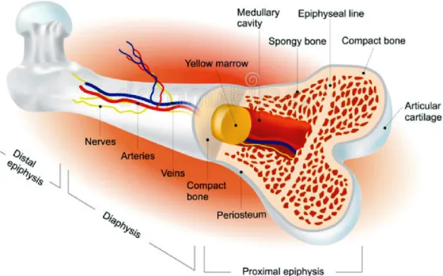
Bone is a mineralized tissue classified into the compact (cortical) bone and the spongy (cancellous or trabecular) bone. Compact bone is a very dense and hard tissue that forms the outer shell around the bones (Figure 1). This tissue provides resistance to pressure and shocks and protects the spongy bone. On the other hand, spongy bone has a relatively porous structure and lies beneath the compact bone (Figure 1). It creates the lightweight nature of bones, supports bone marrow, and shifts in weight distribution. Most bones contain the two types of tissue.
4 However, the distribution and relative amounts of compact and spongy bones are related to the type of bone and its overall functions 2,3.
A long bone (as an example) is usually divided into the diaphysis (consisting mostly of compact bone) and the epiphysis (consisting mostly of spongy bone) (Figure 1).
Figure 1. Schematic representation of a typical adult long bone gross structure. The diaphysis flares outward near the end to form the epiphysis. The outer shell is formed of compact bone covered with the periosteum. Beneath the compact bone is the spongy bone. Inside the diaphysis is the medullary cavity, which has an inner core of yellow marrow (adapted from http://www.istockphoto.com).
The diaphysis is the tubular shaft that links the proximal and distal extremities of bone (Figure 1). The hollow region in the diaphysis is called the medullary cavity, which is filled with red marrow in children or yellow marrow in adults, blood vessels, nerves, and lymphatic vessels, and is lined by a delicate connective tissue membrane known as endosteum (Figure 1). The outer shell of long bones is covered with a dense membrane of connective tissue termed periosteum, except at the articular surfaces (i.e. joints). In long bones, the joints are coated by hyaline cartilage in order to reduce the friction between adjacent and connected bones (Figure 1; see section 1.1.6.2). The periosteum is composed of two distinct layers: an outer fibrous layer and an inner osteogenic (cellular or cambium) layer (Figure
5 2). The outer layer is formed by fibroblasts, collagen and elastin fibers, along with a distinctive nerve and microvascular network. The inner layer of the periosteum is highly cellular, containing fibroblasts, mesenchymal stem cells (MSCs),
osteoprogenitor cells, and osteoblasts, as well as blood vessels, nerves, and
lymphatic vessels 2,3.
The epiphysis is the swollen part at each end of the long bones (Figure 1). It is filled with red marrow between the spaces (cavities) of the spongy bone and contains the epiphyseal line (Figure 1). In a growing long bone between the epiphysis and the diaphysis lies the metaphysis, a narrow portion of bone that contains the epiphyseal (grow) plate. The epiphyseal plate is formed by a layer of hyaline cartilage, which is replaced by compact bone in the early of adulthood, giving rise to the epiphyseal line (see section 1.1.6.2) 2,3.
1.1.2.2. Microstructure of bone – osteon and trabecula
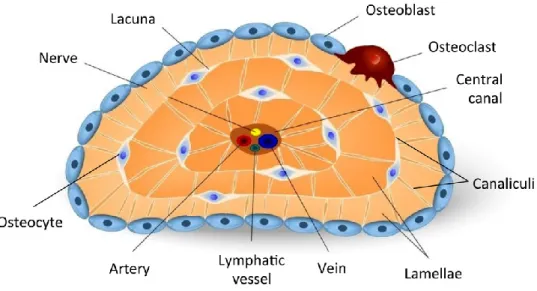
Compact bone and spongy bone are biologically identical, but its microarchitecture is different. The basic structural/functional unit of the compact bone is the osteon or
Haversian system (Figure 2). Each osteon is composed of a single central
(Haversian) canal surrounded by concentric layers of mineralized matrix, called lamellae (Figure 2). These cylindrical structural units are arranged parallel to one another along the long axis of the compact bone. The central canals connect with each other, with the medullary cavity, and with the periosteum, via transverse branches, known as perforating (Volkmann's) canals (Figure 2). Central and perforating canals are lined by endosteum and contain blood vessels, nerves, and lymphatic vessels. Besides the structural function of these canals, they are responsible for the exchange of oxygen/nutrients and removal of metabolic waste products (see section 1.1.5). Between the osteons, the lamellae are called interstitial lamellae, and circumferential lamellae are arranged parallel to the periosteum (Figure 2) 2,3.
In the spongy bone, the microscopic unit is the trabecula. The trabeculae are
composed of lamellae arranged in a honeycomb-like structure (Figure 2). This arrangement creates a considerable amount of open space within the bone filled with marrow, blood vessels, nerves, and lymphatic vessels 2,3.
6 Figure 2. Schematic representation of the distinct microarchitectures of bone tissue. Compact bone is arranged in concentric lamellae around a central canal (osteons). The central canals connect with each other, with the medullary cavity, and with the periosteum,
via perforating canals. Spongy bone is arranged in irregularly lamellae (trabeculae) (adapted
from https://cnx.org/).
1.1.3.
Bone Cells – The O‘ Cells
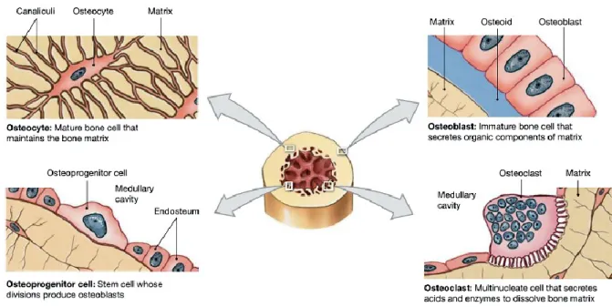
The bone volume is composed by a small amount of bone cells. Nevertheless, these cells play an essential role in bone functions. There are four special types of bone cells, known as the O´cells: osteoprogenitor cells, osteoblasts, osteocytes, and osteoclasts (Figure 3) 1–3.
1.1.3.1. Osteoprogenitor cells
Osteoprogenitor cells are MSCs with the capacity for extensive self-renewal and
osteoblast differentiation. These cells are found in the bone marrow and in the inner
7
1.1.3.2. Osteoblasts
Osteoblasts or bone-forming cells are mononuclear cells with a limited lifespan (Figure 3). Their shape varies from flat to plump, reflecting their level of maturation and cellular activity. Osteoblasts develop from multipotent MSCs in a process called
osteoblastogenesis. During this process, MSCs undergo successive stages of
differentiation with a decreasing proliferation potential, giving rise to pre-osteoblasts and subsequently, osteoblasts. As osteoblasts differentiate, they acquire the ability to form bone tissue. Osteoblasts are then responsible for the synthesis/deposition of the organic components of bone matrix and orchestration of its mineralization (Figure 3;
see section 1.1.4). The organic matrix that has not yet been mineralized is known as
osteoid. Once the osteoid is mineralized, osteoblasts can be buried within the bone
matrix as an osteocyte, become a bone-lining cell (quiescent flat-shaped osteoblasts that cover the bone surfaces), or undergo programmed cell death (i.e. apoptosis).
Osteoblasts are located on the periosteal and endosteal bone surfaces (Figure 3) 1–3.
Figure 3. Bone type of cells: osteoprogenitor cells, osteoblasts, osteocytes, and osteoclasts. Each cell type has a specialized function and is found in different locations in bones (adapted from http://www.istockphoto.com).
1.1.3.3.
Osteocytes
Osteocytes or star-shaped cells are the most common type of bone cell. These terminally differentiated cells result from the trapping of osteoblasts within the bone
8
matrix during the mineralization process. Each osteocyte is located inside a space, termed lacuna, surrounded by lamellae (Figure 4). Osteocytes form long and multiple cytoplasmic processes, known as filopodia, within a network of small channels called canaliculi (Figures 3 and 4). The lacunocanalicular system facilitates the communication of osteocytes with each other and with osteoblasts/bone-lining cells
via gap junctions, which are mainly composed of connexin 43 (Cx43). Osteocytes play an important role in triggering bone remodeling (see section 1.1.7). Like osteoblasts, osteocytes lack mitotic activity 1–3.
Figure 4. Schematic representation of the osteocytic lacunocanalicular system. Osteocytes are present in lacunae between lamellae. They are connected with each other
via numerous cellular filopodial processes extended inside the canaliculi (adapted from
http://www.istockphoto.com).
1.1.3.4. Osteoclasts
Osteoclasts or bone-resorbing cells are multinucleated giant cells with a “foamy”
cytoplasm (Figure 3). These terminally differentiated cells derive from the fusion of
myeloid progenitor cells (i.e. hematopoietic lineage cells (HSCs)) in a process called
osteoclastogenesis. Their differentiation pathway is common to that of macrophages and dendritic cells and is under the influence of several factors. Among these factors, the macrophage colony-stimulating factor (M-CSF), secreted by osteoprogenitor cells and osteoblasts, and receptor for activation of nuclear factor kappa B ligand (RANKL), secreted by MSCs, osteoprogenitor cells, osteoblasts, and osteocytes, are the two main cytokines involved in osteoclast differentiation. The signaling between
9 M-CSF and its receptor CSF-1R is essential for proliferation of osteoclast precursors. The interaction of RANKL/RANK in the presence of M-CSF directly stimulates fusion of osteoclast precursors and activation of mature osteoclasts. Osteoblasts also express a secreted factor called osteoprotegerin (OPG), which acts as a decoy receptor for RANKL to inhibit osteoclastogenesis. Osteoclasts are specialized cells that dissolve the mineralized matrix through the synthesis of acids and specific enzymes (Figure 3). During this process, termed osteolysis, they release stored minerals and soluble factors, which regulate calcium homeostasis and bone remodeling (see section 1.1.7). Osteoclasts are found in the periosteum and the endosteum (Figure 3)1–4.
1.1.4.
Bone Matrix
Bone matrix is composed of ~60% inorganic (mineral) phase, ~25% organic (protein) phase, and ~15% water. The inorganic phase serves as a reservoir for minerals, containing ~99% calcium, ~88% phosphate, and ~50% sodium and magnesium of the human body. This phase is formed by ~95% hydroxyapatite crystals (resulting from the interaction between calcium phosphate and calcium hydroxide -Ca10(PO4)6(OH)2)), and ~5% other inorganic compounds, such as calcium carbonate,
calcium fluoride and magnesium fluoride. These minerals give to the bone its characteristic hardness and the ability to resist compression. The organic phase serves as a depository for cytokines and growth factors and reservoir for lipids. This phase consists of ~90% type I collagen triple helices organised in fibrils, ~8% noncollagenous proteins (glycoproteins, proteoglycans, cytokines, and growth factors), and ~2% lipids. Glycoproteins are the most abundant noncollagenous
proteins in bone and include osteocalcin, alkaline phosphatase (Alp), osteonectin,
RGD (Arg-Gly-Asp) peptide - containing proteins (thrombospondin, fibronectin, vitronectin, osteopontin, and bone sialoprotein), fibrillin, and tetranectin.
Proteoglycans are macromolecules characterized by the covalent attachment of
long-chain polysaccharides (glycosaminoglycans, GAGs) to core protein molecules composed of the leucine-rich repeat sequences (decorin, biglycan, fibromodulin, and osteoadherin). Proteoglycans form a highly hydrated swelled gel-like matrix, which provides the bone with the ability to resist stretching and twisting. Cytokines and growth factors are important modulators of the bone remodeling process (see section 1.1.7) 1–3.
10
1.1.5.
Neurovascular Supply of Bone
Blood supplies oxygen, nutrients and regulatory factors to bone tissue, and also
removes metabolic waste products 5,6.
In long bones (as an example), small arteries of the periosteum (i.e. periosteal arteries) enter the diaphysis through many perforating canals to supply the outer part (1/3) of compact bone (Figure 2). The inner part (2/3) of compact bone and the medullary cavity (including the walls composed of spongy bone) receive nourishment from a large nutrient artery that penetrates the diaphysis through a small opening,
called nutrient foramen (Figure 5). The nutrient artery runs transversely through the
compact bone and on entering the endosteal cavity divides into proximal and distal
branches, which course towards each end of the long bone (Figure 5). Each one of
these two branches divides into several radial branches (Figure 5). The ends of long bones are supplied by the metaphyseal/epiphyseal arteries, which arise from arteries that supply the associated joint (Figure 5). At the region of metaphysis, metaphyseal/epiphyseal arteries anastomose with terminal branches of the nutrient artery. As the blood passes through the different parts of the bone, it is collected by respective vein (nutrient vein, metaphyseal/epiphyseal veins, and periosteal veins)
and carried out of the bone (Figure 5) 5,6.
Nerve fibers accompany the same paths that blood vessels into the bone, where they tend to concentrate in the more metabolically active regions. Innervation is involved in the regulation of blood supply and bone formation. The periosteum is rich in sensory nerve fibers, some of which carry severe pain sensations resulting from a
trauma, fractures, or bone tumors 7–9.
1.1.6.
Bone Formation
There are two processes of bone formation in the fetus and young children, intramembranous ossification and endochondral ossification 10,11.
11 Figure 5. Schematic representation of blood supply of a typical long bone. Blood supply to long bone comes from three sources: periosteal system, nutrient system, and metaphyseal-epiphyseal system (Copyright 2009, John Wiley & Sons, Inc.)
1.1.6.1. Intramembranous ossification
Intramembranous ossification is the source of compact and spongy tissues of flat bones of the skull, the mandible, and the clavicle. This process occurs from sheets of a relatively undifferentiated connective tissue, called embryonic connective tissue or mesenchyme, containing MSCs. Intramembranous ossification occurs about the same time as blood vessels begin to develop (see section 1.1.9). It begins as MSCs proliferate and condense within the mesenchyme, forming small and dense/compact clusters of cells, called ossification centers (Figure 6a). Some of the MSCs support capillary formation, while others differentiate into osteoprogenitor cells and then osteoblasts to form bone tissue. Blood components help osteoblasts to produce and mineralize the osteoid (Figure 6a). As ossification proceeds, some osteoblasts are trapped within the matrix, where they become osteocytes (Figure 6b). The
12 mineralized matrix grows outwards from the ossification centers in small struts termed bony spicules. These spicules eventually fuse with each other giving rise to the trabeculae of the woven bone. As the spicules interconnect they trap blood vessels within the bone (Figure 6c). Periosteum is formed from compact layers of
MSCs lining the periphery of the ossification center (Figure 6c). Osteoblasts of the inner surface of the periosteum, deposit layers of bone matrix and fill spaces between trabeculae, creating a zone of compact bone parallel to the remaining
spongy bone (Figure 6d). Once the rudimentary spongy and compact bones are
formed, the endosteum begins to develop. Crowds of blood vessels near the spongy
bone, condensate and form the red marrow 11,12.
Figure 6. Schematic representation of the intramembranous ossification process. a) MSCs proliferate and condense into centers of ossification, where they differentiate into osteoblasts that secrete osteoid. b) Osteoblasts trapped inside the calcified matrix become osteocytes. c) Bone matrix develops into trabeculae of woven bone. Blood vessels grow into spaces between trabeculae. Mesenchyme at the periphery of the bone matrix condenses and develops into the periosteum. d) Osteoprogenitor cells of the inner layer of the periosteum differentiate into osteoblasts, which produce the compact bone parallel to the existing spongy bone (adapted from www.accessmedicine.com).
1.1.6.2. Endochondral (intracartilaginous) ossification
In endochondral ossification, the MSCs first differentiate into hyaline cartilage (avascular tissue), which is later replaced by bone tissue (highly vascular tissue).
13 This type of ossification is responsible for the formation of most of the bones of the skeleton, particularly short and long bones. Endochondral ossification begins as clusters of MSCs differentiate into chondrocytes (i.e. cartilage cells). Chondrocytes then form a cartilaginous skeletal precursor, whose shape resembles a small version of the bone to be formed. This hyaline cartilage template is covered with a membrane, called perichondrium, which later become the periosteum (Figure 7-1). Some MSCs of the perichondrium differentiate into osteoprogenitor cells and then
osteoblasts to form a bone collar around the diaphysis (Figure 7-2). This periosteal
bone collar supports the growing bone. In the center of the cartilage template,
chondrocytes undergo a degenerative process. This process is characterized by chondrocyte proliferation and differentiation into hypertrophic chondrocytes (Figure 7-2), and ultimately their apoptosis (Figure 7-3). The terminally differentiated chondrocytes stop to produce collagen and other proteoglycans and begin to secrete Alp, which promote the calcification of the surrounding cartilage matrix (Figure 7-2). In addition, these chondrocytes secrete vascular endothelial cell growth factor
(VEGF), which induces the sprouting of blood vessels from the perichondrium (Figure
7-2; see section 1.1.9). As hypertrophic chondrocytes die, they leave cavities within the calcified cartilage matrix (Figure 7-3). A periosteal bud penetrates the diaphysis, previously perforated by osteoclasts, invades these cavities and branch in opposite
directions (Figure 7-3). The periosteal bud contains blood vessels, nerves, and
lymphatic vessels. The blood vessels carry HSCs, osteoprogenitor cells, and other
cells into the cavities. The HSCs will later form the bone marrow. Osteoblasts derived
from osteoprogenitor cells use the calcified cartilage as a scaffold to create spongy bone in the primary ossification center (Figure 7-3). Osteoblasts of the newly formed periosteum deposit compact bone around the spongy bone. Osteoclasts break down
some spongy bone and form the medullary cavity (Figure 7-4). The development of
secondary ossification centers continues somewhat later (after birth) in the epiphyses
by a similar process (Figure 7-4). Bone replaces hyaline cartilage, except the
articular cartilage and epiphyseal plate of long bones. Articular cartilage persists throughout adult life while the epiphyseal plates remain until early adulthood, i.e. until
the bone stops growing (Figure 7-5). The epiphyseal line replaces the epiphyseal
plate and the diaphysis and epiphysis regions fuse together to form a single adult bone in a process known as the epiphyseal closure (Figure 7-5) 10–12.
14 Figure 7. Schematic representation of the endochondral ossification process. 1) A hyaline cartilage model of the respective bone is formed. 2) Around the diaphysis of the cartilage model, a periosteal bone collar is produced by perichondrium. In the center region, the chondrocytes undergo a degenerative process with cell enlargement (hypertrophy) and matrix calcification 3) Chondrocytes die and leave cavities within the calcified cartilage matrix. Blood vessels penetrate the bone collar and invade the cavities in the diaphysis bringing osteoprogenitor cells to this region. These cells become osteoblasts, which form the spongy bone around the cavities. In this way, the primary ossification center is established in the diaphysis. 4) Osteoclasts dissolve some spongy bone to develop the medullary cavity and osteoblasts produce the compact bone beneath the periosteum. Secondary ossification centers appear at the epiphyses. 5) Bone replaces cartilage except the articular cartilage, which persists throughout adult life, and the epiphyseal plate, 6) which is replaced by epiphyseal line in the early of adulthood (adapted from www.accessmedicine.com).
1.1.7.
Bone Remodeling
Bone remodeling (metabolism) is a dynamic process by which old/microdamaged bone is replaced by new bone. This process plays an essential role in calcium homeostasis and ensures the mechanical integrity of the skeleton. An imbalance in the regulation of bone resorption and bone formation results in metabolic bone diseases, such as osteoporosis (bone resorption>bone formation) and osteopetrosis (bone formation>bone resorption). Most of the metabolism occurs at the bone surfaces. Due to the higher surface area of spongy bone compared to cortical bone, most of the turnover takes place at the endosteal surface of spongy bone. Bone
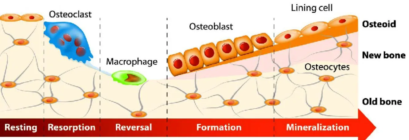
15 remodeling occurs due to coordinated actions of osteoclasts, osteoblasts, osteocytes, and bone lining cells, associated with blood vessels and nerves. Together, they form a temporary anatomical structure, called basic multicellular unit (BMU) or bone remodeling unit (BRU) 13–17.
1.1.7.1. Bone remodeling cycle
Bone remodeling cycle involves a series of highly regulated phases: resting (activation), resorption, reversal (transition), formation, and mineralization (termination) (Figure 8) 13–16.
Figure 8. Schematic representation of the bone remodeling cycle. Bone remodeling consists of a series of sequential phases that are highly regulated. Bone resorption by osteoclasts is followed by the recruitment of osteoblasts and formation of new bone matrix. The newly synthesized matrix is then slowly mineralized (adapted from http://www.istockphoto.com).
The activation of the quiescent bone surface results in the retraction of bone-lining cells by matrix metalloproteinases (MMPs). This activation process is partly regulated by osteocytes, which act as sensors of mechanical loading and biochemical stimuli. Osteoclast precursor cells are then recruited by chemotaxis to the activated surface and fuse to form mature osteoclasts (Figure 8). These osteoclasts attach to the bone surface through a contiguous adhesion belt, known as sealing (clear) zone, which creates an isolated microenvironment beneath the cell. Within the sealing zone, the osteoclast plasma membrane develops into the ruffled border, which acts as the resorptive organelle of these cells (Figure 9). As a consequence, osteoclasts dissolve the inorganic matrix by creating an acidic microenvironment through the secretion of several acids and a large amount of protons (H+), and digest the organic matrix by
16 releasing specific enzymes, such as lysosomal enzymes, tartrate-resistant acid phosphatase (TRAP), cathepsin K, and MMP-9 (Figure 9). Bone degradation products are then removed from the functional secretory domain via transcytotic vesicles (Figure 9). Osteoclast activity results in the formation of resorption pits on bone surface, called as Howship's lacunae (Figure 9). Once the osteoclasts have completed their work, mononuclear cells with an unclear phenotype prepare the surface of Howship's lacunae for bone formation and provide matrix-derived signals, such as transforming growth factor-β (TGF- β) and insulin-like growth factor I and II (IGF-I/II), for osteoblast differentiation and migration (Figure 8) 13–17.
Figure 9. Schematic representation of the osteoclast activity. Osteoclasts form sealing and ruffled borders. Bone resorption is achieved by transport of protons into the resorption lacunae through vacuolar H+-ATPase and secretion of specific enzymes (cathepsin K,
MMP-9 and TRAP). Matrix degradation products are removed from the functional secretory domain
via transcytotic vesicles (adapted from Takahashi et al., BoneKEy Reports, 2014).
The bone remodeling cycle is finished with the synthesis/deposition of organic components of bone matrix by osteoblasts, and its subsequent mineralization (Figure 8). The collagen network is synthesized first followed by incorporation of
non-17 collagenous proteins. The production of type I collagen and formation of the collagenous matrix is followed by an increased expression of ALP. This gradually decreases when matrix mineralization is progressed. Osteocalcin appears at the latest stages of differentiation pathway, approximately at the onset of mineralization. The newly formed bone surface is completely covered with bone-lining cells and maintained until the next remodeling cycle is initiated (Figure 8). The resorption activity in adult human bone takes approximately 3 weeks and the formation
response 3 to 4 months 13–17.
1.1.7.2. Regulatory factors in bone remodeling
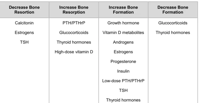
The regulation of bone remodeling is both systemic and local. The main interrelated systemic factors include: genetics; age; nutrition; exercise (mechanical loading); and hormones, particularly thyroid hormones, thyroid-stimulating hormone (TSH), parathyroid hormone (PTH) / parathyroid hormone-related protein (or PTHrP), calcitonin, calcitriol (1,25-dihydroxyvitamin D, 1,25(OH)2D or Vitamin D), growth
hormone, gonadal hormones, insulin, and glucocorticoids. Table 1 summarises the role of the “classic” hormones in bone metabolism 18,19.
As far as local regulation of bone remodeling is concerned, it has been identified a large number of autocrine/paracrine molecules produced by bone cells and soluble factors released from the bone matrix during osteolysis. Important growth factors, cytokines, and prostaglandins acting on bone turnover are tabulated in Table 2, according to its function 18–21.
Another category of molecules called semaphorins (Sema) has been recognized as a modulator of bone remodeling. Sema are a class of secreted and membrane-associated proteins that were originally identified as axonal growth cone guidance molecules. It was found that Sema4D, expressed in osteoclasts, and Sema3A, abundantly expressed in osteoblasts, are important regulators of bone metabolism. During bone resorption, Sem4D inhibits bone formation by binding to its receptor (Plexin-B1) present in osteoblasts 22. On the other hand, Sema3A, secreted by
sensory neurons, promotes bone formation indirectly by modulating the local nerve ingrowth and not by acting directly on osteoblasts (see chapter 1.3) 23.
More recently, microRNAs (miR) have been regarded as one of the most important modulators in bone turnover. miR are short single strand non-coding molecules of RNA (between 18 and 25 nucleotides long) that negatively regulate gene expression either by mRNA degradation or translational silencing. In this context, miR can act as
18 both negative and positive regulators of differentiation of osteoblasts and osteoclasts (Table 3) 24.
Table 1. Hormones involved in bone remodeling.
Decrease Bone
Resortion Increase Bone Resorption Increase Bone Formation Decrease Bone Formation
Calcitonin PTH/PTHrP Growth hormone Glucocorticoids
Estrogens Glucocorticoids Vitamin D metabolites Thyroid hormones
TSH Thyroid hormones Androgens
High-dose vitamin D Estrogens Progesterone
Insulin
Low-dose PTH/PTHrP TSH
Thyroid hormones
PTH, parathyroid hormone; PTHrP, parathyroid hormone-related protein; TSH, thyroid-stimulating hormone
Table 2. Growth factors, cytokines, and prostaglandins involved in bone remodeling. Stimulate bone formation Stimulate bone resorption Inhibit bone resorption Inhibit bone formation Growth factors BMP-2, BMP-4, BMP-5, BMP-6, BMP-7, BMP-9, TGF-β, I, IGF-II, PDGF, VEGF, FGF EGF, PDGF, FGF, GM-CSF, M-CSF TGF-β BMP-3 Cytokines and
Prostaglandins IL-3, IL-13, IL-17 IFN, CT-1
TNF, RANKL, IL-1, IL-6, IL-8, IL-11,
PGE1, PGE2,
PGG2, PGH2, PGI2
OPG, IFNγ, IL-4, Il-10, IL-12, IL-18,
IL-23
BMP, bone morphogenetic protein; CT1- cardiotrophin-1; EGF, epidermal growth factor; FGF, fibroblast growth factor; GM-CSF, granulocyte-macrophage colony-stimulating factor; IFNγ, interferon gamma; IGF, insulin-like growth factor; IL, interleukine; M-CSF, macrophage-colony stimulating factor; OPG, osteoprotegerin; PDGF, platelet-derived growth factor; PG, prostaglandin; RANKL, receptor activator of nuclear factor kappa-B ligand; TGF- β, tumor growth factor-beta; TNF, tumor necrosis factor
19
Table 3. Key miR regulators of bone remodeling
Activate Differentiation Inhibit Differentiation
Osteoblasts
MiR-15b; MiR-17~92; MiR-20a; MiR-29a; MiR-181a; MiR-322;
MiR-335-5p
MiR-17-5p; MiR-30c; MiR-34c; MiR-93; MiR-100; MiR-125b; MiR-133a; MiR-135a; MiR-137;
MiR-138; MiR-141; MiR-200a; 143; 182; MiR-204/211; MiR-205; MiR-206; MiR-208; MiR-217; MiR-218; MiR-338; MiR-542-3p; MiR-637;
MiR-764-5p
Osteoclasts 21; 29b; 31; MiR-34a; MiR-148a; MiR-223
MiR-29b; MiR-125a; MiR-146a; MiR-155; MiR-223; MiR-503
Adapted from Alečković M and Kang Y, BoneKEy Reports, 2015.
1.1.8.
Canonical Wnt Signaling Pathway in bone formation and
remodeling
The canonical Wnt pathway (Wnt/β-catenin pathway) regulates many cellular activities, such as proliferation, differentiation, maturation, migration, survival, and apoptosis. It is widely demonstrated that this pathway is critical for osteoblast differentiation and function and consequently, for bone formation and metabolism. Wnt/β-catenin pathway is also important in adipogenesis, chondrogenesis and hematopoiesis and may be stimulatory or inhibitory at different stages of osteoblastogenesis 25–28.
The central function of the canonical Wnt pathway is the regulation of β-catenin (encoded by Ctnnb1) stabilization. β-catenin is a dual-function protein that coordinates cell-cell adhesion and acts as an intracellular signal transducer in the Wnt/β-catenin pathway. β-catenin binds to cadherin and α-catenin at the plasma membrane, and this cadherin-catenin complex links to the actin filaments and several actin-binding proteins to promote cell-cell adhesion (Figure 10) 25–28.
20 When Wnt/β-catenin signaling is not activated, β-catenin is degraded after being recruited to a complex composed of axin, adenomatous polyposis coli (APC), glycogen synthase kinase (GSK) 3β, and other proteins (Figure 10). Axin and APC act as scaffold proteins promoting the interaction of β-catenin with GSK-3β. The phosphorylation of β-catenin by GSK-3β targets it for ubiquitination and subsequently, recognition and constitutive degradation by the ubiquitin-proteasome system (Figure 10) 25–28.
When a Wnt secreted glycoprotein (e.g. Wnt3a and Wnt10b) binds to its Frizzled (Fzd) receptor and co-receptor low-density lipoprotein receptor-related protein (LRP) - 5/6, axin and the cytoplasmic protein disheveled (Dsh) are recruited and tethered to the ligand-receptor complex (Figure 10). Axin binds to the phosphorylated LRP5/6 and disables the degradation complex. Phosphorylated Dsh binds to the Fzd receptor and transduces a signal for inhibition of the GSK3β activity, which leads to accumulation of nonphosphorylated β-catenin in the cytosol (Figure 10). The stabilized β-catenin is then translocated into the nucleus to activate the transcriptional expression of Wnt-related genes via lymphoid enhancer binding factor (Lef) and T cell factors (Tcf) (Figure 10). Runt-related transcription factor 2 (Runx2) is a direct target gene of β-catenin/Tcf/Lef. Wnt/β-catenin signaling activates expression of Runx2 in MSCs for the control of osteoblast differentiation and bone formation. It determines the osteoblastogenesis at the early stage and inhibits it at the late stage. Runx2 interacts with other transcription factors, especially osterix, to regulate the transcriptional expression of important bone phenotyping genes, such as collagen type I alpha 1 chain (Col1a1), Alp, and bone gamma-carboxyglutamic acid-containing protein (Bglap, encoding osteocalcin). Tumor necrosis factor ligand superfamily member 11 (Tnfrsf11b), encoding the anti-osteoclastogenic factor OPG, is known to be another Wnt-related gene. The canonical Wnt signaling suppresses indirectly osteoclast differentiation and bone resorption through upregulation of OPG in osteoblasts 25–28.
The amplitude of the Wnt/β-catenin signaling is fine-tuned in part via negative feedback by secreted extracellular antagonists, such as secreted Fzd-related peptides (sFRPs), Wnt inhibitory factor (Wif) - 1, dickkopf (Dkk) 1/2, and sclerostin.
While sFRPs and Wif-1 interact directly with Wnt proteins, Dkk 1/2 and sclerostin
bind to the extracellular domains of LRP5/6, thus preventing signaling activation and
21 Figure 10. Schematic representation of the canonical Wnt signaling pathway. β-catenin forms a cadherin-catenin complex at the plasma membrane that regulates cell-cell adhesion. (Left panel) In the absence of Wnt signaling, β-catenin is prone to degradation, by forming a complex with axin, APC and GSK3-β. β-catenin is phosphorylated by GSK3-β that marks it for ubiquitination and subsequent degradation by the proteasome. (Right panel) In the presence of Wnt signaling, β-catenin is uncoupled from the degradation complex, accumulates and translocates into the nucleus, where it acts as a co-activator of Lef/Tcf transcription factors (adapted from Reya T and Clevers H, Nature, 2005).
The canonical Wnt signaling cooperates with a number of other bone formation signaling pathways, such as PTH/PTHrP, bone morphogenetic proteins (BMPs)/TGF-β, and Indian hedgehog.
22
1.1.9.
Angiogenesis/Vascularization in bone formation and remodeling
Bone formation and remodeling processes require the action of both intrinsic and extrinsic inductive factors produced from multiple cell types, which function in a hierarchical and temporal fashion to control angiogenesis and osteogenesis. Endothelium (i.e. the inner lining of blood vessels) is an integral part of bone tissue, and its interaction with bone cells is crucial for skeletal development, homeostasis, and healing 30. The interplay between endothelial and bone cells has been
extensively investigated, and numerous modes of cell communication have been proposed, specifically via secretion of humoral factors, growth factors, chemokines, and direct cell-cell contact through gap junctional proteins 31. Among the growth
factors, VEGF expressed in endothelial cells plays a critical role in the orchestration of bone formation and remodeling, by increasing the production of BMPs 32. In
addition, other factors secreted by endothelium, such as endothelin-1 and angiotensin-II, are also able to induce bone formation 33–35.
Previous studies have shown that angiogenesis is preceded by peripheral innervation during bone formation and regeneration 31, suggesting that peripheral nervous
system controls these processes through the local release of neuronal mediators. A recent study indicates that sonic hedgehog, a traditional neurogenic morphogen, inversely regulates the vascular morphogens angiopoietin-1 and angiopoietin-2, thus contributing to blood vessel growth, maturation and stabilization in a neurovascular network 36.
These and other studies provide a robust body of evidence that bone is regulated by different tissues/systems, evidencing its complexity and dynamic nature 29. In this
dissertation we focused on the regulation of osteoblast and bone formation by the sensory nervous system.
23
SENSORY NERVOUS SYSTEM
.1.2
The sensory nervous system is the part of the peripheral nervous system responsible for processing sensory information. Sensory neurons carry this information as a nerve impulse from the viscera, sense organs, muscles, bones and joints towards the
central nervous system (CNS), where the information is perceived and interpreted. Despite this classic function, a specific subgroup of sensory neurons also has the
ability to release neuropeptides from their peripheral endings to regulate organ/tissue
activities 37–39.
1.2.1.
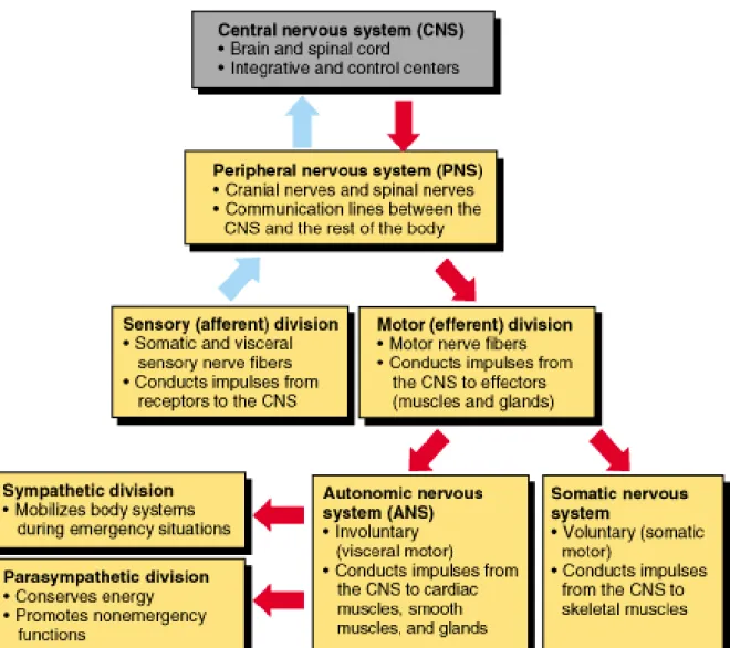
Functional Organization of the PNS
The peripheral nervous system (PNS) consists of the nervous tissue that lies outside the brain and spinal cord (i.e. outside the CNS). It forms the communication network between the CNS and rest of the body. The CNS interacts with the PNS through 12 of cranial nerves, which connect the brain to areas of the head and neck, and 31 pairs of spinal nerves, which connect the spinal cord to the rest of the body. The spinal nerve emerges from the spinal cord between adjacent vertebrae through an opening called intervertebral foramen. Each spinal nerve is formed by dorsal root housing afferent (sensory) neurons and ventral root carrying efferent (motor) neurons. These two roots are actually parts of the two major divisions of the PNS:
sensory, or afferent, and motor, or efferent, divisions (Figure 11). The sensory
division consists of afferent neurons that carry sensory information from receptors in the periphery of the body to the CNS (Figure 11). The motor division contains efferent neurons that carry motor information from the brain to the rest of the body.
This division is subdivided into somatic nervous system and autonomic nervous
system (Figure 11). The somatic nervous system includes the voluntary motor neurons and transmits signals from the CNS to skeletal (striated) muscles (Figure 11). It is responsible for stimulating muscle contraction (conscious activities). The autonomic nervous system includes the involuntary motor neurons and transmits signals from the CNS to cardiac muscle, smooth muscles, and glands (Figure 11). It
is responsible for secretion of chemical signals from the glands and maintains the
24 (subconscious activities). There are two subdivisions of the autonomic nervous system, the sympathetic and parasympathetic (Figure 11). The sympathetic nervous system is responsible for mobilization of energy and resources during times of stress and arousal, while the parasympathetic nervous system acts as an antagonist that
returns the body to its normal resting state (Figure 11). This antagonistic functional
relationship serves as a balance to help maintain homeostasis 37–39.
Figure 11. Functional organization of the peripheral nervous system. The PNS is divided into sensory (afferent) and motor (efferent) divisions. The afferent division sends sensory signals to the CNS and the efferent division receives motor signals from the CNS. The efferent division can be subdivided into two groups: autonomic and somatic nervous systems. The autonomic nervous system itself can be divided into the sympathetic and parasympathetic nervous systems (adapted from www.apsubiology.org)
25
1.2.2.
Basic Function and Structure of a Neuron
The nervous system contains two types of cells: glial cells and neurons. The glial cells protect and nourish the neurons and are the key element to support their
function. Neurons are considered to be the basic working unit of the nervous tissue.
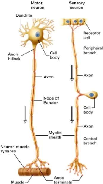
These specialized cells receive and transmit signals to other cells.They have a main
part called cell body or soma and extensions of their membranes, generally referred to as processes. The cell body contains the nucleus and cytoplasm with most of the major organelles (Figure 12) 37–39.
Figure 12. Schematic representation of neuron structure. (Left) A motor neuron with a single axon projected from the cell body, which is located in the ventral root ganglion, to the effector cell. (Right) A sensory neuron with an axon that divides into a central and peripheral branch just after it leaves the cell body, which is located in the dorsal root, trigeminal root, and nodose ganglia. The peripheral branch carries the electrical impulse from the receptor cell to the cell body; the central branch and axon terminals carry the impulse from the cell body to the CNS (adapted from Lodish H, et al. New York: W. H. Freeman; 2000).
26 There is one important process called an axon, which is the fiber that emerges from the cell body and projects to target cells (Figure 12). The single axon can branch repeatedly into axon terminals to communicate with multiple target cells via synapses (Figure 12). The neuron transmitting the signal is termed the presynaptic neuron, and the neuron receiving the signal is called the postsynaptic neuron. There are two different types of synapses, electrical and chemical. At electrical synapses, current flows directly and passively from pre- to postsynaptic cells through specialized membrane channels (gap junctions) (Figure 13A). At chemical synapses, the vesicular release of chemical messengers (neurotransmitters) by a presynaptic cell into the synaptic cleft, produces secondary current flow in a postsynaptic cell by activating specific receptors on the plasma membrane (Figure 13B). The current flow switches the postsynaptic membrane from an internal negative charge to a positive charge state. This change is known as depolarization, which generates an action potential or nerve impulse. This depolarization of the membrane is followed by a rapid repolarization, returning the membrane potential to the resting state 37–39.
Another type of process extended from the cell body is the dendrite (Figure 12). Dendrites are usually highly branched and are responsible for receiving most of the information from axon terminals of other neurons (Figure 12) 37–39.
The specialized structure where the axon emerges from the cell body is known as the axon hillock or initial segment (Figure 12). This structure integrates signals from multiple synapses 37–39.
Many axons are wrapped with a layered myelin sheath, which acts as an insulator and speeds the transmission of the nerve impulse along the axon (Figure 12). In the PNS, this sheath is made of specialized cells known as Schwann cells. Each gap uncovered by myelin is termed a node of Ranvier and is critical for myelin functionalization. The action potential jumps between axonal nodes of Ranvier to increase the speed of its propagation (Figure 12) 40.
In the PNS, a cluster of neuron cell bodies is referred to as a ganglion and a bundle
27 Figure 13. Schematic representation of electric and chemical synapses. (A) At an electrical synapse, ions flow directly and passively through gap junctions resulting in a localized depolarization of the postsynaptic membrane. (B) At a chemical synapse, when the neurotransmitters released by presynaptic cell bind to specific receptors on the postsynaptic membrane, the ligand-gated ion channels open and ions cross the membrane changing its charge (adapted from Purves D, et al. Sunderland (MA): Sinauer Associates; 2004).
1.2.3.
Types of Sensory Receptors
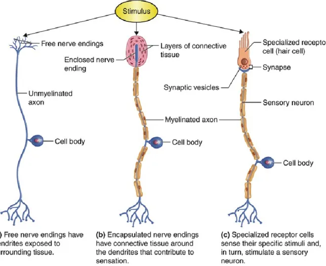
Sensory receptors are either specialized endings of sensory neurons or separate cells that signal to sensory neurons. They are adapted to respond to specific stimuli and to convert them into nerve impulses that are transmitted to the CNS. Different types of sensory neurons have different sensory receptors. Sensory receptors can be classified on the basis of their morphology or function. Morphologically, there are three groups of sensory receptors: free nerve endings, encapsulated nerve endings, and specialized transducing cells. Free nerve endings are simply free dendrites extend into a tissue (Figure 14a). This type of sensory receptor is sensitive to heat, cold, and tissue injury. An encapsulated nerve ending is a nerve ending wrapped in a round capsule of connective tissue (Figure 14b). Mechanical forces stimulate this
28 type of sensory receptor. Specialized transducing cells detect stimuli from special senses such as vision, hearing, smell, taste, and balance (Figure 14c). Each of the specialized cells is sensitive to a unique special sense. 41
Functionally, there are three major classes of sensory receptors: mechanoreceptors, nociceptors, and thermoreceptors. Mechanoreceptors are free nerve endings that respond to mechanical stimuli, such as touch, vibration, stretch, pressure, and movement. Nociceptors are unspecialized free nerve endings that respond to noxious stimuli, including extreme temperatures, acidic pH, and tissue damage, by sending pain signals to the CNS. Thermoreceptors monitor changes in temperature inside the body and in its surroundings 37–39.
Figure 14. Schematic representation of sensory receptor types. (a) Simply receptors are neurons with free nerve endings. (b) Complex neural receptors have nerve endings enclosed in connective tissue capsules. (c) Most special senses receptors are cells that release neurotransmitter onto sensory neurons, initiating an action potential (adapted from www.pasadena.edu).
29
1.2.4.
Classification of the Sensory Nerve Fibers
According to the Lloyd-Hunt classification, the sensory nerve fibers can be classified into type I-fibers, with diameters from 13 to 20 µm (80-120 m/s conduction velocity); type II-fibers, with diameters between 6 and 12 µm (35-75 m/s conduction velocity); type III-fibers, with diameters from 1 to 5 µm (5.0-30 m/s conduction velocity); and type IV-fibers represented by the unmyelinated nerve fibers, with diameters between 0.2 and 1.5 (0.5-2.0 m/s conduction velocity) (Table 4). Type I-fibers are frequently subdivided in type Ia and Ib to differentiate among afferences from muscle spindles and Golgi tendons. The Erlanger-Gasser classification is used to define both afferent and efferent fibers. Thus, type I-fibers represents the Aα-fibers of the Erlanger-Gasser classification; type II-fibers equivalents to the Aβ-fibers; type III-fibers corresponds to the Aδ-fibers; and type IV-fibers represents C fibers (Table 4) 42.
Table 4. Classification of sensory nerve fibers.
Lloyd-Hunt
Classification Erlanger-Gasser Classification Myelin
Fiber Diameter (μm) Conduction Velocity (m/s) Type of Receptor Supplied
Ia and Ib Aα Yes 13-20 80-120 Mechanoreceptor
II Aβ Yes 6-12 35-75 Mechanoreceptor
III Aδ Yes 1-5 5.0-30 Mechanoreceptor Thermoreceptor Nociceptor
IV C No 0.2-1.5 0.5-2.0 Mechanoreceptor, Thermoreceptor Nociceptor Adapted from www.clinicalgate.com
1.2.5.
Dual Afferent and “Efferent” Function of Sensory Neurons
Sensorimotor neurons are a subpopulation of polymodal nociceptive sensory neurons (i.e. respond to painful thermal, mechanical, and chemical stimuli) with a dual afferent and efferent function. They contain predominantly C-fibers and a