HAL Id: inserm-02274747
https://www.hal.inserm.fr/inserm-02274747
Submitted on 30 Aug 2019
HAL is a multi-disciplinary open access archive for the deposit and dissemination of sci-entific research documents, whether they are pub-lished or not. The documents may come from teaching and research institutions in France or abroad, or from public or private research centers.
L’archive ouverte pluridisciplinaire HAL, est destinée au dépôt et à la diffusion de documents scientifiques de niveau recherche, publiés ou non, émanant des établissements d’enseignement et de recherche français ou étrangers, des laboratoires publics ou privés.
Essential role of GEXP15, a specific Protein
Phosphatase type 1 partner, in Plasmodium berghei in
asexual erythrocytic proliferation and transmission
Thomas Hollin, Caroline de Witte, Aline Fréville, Ida Chiara Guerrera, Cerina
Chhuon, Jean-Michel Saliou, Fabien Herbert, Christine Pierrot, Jamal Khalife
To cite this version:
Thomas Hollin, Caroline de Witte, Aline Fréville, Ida Chiara Guerrera, Cerina Chhuon, et al.. Essen-tial role of GEXP15, a specific Protein Phosphatase type 1 partner, in Plasmodium berghei in asexual erythrocytic proliferation and transmission. PLoS Pathogens, Public Library of Science, 2019, 15 (7), pp.e1007973. �10.1371/journal.ppat.1007973�. �inserm-02274747�
Essential role of GEXP15, a specific Protein
Phosphatase type 1 partner, in Plasmodium
berghei in asexual erythrocytic proliferation
and transmission
Thomas HollinID1☯, Caroline De WitteID1☯, Aline Fre´villeID1, Ida Chiara Guerrera2, Cerina Chhuon2, Jean-Michel Saliou
ID1, Fabien HerbertID1, Christine Pierrot1, Jamal KhalifeID1*
1 Univ. Lille, CNRS, Inserm, CHU Lille, Institut Pasteur de Lille, U1019 - UMR8204 – CIIL - Center for
Infection and Immunity of Lille, Lille, France, 2 Proteomics platform 3P5Necker, Universite´ Paris Descartes -Structure Fe´de´rative de Recherche Necker, INSERM US24/CNRS UMS3633, Paris, France
☯These authors contributed equally to this work.
*jamal.khalife@pasteur-lille.fr
Abstract
The essential and distinct functions of Protein Phosphatase type 1 (PP1) catalytic subunit in eukaryotes are exclusively achieved through its interaction with a myriad of regulatory part-ners. In this work, we report the molecular and functional characterization of Gametocyte EXported Protein 15 (GEXP15), a Plasmodium specific protein, as a regulator of PP1. In
vitro interaction studies demonstrated that GEXP15 physically interacts with PP1 through
the RVxF binding motif in P. berghei. Functional assays showed that GEXP15 was able to increase PP1 activity and the mutation of the RVxF motif completely abolished this regula-tion. Immunoprecipitation assays of tagged GEXP15 or PP1 in P. berghei followed by immu-noblot or mass spectrometry analyses confirmed their interaction and showed that they are present both in schizont and gametocyte stages in shared protein complexes involved in the spliceosome and proteasome pathways and known to play essential role in parasite devel-opment. Phenotypic analysis of viable GEXP15 deficient P. berghei blood parasites showed that they were unable to develop lethal infection in BALB/c mice or to establish experimental cerebral malaria in C57BL/6 mice. Further, although deficient parasites produced gameto-cytes they did not produce any oocysts/sporozoites indicating a high fitness cost in the mos-quito. Global proteomic and phosphoproteomic analyses of GEXP15 deficient schizonts revealed a profound defect with a significant decrease in the abundance and an impact on phosphorylation status of proteins involved in regulation of gene expression or invasion. Moreover, depletion of GEXP15 seemed to impact mainly the abundance of some specific proteins of female gametocytes. Our study provides the first insight into the contribution of a PP1 regulator to Plasmodium virulence and suggests that GEXP15 affects both the asexual and sexual life cycle.
a1111111111 a1111111111 a1111111111 a1111111111 a1111111111 OPEN ACCESS
Citation: Hollin T, De Witte C, Fre´ville A, Guerrera IC, Chhuon C, Saliou J-M, et al. (2019) Essential role of GEXP15, a specific Protein Phosphatase type 1 partner, in Plasmodium berghei in asexual erythrocytic proliferation and transmission. PLoS Pathog 15(7): e1007973.https://doi.org/10.1371/ journal.ppat.1007973
Editor: Michael J Blackman, Francis Crick Institute, UNITED KINGDOM
Received: May 20, 2019 Accepted: July 10, 2019 Published: July 26, 2019
Copyright:© 2019 Hollin et al. This is an open access article distributed under the terms of the
Creative Commons Attribution License, which permits unrestricted use, distribution, and reproduction in any medium, provided the original author and source are credited.
Data Availability Statement: Data are available via ProteomeXchange with identifiers PXD013814 and PXD013874.
Funding: This work is funded by the University of Lille, CNRS and Institut Pasteur de Lille. The funders had no role in study design, data collection and analysis, decision to publish, or preparation of the manuscript.
Competing interests: The authors have declared that no competing interests exist.
Author summary
In the absence of an effective vaccine and the emerging resistance to artemisinin combina-tion therapy, malaria is still a significant threat to human health. Increasing our under-standing of the specific mechanisms of the biology ofPlasmodium is essential to propose
new strategies to control this infection. Here, we demonstrated that GEXP15, a specific protein inPlasmodium, was able to interact with the Protein Phosphatase 1 and regulate
its activity. We showed that both proteins are implicated in common protein complexes involved in the mRNA splicing and proteasome pathways. We reported that the deletion of GEXP15 leads to a loss of parasite virulence during asexual stages and a total abolish-ment of the capacity of deficient parasites to develop in the mosquito. We also found that this deletion affects both protein phosphorylation status and significantly decreases the expression of essential proteins in schizont and gametocyte stages. This study character-izes for the first time a novel molecular pathway through the control of PP1 by an essential and specificPlasmodium regulator, which may contribute to the discovery of new
thera-peutic targets to control malaria.
Introduction
Convergent findings from several studies indicate that protein dephosphorylation by phospha-tases governs key fundamental processes in the biology of eukaryotic cells including Plasmo-dium falciparum [1]. Among these phosphatases, the Protein Phosphatase type 1 catalytic subunit (PP1c) seems to play a pivotal role in the development and growth ofPlasmodium
blood stage parasites [2,3]. In addition, it has been reported in eukaryotes that several con-served PP1c interacting proteins (PIPs) endowed with phosphatase regulatory functions are as essential as PP1c itself [4–7]. In mammalian cells, hundreds of PIPs have been identified and classified as regulators and substrates, capable of targeting PP1c to particular cellular organ-elles [8,9]. The combination of PP1c with an extensive range of PIPs contributes to the consti-tution of specific PP1 network, playing an effective role as a hub in diverse cellular functions.
Our earlier studies onP. falciparum revealed the expression of four conserved putative
regulators (PfLRR1, Pf Inhibitor 2, Pf Inhibitor 3 and Pfeif2β) which have been extensively explored to define their functions [10–15]. In this context, biochemical and structure-activity relationship studies demonstrated that these regulators bind to recombinant PfPP1 and three of them regulate its activityin vitro. Several binding sites have also been defined by
structure-interaction studies using mutated recombinant proteins and derived peptides, showing the involvement of LRR/LRR cap motifs for PfLRR1 and the consensus RVxF motif for PfI2, PfI3 and Pfeif2β. In addition, knock-out approaches suggested their essentiality in the life cycle of blood stage parasites. Finally, these interactions seem to be indispensable since disrupting the interaction between PP1c and PfI2, PfI3 or PfLRR1 by the use of interfering peptides that inhibit complex formation led to parasite growth inhibitionin vitro [12–14]. This proof of con-cept offers a robust basis to consider PP1c interactions as valuable therapeutic targets.
Based on the above data and on the atypical life cycle ofPlasmodium with ~60% of
unknown genes in its genome [16], including specific proteins submitted to reversible phos-phorylation, our working hypothesis was thatPlasmodium must express specific partners,
reg-ulators and/or transporters which direct PP1 activity. In order to identify PfPP1 interactors, extensive screens for PfPP1 binding proteins were carried out, using the yeast two-hybrid (Y2H) system combined with direct interactions studies using recombinant proteins [17]. A total of 31 proteins in the correct translational reading frame with the Gal4 activating domain
were identified. Upon close inspection of the amino acid sequences of binding regions of these proteins, six clones were identified exhibiting a potential RVxF binding motif with a more restrictive and specific consensus sequence of PP1 interactors [17,18]. Indeed, we have used the PfPP1 interactome to reevaluate the features of flanking residues of the RVxF motif. We observed enrichment for basic amino acids (R and K) at the N-terminal side of this binding motif.
We prioritized candidate genes that werePlasmodium-specific with a robust interaction
with PfPP1. One of these genes, formerly described asP. falciparum Gametocyte EXported
Protein 15 (PfGEXP15) (PF3D7_1031600), was repeatedly detectable under stringent condi-tions of binding to PfPP1 throughout the Y2H screens. In addition, the presence of a potential restrictive binding motif was identified in the interacting region of PfGEXP15 [17]. PfGEXP15 has been classified in the GEXP cluster, grouping different proteins predicted to be exported and detected only in the proteome of early gametocytes or being overexpressed at this stage [19]. However, interestingly, PfGEXP15 was also detected in the proteomes of asexual stages and sporozoites suggesting its involvement at different steps of the parasite life cycle [20].
In this work, we confirm that GEXP15 interacts with PP1 and showed its capacity to regu-late the dephosphorylation activity of PP1 via a major contribution of RVxF binding motif in
P. berghei. Interatomic analysis revealed that the GEXP15-PP1 are part of two protein
com-plexes involved in mRNA splicing and proteasome pathways, known to play key roles in para-site development. Further, for the first time, we demonstrate that the complete disruption of GEXP15 generates attenuated parasites both for asexual and sexual growth in mice and mos-quitoes respectively, suggesting specific and essential functions. Finally, we provide evidence that proteome and phosphoproteome of deficient schizont and gametocyte parasite stages are impacted at different levels arguing for an involvement of GEXP15 in these processes.
Results
Interaction of
Plasmodium GEXP15 with PP1 and binding motifs
In order to extend and to further characterize the GEXP15 that we previously reported as a partner of PP1 inP. falciparum [17], its ortholog inP. berghei (PBANKA_0515400) was
inves-tigated. The predicted PbGEXP15 protein (656 aa) shares an overall identity of 41% with PfGEXP15 and both contain two putative consensus RVxF binding motifs to PP1 (S1 Fig). The construct of PbGEXP154–590, spanning almost the full-length sequence was tested in the Y2H
system in which PfPP1c was used as bait as it exhibits 99% identity with PbPP1. We observed that only the diploids expressing PbGEXP15 and PfPP1c were able to grow on selective and stringent media indicating their ability to specifically interact with each other (S2A Fig). Con-trol constructs (empty vector, vector expressing unrelated gene) did not show any growth. To better define the interacting regions, constructs with PbGEXP154–178containing the
first RVxF binding motif to PP1 or PbGEXP15446–596comprising the second one were used.
PbGEXP154–178revealed the same specific interaction with PfPP1c, confirming the data
described above (S2B Fig). In the case of PbGEXP15446–596with the second RVxF motif
no yeast growth was observed, suggesting that it is a random sequence and a non-canonical binding site (S2B Fig). To validate the involvement of the first RVxF binding motif present in PbGEXP15, site directed mutagenesis was carried out to obtain V34A and F36A (PbGEXP154–178 KKKKKAQA). Interaction with PfPP1c was not observed with this mutant,
confirming that the native sequence represents a genuine RVxF binding motif (S2B Fig). The main contribution of this RVxF motif was reinforced by the lack of growth on high stringency selection in the presence of PfPP1c mutated at its docking site by replacing F255 and F256 by two alanine residues (S2C Fig) [18,21–23].
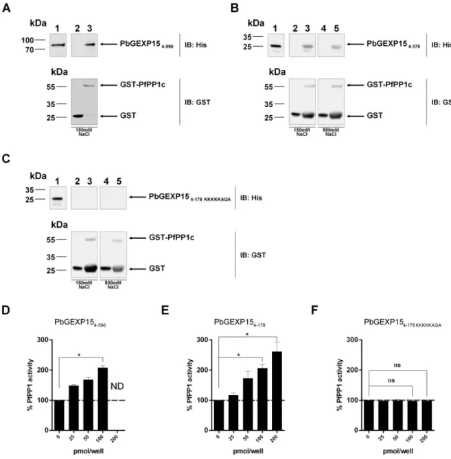
To further support the PbGEXP15-PP1 interaction, GST-PfPP1c pull-down experiments with wild-type and mutated His-PbGEXP15 recombinant proteins were carried out. Western blot analyses showed that PbGEXP154–590(Fig 1A), and PbGEXP154–178(Fig 1B) bound to
GST-PfPP1c but not to GST alone, while the mutated PbGEXP154–178 KKKKKAQArevealed no
Fig 1. Direct interaction of PbGEXP15 with PP1c and its effect on the phosphatase activityin vitro. A, B and C represent GST-pull down
experiments. A, Immunoblot (IB) represents the input positive control (500ng) in lane 1 and the GST-pull down of 6-His PbGEXP154–590
with GST alone or PfPP1c-GST in lanes 2 and 3 respectively and revealed with mAb anti-His (upper panel) and anti-GST (lower panel). B, Immunoblot (IB) represents the input positive control (500ng) in lane 1 and the GST-pull down of 6-His PbGEXP154–178with GST alone or
PfPP1c-GST in lanes 2 and 3 respectively in the presence of 150 mM NaCl and revealed with mAb anti-His (upper panel) and anti-GST (lower panel). Lanes 4 and 5 represent the GST-pull down as in lanes 2 and 3 in the presence of 500 mM NaCl. C, 6-His PbGEXP154–178 KKKKKAQA
was incubated in the same conditions as in B. Figures D, E and F show the impact on phosphatase activity. PfPP1c was pre-incubated for 30 min at 37˚C with different concentrations of PbGEXP154–590(D), PbGEXP154–178(E) and PbGEXP154–178 KKKKKAQA(F) before the
addition of pNPP. The optical density was measured after 1h at 37˚C. Results indicate the mean± SD of the % of relative increase from two independent experiments performed in duplicate. Mann–WhitneyU test was performed for 100 and 200 pmol/well of recombinant proteins
compared to control,�p<0.05. ND: Not determined. ns: no significant.
https://doi.org/10.1371/journal.ppat.1007973.g001
binding to GST-PfPP1c (Fig 1C). In addition, the interaction of PbGEXP154–178with PfPP1c
was tested under stringent conditions (500 mM NaCl) and remained detectable. These data showed that GEXP15 physically and strongly binds to PP1c via the RVxF motif.
Effect of GEXP15 on the activity of PP1
Based on the ability of recombinant GEXP15 to interact directly with PP1c, its effect on the phosphatase activity was assessed. As depicted inFig 1D and 1E, using a quantity of PfPP1c generating a linear release of phosphate from pNPP substrate, the addition of either PbGEXP154–590or a shorter PbGEXP154–178protein strongly increased the
dephosphoryla-tion activity of PfPP1c in a concentradephosphoryla-tion-dependent manner. A two-fold increase was observed at 100 pmol/well with both versions of PbGEXP15 proteins, suggesting that the main activating region of PfPP1c is carried by the N-terminal moiety of GEXP15. The use of the PbGEXP154–178 KKKKKAQAmutant abolished the regulatory effect on PP1 activity (Fig 1F).
These data exclude any non-specific activation of PfPP1c and support the major role of RVxF in the function of GEXP15.
Localization of PbGEXP15 and PbPP1 and detection of the complex in
P.
berghei
To follow up the localization of PbGEXP15 in blood stages, we generated in GFP-P. berghei
lines [24], parasites expressing PbGEXP15-mCherry or PbPP1-mCherry (S3A and S3B Fig). The expression of these tagged proteins was checked by immunoblots using anti-mCherry antibody (S3C and S3D Fig). Examination of PbGEXP15-mCherry by immunofluorescence assays showed a distribution in the cytoplasm of all parasite stages examined along with clear punctate localization, suggesting potential cytoplasmic organelle structures (Fig 2A). Further, the PbGEXP15 signal clearly exhibited a pattern adjacent to and in the nucleus of trophozoite and gametocyte stages. With respect to PbPP1, the signal was observed throughout the cyto-plasm with fluorescence partially overlapping DNA (Fig 2B). Earlier works reported similar distributions for GEXP15 and PP1 inP. falciparum [10,25], suggesting that the two proteins localize to the same compartments and could interactin vivo with each other. To confirm
this, an anti-PbGEXP15 antibody raised against the recombinant protein and recognizing the native protein (~100kDa) (S3E Fig) was tested on eluates immunoprecipitated from PbPP1-mCherry parasite extracts with anti-mCherry antibody. As shown inFig 2C, immuno-blot analysis revealed the presence of PbGEXP15. These data, together with the results reported above, strongly support a physical interaction within the parasite.
Identification of PbGEXP15 interacting proteins and common pathways
with PbPP1
In order to provide new information about the functional pathways involving GEXP15, it was important to better define the supramolecular architecture of PbGEXP15 complexes. To this end and to characterize a potential dynamic interactome of GEXP15, we performed global immunoprecipitation of PbGEXP15-mCherry obtained from schizont and gametocyte soluble extracts using anti-mCherry antibody followed by mass spectrometry analysis (IP/ MS). With respect to the schizont stages, bait recovery from the IP/MS of 3 biological repli-cates yielded between 72 and 232 spectral counts with an average of 47% coverage, support-ing the selectivity of this approach (S1 Table). Results were validated and filtered if proteins were detected in at least two biological replicates out of three with � 2 peptides and with pep-tides and spectra � 2 fold compared with the control parental strain. In total, 18 proteins
Fig 2. Localization and interactomes of PbGEXP15 and PbPP1 inP. berghei. Immunofluorescence assay of PbGEXP15-mCherry (A) and
PbPP1-mCherry (B) on early trophozoite (ET), late trophozoite (LT), schizont (S) and gametocyte (G) stages. The PbGEXP15 or PbPP1 were labeled with anti-mCherry (red) and were detected within the parasite (GFP signal). Parasite nuclei are stained with DAPI (blue). Merged images represents the composite of PbGEXP15-mCherry or PbPP1-mCherry along with GFP. Scale bar: 5μm. (C) Detection of endogenous PbGEXP15 and PbPP1-mCherry from parental (lanes 1 and 3) and transfectedP. berghei parasites (lanes 2 and 4). The different
soluble protein extracts (lanes 1 and 2) were used with mCherry-beads to immunoprecipitate (IP) PbPP1-mCherry (lanes 3 and 4). Immunoblot (IB) was performed using anti-mCherry (upper panel) then stripped and probed with anti-GEXP15 (lower panel). Note that the signal corresponding to PbPP1-mCherry is still detectable after stripping and reprobing with anti-GEXP15 antisera. (D)
Immunoprecipitations followed by mass spectrometry analysis (IP/MS) identified two common pathways between PbGEXP15 and PbPP1 (in red). The RNA splicing network is represented in blue octagons and the proteasome in green triangles. Interactions from IP/MS in PbGEXP15-mCherry and PbPP1-HA schizonts are indicated in solid lines and those coming from PbGEXP15-mCherry gametocytes in dotted lines. To complete the network, theP. berghei orthologs to the partners of PP1 previously identified in P. falciparum are included and
represented in dash-dotted lines [17]. The image was generated with Cytoscape 3.
https://doi.org/10.1371/journal.ppat.1007973.g002
were identified including PbPP1 (S1 Table). This supports the western blot analysis, con-firms the endogenous interaction between the two proteins and demonstrates the reliability of the IP/MS approach. According to their GO annotations, the majority of the partners (12/ 18) are linked to mRNA splicing (6 proteins) or proteasome core complex (6 proteins) indi-cating at least two networks around PbGEXP15. In the case of the IP/MS in gametocytes, results from two biological replicates have been filtered as described above and we identified 37 proteins (S2 Table). We noticed an overlap of 6 partners already detected in the schizont stage and being mainly involved in splicing and the proteasome. In addition to these pro-teins, 8 novel members of the proteasome core were identified as well as DDX6 and SmD3 in the mRNA splicing complex confirming the link of PbGEXP15 with these pathways. Regard-ing PbPP1, it was clearly detected in one replicate and at the limit of cut-off criteria in the second replicate (S2 Table, sheet 2).
To further identify potential shared pathways in the PP1-GEXP15 complexes, we took advantage of aP. berghei strain expressing PbPP1-HA and completed the PP1 interactome
in schizont stages obtained by IP/MS [23]. In this initial work, we confirmed that PbPP1-HA binds with the two most conserved regulators LRR1 and inhibitor 2, interactions previously reported inP. falciparum [10,12], and a novel regulator designated as RCC-PIP [23]. In this study, further analysis of the PbPP1 interactome was performed. As could be expected given the high number of biological processes implicating PP1, a total of 178 proteins, including PbGEXP15, were identified in this IP/MS based on the cut-off criteria used above (S3 Table). These data revealed that the most important network corresponds to the 60S and 40S ribo-somal proteins with the detection of 21 and 18 partners respectively and is consistent with out-comes obtained in different organisms [26–31]. When these data were compared with those obtained with the PfPP1 interactome [17], we observed 18 overlapping proteins. However, other partners can be added since they share similar functions such as HSP/chaperones, 60/ 40S ribosomal proteins, histones and transcription factors.
Most interestingly, comparative analysis of GEXP15 and PP1 interacting proteins revealed that both proteins are part of common protein complexes (Fig 2D). Indeed, 5 and 10 proteins identified in the PbPP1 interactome are known to be part of the mRNA splicing and the pro-teasome complexes respectively, suggesting that the GEXP15/PP1 complex is a component of two different networks. Of note, previous studies have highlighted the importance of PP1 in these processes in various organisms, includingP. falciparum [17,26,32–35].
Disruption of the GEXP15 gene
To investigate the function of GEXP15, a complete disruption ofgexp15 in P. berghei by
double homologous recombination was attempted. A construct comprising 5’ and 3’ UTRs of
pbgexp15 flanking the pyrimethamine-resistance cassette was used for selection after parasite
transfection (S4A Fig). Among the pyrimethamine-resistant blood parasites, two clones were selected (Δgexp15cl1 and Δgexp15cl2) and the presence of the double crossover was confirmed along with the absence of the wild locus (S4A Fig). We also performed immunoblot experi-ments using anti-GEXP15 antisera on schizont parasites. PbGEXP15 protein was only detected in parental, but not in theΔgexp15 parasites, demonstrating a lack of PbGEXP15 protein expression in these clones (S4B Fig).
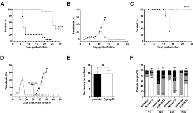
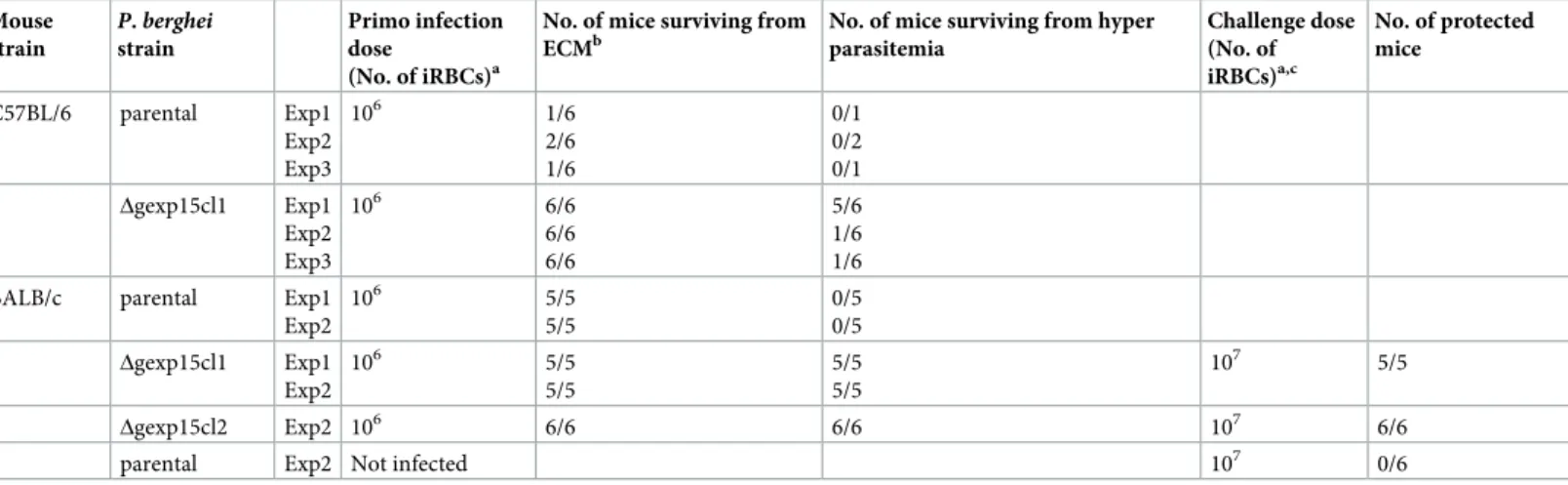
To explore the phenotype(s) of these parasites in more detail, we used two different mouse malaria models, C57BL/6 for experimental cerebral malaria (ECM) and BALB/c for malaria-linked pathologies (severe anemia, hyperparasitemia). The survival was based on euthanizing mice according to criteria described in the Materials and Methods section. When C57BL/6 mice were infected with the parental strain (106parasites), about 80%
succumbed within 6–8 days post-infection from ECM (Fig 3A,Table 1) due to blood-brain barrier disruption as evidenced by Evans blue staining (S4C Fig). In contrast, none of C57BL/6 mice infected with theΔgexp15 parasites exhibited any ECM symptoms and 61% succumbed exclusively to hyperparasitemia between days 20–25 post-infection. In the case of BALB/c mice, as expected, all mice succumbed from hyperparasitemia before day 15 after infection with 106parental parasites (Fig 3B). Importantly, all BALB/c mice infected with Δgexp15 survived infection with a rapid clearance of all blood parasites after a peak of between days 10 to 12 (Fig 3B and 3C). Given these data, we further tested whether
Δgexp15cl1 infected BALB/c mice that survived the infection could be protected from a sec-ondary infection with parental parasites. As depicted inFig 3DandTable 1, mice challenged with 107infected red blood cells (iRBC) showed either low parasitemia (<1%) that was quickly cleared or no detectable infection up to 40 days post reinfection, while mice in the control group succumbed to the infection. A similar result was observed withΔgexp15cl2 (Table 1). These results suggest that the infection of BALB/c withΔgexp15 induces a potent protective response against parental parasites.
Fig 3. Effect of PbGEXP15 knock-out on parasite virulence and fitness. (A) C57BL/6 mice were intraperitoneally inoculated with 106of parental (black circles) orΔgexp15 (white circles) infected red blood cells (iRBCs). The cumulative survival rates of three independent experiments (n = 6/experiment, seeTable 1) are indicated (Log-rank test,����p<0.0001). (B) BALB/c mice were inoculated in the same conditions and the course of infection was observed by blood smears. The results of one representative experiment out of two are shown as the mean parasitemia± SEM (n = 5/experiment, seeTable 1) (Wilcoxon test,� �p<0.01). (C) Cumulative survival rates of BALB/c mice were indicated in a second graph (Log-rank test,����p<0.0001). (D) BALB/c mice were inoculated with 106ofΔgexp15 iRBCs at day 0 (white
circles). After parasite clearance, the mice (white circles) and matched naïve mice of the same age (black circles) were inoculated with 107of
parental iRBCs at day 30 (black arrow). The results of one out of two representative experiments are shown as the mean parasitemia± SEM (n = 11 in total, seeTable 1) (Wilcoxon test,��p<0.01). (E) Number of merozoites per schizont obtained afterin vitro culture of parental andΔgexp15 parasites. Data represent mean ± SD of two independent experiments (n = 60). No significant (ns) difference was observed (Mann–WhitneyU test). (F) Parental and Δgexp15 parasites were synchronized in vitro and mice were intravenously infected. After 1, 22, 25
and 29h post-infection, blood smears were performed and the percentages of rings (in white), trophozoites (in grey) and schizonts (in black) were determined. Data shown represent means± SD of two independent experiments in duplicate (Tukey’s multiple comparison test, no significant (ns),���p<0.001,����p<0.0001 compared to parental at the same timing).
https://doi.org/10.1371/journal.ppat.1007973.g003
Intraerythrocytic development of
Δgexp15 parasites
To assess the effects of deficiency of PbGEXP15 expression on parasite growth, we investigated their intraerythrocytic development. First, the number of merozoites per schizont observedin vitro did not significantly differ between parental and Δgexp15 parasites (Fig 3E). Thereafter, the growth rate was followed in mice infected intravenously with purified schizonts obtained from overnight cultures of parental andΔgexp15 parasites. At 1h post-infection, as shown in
Fig 3F, both parental andΔgexp15 parasites exhibited ~80% rings suggesting a similar ability to invade. After 22h, a slight delay in the maturation ofΔgexp15 parasites was observed with 64% of trophozoites and 30% of schizonts versus 41% and 50% respectively in parental para-sites. At 25h, the examination of parental parasites showed 63% rings while theΔgexp15 para-sites presented 15% rings (p<0.0001). The follow up at 29 h post-infection underscored the delay in transition (85% trophozoites in parental versus 32% inΔgexp15, p<0.0001). These data clearly indicate that the depletion of PbGEXP15 protein delays the intraerythrocytic growth ofP. berghei.
Role of GEXP15 in transmission to the mosquito
Consistent with the first proteomic analysis [19] and this study showing the expression of GEXP15 in gametocytes, we examined whether PbGEXP15 is essential at this stage. Unexpect-edly, we noticed that the gametocytemia ofΔgexp15 parasites did not differ significantly when compared to parental parasites (Fig 4A) and the deletion seemed to have no effect on the number of exflagellation centers of male gametocytes (Fig 4B). These results indicate that PbGEXP15 did not affect the early male gametocyte development.
In order to examine the role of PbGEXP15 during the mosquito stages and due to the diffi-culties to obtain reproducible and reliable data fromin vitro ookinete conversion assays with
the pG230 line that showed a very low conversion efficiency to ookinetes [36], we used paren-tal orΔgexp15 infected mice to feed Anopheles stephensi mosquitoes. Two independent experi-ments were performed and the dissection of the mosquito midguts at day 9 confirmed that 80% of blood meals were positive with parental parasites (Fig 4C). Interestingly, in contrast to parental parasites,Δgexp15 parasites failed to initiate the formation of oocysts. This result was Table 1. Time course of parental andΔgexp15 parasites infection in C57BL/6 and BALB/c mice and protection of BALB/c mice challenged with parental parasites. Mouse strain P. berghei strain Primo infection dose (No. of iRBCs)a
No. of mice surviving from ECMb
No. of mice surviving from hyper parasitemia Challenge dose (No. of iRBCs)a,c No. of protected mice C57BL/6 parental Exp1 Exp2 Exp3 106 1/6 2/6 1/6 0/1 0/2 0/1 Δgexp15cl1 Exp1 Exp2 Exp3 106 6/6 6/6 6/6 5/6 1/6 1/6
BALB/c parental Exp1
Exp2 106 5/5 5/5 0/5 0/5 Δgexp15cl1 Exp1 Exp2 106 5/5 5/5 5/5 5/5 107 5/5 Δgexp15cl2 Exp2 106 6/6 6/6 107 6/6
parental Exp2 Not infected 107 0/6
aParasites were inoculated by intraperitoneal injection of infected red blood cells (iRBCs).
bSurvival linked to ECM was assessed by specific clinical symptoms at low parasitemia, between 6–8 days post-infection (p.i) and by brains staining using Evans blue. cMice were challenged with parental parasites at day 30 post primo-infection.
confirmed by the lack ofΔgexp15 sporozoites in the salivary glands observed at day 18 whereas 22,000 sporozoites, on average, were detected with parental parasites (Fig 4D). We conclude that PbGEXP15 is essential for parasite development in the mosquito.
Quantitative proteomic and phosphoproteomic analyses of
Δgexp15
schizonts
Previous studies showed that phosphatase inhibitors, comprising those acting on PP1, are toxic to cells and that the uncontrolled activity of the catalytic phosphatase subunits could cause apoptotic cell death [37]. Given the capacity of PbGEXP15 to interact with and regulate the dephosphorylation activity of PP1, we explored whether PbGEXP15 depletion could Fig 4. Essentiality of PbGEXP15 in the parasite development in mosquito. (A) Comparison of gametocytemia from parental andΔgexp15 parasites. Mean ± SEM of the number of gametocytes is indicated for two independent experiments (n = 8). No significant difference (ns) was observed (Mann–WhitneyU test). (B) Comparison of
exflagellation from parental andΔgexp15 parasites. Mean ± SEM of the number of exflagellation centers is indicated from two independent experiments (n = 6). No significant difference (ns) was observed (Mann–WhitneyU test). (C)
Comparison of midgut oocysts from parental andΔgexp15 parasites (n = 20). Mosquito midguts were dissected at day 9 and average oocyst number± SEM is indicated from two independent experiments (Mann–Whitney U test,���� p<0.0001). (D) Quantification of sporozoites from mosquitoes infected with parental andΔgexp15 parasites. Salivary glands were dissected at day 18 and sporozoites were manually counted. Bars indicate mean± SEM of the number of sporozoites per infected mosquito from 4 technical replicates of two independent experiments (Mann–WhitneyU test,
�p<0.05).
https://doi.org/10.1371/journal.ppat.1007973.g004
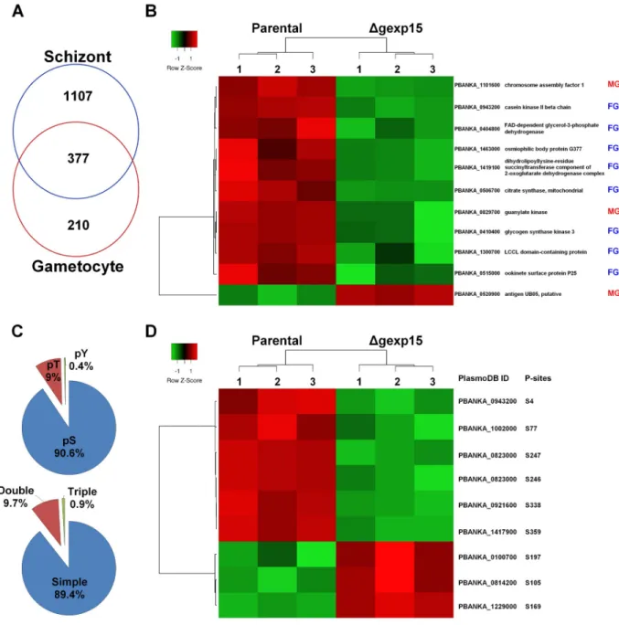
change the global phospho-proteomic patterns ofΔgexp15 parasites. To investigate this, paren-tal andΔgexp15cl1 schizonts were compared to assess proteomic and phosphoproteomic pro-files. The quantitative experiments were performed on four biological replicates and three technical replicates.
First, for the whole proteome analysis, we identified 2188Plasmodium proteins across
sam-ples corresponding to 43% of the predicted proteome ofP. berghei. We retained 1484 Plasmo-dium proteins that were reliably quantified in four biological replicates in at least one group
(S4 Table). In this global proteome, PbPP1 was detected and no difference was observed between levels in parental andΔgexp15 parasites. We next focused on proteins with significant changes in parental andΔgexp15 proteomes (FDR<0.05). The results indicate a total of 106 proteins (accounting for ~ 7% of the total proteins identified) whose abundance is significantly altered, of which 27 and 79 showed a significant increase and decrease respectively in compari-son to parental parasites (Fig 5A and 5BandS4 Table). An enrichment analysis of the biologi-cal processes was then performed for these proteins versus the global proteome (Fig 5C). Interestingly, 16 proteins playing a role in the pathogenicity, such as RONs and MSPs, were detected with a significant decrease in theΔgexp15 parasites and correspond to the highest sig-nificant enrichment (5.6-fold, p<0.001) (Fig 5B and 5C). We also noticed an under-represen-tation of proteins involved in translation (0.13-fold, p<0.01) while 3 AP2 transcription factors were enriched among the up-regulated proteins (5.25-fold, p<0.05) (Fig 5B and 5CandS4 Table). Of note, many proteins involved in these three biological processes have already been described to interact with PP1 (this study and [17,32,38]). In total, 12% (13/106) of the altered proteins, such as AMA1, RON-2, -4 and -5, are detected in thePlasmodium interactomes of
PP1 (S5A FigandS4 Table). Collectively, these data indicate that the knock-out ofpbgexp15
impacted the expression of several PP1 partners and would tend to confirm the commonality of signaling pathway(s) between GEXP15 and PP1.
Next, we explored the phosphoproteome of parental andΔgexp15 parasites. In total 2460 different phosphorylation sites ofP. berghei were identified and quantified belonging to 780 Plasmodium proteins (Fig 6AandS5 Table). We observed significant changes for 166 phos-phopeptides corresponding to 118 proteins (FDR<0.01) (Fig 6BandS5 Table). Levels of phosphorylation of most of the phosphosites (143 phosphosites) were lower inΔgexp15 when compared to the phosphoproteome of the parental parasites. The analysis showed a significant enrichment in proteins acting on RNA metabolism (1.9-fold, p<0.01) with 11 out of 12 phos-phosites hypophosphorylated inΔgexp15 parasites (Fig 6C). We also noticed the hypopho-sphorylation of 8 proteins engaged in transcriptional regulation (including AP2 transcription factors), 5 proteins playing a role in post-translational modifications/chaperones and 4 pro-teins involved in trafficking (S5 Table). A smaller set of 19 proteins was found to be hyperpho-sphorylated in theΔgexp15 parasites but most of these proteins have an unknown function. We also observed that overall 18% (21/118) of the phosphoproteins showing significant changes in phosphorylation were previously reported in thePlasmodium interactomes of PP1
suggesting that the phenotypes observed above could be due to a deregulation of the PbPP1 activity (S5A Fig).
Quantitative proteomic and phosphoproteomic analyses of
Δgexp15
gametocytes
To explore more deeply the role and the essentiality of GEXP15 in sexual and mosquito stages, we purified parental andΔgexp15cl1 gametocytes for proteomic and phosphoproteomic stud-ies. The quantitative experiments were performed on three biological replicates and three tech-nical replicates.
Fig 5. Differential proteome of parental andΔgexp15 schizonts in P. berghei. (A) Hierarchical clustering and heatmap of proteins with a significant differential abundance in parental andΔgexp15 schizonts (FDR<0.05, S0 = 0.1). (B) Volcano plot representation of proteins quantified by mass spectrometry in parental andΔgexp15 schizonts (FDR<0.05, S0 = 0.1). AP2 transcription factors are indicated as blue squares and proteins involved in the pathogenicity as red dots. (C) Functional enrichment of parental andΔgexp15 schizonts proteomic profiles. Fold enrichment was performed on the 106 filtered proteins relative to allP. berghei proteins detected in the proteome. The x-axis
represents the fold enrichment (log2) for the indicated function (Hypergeometric test,��p<0.01,����p<0.0001).
https://doi.org/10.1371/journal.ppat.1007973.g005
Firstly, we determined the gametocyte proteome and identified 587Plasmodium proteins
that were reliably quantified in three biological replicates in at least one group (S6 Table). As depicted inFig 7A, 64% of these proteins are also detected in the schizont proteome, a percent-age similar to previous overlaps inP. berghei (50%) and P. falciparum (59%) [39,40]. Signifi-cant changes in parental andΔgexp15 proteomes (FDR<0.05) were observed for 11 proteins, with a significant decrease in abundance for 10 proteins in mutant parasites (Fig 7BandS6 Table). One of these proteins, PbGSK3 (PBANKA_0410400) that decreased significantly in Δgexp15 gametocytes and for which commercial antisera were available was tested by western Fig 6. Differential phosphoproteome of parental andΔgexp15 schizonts in P. berghei. (A) Distribution of phospho-Ser (pS), phospho-Thr (pT), and phospho-Tyr (pY) residues and multiplicity of phosphosites detected in schizonts (n = 2460). (B) Hierarchical clustering and heatmap of phosphosites with a significant differential in parental and Δgexp15 schizonts (FDR<0.01, S0 = 0.1). (C) Functional enrichment of parental and Δgexp15 schizont
phosphoproteomic profiles. Fold enrichment was performed on the 166 filtered phosphosites relative to allP. berghei
phosphosites detected in the phosphoproteome. The x-axis represents the fold enrichment (log2) for the indicated
function (Hypergeometric test,��p<0.01).
blot experiments in two independent biological samples. Results indicated a drastic decrease of this protein inΔgexp15 gametocytes when compared to the parental strain, validating the proteomic analysis (S5B and S5C Fig).
Interestingly, 8 proteins out of the 11 have been described being enriched in female gameto-cytes and the others 3 in male gametogameto-cytes [39–42]. Among these proteins, we detected G377 (PBANKA_1463000) which is localized in the osmiophilic bodies of female gametocytes and Fig 7. Differential proteome and phosphoproteome of parental andΔgexp15 gametocytes in P. berghei. (A) Venn diagram of proteins detected in the proteomes of schizonts and gametocytes. (B) Hierarchical clustering and heatmap of proteins with a significant differential abundance in parental andΔgexp15 gametocytes (FDR<0.05, S0 = 0.1). The sex enrichment in gametocyte stage is indicated for each protein. MG: male gametocyte, FG: female gametocyte [40–42]. (C) Distribution of phospho-Ser (pS), phospho-Thr (pT), and phospho-Tyr (pY) residues and multiplicity of phosphosites detected in gametocytes (n = 444). (D) Hierarchical clustering and heatmap of phosphosites with a significant differential in parental andΔgexp15 gametocytes (FDR<0.01, S0 = 0.1). The position and the residue of the phosphosites are indicated.
https://doi.org/10.1371/journal.ppat.1007973.g007
plays a role in the formation of these organelles and in the induction of gametogenesis with a delay in female gamete egress [43,44]. Furthermore, diverse studies have demonstrated that P25 (PBANKA_0515000) and Lap2 (PBANKA_1300700) play important roles in parasite transmission by mosquitoes and more precisely in the ookinete and oocyst stages [45–48]. Altogether, these data confirmed the phenotypic analyses observed and suggested that the deletion ofpbgexp15 impacted key proteins in mosquito transmission. Concerning the
phos-phoproteome of parental andΔgexp15 parasites, we identified 444 different phosphorylation sites ofP. berghei belonging to 278 proteins and we observed significant changes for only 9
phosphopeptides corresponding to 8 proteins (FDR<0.01) (Fig 7C and 7DandS7 Table). Unlike the proteome, these proteins did not seem to exhibit features linked to sexual differenti-ation similar to those shown above. Only two proteins are clearly described to be enriched in male and one in female gametocytes (S7 Table).
Discussion
The functional diversity of the PP1 catalytic subunit, an essential phosphatase enzyme, is now clearly attributable to more than 200 regulators that have been described in diverse eukaryotic organisms [8,9]. To date, only four conserved regulators of PP1 have been identi-fied and characterized inP. falciparum [10–12,15]. A more recent study, using an Y2H screening to examine the global PP1 interactome inP. falciparum, identified GEXP15
as a potent regulatory partner of PP1 [17]. Here we confirmed the direct interaction of PbGEXP15 with PP1 and demonstrated its capacity to control the phosphatase activity. These results extend our previous data showing the capacity of conserved regulators to affect PP1 activity to a specific protein expressed byPlasmodium. Structure-activity studies
indi-cate a major contribution of the well-known RVxF consensus binding motif to the function of GEXP15. Moreover, a short N-terminal region of GEXP15 was able to bind PP1 and to increase its phosphatase activity in a similar manner to that observed with the full length pro-tein. The mutation of the RVxF motif, present in this N-terminal region, completely abol-ished this regulation. These data suggest that this region seems to carry the regulatory function of GEXP15 on PP1 activity.
To further explore the functional role of GEXP15, we examined the impact of its deletion in the rodent malaria parasiteP. berghei. Phenotypic analyses of these deficient parasites revealed
a drastic effect on their development both in mice and mosquitoes. Indeed, while BALB/c mice infected with parental parasites succumbed to infection from hyperparasitemia, they were able to efficiently clearΔgexp15 parasites. This could be linked at least in part to the retarded multiplication that we observed. Further, surviving mice exhibited a protection against a secondary challenge by parental parasites, suggestive of a role of these deficient para-sites in inducing protective responses.
When C57BL/6 mice susceptible to ECM were tested,Δgexp15 parasites were found to be unable to induce ECM. Investigations on ECM in the mouse model indicated that it is a com-plex process involving both the parasite and the host molecules including proinflammatory cytokines [49–52]. However, when the outcomes of infections byΔgexp15 parasites of C57BL/ 6 and BALB/c mice (prototypical Th1 and Th2-type strains respectively) were compared, we observed a higher rate of mortality in C57BL/6 (ECM model) due to hyperparasitemia than in BALB/c (hyperparasitemia model), suggesting a difference in the immune responses raised by these hosts towards the infection withΔgexp15 parasites. Earlier studies mainly focused on phenotypic analyses have shown similar data obtained with targeted disruption of hmgb2 described as a pro-inflammatory protein [53], MSP7 involved in invasion of erythrocytes [54] or plasmepsin-4 contributing to hemoglobin digestion [55]. Although, the parasite derived
molecules triggering host responses are still largely unknown, the use ofΔgexp15 parasites might contribute for a better understanding of the protective mechanisms against ECM.
Of note, the essentiality of GEXP15 in the blood stages was further supported by a recent study using a piggyBac transposon inserted randomly inP. falciparum genome [56] in which they did not obtain viable parasites with a disruptedpfgexp15 gene despite the presence of 35
potential insertion sites.
For an in-depth dissection of the biological functions of GEXP15, we performed quantita-tive proteomic and phosphoproteomic analyses. To the best of our knowledge, our proteomic study is the first in which a PP1 partner has been suppressed inPlasmodium.
Of particular interest are low abundance proteins in schizonts that are members of the AP2 transcription factor family, which play a role in the regulation of gene expression, and invasion proteins including RONs and AMA1. The low abundance of three AP2 transcription factors, that have been suggested to be essential [57,58], could explain the general down-regulation observed for the proteins whose expression varies inΔgexp15 parasites. These results highlight the role played by GEXP15 and could explain the attenuated virulence ofΔgexp15 parasites in blood stages.
In our global phosphoproteomic analysis in schizonts, the data indicate the hyperphosphor-ylation of 19 proteins inΔgexp15 when compared to parental parasites. This could be expected as the lack of GEXP15, an activator of PP1in vitro, may lead to a decrease in PP1 activity and
consequently an increase of phosphorylation of target proteins. Should this be the case, these hyperphosphorylated proteins could be considered as potential substrates of the complex PP1-GEXP15. Interestingly, the RON2 protein, present in lower abundance inΔgexp15 para-sites, was found to be hyperphosphorylated, possibly attenuating its known function in inva-sion [59–63]. However, synchronizedΔgexp15 parasites did not show any delay in the first invasive cycles, suggesting a functional overlap and/or compensation between proteins involved in invasion [64] or that the induced defect did not attain a sufficient threshold to interrupt the invasion at least during the first cycles. On the other hand, the data suggest an unanticipated role of GEXP15 in the phosphorylation process. Indeed, we observed a drop in the phosphorylation levels of 100 proteins when compared to controls. These data could be explained either by free and uncontrolled PP1 capable of non-specifically dephosphorylating many and diverse substrates in the absence of GEXP15 and/or by an inhibitory role of GEXP15 on PP1 activity inPlasmodium. This latter possibility cannot be excluded as in vitro
experiments, which showed a positive effect of GEXP15 on PP1 activity, were performed with the non-natural pNPP substrate. In addition, any post-translational modifications of GEXP15
in vivo could affect its function, which may be different from that observed with the
recombi-nant protein. In this context, it has been shown that the phosphorylation status of Inhibitor-1, a well-known regulator of human PP1, differentially alters its function [65–67].
Concerning the proteome in gametocyte stages, we detected 11 proteins whose expression varies inΔgexp15 parasites. Among these proteins, eight, described to be overexpressed in female gametocytes, are present at lower levels in these deficient parasites. These data along with the above observations indicate that the lack of GEXP15 impacts neither the number of gametocytes nor the exflagellation of male gametocytes and strongly suggest that GEXP15 could function downstream in female gamete formation/egress/fertility or zygote-to-ookinete development. Supporting this is the under-abundance of the G377 protein inΔgexp15 that has been reported as a key actor in female egress/emergence (43, 44). Additional proteins could also contribute to the observed phenotype include Lap2, demonstrated as being important in the mosquito transmission [47,48] or AP2-O2 that was hypophosphorylated on 2 serines in Δgexp15 schizonts compared to the parental parasites. This latter transcription factor is described as being crucial in the development of ookinetes and oocysts inP. berghei [58] while
inP. yoelii, a knock-out of AP2-O2 did not seem to affect the gametocytes and ookinetes but
only the number of oocysts and sporozoites [68]. In this study, unfortunately, the pG230 line used to generateΔgexp15 parasites exhibits very low conversion efficiency in vitro, hampering these studies. Of note, transfections of two otherP. berghei strains did not allow the generation
of viable knock-out or stable inducible knock-down parasites. Whatever the explanation, it is clear that the depletion of GEXP15 led to a complete abolition of oocyte/sporozoite formation
in vivo. Taken together, our observations suggest that while the lack of GEXP15 expression
could be transiently compensated in intraerythrocytic growth in the blood, this compensation seems to be insufficient with a high fitness cost in the mosquito.
Interestingly, an earlier study suggested that GEXP15 was a potential orthologue of human CD2BP2 (CD2 Cytoplasmic Tail Binding Protein 2) [69]. CD2BP2 has been described to inter-act with splicing finter-actors and PP1 through a GYF domain [34] and an RVxF motif respectively [35]. The sequence analysis of PbGEXP15 reveals only 14% identity with HsCD2BP2, but it presents a GYF like-domain, even if this does not match perfectly the consensus sequence [70]. However, although the IP/MS of PbGEXP15-mCherry demonstrated that several splicing fac-tors are potential partners of GEXP15, its contribution in the splicing function inPlasmodium
requires further investigation in the future.
In conclusion, the results shown here indicate that the viability of parasites in the absence of GEXP15 expression is accompanied by major alterations that could contribute to the aviru-lent phenotype of these parasites and their incapacity to produce oocysts. These alterations could affect spliceosome and proteasome pathways along with extensive changes to the phos-phorylation patterns ofΔgexp15 parasites that may be linked to an uncontrolled PP1, capable of dephosphorylating inappropriate substrates. Additional studies are required to examine each novel aspect of the phenotypes of theseΔgexp15 parasites and the potential alteration of the RNA splicing pathway.
Materials and methods
Plasmids
Plasmids pGADT7/pGBKT7, pETDuet-1 and pGEX4T3 were purchased from Clontech, Novagen and GE Healthcare Life Sciences. The plasmids pBS-DHFR and pL1886 were kindly provided by Drs R. Tewari and B. Franke-Fayard respectively. The primers used are described inS8 Table.
Ethics statement
Mice were housed in an Animal Biosafety Level 2 facility at the Institut Pasteur de Lille and maintained in accordance with the French National Guidelines for Use of Animals for Scien-tific Purposes which is also in line with EU Directive 2010/63/EU. Experimental protocols performed in this study were reviewed and approved by the Comite´ d’Ethique C2EA-75 en Expe´rimentation Animale Nord-Pas de Calais-France (project number: 00527.04).
Animals
Infections and antiserum production were performed in CD1 male mice (30g) (Charles River). BALB/c (10 weeks) and C57BL/6 (4-5weeks) male mice (Janvier Labs) were sorted randomly into groups of 5–6 animals and used for hyperparasitemia and ECM comparison between parental andΔgexp15 parasites. The duration of experiments was strictly limited and constant monitoring of infected mice was carried out. When parasitemia was about 60% accompanied with weight body loss, mice were euthanized by CO2inhalation. Mice susceptible to ECM
were euthanized by CO2inhalation if they displayed paralysis, convulsions/fits or coma. For
the disruption of blood brain barrier, 100μl of 2% Evans blue dye in PBS were injected intrave-nously in C57BL/6 infected mice at day 6 p.i. Mice were then euthanized by CO2inhalation 1h
post-injection and brains were recovered.
Yeast two-hybrid assays
PbGEXP154–590, PbGEXP154–178, PbGEXP15446–596were amplified by PCR onP. berghei
gDNA with primers p1-p2, p1-p3, p4-p5 respectively (S8 Table) and cloned into the pGADT7 vector (Clontech) using the In-Fusion HD Cloning system (Clontech) according to the manu-facturer’s instructions. Gal4-DBD-PfPP1c and PfPP1c F255A/F256A were previously cloned [12,23]. PbGEXP154–178 KKKKKAQAwas obtained by PCR-based site directed mutagenesis
with Isis DNA polymerase (MP Biomedicals) and pGADT7-PbGEXP154–178as template and
primers p6-p7. pGADT7 and pGBKT7 constructs were transformed into Y2H Gold and Y187 yeast strains (Clontech) respectively, and the yeasts were spread on Synthetic Defined agar medium lacking leucine (SD-L) or lacking tryptophane (SD-W) respectively and grown at 30˚C for 3–5 days. Different mating experiments were performed and spread on selective media SD-LW. They were restreaked on more stringent media SD-LWH and SD-LWHA (L: Leucine, W: Tryptophan, H: Histidine, A: Adenine) after dilutions at 1:1, 1:25, 1:50. Diploids were incubated for 4–6 days at 30˚C. Empty vectors pGADT7 or pGBKT7 and pGBKT7-Lami-nin were used as negative controls.
In order to checkPlasmodium gene expression in yeast, RT-PCRs were performed. Total
RNA was isolated from cultured yeasts (OD = 0.5) after flash freezing and using TRIzol Reagent (Thermo Fisher Scientific) with glass beads for 45 min at 65˚C with occasional vortex-ing. RNA (5μg) was treated with DNAse I (Thermo Fisher Scientific) and DNA contamina-tion was checked using an Agilent 2100 Bioanalyzer and by RT-PCR on intronic gene of yeast
tub1. cDNA was synthesized using SuperScript III First-Strand Synthesis SuperMix (Thermo
Fisher Scientific) according to the manufacturer’s instructions. Amplification of transcripts was carried out by PCR using the Advantage 2 Polymerase Mix (Clontech) and the following primers (S8 Table): p13-p14 for TUB1, p11-p12 for PfPP1c and Gal4-DBD-PfPP1c F255A F256A, p8-p9 for Gal4-AD-PbGEXP154–590, Gal4-AD-PbGEXP154–178and
Gal4-AD-PbGEXP154–178 KKKKKAQA, and p8-p10 for Gal4-AD-PbGEXP15446–596.
Recombinant protein expression and antiserum production
The coding regions of PbGEXP154–590and PbGEXP154–178were obtained by PCR with the
primers p15-p16 and p15-p17 respectively (S8 Table) and cloned into pETDuet-1 (Novagen) using the In-Fusion HD Cloning system (Clontech). PbGEXP154–178 KKKKKAQAwas obtained
by PCR-based site directed mutagenesis with Isis DNA polymerase (MP Biomedicals), pET-Duet-1-PbGEXP154–178as template and primers p6-p7.
GST, GST-PfPP1c and PfPP1c were produced as previously described [11,15]. All recombi-nant GEXP15 expressions were carried out in One Shot1BL21 Star™ (DE3) Chemically Com-petentE. coli cells (Life Technologies) in the presence of 0.5 mM IPTG at 16˚C overnight.
Cells were harvested in non-denaturing buffer (20 mM Tris, 500 mM NaCl, 20 mM imidazole and protease inhibitor cocktail (Roche), pH 7.5) followed by sonication and ultracentrifuga-tion. Pellets were resuspended and centrifuged in denaturing buffer (20 mM Tris, 500 mM NaCl, 6 M guanidine, 20 mM imidazole and protease inhibitor cocktail (Roche), pH 7.5). Ni2+-NTA agarose beads (Macherey Nagel) were used to purify the recombinant proteins as previously described [12]. SDS-polyacrylamide gels were blotted onto nitrocellulose and probed with His antibody (1:2000 dilution) (Qiagen) followed by HRP-labeled
mouse IgG (1:50000 dilution). Chemiluminescence detection with SuperSignal™ West Dura Extended Duration Substrate (Life Technologies) was carried out. Recombinant proteins were quantified with Pierce™ BCA Protein Assay Kit (Life Technologies).
The purified PbGEXP154–178was used to produce antisera as previously described [12] in
CD1 mice.
GST pull-down assays
Glutathione-Sepharose beads (Sigma-Aldrich) coupled with GST-PfPP1c were incubated over-night at 4˚C with 2μg of PbGEXP154–590, PbGEXP154–178or PbGEXP154–178 KKKKKAQA
in 20 mM Tris, 150 or 500 mM NaCl, 0.2 mM EDTA, 20 mM HEPES, 1 mM MnCl2, 1 mM
DTT, 0.1% Triton X-100, 10% glycerol, protease inhibitor cocktail (Roche) and pH 7.5. After 5 washes of the beads with the same buffer, proteins bound to the beads were analyzed by 4–20% SDS–PAGE followed by immunoblotting with anti-His (1:2000) or anti-GST mAb (1:2000) (Invitrogen) as described above.
Effect of GEXP15 on PfPP1 activity
The role of GEXP15 on the activity of PfPP1c was investigated using the p-nitrophenyl phosphate (pNPP) assay. Different amounts of PbGEXP154–590, PbGEXP154–178and
PbGEXP154–178 KKKKKAQAwere preincubated with 40 pmol of PfPP1c for 30 min at 37˚C.
The enzymatic reaction was initiated by the addition of pNPP substrat (Sigma-Aldrich) to the reaction medium and the absorbance was measured at 405 nm (Thermo Scientifc Multiskan FC). The lack of phosphatase activity of recombinant GEXP15 alone was checked according to the described procedure in the absence of PP1. Two independent experiments were carried out in duplicate.
Generation and analysis of
P. berghei transgenic parasites
In order to tag PbPP1c with mCherry,pbpp1c was amplified with primers p18-p19 (1324 bp)
(S8 Table). The insert was subcloned into pL1886 plasmid. The construct was linearized by Tth111I before transfection. The same plasmid was used for PbGEXP15-mCherry. The 3’ region ofpbgexp15 was amplified with primers p22-p25 and p24-p23 (1332 bp) and a silent
mutation was introduced by PCR-based mutagenesis in order to obtain a BsmI site, used to linearize the plasmid.
For the knock-out ofpbgexp15, PCR amplifications were generated with the 5’ and 3’ UTR
regions with primers p26-p27 (847 bp), p28-p29 (695 bp) andP. berghei gDNA as template.
The inserts were subcloned into pBS-DHFR plasmid [71] and the construct was linearized by XbaI-ApaI before transfection.
Linearized pL1886 plasmids and pBS-DHFR plasmid were transfected by electroporation as previously described [72] inP. berghei ANKA GFP line [24] and pG230 line [36] respectively, kindly provided by Drs O. Silvie and N. Philip. Transfected parasites were inoculated in CD1 mice and positively selected by pyrimethamine in the drinking water, 30h after transfection [72].
Parasitized erythrocytes were lysed with Red blood cell Lysing buffer (Sigma-Aldrich) fol-lowed by the use of KAPA Express Extract kit (KAPA BioSystems) to extract DNA (manufac-turer’s instructions). Primers p20-p21 and p15-p21 were used to genotypepbpp1c-mCherry
andpbgexp15-mCherry respectively (S8 Table). Deletion ofpbgexp15 was verified by
diagnos-tic PCR using primers p30-p31 and p32-p33 and positivepbgexp15 knock-out parasites were
Immunofluorescence assays
Blood from mice infected with PbPP1-mCherry or PbGEXP15-mCherry parasites was fixed with 4% paraformaldehyde and 0.0075% glutaraldehyde for 10 min at 4˚C. After PBS washing, cells were sedimented on Poly-L-lysine coated coverslips overnight then permeabilized and saturated with PBS, 0.5% Triton X-100 and 1% BSA for 30min. Anti-RFP pAb (MBL, PM005) was diluted 1:500 in PBS BSA 1% and applied for 1h at 37˚C. Coverslips were washed with PBS and incubated with Goat anti-Rabbit IgG (H+L) Cross-Adsorbed, Alexa Fluor 594 (Invitrogen, A11012) in PBS BSA 1% at 1:1000 in addition to DAPI (1μg/ml) for 1h at 37˚C. The coverslips were mounted in Mowiol and confocal imaging was performed with an LSM880 microscope (Zeiss). Images were treated with ImageJ.
Purification of schizonts and gametocytes in
P. berghei
To obtain schizonts, blood from infected mice was incubated 20h at 37˚C in RPMI1640 culture medium supplemented with 0.4% AlbuMAX™ II Lipid-Rich BSA (Life technologies), then schizonts were purified on a 55% Nycodenz gradient. Gametocytes purification was performed as previously described [73]. Briefly, CD1 mice were treated with phenylhydrazine by the intraperitoneal route (200μl, 6mg/ml, Sigma-Aldrich) 2 days pre-infection then treated with sulfadiazine (20mg/ml in drinking water, Sigma-Aldrich) 3 days infection. At day 5 post-infection, blood was collected by cardiac puncture and gametocytes purified on a 48% Nyco-denz column in coelenterazine buffer. Purifications were higher than 95% for schizonts and gametocytes.
Immunoprecipitation and mass-spectrometry
Purified schizonts or gametocytes of PbPP1-HA, PbPP1-mCherry, PbGEXP15-mCherry and parental wild-type parasites used as control, were suspended in 50 mM Tris, 0.5% Triton X-100 and protease inhibitor cocktail (Roche), pH 8. After 10 freeze-thaw cycles and sonication, soluble fractions were obtained after repeated centrifugations at 13000 rpm at 4˚C. Anti-HA agarose beads (Life Technologies) or RFP-Trap1_A beads (Chromotek) were mixed overnight at 4˚C with parasite soluble extracts in 20 mM Tris, 150 mM NaCl, 0.5% Triton X-100 and pro-tease inhibitor cocktail (Roche), pH 7.5. Beads were washed and elution was performed in Laemmli buffer. Then after 3 min at 95˚C, samples were loaded on a 4–20% SDS-PAGE for western blot or mass spectrometry analyses. Western blots were carried out as described above and probed with anti-RFP pAb (1:1000, MBL) followed by goat anti-rabbit IgG-HRP (1:20000, Sigma-Aldrich). Then, the membrane was stripped and probed with mouse sera anti-GEXP15 (1:100) followed by Mouse TrueBlot1Ultra: Anti-Mouse Ig HRP (1:2000, eBioscience). For the Mass-spectrometry analysis, electrophoretic migration, tryptic digestion and nanoLC-MSMS analysis were performed as previously described [74]. Raw data collected during nanoLC-MS/MS analyses were processed and converted into�.mgf peak list format with Proteome Discoverer 1.4 (Thermo Fisher Scientific). MS/MS data were interpreted using the search engine Mascot (version 2.4.0, Matrix Science, London, UK) installed on a local server. Searches were performed with a tolerance on mass measurement of 0.2 Da for precur-sor and 0.2 Da for fragment ions, against a composite target decoy database (2�22,202 total entries) built withMus musculus Uniprot database (10090–17,008 entries), Plasmodium ber-ghei PlasmoDB database (Release 41.0–5 December 2018–5,076 entries) fused with the
sequences of PbPP1-HA or PbGEXP15-mCherry, recombinant trypsin and a list of classical contaminants (118 entries). Cysteine carbamidomethylation, methionine oxidation, protein N-terminal acetylation, and cysteine propionamidation were searched as variable modifica-tions. Up to one trypsin missed cleavage was allowed. For each sample, peptides were filtered
out according to the cut-off set for proteins hits with one or more peptides longer than nine residues. Ion and identity score were fixed to obtain a 1% false positive rate.
Asexual and sexual development assays
In order to evaluate the number of merozoites per schizont, purified schizonts fromP. berghei
cultures were fixed in 4% paraformaldehyde and 0.0075% glutaraldehyde for 10 min at 4˚C then incubated 30 min with DAPI. The nuclei were counted with Leica Leitz DMRB fluores-cence microscope. To measure growth rate, purified mature schizonts were intravenously injected in CD1 mice. Smears were performed at 1h, 22h, 25h and 29h post-infection and rings, trophozoites and schizonts were counted under the microscope after Giemsa staining.
For sexual development, the gametocytemia was determined by microscopy. Exflagellation was assessed after 10–12 min of incubation at 21˚C in RPMI 1640 with 25 mM HEPES and 10% fetal calf serum, pH 8 [75]. The exflagellation centers were counted under slide-coverslip by microscopy using a 40x objective.
Mosquito transmission
Anopheles stephensi mosquitoes were maintained at the insectarium of the Institut Pasteur de
Lille. They are reared at 19˚C and 75–80% humidity under 12/12 hour light/dark cycle. Female mosquitoes (4 to 6 days) were fed on anaesthetized infected CD1 mice once gametocytes had been observed. The presence of oocysts in the midgut was checked at day 9 post blood meal and dissection of salivary glands was assessed at day 18. Fifteen salivary glands were pooled and homogenized per technical replicate. Sporozoite counts were determined with a Kova slide. The experiments were performed twice independently.
Sample preparation and mass spectrometry
P. berghei schizonts or gametocytes were purified as described above and treated with 0.15%
saponin to avoid host contamination. Soluble proteins were extracted in RIPA buffer (Thermo Fisher Scientific), Halt™ Protease and Phosphatase Inhibitor Cocktail (Thermo Fisher Scien-tific) and DNase I (Thermo Fisher ScienScien-tific). Proteins were quantified with the Pierce™ BCA Protein Assay Kit (Life Technologies). 82μg of proteins for the schizonts and 100 μg of pro-teins for the gametocytes were first reduced with 0.1 M DTT final concentration at 60˚C for 1h. MS sample preparation was performed using a FASP method (filter aided sample prepara-tion) according to Lipeckaet al [76]. We set aside around 10μg of the digested proteins for the analysis of total proteomes, while the remaining samples were used for phosphopeptide enrichments. The expression of PbGSK3 was examined by western blot on purified gametocyte extracts and probed with anti-PfGSK3 (1:1000 dilution, Covalab, pab0250) and anti-Actin1 (1:2000 dilution) followed by HRP-labeled anti-rabbit (1:20000 dilution) and HRP-labeled anti-mouse (1:20000 dilution) respectively. Relative quantification of PbGSK3 in parental and Δgexp15 gametocytes was normalized using PbActin-1.
Phosphopeptide enrichment by titanium dioxide (TiO
2)
Phosphopeptide enrichments were carried out using Titansphere TiO2Spin tips (3 mg/200μL,
Titansphere PHOS-TiO, GL Sciences Inc) on the digested proteins for each biological repli-cate. Briefly, the TiO2Spin tips were conditioned with 20μL of solution A (80% acetonitrile,
0,4% TFA), centrifuged at 3,000 g for 2 min and equilibrated with 20μL of solution B (75% acetonitrile, 0,3% TFA, 25% lactic acid) followed by centrifugation at 3,000 g for 2 min. Pep-tides were dissolved in 20μL of solution A, mixed with 100 μL of solution B and centrifuged at
1,000 g for 10 min. Sample was applied back to the TiO2Spin tips twice more in order to
increase the adsorption of the phosphopeptides to the TiO2. Spin tips were washed sequentially
with 20μL of solution B and twice with 20μL of solution A. Phosphopeptides were eluted by the sequential addition of 50μL of 5% NH4OH and 50μL of 5% pyrrolidine. Centrifugation
was carried out at 1,000 g for 5 min. Phosphopeptides were purified using GC Spin tips (GL-Tip, Titansphere, GL Sciences Inc). Briefly, the GC Spin tips were conditioned according to manufacturer’s instructions, then eluted phosphopeptides from the TiO2Spin tips were
added to the GC Spin tips and centrifuged at 1,000 g for 5 min. GC Spin tips were washed with 20μL of 0.1% TFA in HPLC-grade water. Phosphopeptides were eluted with 70 μL of 80% ace-tonitrile, 0.1% TFA (1,000 g for 5 min) and vacuum dried.
nanoLC-MS/MS protein identification and quantification
Peptides for the analysis of total proteomes were resuspended in 0.1% TFA in HPLC-grade water, 10% acetonitrile and 500 ng of each sample was injected in a nanoRSLC-Q Exactive PLUS (RSLC Ultimate 3000, Thermo Scientific). Phosphopeptides were resuspended in 42μL of 0.1% TFA in HPLC-grade water and 5μL of each sample was injected into the mass spec-trometer. Samples were loaded onto aμ-precolumn (Acclaim PepMap 100 C18, cartridge, 300μm i.d.×5 mm, 5 μm, Thermo Scientific), and were separated on a 50 cm reversed-phase liquid chromatographic column (0.075 mm ID, Acclaim PepMap 100, C18, 2μm, Thermo Sci-entific). Chromatography solvents were (A) 0.1% formic acid in water, and (B) 80% acetoni-trile, 0.08% formic acid. Samples were eluted from the column with the following gradient: 5% to 40% B (120 min), 40% to 80% (6 min). At 127 min, the gradient returned to 5% to re-equili-brate the column for 20 min before the next injection. One blank was run between biological replicates to prevent sample carryover. Samples eluting from the column were analyzed by data dependent MS/MS, using the top-10 acquisition method. Peptides and phosphopeptides were fragmented using higher-energy collisional dissociation (HCD). Briefly, the instrument settings were as follows: resolution was set to 70,000 for MS scans and 17,500 for the data dependent MS/MS scans in order to increase speed. The MS AGC target was set to 3.106 counts with maximum injection time set to 200 ms, while MS/MS AGC target was set to 1.105 with maximum injection time set to 120 ms. The MS scan range was from 400 to 2000 m/z. Dynamic exclusion was set to 30 sec duration.
The MS files were processed with the MaxQuant software version 1.5.8.3 and searched with Andromeda search engine against the database ofMus musculus from swissprot 07/
2017 andPlasmodium berghei ANKA from PlasmoDB (v37) [20]. To search parent mass and fragment ions, we set a mass deviation of 4.5 ppm and 20 ppm respectively. Strict specificity for trypsin/P cleavage was required, allowing up to two missed cleavage sites. Carbamido-methylation (Cys) was set as a fixed modification, whereas oxidation (Met) and N-term acet-ylation were set as variable modifications. For the analysis of MS files issuing from of TiO2 enrichment, the variable modification of phosphorylation on S, T and Y were also added. The false discovery rates (FDRs) at the protein and peptide level were set to 1%. Scores were calculated in MaxQuant as described previously [77]. Match between runs was allowed. The reverse hits were removed from MaxQuant output. Proteins were quantified according to the MaxQuant label-free algorithm [77,78] using LFQ intensities and phosphopeptides according to intensity. Protein quantification was obtained using at least 2 peptides per protein.
Statistical and bioinformatic analysis, including volcano plots and clustering, were per-formed with Perseus software (version 1.6.0.7) freely available atPerseus[79]. For statistical comparison we set two groups:P. berghei parental and Δgexp15. Each group contained four