HAL Id: hal-03182668
https://hal.u-pec.fr//hal-03182668
Submitted on 26 Mar 2021
HAL is a multi-disciplinary open access
archive for the deposit and dissemination of
sci-entific research documents, whether they are
pub-lished or not. The documents may come from
teaching and research institutions in France or
abroad, or from public or private research centers.
L’archive ouverte pluridisciplinaire HAL, est
destinée au dépôt et à la diffusion de documents
scientifiques de niveau recherche, publiés ou non,
émanant des établissements d’enseignement et de
recherche français ou étrangers, des laboratoires
publics ou privés.
Coumarin from Its Microwave-Determined Structure
Ha Vinh Lam Nguyen, Jens-Uwe Grabow
To cite this version:
Ha Vinh Lam Nguyen, Jens-Uwe Grabow. The Scent of Maibowle – π Electron Localization in
Coumarin from Its Microwave-Determined Structure. ChemPhysChem, Wiley-VCH Verlag, 2020, 21
(12), pp.1243-1248. �10.1002/cphc.202000234�. �hal-03182668�
1
The scent of maibowle –
electron localization in coumarin from
its microwave-determined structure
Ha Vinh Lam Nguyen*
[a]and Jens-Uwe Grabow
[b]This work is dedicated to T. Bell.
Abstract: The microwave spectra of the natural substance coumarin,
a planar aromatic molecule with the specific scent of maibowle, a popular fruit punch served in spring and early summer, were recorded using a molecular jet Fourier transform microwave spectrometer working in the frequency range from 4.0 to 26.5 GHz. The rotational constants and centrifugal distortion constants were determined with high precision, reproducing the spectra to experimental accuracy. The spectra of all singly-substituted 13C and 18O isotopologues were observed in their natural abundances to
determine the experimental heavy atom substitution rs and
semi-experimental equilibrium reSE structures. The experimental bond
lengths and bond angles were compared to those obtained from quantum chemical calculations and those of related molecules reported in the literature with benzene as the prototype. The alternation of the C-C bond lengths to the value of 1.39 Å found for benzene reflects the localization of electrons in coumarin, where the benzene ring and the lactone-like chain −CH=CH−(C=O)−O− are fused. The large, negative inertial defect of coumarin is consistent with out-of-plane vibrations of the fused rings.
1. Introduction
Coumarin is an aromatic molecule with the structural formula C9H6O2. With its specific, pleasant odor and its bitter taste,
coumarin is a natural substance present in many plants, e.g. in vernal grasses, yellow melilot, woodruff, date, and vietnamese cinnamon [1]. It is particularly used as fragrance in the perfumery as well as in cuisine, most notably to give maibowle its very specific woodruff aroma [2].
Coumarin is produced synthetically i.e. in a condensation reaction between acetic anhydride and salicylaldehyde using Perkin’s synthesis. From nature, the starting material for coumarin in plants is cinnamic acid, and coumarin is formed by hydroxylation, glycosidation, and cyclization. Itself being of moderate size, coumarin is a common structural part of larger biological molecules. Many molecules which have important roles in medicine, rodenticide precursors, laser dyes, perfumes, and aromatizes contain coumarin as a substructure, such as the prescriotion drug warfarin which is an anticoagulant used to inhibit formation of deep vein thrombosis, blood clots, and
pulmonary embolism. 4-hydroxycoumarins are a type of vitamin K antagonist, which block the regeneration and recycling of this vitamin.
The structure of benzene, well-established by X-ray diffraction, possesses a constant C–C–C angle of 120° and a constant C–C distance of 1.39 Å between six carbon atoms, which is shorter than a C–C single bond of about 1.47 Å but greater than a C–C double bond of 1.35 Å. This is a measure for the perfect -electron delocalization in benzene. It is known that substitutions on the benzene ring can alter structural parameters such as bond lengths and bond angles at the substituted positions. As a consequence, they break the perfect -electron delocalization, making some regions of the aromatic system richer on -electrons than the others. The use of pulsed molecular jet Fourier Transform microwave spectrometers has led to significant improvements in sensitivity and accuracy compared to absorption cells, enabling us to measure and study the isotopic species of all singly-substituted 13C- and 18
O-isotopologues of coumarin. Subsequently, bond lengths and bond angles can be precisely determined to deduce information about the -electron location in coumarin.
2. Quantum Chemical Calculations
Constrained by the rigidity of the aromatic ring, coumarin can only possess one possible, planar structure as indicated in Figure 1. Previous microwave spectroscopic studies on similar systems reported in the literature, such as those on the polycyclic aromatic hydrocarbons (PAHs) azulene, acenaphthene, acenaphthylene, and fluorene [3], polycyclic aromatic nitrogen heterocycles (PANHs) phenanthridine, acridine, and 1,10-phenanthroline [4], as well as a series of alkylnaphthalenes [5] consistently suggested that the structures of such molecules can be calculated well with the B3LYP method. The basis set was chosen to be either Dunning’s cc-pVTZ [6] or Pople’s 6-311+G(d,p) and 6-311++G(d,p) [7]. From our own experiences [8-11] and studies in the literature [12-15], the MP2/6-311G++(d,p) level of theory has proven its reliability in calculating rotational constants of aromatic-ring containing molecules for assignment purposes. Therefore, quantum chemical calculations at this level were carried out with the
Gaussian 16 program [16] and the ab initio rotational constants A = 2819.5 MHz, B = 880.0 MHz, C = 670.7 MHz were used as
starting values to predict the microwave spectrum of coumarin. For comparison, the obtained structure was re-optimized at the CAM-B3LYP-D3BJ/cc-pVTZ level of theory using Grimme’s dispersion correction [17], Becke-Johnson damping [18], as well as the electron mass corrections which are important in determining the equilibrium geometry of planar, aromatic molecules. We also performed calculations at the MP2/6-31G(d,p) level, since it has yielded rotational constants with [a] Dr. Ha Vinh Lam Nguyen
Laboratoire Interuniversitaire des Systèmes Atmosphériques (LISA), CNRS UMR 7583, Université Paris-Est Créteil, Université de Paris, Institut Pierre Simon Laplace, 61 avenue du Général de Gaulle, F-94010 Créteil cedex, France. * E-mail: lam.nguyen@lisa.u-pec.fr [b] Prof. Dr. Jens-Uwe Grabow
Institut für Physikalische Chemie und Elektrochemie, Lehrgebiet A, Callinstrasse 3a, D-30167 Hannover, Germany
Supporting information for this article is given via a link at the end of the document.
2
values remarkably close to those of the experimental ones forthe related molecules quinoline and isoquinoline [19], as well as for some other molecules containing an aromatic ring [20-22]. The Cartesian coordinates of the optimized structure are available in Table S-1 in the Supporting Information. Anharmonic frequency calculations were performed to obtain vibrational ground state rotational constants and centrifugal distortion constants.
Figure 1. Structure of coumarin optimized at the MP2/6-311++G(d,p) level of
theory (see text).
3. Microwave Spectrum
3.1. Spectral assignment
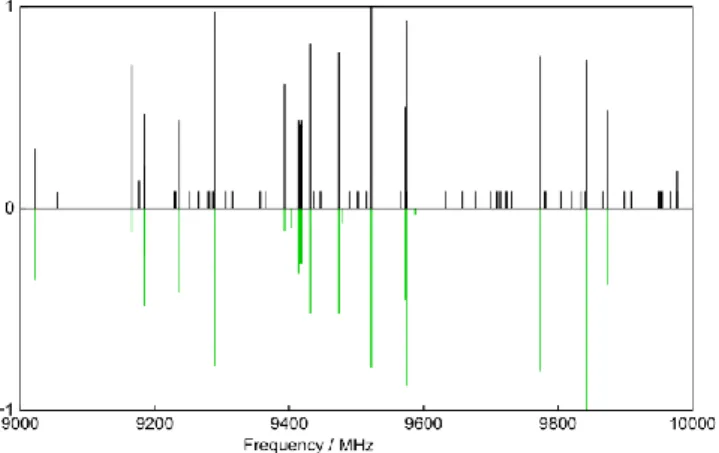
At the beginning of the experiment, a broadband scan in the frequency range from 8 to 11 GHz was carried out, where about 20 intense lines and many much weaker lines are observed. A portion of the survey spectrum is shown in Figure 2. The dipole moment components calculated with the MP2 method are µa = 4.88, µb = 3.14, and µc = 0.0 D. Therefore, strong a- and b-type, but not c-type transitions were expected in the rotational spectrum. We started our assignment with a-type J’’ = 5 ← J’ = 4 transitions, because - from our experiences - the frequencies predicted for a-type lines are often quite reliable. The assignment was straightforward. Afterwards, b-type lines were also assigned and all 20 strong lines found in the scan could be fitted with the program XIAM [23] using a semi-rigid rotor model where the Hamiltonian only consists of rotational and quartic centrifugal distortion terms of Watson’s S reduced Hamiltonian.
Figure 2. A portion of the broadband scan of coumarin. The frequencies are
given in MHz; the intensity in arbitrary unit. The upper trace shows the experimental spectrum with all intense lines belonging to the parent species. For the weaker transitions arising from the nine 13
C isotopologues, only the line positions are indicated as satellite bands in black with constant intensity. The calculated spectrum of the main isotopologue is given in maibowle green on the lower trace.
With the prediction refined from this fit, further transitions out of the scan region could be identified and measured directly. A typical spectrum is presented in Figure 3. The final fit, given as
12C in Table 1, contains 162 lines with J 12 and K
a 7 and shows an excellent standard deviation of 1.3 kHz. A list of all measured lines can be found in Table S-2 in the Supporting Information.
After the main isotopic species was assigned, many weak lines remained in the spectrum. We assumed that they belong to the spectra of the nine 13C isotopologues of coumarin. The
Cartesian coordinates of the optimized structure in Figure 1 were taken, and the mass of the individual carbon atoms was adjusted to calculate the rotational constants of all 13C
isotopologues. The predicted 13C-frequencies were
correctedwith the differences between the frequencies predicted with the Be constants obtained at the MP2/6-311++G(d,p) level and the experimental frequencies of the parent species, leading to straightforward assignments of all 13C isotopologues.
Transitions with J 7 and Ka 3 were measured. Due to the
Table 1. Spectroscopic parameters of the parent species, nine 13C-isotopologues, and two 18O-isotopologues of coumarin (for atom numbering, see Figure 1).
Par.[a] Unit 12C 13C(1) 13C(2) 13C(3) 13C(4) 13C(5) Calc.[b]
A MHz 2830.370692(65) 2813.97656(22) 2796.09675(75) 2825.65025(21) 2819.79587(39) 2805.77814(90) 2811.528 B MHz 885.581280(30) 875.01639(12) 882.926957(42) 885.492575(96) 885.157599(76) 880.200278(50) 875.068 C MHz 674.795663(28) 667.72513(12) 671.296091(25) 674.475742(96) 673.947569(48) 670.272915(30) 667.675 DJ kHz 0.00893(13) 0.00826 DJK kHz 0.02296(53) 0.0173(56) 0.0174(45) 0.0174(51) 0.0249(98) 0.0140(54) 0.02141 DK kHz 0.1687(24) 0.1627 d1 kHz 0.002443(73) 0.00242(74) 0.00407(68) 0.002374 d2 kHz 0.000432(30) 0.000406 [c] kHz 1.3 0.7 0.6 0.7 1.2 0.7 N[d] 162 14 13 14 14 13 13C(6) 13C(7) 13C(8) 13C(9) 18O(10) 18O(11) A MHz 2828.61467(58) 2784.61352(67) 2807.11546(18) 2829.70926(24) 2796.76921(22) 2814.8637(80) B MHz 872.67458(27) 885.03645(51) 880.276698(93) 878.78450(13) 882.555582(10) 855.26121(51) C MHz 667.17917(32) 671.84377(55) 670.394192(94) 670.80478(13) 671.123344(7) 656.18790(24) DJ kHz 0.0117(33) 0.0146(43) DJK kHz 0.0243(45) 0.0139(63) d1 kHz -0.0126(24) -0.00230(60) -0.00285(83) [c] kHz 1.8 2.2 0.6 0.8 0.1 3.5 N[d] 14 12 14 13 6 7
[a] All parameters refer to the principal axis system. Watson’s S reduction in Ir
representation was used. Standard errors in parentheses are in the unit of the least significant digits. For all 13
C and 18
O-isotopologues, the values of centrifugal distortion constants which are not fitted are fixed to those of the parent species. [b]
Ground state rotational constants and centrifugal distortion constants from anharmonic frequency calculations at the MP2/6-31G(d,p) level of theory. [c]
Standard deviation of the fit. [d]
3
Figure 3. A typical 𝐽′′𝐾𝑎′′𝐾 𝑐 ′′← 𝐽′𝐾 𝑎 ′𝐾𝑐′ rotational transition of coumarin. The line
width is 14 kHz (full width at half height). The doublet arises from the Doppler effect; the mean frequency is 9575.3680 MHz. For this spectrum, 1000 free-induction decays were co-added and averaged.
small number of observable lines, only one or two quartic centrifugal distortion constants could be determined while the values of the other distortion constants were fixed to those of the parent species. The spectroscopic parameters of the 13C
isotopologues are summarized in Table 1 along with those of the parent species; the frequency lists are given in Table S-3 in the Supporting Information.
To complete the heavy atom structure, we also searched for the two 18O-isotopologues. Because of the low natural
abundances of 0.2%, transitions of these species were not visible in the broadband scan given in Figure 2 where only 200 co-added decays were taken per each scan step of 0.25 MHz. Therefore, scans of ±2 MHz around the corrected frequencies of three a-type transitions 606 505, 615 514, and 707 606 were
performed by steps of 0.2 MHz with 10000 co-added decays at each step. In total, 6 lines could be found for the 18O(10) and 7
lines for the 18O(11) species. In the fits, all quartic centrifugal
distortion constants were fixed to the values of the parent species.
3.2. Structure determinations
The rotational constants of the parent species as well as those of the 13C and 18O-isotopologues given in Table 1 were
input in the programs KRA and EVALavailable at the PROSPE website [24] to determine the heavy atom substitution rs structure of coumarin using Kraitchman’s equations [25]. Since the atom coordinates appear squared in these equations, their signs are ambiguous and were taken from the optimized geometry. Costain’s rule was used to determine the uncertainties [26]. The atom coordinates output by the program KRA are given in Table 2; the bond lengths and bond angles obtained from the program EVAL are presented in Table 3 and Figure 4. For the EVAL input, all values of the c-coordinates were set to zero.
Though the substitution rs structure is quite often reported in the literature, it is known that this structure determination is not reliable for large molecules such as coumarin exhibiting one or more atoms near one of the principal axes and Costain's rule is over-optimistic [27]. For this reason, the semi-experimental equilibrium reSE structure might be a better alternative. The
Table 2. Experimental atom positions from the substitution rs and
semi-experimental equilibrium reSE structures of coumarin. Due to planarity, all
c-coordinates were set to zero. The equilibrium atom positions re are optimized at
the MP2/6-31G(d,p) level of theory. Standard errors in parentheses are in the unit of the least significant digits.
rs rseSE a/Å b/Å a/Å b/Å C(1) 2.62603(57) –1.0306(15) 2.62084(57) –1.0299(15) C(2) 1.3084(12) –1.4853(11) 1.3114(12) –1.4832(11) C(3) 0.2392(63) –0.5471(28) 0.2690(56) –0.5512(28) C(4) 0.5230(29) 0.8199(19) 0.5292(29) 0.8211(19) C(5) 1.86732(80) 1.2587(12) 1.86656(80) 1.2578(12) C(6) 2.90977(52) 0.3362(45) 2.90311(52) 0.3402(45) C(7) –0.5937(26) 1.71746(88) –0.6009(26) 1.71428(88) C(8) –1.85403(81) 1.2235(12) –1.84860(81) 1.2224(12) C(9) –2.10449(71) –0.2061(73) –2.10317(71) –0.2182(69) O(10) –0.99158(16) –1.0438(15) –0.9969(16) –1.0411(15) O(11) –3.19574(47) –0.7276(21) –3.19019(47) –0.7247(21) re r0eSE C(1) 2.63179 –1.03739 –2.6210(47) –1.030(13) C(2) 1.31644 –1.48807 –1.3119(96) –1.4834(87) C(3) 0.28051 –0.55162 –0.276(13) –0.551(22) C(4) 0.54065 0.82802 –0.532(18) 0.821(16) C(5) 1.87911 1.26021 –1.8668(67) 1.258(11) C(6) 2.91574 0.33681 –2.9032(43) 0.342(20) C(7) –0.59127 1.71955 0.597(19) 1.7149(75) C(8) –1.85094 1.22513 1.8483(65) 1.223(11) C(9) –2.11872 –0.21095 2.1029(64) –0.223(23) O(10) –1.00008 –1.04683 0.9965(65) –1.0416(66) O(11) –3.21970 –0.72626 3.1898(22) –0.7236(94) H(12) 1.06981 –2.54115 –1.069(30) –2.531(68) H(13) 3.44088 –1.75623 –3.427(56) –1.743(44) H(14) 3.94306 0.67613 –3.923(69) 0.683(30) H(15) 2.08683 2.32383 –2.068(22) 2.315(68) H(16) –0.41946 2.78996 0.424(43) 2.777(68) H(17) –2.72393 1.86131 2.718(58) 1.856(40)
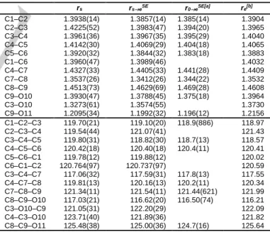
Table 3. Bond lengths (in Å) and bond angles (in degrees) of coumarin
deduced from the substitution rs, semi-experimental equilibrium reSE, and re
equilibrium structures. Standard errors in parentheses are in the unit of the least significant digits.
rs rseSE r0eSE[a] re[b] C1–C2 1.3938(14) 1.3857(14) 1.385(14) 1.3904 C2–C3 1.4225(52) 1.3983(47) 1.394(20) 1.3965 C3–C4 1.3961(36) 1.3967(35) 1.395(29) 1.4040 C4–C5 1.4142(30) 1.4069(29) 1.404(18) 1.4065 C5–C6 1.3920(32) 1.3844(32) 1.383(18) 1.3883 C1–C6 1.3960(47) 1.3989(46) 1.4032 C4–C7 1.4327(33) 1.4405(33) 1.441(28) 1.4409 C7–C8 1.3537(26) 1.3412(26) 1.344(22) 1.3532 C8–C9 1.4513(73) 1.4629(69) 1.469(28) 1.4608 C9–O10 1.3930(47) 1.3788(45) 1.375(18) 1.3964 C3–O10 1.3273(61) 1.3574(55) 1.3730 C9–O11 1.2095(34) 1.1992(32) 1.196(12) 1.2156 C1–C2–C3 119.70(21) 119.10(20) 118.9(886) 118.97 C2–C3–C4 119.54(44) 121.07(41) 121.43 C3–C4–C5 119.80(31) 118.82(30) 118.7(13) 118.57 C4–C5–C6 120.42(18) 120.40(18) 120.4(11) 120.41 C5–C6–C1 119.78(12) 119.88(12) 120.02 C6–C1–C2 120.764(97) 120.737(97) 120.59 C3–C4–C7 117.06(32) 117.59(31) 117.8(13) 117.55 C4–C7–C8 119.81(13) 120.16(13) 120.2(11) 120.34 C7–C8–C9 121.34(11) 121.54(11) 121.44(621) 121.99 C8–C9–O10 117.03(21) 116.62(20) 116.50(74) 116.21 C3–O10–C9 121.05(31) 122.20(29) 122.09 C4–C3–O10 123.71(40) 121.89(36) 121.82 C8–C9–O11 125.48(38) 125.00(36) 124.7(16) 125.64
[a] All hydrogen atom’s bond lengths were fitted to be 1.076(74) Å but
constrained to be the same. [b] Geometry optimized at the MP2/6-31G(d,p)
level of theory.
determination is shown i.a. in Refs. [20,27-32]. For a comparison with the rs structure, we first determined the so-called rseSE structure, where the experimental ground state rotational constants A0, B0, and C0 of the parent species and all
isotopologues given in Table 1 are rovibrationally corrected from the ab initio cubic force field at the MP2/6-31G(d,p) level of
4
theory before inputting them in the program KRA [24] (for details,see Table S-4 in the Supporting Information). Nevertheless,
rseSE is a partial structure determination since only spectra of heavy atoms could be measured and the locations of hydrogen atoms remained unknown. However, hydrogen coordinates predicted by quantum chemistry can be fitted together with the corrected A0, B0, and C0 rotational constants using the program
STRFIT [33], yielding the r0eSE structure. In the fit, the hydrogen atom locations were varied, but all CH bond lengths are constrained to be the same. The atom coordinates, bond lengths, and bond angles of both reSE structures are also given in Tables 2 and 3.
Figure 4. Experimental bond lengths (blue, in Å) and bond angles (red, in
degrees) of coumarin from the semi-experimental equilibrium rseSE structure.
Bond lengths from the equilibrium re structure calculated at the
MP2/6-31G(d,p) level of theory (purple, in Å) are also given for comparison. Standard errors in parentheses are in the unit of the least significant digit.
4. Discussion
4.1. Molecular geometry
The well-established structure of benzene determined by X-ray diffraction shows the same bond length of 1.39 Å between six carbon atoms, which is shorter than a C–C single bond of about 1.47 Å but greater than a C–C double bond of 1.35 Å. This constant, intermediate C–C distance in benzene implies a perfect electron delocalization where the electrons are distributed equally within the ring. Examinations of other aromatic compounds related to benzene, e.g. naphthalene, anthracene, and phenanthrene, disclose that for example in naphthalene, four bonds are shorter (1.37 Å) and have about two times greater double bond character than the other six bonds (1.42 Å) [34]. Obviously, the electron delocalization is perturbed by the less symmetric structure compared to that of benzene. In coumarin, the perturbations of bond lengths in benzene are even more pronounced due to the insertion of the lactone moiety into the ring fused with benzene, which breaks the perfect delocalization of benzene due to angle strains (known as the Mills-Nixon effect [35]). While the distance between the carbon atom in the benzene part of coumarin are essentially equal to the value of 1.39 Å reported for solid benzene (see Figure 4 for atom numbering and values), the electron delocalization is significantly perturbed in the other half part containing the lactone group, and is clearly reflected in the bond lengths. The distance of 1.341(3) Å between C(7) and C(8) as well as 1.357(6) Å between C(3) and O(10) implies that both C7–C8 and C9–O10 have a great double bond character. The
C9–O10 bond length of 1.379(5) Å is similar to the values of the C–C bond lengths in the benzene part. However, it is greater than the value of 1.35 Å found for the (O=)C–O bond of s-cis-methacrylic acid (MAA) and s-cis-methyl methacrylate (MMA) [32], marked in blue in Figure 5. The C8–C9 bond length of 1.4678 Å reflects slightly less single bond character than the comparable (O=)C–C bond of MAA and MMA (1.48 Å), shown in red in Figure 5. These differences can be also explained by angle strains at the oxygen atom arising from ring fusion, which does not affect much the length of the C7–C8 bond (marked in green in Figure 5, 1.34 Å in coumarin, MAA, and MMA). Finally, the C9–O10 bond is also not perturbed and has the same length as that found for MAA and MMA, as well as that of 2-thiophenecarboxaldehyde (2TPC) [28] (1.20 Å in all cases). We note that all values of MAA, MMA, and 2TPC also refer to the
reSE structure, and thus ensure a reasonable comparison without influence of vibrations.
Figure 5. Comparison of some bond lengths of coumarin (1) with those of the
open chain molecules s-cis-methacrylic acid (2) and s-cis-methyl methacrylate (3) (see text).
4.2. Inertial defect
With all atoms located on the ab-plane, we expected an inertial defect very close to zero, as observed for e.g. pyrrole (0.017 uÅ2) [36], oxazole (0.056 uÅ2) [37], 2TPC (0.008 uÅ2) [28], and
fluorobenzene (0.033 uÅ2), but surprisingly found
c = Ic – Ia – Ib = –0.294 uÅ2. Such negative, large values have been observed
for many molecules related to coumarin such as the PAHs [3] and PANHs [4] mentioned in Section 2, as well as 9-cyanoanthracene [38], quinolone [19], and isoquinoline [19], which could be traced back to significant out-of-plane vibrations of the rings [39]. The small, positive inertial defect is well-known as a judgement for the planarity of small molecules. The abnormally large, negative inertial defect due to out-of-plane ring motions has been found in a considerable number of molecules containing aromatic rings [40].
4.3. Theory versus experiment
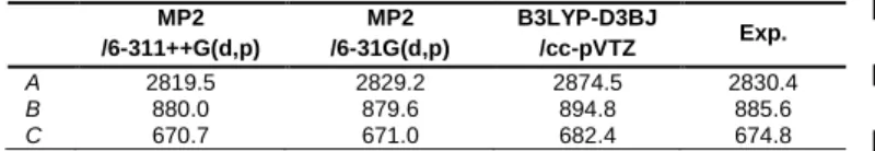
The equilibrium rotational constants Be calculated at the MP2/6-31G(d,p) level of theory are in remarkable agreement with the experimental results, as shown in Table 4. Though comparing predicted Be constants with experimental B0 constants is not physically meaningful, it shows that the MP2/6-31G(d,p) level offers cost-efficient calculations with sufficient accuracy for spectral assignment purposes. The MP2/6-311++G(d,p) is a higher level than MP2/6-31G(d,p) and therefore, purely from theoretical perspective, more accurate. However, the errors of the MP2/6-31G(d,p) level compensate with the missing Be B0 corrections in a way, that the Be calculated rotational constants accidentally match the B0 experimental constants. Therefore, this level is useful for assigning microwave spectra of aromatic molecules like coumarin.
5
Table 4. Equilibrium rotational constants Ae, Be, Ce (in MHz), vibrational
ground state rotational constants A0, B0, C0 (in MHz) of coumarin calculated at
different levels of theory.
MP2 /6-311++G(d,p) MP2 /6-31G(d,p) B3LYP-D3BJ /cc-pVTZ Exp. A 2819.5 2829.2 2874.5 2830.4 B 880.0 879.6 894.8 885.6 C 670.7 671.0 682.4 674.8
5. Conclusions
The interplay between high-resolution pulsed jet microwave spectroscopy and quantum chemistry yields accurate molecular parameters of coumarin in the gas phase. The spectra of all singly-substituted heavy atom isotopologues were assigned, enabling the determination of the substitution rs and semi-experimental equilibrium reSE structures. The resulting bond lengths and bond angles were used to evaluate the perturbation in the electron delocalization within the aromatic ring. The large and negative value found for the inertial defect of coumarin could be explained by out-of-plane motions of the ring, as observed for other related molecules. Comparison of the experimental and calculated rotational constants has shown that error compensations at the MP2/6-31G(d,p) level result in accidentally good values for spectral assignment purposes.
Experimental Section
The rotational spectra were measured with a coaxially oriented beam-resonator arrangement (COBRA) spectrometer operating in the frequency range from 4 to 26.5 GHz. Details of the spectrometer were described elsewhere [41] already. Coumarin was purchased from Merck Schuhard, Hohenbrunn, Germany, with a stated purity of over 98 % and used without further purification. For the measurements, the solid substance coumarin was placed onto some glass wool which was put near the expansion nozzle inside the soleniod valve and heated to 75°C. Helium at a pressure of approximately 200 kPa flows over the sample slowly to saturate. The helium-coumarin mixture was expanded through the pulsed nozzle into the cavity.
Acknowledgements
This work was supported by the Agence Nationale de la Recherche ANR (project ID ANR-18-CE29-0011). J.-U.G. thanks the Deutsche Forschungsgemeinschaft (DFG) and the Land Niedersachsen for funds.
Keywords: microwave spectroscopy • rotational spectroscopy •
coumarin • isotopologue • structural determination
[1] Y.-H. Wang, B. Avula, N. P. D. Nanayakkara, J. Zhao, I. A. Khan, J.
Agric. Food Chem. 2013, 61, 4470–4476.
[2] J. Daphi-Weber, H. Raddatz, R. Müller, Untersuchung von Riechstoffen – Kontrollierte Düfte, Gesellschaft Deutscher Chemiker, 2010, ISBN 978-3-936028-64-5.
[3] S. Thorwirth, P. Theulé, C. A. Gottlieb, M. C. McCarthy, P. Thaddeus,
Astrophys. J. 2007, 662, 1309–1314.
[4] D. McNaughton, P. D. Godfrey, R. D. Brown, S. Thorwirth, J.-U. Grabow, Astrophys. J. 2008, 678, 309–315.
[5] E. G. Schnitzler, B. L. M. Zenchyzen, W. Jäger, Astrophys. J. 2015, 805, 141.
[6] T. H. Dunning Jr., J. Chem. Phys. 1989, 90, 1007.
[7] R. Ditchfield, W. J. Hehre, J. A. Pople, J. Chem. Phys. 1971, 54, 724– 728.
[8] R. Hakiri, N. Derbel, H. V. L. Nguyen, H. Mouhib, Phys. Chem. Chem.
Phys. 2018, 20, 25577–25582.
[9] V. Van, W. Stahl, H. V. L. Nguyen, ChemPhysChem 2016, 17, 3223– 3228.
[10] V. Van, J. Bruckhuisen, W. Stahl, V. Ilyushin, H. V. L. Nguyen, J. Mol.
Spectrosc. 2018, 343, 121–125.
[11] L. Ferres, H. Mouhib, W. Stahl, M. Schwell, H. V. L. Nguyen, J. Mol.
Spectrosc. 2017, 337, 59–64.
[12] J. C. López , V. Cortijo , S. Blanco, J. L. Alonso, Phys. Chem. Chem.
Phys. 2007, 9, 4521–4527.
[13] A. R. Conrad, N. Z. Barefoot, M. J. Tubergen, Phys. Chem. Chem.
Phys. 2010, 12, 8350–8356.
[14] C. Calabrese, A. Maris, L. Evangelisti, W. Caminati, S. Melandri,
ChemPhysChem 2013, 14, 1943–1950.
[15] Y. Jin, T. Lu, Q. Gou, G. Feng, J. Mol. Struct. 2020, 1205, 127632. [16] M. J. Frisch, G. W. Trucks, H. B. Schlegel, G. E. Scuseria, M. A. Robb,
J. R. Cheeseman, G. Scalmani, V. Barone, G. A. Petersson, H. Nakatsuji, X. Li, M. Caricato, A. V. Marenich, J. Bloino, B. G. Janesko, R. Gomperts, B. Mennucci, H. P. Hratchian, J. V. Ortiz, A. F. Izmaylov, J. L. Sonnenberg, D. Williams-Young, F. Ding, F. Lipparini, F. Egidi, J. Goings, B. Peng, A. Petrone, T. Henderson, D. Ranasinghe, V. G. Zakrzewski, J. Gao, N. Rega, G. Zheng, W. Liang, M. Hada, M. Ehara, K. Toyota, R. Fukuda, J. Hasegawa, M. Ishida, T. Nakajima, Y. Honda, O. Kitao, H. Nakai, T. Vreven, K. Throssell, J.A. Montgomery, Jr., J. E. Peralta, F. Ogliaro, M. J. Bearpark, J. J. Heyd, E. N. Brothers, K. N. Kudin, V. N. Staroverov, T. A. Keith, R. Kobayashi, J. Normand, K. Raghavachari, A. P. Rendell, J. C. Burant, S. S. Iyengar, J. Tomasi, M. Cossi, J. M. Millam, M. Klene, C. Adamo, R. Cammi, J. W. Ochterski, R. L. Martin, K. Morokuma, O. Farkas, J. B. Foresman, D. J. Fox, Gaussian 16, Revision B.01, Inc., Wallingford CT, 2016.
[17] S. Grimme, J. Antony, S. Ehrlich, H. Krieg, J. Chem. Phys. 2010, 132, 154104.
[18] S. Grimme, S. Ehrlich, L. Goerig, J. Comp. Chem. 2011, 32, 1456– 1465.
[19] Z. Kisiel, O. Desyatnyk, L. Pszczółkowski, S. B. Charnley, P. Ehrenfreund, J. Mol. Spectrosc. 2003, 217, 115–122.
[20] H. V. L. Nguyen, J. Mol. Struct. 2020, 1208, 127909.
[21] L. Ferres, W. Stahl, H. V. L. Nguyen, Mol. Phys. 2016, 114, 2788–2793. [22] L. Ferres, H. Mouhib, W. Stahl, H. V. L. Nguyen, ChemPhysChem 2017,
18, 1855–1859.
[23] H. Hartwig, H. Dreizler, Z. Naturforsch. 1996, 51a, 923–932. [24] Z. Kisiel, PROSPE-Programs for ROtational SPEctroscopy. <http://info.ifpan.edu.pl/∼kisiel/prospe.htm>.
[25] J. Kraitchman, Am. J. Phys. 1953, 21, 17–24.
[26] C. C. Costain, Trans. Am. Crystallogr. Assoc. 1966, 2, 157–164. [27] M. Juanes, N. Vogt, J. Demaison, I. León, A. Lesarri, H. D. Rudolph,
Phys. Chem. Chem. Phys. 2017,19, 29162–29169
[28] R. Hakiri, N. Derbel, W. C. Bailey, H. V. L. Nguyen, H. Mouhib, Mol.
Phys. 2020. DOI: 10.1080/00268976.2020.1728406
[29] J. Demaison, M. K. Jahn, E. J. Cocinero, A. Lesarri, J.-U. Grabow, J.-C. Guillemin, H. D. Rudolph, J. Phys. Chem. A 2013, 117, 2278–2284. [30] J. Demaison, A. G. Császár, L. D. Margulès, H. D. Rudolph, J. Phys.
Chem. A 2011, 115, 14078–14091.
[31] B. J. Esselman, B. K. Amberger, J. D. Shutter, M. A. Daane, J. F. Stanton, R. C. Woods, R. J. McMahon, J. Chem. Phys. 2013, 139, 224304.
[32] S. Herbers, P. Kraus, J.-U. Grabow, J. Chem. Phys. 2019, 150, 144308. [33] Z. Kisiel, J. Mol. Spectrosc. 2003, 218, 58−67.
[34] D. W. J. Cruickshank, R. A. Sparks, Proc. R. Soc. Lond. A 1960, 258 [35] A. Stanger, J. Am. Chem. Soc. 1991, 113, 8277–8280
[36] L. Nygaard, J. T. Nielsen, J. Kirchheiner, G. Maltesen, J. R. Andersen, G. O. Sorensen, J. Mol. Struct. 1969, 3, 491–506.
[37] A. Kumar, J. Sheridan, O. L. Stiefvater, Z. Naturforsch. 1978, 33a, 145– 152.
6
[38] Y. Ohshima, R. Kanya, Y. Sumiyoshi, Y. Endo, J. Mol. Spectrosc. 2004,223, 148–151.
[39] M. K. Jahn, J.-U. Grabow, M. J. Travers, D. Wachsmuth, P. D. Godfrey, D. McNaughton, Phys. Chem. Chem. Phys. 2017, 19, 8970–8976. [40] T. Oka, J. Mol. Struct. 1995, 352–353, 225–233.
[41] J.-U. Grabow, W. Stahl, H. Dreizler, Rev. Sci. Instrum. 1996, 67, 4072– 4084
7
FULL PAPER
The interplay of microwave rotational spectroscopy and quantum chemistry leads to highly accurate geometry parameters of coumarin. Perturbations of the electron delocalization are reflected in the bond lengths and bond angles. The large, negative inertial defect indicates pronounced out-of-plane motions of the ring.
Ha Vinh Lam Nguyen*, Jens-Uwe Grabow
Page No. – Page No.
The scent of maibowle – electron localization in coumarin from its microwave-determined structure