*Corresponding author: Georgette B. Salieb-Beugelaar, Nanomedicine Research Lab CLINAM, University Hospital Basel, Bernoullistrasse 20, Basel, CH-4056, Switzerland; and The European Foundation for Clinical Nanomedicine (CLINAM), Alemannengasse 12, CH-4016 Basel, Switzerland, E-mail: [email protected] Bei Zhang: Nanomedicine Research Lab CLINAM, University Hospital Basel, Bernoullistrasse 20, Basel, CH-4056, Switzerland
Maurice M. Nigo: Nanomedicine Research Lab CLINAM, University Hospital Basel, Bernoullistrasse 20, Basel, CH-4056, Switzerland; and ISTM-Nyankunde, B.P. 55, Bunia, Democratic Republic Congo Sieghard Frischmann: MAST Diagnostica GmbH, Feldstraße 20, DE 23858 Reinfeld, Germany
Patrick R. Hunziker: Intensive Care Clinic, University Hospital Basel, Petersgraben 4, CH-4031 Basel, Switzerland; Nanomedicine Research Lab CLINAM, University Hospital Basel, Bernoullistrasse 20, Basel, CH-4056, Switzerland; and The European Foundation for Clinical Nanomedicine (CLINAM), Alemannengasse 12, CH-4016 Basel, Switzerland
Critical Review
Georgette B. Salieb-Beugelaar*, Bei Zhang, Maurice M. Nigo, Sieghard Frischmann
and Patrick R. Hunziker
Improving diagnosis of pneumococcal
disease by multiparameter testing and
micro/nanotechnologies
DOI 10.1515/ejnm-2016-0012
Received April 25, 2016; accepted May 12, 2016; previously published online June 3, 2016
Abstract: The diagnosis and management of
pneumococ-cal disease remains challenging, in particular in children who often are asymptomatic carriers, and in low-income countries with a high morbidity and mortality from febrile illnesses where the broad range of bacterial, viral and par-asitic cases are in contrast to limited, diagnostic resources. Integration of multiple markers into a single, rapid test is desirable in such situations. Likewise, the development of multiparameter tests for relevant arrays of pathogens is important to avoid overtreatment of febrile syndromes with antibiotics. Miniaturization of tests through use of micro- and nanotechnologies combines several advan-tages: miniaturization reduces sample requirements, reduces the use of consumables and reagents leading to a reduction in costs, facilitates parallelization, ena-bles point-of-care use of diagnostic equipment and even reduces the amount of potentially infectious disposables,
characteristics that are highly desirable in most health-care settings. This critical review emphasizes our vision on the importance of multiparametric testing for diagnos-ing pneumococcal infections in patients with fever and examines recent relevant developments in micro/nano-technologies to achieve this goal.
Keywords: immunoassays; microfluidics;
nanodiagnos-tics; nanotechnology; pneumococcal disease; point-of-care; rapid tests.
Introduction
Streptococcus pneumoniae (pneumococcus) is a
Gram-positive bacterium that is a major cause of infectious diseases including pneumonia, empyema, sepsis, otitis media and meningitis. Pneumococcal infections occur more frequently in the elderly or the very young. Infec-tion may point to an underlying compromise of the host’s immune system but infection can also result from an overwhelming inoculum or a particularly virulent strain. Asymptomatic carriage in the nasopharynx occurs in children below the age of 10 years with a reported preva-lence of 30%–60% but is also found in 1%–10% of adults (1). In economically deprived populations and in devel-oping countries, these carriage rates are often higher (2). Here, the detection of the pathogen in the nasopharynx alone, is therefore not sufficient to diagnose a clini-cal infection. Thus, another parameter is required here to identify the pathogen which emphasizes the need of multiparameter tests.
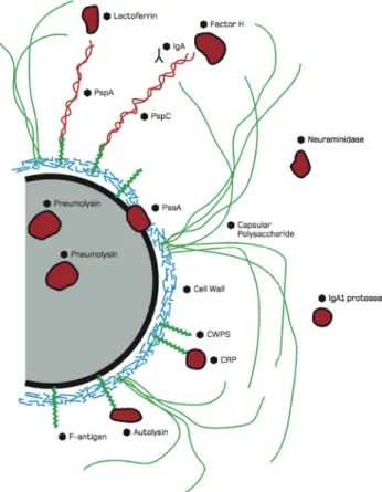
Streptococcus pneumoniae has several virulent factors
(Figure 1 and Table 1) and at least 93 different serotypes are identified (14) the prevalence of which changes over time and differs between geographic locations (15), thus adding some critical potential to the challenge of reliable diagno-sis using a single diagnostic assay. Diagnodiagno-sis is important
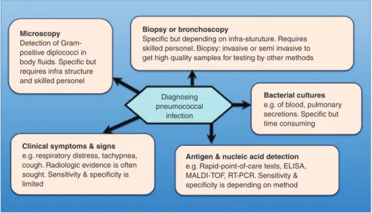
because untreated disease (e.g. pneumonia, sepsis, men-ingitis) may be lethal. Specific treatment is available and may be life-saving when applied early. Current diagnostic options are shown in Figure 2. A clinical syndrome com-patible with pneumococcal disease is typically worked up with radiographs and antigen testing in developed coun-tries, but in resource poor councoun-tries, diagnosis of pneu-monia, meningitis and sepsis is often based on clinical algorithms only (16). The resulting overuse of antibiotics is believed to contribute to the development of antibacterial drug resistance.
In this critical review we briefly discuss common diagnostic methods (detection of antigens, nucleic acids and serologic response) and illustrate these with relevant achievements (including recent) that were felt to be impor-tant. This will be followed by the timeline of testing in a clinical context. In the final part of this work, we present emerging diagnostics that use micro/nanotechnology such as micro/nanoparticles, microsensors, and microfluidic devices. Here, the focus is also on new techniques that might lead to a new era of diagnostics for pneumococcal infections. Figure 1: Schematic representation of S. pneumoniae and its virulent factors [with permission from textbook in Diagnosis, Serotyping, Virulence Factors and Enzyme-linked Immunosorbent Assay (ELISA) for Measuring Pneumococcal Antibodies.” (2nd version) Statens Serum Institut, Denmark (3)].
Original articles and reviews published from January 2000 to April 2016 were identified by keyword search in PubMed, ScienceDirect and Scopus using the keywords “pneumococcus”, “pneumococcal infection”, “pneumo-coccal antigens”, “nanotechnology” and following links in identified articles. Articles cited in the review were selected by the authors based on their perceived relevance for the target audience of this journal.
Microscopy and blood culture
The microscopic appearance and colony morphology is used in most clinical laboratories to identify S.
pneumo-niae. Under the microscope, the Gram-positive bacteria
is visible as slightly pointed cocci that are usually found in pairs, but also in short chains or as single bacteria. On blood agar plates, most serotypes of this microorganism form small round doughnut shaped colonies. Serotype 3 and 37 typically form large mucoid colonies (17).
Streptococcus pneumoniae is capable of producing
α-hemolysis on blood agar plates, which produces a green color. Other species as Streptococcus mitis and
Streptococ-cus oralis also have this characteristic and they are often
referred as the viridans group (18). Thus additional specific characteristics of S. pneumoniae are required to exclude most viridans species such as optochin susceptibility and catalase negativity (18, 19). Bile solubility might be used, however, some exceptions are known (20). Thus, identifying
S. pneumoniae on phenotype alone is not adequate enough.
Detecting antigens, nucleic acids
and serology
Antigens
Teichoic acids or cell wall polysaccharides
Teichoic acids, found in Gram-positive bacteria, are bac-terial polysaccharides of ribitol-, or glycerol-phosphate that are linked via phosphodiester bonds. These anionic glycopolymers, known as cell wall polysaccharide (CWPS), play a key role in antibiotic resistance (21). CWPS can be detected by the commercial Binax NOW (Alere, Global) rapid urinary antigen test, with a clinical test performance reported in a meta-analysis (22). The majority of included patients were adults and had suspected community- acquired pneumonia (CAP); data comparing the Binax NOW test against any reference test were analyzed. In
Table 1: Top, a description of important virulent factors of S. pneumoniae.
Virulent factor Molecular weight Description Type
Capsular polysaccharide (CPS) – Outermost layer of S. pneumonia ±200–400 nm thick; prevents
phagocytosis; 93 serotypes (4) Polysaccharide Theichoic acids: also called cell
wall polysaccharide (CWPS) – Common cell wall polysaccharide; tetrasaccharide units, joined together through ribitol phosphate diester and contains one or two phosphocholine subsituents (4)
Polysaccharide Lipoteichoic acid – Also called F-antigen; polysaccharide part (same as CWPS)
linked to diacylated glycerol via glucose residue (4) Polysaccharide Pneumolysin 53 kDa Pore-forming protein capable of causing lysis and activation of
complement (5) Protein
Pneumococcal surface protein A
(PspA) 67–99 kDa Bound to cell surface through CWPS/choline attached; inhibits opsonisation and phagocytosis through binding complement component C3 (6, 7)
Protein Pneumococcal surface protein C
(PspC) 75 kDa Bound to cell surface through CWPS/choline attached protein; mediates adherence to host by recognizing sialic acid on epithelial cells (8, 9)
Protein Pneumococcal surface adhesin A
(PsaA) 37 kDa Membrane bound; ABC-type transport protein complex; transports Mn2+, plays role in attachment to host cell and virulence (10)
Lipoprotein N-acetylmuramoyl-L-alanine
amidase (Autolysin) 36 kDa In cell envelope, degrades peptidoglycan in cell wall leading to lysis and release of pneumolysin. Is activated by bile (11) Enzyme Neuraminidase A and B [NanA and
NanB) 108 kDa (NanA) 75 kDa (NanB) Sialidase that cleaves the terminal sialic acids from glycoproteins, glycolipids and oligosaccharides on the cell surface and in the mucus (12)
Enzyme IgAI protease – Capable of cleaving human immunoglobulin A1 (IgA1) (13) Enzyme
O O O O O O O O O O
CWPS. Tetrasaccharide units. R=H or COCH3
OCH2CH2N(CH3)2 O H H H H H H H H H H H H H H H H H H H OH OH OH O O P P n O– O– H H NHR NHAc NHAc NHR CH3 H OH OH OH OH OH HO
Protein structure pneumolysin On the bottom (right), the structure formula of the tetrasaccharide units of CWPS and (left) a 3D protein structure of pneumolysin [image with permission from the RCSB PDB (www.rcsb.org) of PDB ID 2bk1. [Tilley SJ, Orlova EV, Gilbert RJ, Andrew PW, Saibil HR. “Structural basis of pore formation by the bacterial toxin pneumolysin.” Cell 2015;121:247–56].
children, a high rate of false positives due to the carrier status of pneumococci was found, rendering the test less useful in this setting. In 12 out of the 27 studies, the ref-erence standard was a combination of blood culture, sputum (culture or smear) and culture of another respira-tory sample (e.g. nasopharyngeal, transthoracic needle aspirate). Bivariate meta-analysis resulted in an overall sensitivity of 68.5% and a specificity of 84.2% for the Binax NOW test. The inclusion of all 27 selected studies with positive blood culture alone as reference test yielded a sen-sitivity of 74.0% and a specificity of 97.2%. Other investi-gators reported that false negative results may arise with low CWPS levels while false positive results may result due
to cross-reaction, for example shortly after an infection, in nasopharyngeal carriage or in the presence of S. mitis and/ or S. oralis species carrying closely related CWPS (23).
Capsular polysaccharides (CPS)
Capsular polysaccharides (CPS) form the outermost layer of the pneumococcal wall and their variability results in 93 known serotypes. CPS are detectable in the urine of patients and asymptomatic carriers during, after infection and even after vaccination. Antibodies against CPS are protective rendering CPS suitable as vaccines.
Microscopy Detection of Gram-positive diplococci in body fluids. Specific but requires infra structure and skilled personel
Biopsy or bronchoscopy
Specific but depending on infra-sturuture. Requires skilled personel. Biopsy: invasive or semi invasive to get high quality samples for testing by other methods
Clinical symptoms & signs e.g. respiratory distress, tachypnea, cough. Radiologic evidence is often sought. Sensitivity & specificity is limited
Antigen & nucleic acid detection e.g. Rapid-point-of-care tests, ELISA, MALDI-TOF, RT-PCR. Sensitivity & specificity is depending on method
Bacterial cultures e.g. of blood, pulmonary secretions. Specific but time consuming Diagnosing
pneumococcal infection
Figure 2: A schematic overview of the different possibilities to diagnose pneumococcal disease.
A multiplexed immunoassay was developed, employ-ing microspheres and based on the Luminex system, which permits detection of 13 different serotypes in urine with a limit of detection of 0.6–8.8 pg/mL (24). A clinical sensitivity and specificity of 97% and 100% was achieved when testing urine samples in the subset of patients with a positive blood culture for S. pneumoniae and an X-ray confirmed CAP. Pickering and Hill (25) present a protocol to measure the antibodies after vaccination with the 23 valent pneumococcal polysaccharide vaccine based on the Luminex xMAP (Luminex Corporation, Austin, TX, USA) microsphere-based liquid assay. Sensitivities and specificities are not included.
Serotyping pneumococci by CPS detection in urine or other fluids such as nasopharyngeal secretions is also possible with the latex agglutination test. A protocol for agglutination reagents with serotype specific antibod-ies from antisera on polystyrene latex particles is avail-able (26). This low-cost protocol is easy to use, includes quality control and allows storage for a year at 2–8°C, and is therefore suited also for resource-constrained settings.
Pneumolysin
The intracellular pore forming toxin pneumolysin is a 53 kDa conserved protein released upon LytA (autolysin) lysis that contributes to the invasiveness of the strain. Pneumolysin detection in sputum, urine, cerebrospinal fluid and blood indicates pneumococcal infection (27).
The value of pneumolysin detection in urine was investigated and compared to the Binax NOW test in adults and children in another investigation (28). The pneumoly-sin ELISA required 3 h procespneumoly-sing time. One hundred and eight patients with blood culture confirmed pneumococcal infections were tested. In adults, sensitivity and specificity were 56.6% and 92.2%, respectively. Pneumolysin concen-tration in urine decreased after the initiation of treatment. Notably, pneumolysin was not detected in children with only nasopharyngeal colonization, leading to enhanced specificity. In colonized children, sensitivity and speci-ficity of pneumolysin was 62.5% and 94.4%, respectively, while Binax NOW had a sensitivity and specificity of 87.5% and 27.8%, respectively. For non-colonized children, the sensitivity and specificity of the pneumolysin ELISA was 68.7% and 94.1%, respectively, while the Binax NOW test had a sensitivity of 93.7% and a specificity of 41.2% (28).
In another investigation, the Binax NOW test was used to detect pneumolysin in cerebrospinal fluid (CSF) and compared to culture and latex agglutination (29). 1173 CSF samples of five countries in Africa and Asia, collected from patients between 1 and 59 months old. From these patients, n = 69, were confirmed by culture on pneumococcal meningitis and the Binax NOW test was found to be positive in 98.6%. By using the test on culture positive samples for bacterial meningitis caused by other pathogens (n = 125), the test was negative in 99.3%. When only the latex agglutination tests and culture was used, pneumococci were detected from 7.4% in Asia to 15.6% in Africa, including the Binax Now test resulted in 16.2% in Africa (Nigeria) to 20% in Asia (Bangladesh).
These results suggested the underestimation of pneumo-cocci infections in the past.
Other pneumococcal antigens
Pneumococcal surface adhesin A is an immunogenic, 37 kDa surface lipoprotein expressed by all pneumococci. It is highly conserved among pneumococcal serotypes, and is also present in S. anginosus, S. mitis and S. oralis with a sequence similarity of 90%, 94% and 95% com-pared to S. pneumoniae serotype 6B (30), limiting its diag-nostic value.
Others investigated surface proteins are the pneumo-coccal surface protein A (PspA), family 1 and 2, and the pneumococcal surface protein C (PspC). PspC, present in 75% of pneumococcal strains, is related to PspA, but has a specific N-terminal region interfering with the comple-ment system through binding of the complecomple-ment factor H. The Hic protein, (factor H-binding inhibitor of comple-ment), is a PspC variant mainly found in serotype 3 (31). The homology between these proteins renders their spe-cific discrimination difficult and may limit their value as a diagnostic biomarker.
The triggering receptor expressed on myeloid cells (sTREM1), has been reported as a promising marker for infection (32), and has a decent correlation with blood culture positivity in a univariate meta-regression analysis of 13 studies. The pooled sensitivity and specificity was 84% and 77%, respectively, for the diagnosis of lower res-piratory tract infection with similar diagnostic accuracy for community-acquired infection and hospital acquired infection in subgroup analysis.
The search for new potential biomarkers by using mass spectroscopy-based proteomic analysis in African children provided two valuable candidates: lipocalin-2 and haptoglobin (33). Lipocalin-2 discriminated non-severe and non-bacterial from non-severe and bacterial pneu-monia and the authors hypothesize that the use of this marker, embedded in a point-of-care-test in combination with haptoglobin measurement, could distinguish severe pneumonia from malaria and viral lung infection.
Nucleic acids
Pneumococcal nucleic acids can be detected by amplifi-cation technologies. The polymerase chain reaction (PCR) amplifies a selected region of the target gene, but is chal-lenged by the homology of genes between species and the occurrence of false positive results in asymptomatic
carriers. Strain-specific primers focusing on non- homologous DNA regions may improve the specificity of amplification tests. An example of pitfalls of homology among bacterial species, is the pneumolysin gene ply as reported by Song et al (17). Samples originating from the lower respiratory tract of patients with pneumococcal disease were tested and yielded sensitivities between 68% and 100% and poor specificities. However, throat swabs of patients with community acquired pneumonia and from control subjects resulted in similar rates of test positivity of ~55%–58% for the ply gene (34). Another example is the
lytA gene that is present in pneumococci as well as in
strep-tococci, but varies less among streptococci. Others found that in ~2% of the investigated pneumococcal strains a mutation is present that is resulting in a negative PCR test (20). This test is still applicable in routine laboratories but needs confirmation in case of a negative result. To distin-guish between α-hemolytic streptococci and pneumococci the bile test might be used. The cell wall of pneumococci is bile soluble (and therefore resulting in lysis), where as all α-hemolytic streptococci are not. However, the 2% pneu-mococci carrying the mutation in the lytA gene are also bile insoluble and thus providing a false negative result.
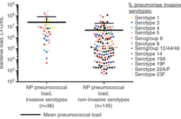
Thus, strain specific primers may be of high impor-tance for certain microorganisms and might improve the sensitivity and specificity of an amplification test. A high-throughput method for serotype specific quantifi-cation and molecular serotyping of pneumococci using a nanofluidics based RT-PCR system was developed (35). Primer pairs were designed such that over 50 serotypes are covered. Their performance is comparable to the conventional PCR-based assays. Recently, a novel quan-titative real-time PCR was developed to detect 40 major pneumococci serotypes worldwide directly in blood and nasopharyngeal specimens (36). This work presents newly designed primers and probes based on targets that were already described by Pai et al. (37). The most frequent serotypes found in Brazilian, French and South African (see Figure 3) samples were 14, 1 and 7A/F and 3 and 19F, respectively. In case both blood and nasopharynx samples were available, the serotype in blood was always present together with other serotypes in the nasopharynx. This highly sensitive assay was capable of detecting < 100 CFU/mL and opens the door to large-scale epidemiologi-cal studies of pneumococci. Other scientists (38) pub-lished a novel quantitative PCR assay for the detection of S. pneumoniae. The target is the competence regulator gene comX, which exists in duplicate copies in S.
pneu-moniae and not in other bacterial species. Validation of
the assay was done on DNA extracted from serum of 30 patients with blood cultures positive for S. pneumoniae
and 51 serum samples positive for other bacteria. The LytA quantitative PCR assay was used to compare. The clini-cal sensitivity was 47% for both assays and the diagnos-tic specificity was 98.2% and 100% for LytA and comX, respectively (38).
Serology
The detection of serotype specific antibodies (CPS) can be done by ELISA, the Neufeld Quellungs test and the latex agglutination test [see also Streptococcus
pneu-moniae Textbook, Statens Serum Institut, Copenhagen,
Denmark (3)].
Immune response against, for example, PspA, PspC and Hic are known, but titers do not sufficiently discrimi-nate patients from healthy controls, with the exception of PspA that shows a significant titer increase in the conva-lescence phase. Cross-reactivity between the antibodies for PspA and PspC are also reported (39, 40). Multiparam-eter testing is probably required for correct result interpre-tation in this situation.
A validation study for a multiplexed set of antigens causing respiratory tract disease was performed where fluorescent bead based multiplexed immunoassay quan-tifies IgG against a panel of microorganisms including
S. pneumoniae, Haemophilus influenzae and Moraxella catarrhalis. For each marker a singleplex measurement
was compared with the multiplex assay, and the pneu-mococci multiplex was compared with an ELISA of 22
109 108 107 106 105 104 103 Bacter
ial load, CFU/mL
102
Mean pneumococcal load NP pneumococcal load, invasive serotypes (n=36) NP pneumococcal load, non-invasive serotypes (n=145) S. pneumoniae invasive serotypes: Serotype 1 Serotype 3 Serotype 4 Serotype 8 Serotype 5 Serotype 14 Serotype 19A Serotype 19F Serotype 22A/F Serotype 23F Serogroup 6 Serogroup 12/44/46
Figure 3: Pneumococcal load distribution in nasopharyngeal samples from the South African cohort.
Each plot represents a sample and the mean bacterial concentra-tion for each group is indicated by the bold bars (Student’s t-test, p < 0.001). Invasive and non-invasive serotypes are compared upon presence in nasopharyngeal samples en whole blood. [Messaoudi et al. (36); http://dx.doi.org/10.1371/journal.pone.0151428.g002.]
pneumococcal antibodies containing serum samples that had been analyzed previously. The reproducibility, speci-ficity and correlation were validated for six pneumococ-cal antigens [Ply, PspA1, PspA2, choline binding protein A (CbpA), pneumococcal choline binding protein A (PcsA) and pneumococcal histidine triad protein D (PhtD)]. The assay had a specificity above 92% and some cross-reac-tivity between PspA1, PspA2 and CbpA was reported. The serum samples of 50 children were also examined and revealed a wide range of antibody concentrations and increases in samples of recovering patients (41).
Time line of testing
Applying a test in the right time slot of infection is critical for its clinical benefit and its interpretation. Blood culture testing is more sensitive early in infection, but becomes rapidly negative after treatment with antibiotic therapy. Recently, a strategy was proposed to identify various bac-teria including pneumococci within 6 h after signaling growth in blood culture (42). Directly from the positive blood cultures, rapid agglutination tests were performed and showed a very high predictive value. Such early diag-nosis allows faster treatment adjustment in serious bacte-rial infection. Negative tests were repeated after colonies grew for further 4–6 h.
DNA based detection of pneumococci in serum was found to be particularly sensitive when the disease was more severe than uncomplicated pneumonia, and claimed that such testing gives additional diagnostic ben-efits to blood cultures including: 1) better disease severity assessment, 2) antibiotic streamlining and 3) detection of invasive pneumococcal disease after initiation of antibiotics (43).
Others investigated the use of a urinary test after start-ing an empiric antibiotic treatment and concluded that this has a potential to guide the right choice of medical treatment of pneumococcal disease in adults at an earlier stage (44). From this we conclude that combining existing and new test modalities may lead to new improved diag-nostic strategies/management.
Towards multiparameter testing
Multiparameter testing includes the notion of determin-ing multiple parameters for a particular infectious agent, and also the concept of testing for multiple pathogens in a single test. An important technique here, which evolved
rapidly last decade, is the use of matrix-assisted laser desorption ionization time-of-flight (MALDI-TOF) mass spectrometry (MS) in the clinic. This technique enables the rapid detection of bacteria and fungi in < 1 h when starting with pure bacteria culture. The advantage is that there is no knowledge of the pathogen required in advance, however, the disadvantage is that a pure culture is required and with this sufficient equipped laboratory and trained personnel. Thus, for most resource poor coun-tries this is not an option. Fall et al. (45), presented the use of MALDI-TOF in Dakar (Senegal) where it was success-fully used to identify species causing infectious diseases in tropical Africa.
While testing in febrile disease in developed coun-tries often yields an abundance of parameters from a fully equipped central lab as mentioned above, multiparameter testing in a simple and inexpensive fashion is a pressing need in particular for these countries. Mortality and mor-bidity resulting from infectious diseases are still highest in sub-Saharan Africa and in low-income countries of south-east Asia. The relative frequency of fever episodes due to pneumococcal disease, malaria, typhoid fever, dengue, or chikungunya may change with age, with season, as a function of geography, and may be related to local disease outbreaks. Currently, blind treatment is associated with high costs and microbial resistance development, while systematic testing is economically unrealistic in many areas. The recent emergence of global viral threats includ-ing new influenza subtypes, dengue virus, Middle East respiratory syndrome coronavirus and Ebola add an addi-tional urgency to the development of rapid, bedside, low-cost tests that discriminate between multiple alternative causes of fever.
There is evidence that availability of multiple diag-nostic parameters may enhance the diagnosis of pneu-mococal disease. The combination of a positive urinary Binax Now antigen test combined with measurement of CRP and PCT in plasma was evaluated in children ( < 6 years) (46). Diagnostic accuracy for predicting pneu-mococcal CAP (without positive urine test) for PCT alone (cut-off ≥ 1.5 ng/mL) was limited, having a sensitivity of 94.4% and a specificity of 52.6% whereas for CRP (cut-off ≥ 100 mg/L) the sensitivity was 91.9% and the specific-ity 60.5%. In combination with a positive urine test (same cut-offs), sensitivity and specificity were 65.5% and 85.7% for PCT, and 65.5% and 88.6% for CRP, respectively. Thus, in case of an emergency consultation such a combination is a useful tool to predict presumed pneumococcal CAP. Applied to examine pleural fluid samples of children, the Binax NOW had a sensitivity of 71%–96% and a specific-ity of 71%–100% for pneumococcal empyema (47–50).
Recent publications show progress in miniaturization of diagnostic technologies, paving the way to multiparam-eter testing by integration of multiple assays into a single device or test substrate [for further reading see (51, 52)].
Micro/nanotechnology diagnostic
innovations
Both micro- and nanotechnology has significantly con-tributed to new therapies, drugs and diagnostics during the last 5–10 years. In this paragraph, we only focus on new developments that might be used to improve or to develop new diagnostics for the detection of a pneumo-coccal infection.
The separation of bacteria from blood (or urine) may be done by, e.g. filtration, centrifugation and sedimenta-tion. However, the key limitation for S. pneumoniae is that the number in colony forming units (i.e. viable bacteria) in blood cultures is known to be low and high sensitiv-ity is improbable except when a large volume of blood is separated. The use of microfluidic devices may largely improve the capability to separate or concentrate bacte-ria prior the use of other diagnostic methods and tests. For example, the work of Park et al. (53), who developed a microfluidic device for the continuous dielectropho-retic separation and concentration of bacteria from crude biologic samples. They showed a 104 concentration of target cells and provided a separation efficiency of 94.3% in human CSF and 87.2% in blood for Escherichia coli. Ai et al. (54) used a microfluidic device and surface acoustic waves across the channels and demonstrated the separa-tion of E. coli bacteria from peripheral blood. Another pos-sibility is size-based cell sorting by the use of an ordered array of obstacles. This is also called deterministic lateral displacement. A combination of grooves and protrusions in a microfluidic device could successfully separate, 3D spherical particles, red blood cells (2D planar shaped) and rod shaped bacteria. The bacteria investigated in this work were: E. coli (rod-shaped or bacillus),
K. pneu-moniae (rod shaped), S. epidermidis (spherical or coccus)
and Pseudomonas aeruginosa (rod-like or coccobacillus) (55). The final example is the work of Kang et al. (56) who presented a method to selectively detect bacteria directly from milliliters of diluted blood in one step. Droplet encapsulation of diluted blood was done by using a micro-fluidic device and a high throughput 3D particle counter system. Specific DNA enzymes were used as sensor to the selected target E. coli. The fluorophore attached the DNA enzyme will be released from its quencher upon binding
and subsequently generate a fluorescent signal. The limit of detection was 1–10 CFU/mL. To conclude, the use of microfluidic techniques to separate and/or concentrate and detect bacteria is promising but needs to be further developed for challenging bacteria as S. pneumococci. In addition, the above mentioned is mainly applicable in developed countries.
Micro- and nanoparticles are nowadays widely used in lateral flow tests (rapid tests) that are in most cases based on sandwich immunoassays. Here, the presence of an antigen or antibody in a liquid sample (e.g. blood, serum, plasma, urine and CSF) leads to the immobiliza-tion of micro/nanoparticles and a visible signal. Exam-ples of used particles are gold nanoparticles, colored latex beads, magnetic particles, carbon nanoparticles. Important here is that all materials should retain their properties once conjugated to biomolecules and should be easily detectable at a concentration near the diagnostic threshold. [For further reading, see Refs. (57–59).] Lateral flow tests for the detection of pneumococcal already exist, however, an option would be to develop multiparameter tests as mentioned in the previous paragraph (46).
Wu and coworkers (60) presented a novel detection system based on quantum dots and microbeads that was developed to detect target DNA of pathogenic bacteria. On microbeads DNA hairpin structures are coupled. The composition of these hairpin oligonucleotides includes: a poly-T linker, a Tag sequence, the barcode region and an anti-Tag sequence. The barcode region is composed of four words each of four nucleotides (61) and has a unique design used for each species. Then, two types of probes are used: the internal probe, which has a complementary sequence to the barcode region and a reporter probe that has a sequence complementary to the anti-Tag region. Both probes have quantum dots in two different emission spectra. Legionella spp. is used as an example microorganism. The method exists of four steps, 1) the DNA hairpin structures on the microbeads will be denatured at 95°C, then 2) the provided linear oli-gonucleotide microbeads will hybridize with the dena-tured DNA samples (Legionella spp.) and will prevent the recovery of the hairpin structures, 3) the linear oligo-nucleotides will now be able to hybridize with both the internal as the reporter probes. When the target DNA is not present, the hairpin structure will recover. The fluo-rescence intensity of the reporter probes may be used to quantify the target DNA. The limit of detection was 0.1 ng of the extracted DNA and 10 CFU/test. This method is very useful for the multiplex detection of serotypes of
S. pneumoniae, however, will only be available for
well-equipped laboratories.
Veigas et al. (62) showed for the first time that by functionalizing a single Au-nanoprobe with multiple sequences, in the presence or absence of two pathogens could be determined in a single test. The selected targets were the conserved region of the Mycobacterium
tubercu-losis rpoB gene and Plasmodium 18S ribosomal RNA (18S
rRNA). After a multiplex PCR reaction, a colorimetric assay is performed by heating each sample up to 95°C (concen-tration 60 μg/mL) and cooled down to 25°C in the pres-ence of the functionalized Au-probes. The assay consist of a blank (sample without DNA), non-related control DNA and the samples. MgCl2 in a pre-determined concentration was added to the samples after 30 min for color develop-ment. After measurement by UV/Vis spectroscopy, the aggregation profiles for each Au-nanoprobe were ana-lyzed by comparing the absorption ratio 525 nm/600 nm. This method showed the capability of these particles. As also suggested by the authors, replacing the PCR by loop mediated isothermal amplification (LAMP) and the use of a cheap disposable platform (paper) as was already pre-sented in another investigation of the authors (63) will simplify the use of this method in resource poor areas.
LAMP assays are already developed and integrated on microfluidic platforms as described by Luo et al. (64). They showed that the differentiation of bacterial strains in res-piratory tract infections could also be done in multiplexed nucleic acid detection assays. The developed microfluidic device for LAMP was successfully used for the detection of
H. influenza, K. pneumonia and M. tuberculosis with limits
of detection of 17, 16 and 28 copies per μL, respectively. Methylene blue was used to electrochemically indicate the presence of double stranded DNA (65). This operation-ally simple and potentioperation-ally fast and cost-effective device analysed multiple genes qualitatively and quantitatively paves the way to multiplexed assays including other organisms as S. pneumoniae.
From particles, we move to silver nanorods and surface enhanced Raman scattering (SERS). Kotanen et al. (66) characterized and evaluated a handheld SERS system and showed that it can identify bacteria in pooled human sera. Different bacteria (Acinetobacter baumannii, P.
aer-uginosa, K. pneumoniar, E. coli and Staphylococcus aureus)
were individually inoculated into pooled human sera. Lysis filtration was performed to separate and isolate the bacteria. Silver nanorod substrates were incubated with the bacterial samples for 3 h at 60°C. The molecular finger-print of each species was determined by using partial least squared differential and principal component analysis. With the device, they could were able identify bacteria at the species level from both serum as culture samples with a limit of detection of 109 CFU/mL. Identification at strain
level was not obtained. Therefore, it was suggested that the spectro meter instrumentation needs to be at an equal level with the measurement sensitivity of the used nanoparti-cles which are known to be capable of sufficient enhance-ment factors to discriminate in between strains (67).
Even when we estimate that the actual production and consumable costs are too high for developing countries, we do see a potential for this portable device for future use in resource poor areas. Battery based hand-held spec-trometers with capacities to transfer the provided bacterial fingerprints by satellite based Internet to a central labora-tory is not unrealistic. For further reading about isolation and identification of bacteria by means of Raman spectros-copy, see Ref. (68).
On-chip microbial growth was combined with sensi-tive and specific surface plasmon resonance detection (SPR) of target antigen binding to an immobilized detec-tion antibody (69). The fluidic-less device has the capac-ity to culture-capture and measure bacteria as a result of coupling a microarray to a surface resonance imager. This enables the label free and real-time monitoring of bacte-ria with a series of immobilized ligands. Different types of viable cells growing and dividing on chip were successfully detected by SPR imaging with a limit of 2.8±19.6 CFU/mL in the initial sample. On-chip, S. enterica, S. pneumoniae and E. coli O157:H7 was assayed and quantitative deter-mination of the initial contaminating bacteria was pos-sible (validated with standard plate counting data). The method developed proved to produce within only a few hours (with liquid or solid samples), similar results to cul-turing methods (sensitivity and specificity) that were run in parallel and over several days. The developed biosensor has the advantage of being simply to use, needs minimal
supplementary handling and is promising for the future use in resource low areas. More details about SPR can be found in Ref. (70).
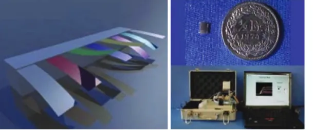
A nanomechanical olfactory sensor system for the detec-tion of volatiles was developed and tested (see Figure 4) (71, 72). This device consists of an array of silicon cantilevers with differential surface functionalization by polymers that swell upon exposure to various vapor mixtures (containing volatile molecules such as alcanes, alcohols, aldehydes). The complex nanomechanical response pattern was fed into an artificial neuronal network for pattern recognition. The diagnosis of respiratory failure was shown with a sensitiv-ity of 83%, a specificsensitiv-ity of 95% and a diagnostic accuracy of 89%. This work is followed by others who developed an electronic nose-on-chip to detect metabolites generated during infection capable for the diagnosis of ventilator-asso-ciated pneumonia (73). Using a kernel learning method, a classification accuracy for the infected samples of 100% was reported. This device might be used for rapid diagnosis and might even be usable in resource poor areas.
Multiparameter testing may require expertise for correct interpretation, which may depend on specific clin-ical settings, on specific markers combinations and poten-tially on differential parameter thresholds. Advanced analysis strategies implemented in software and accom-panied by suited user interfaces will therefore be an important complement to the actual biomarker tests.
Conclusion
The diagnosis and management of pneumococcal disease remains challenging, in particular in children who are
Figure 4: Left, a differentially polymer coated cantilever array.
Any changes in the surface tension lead to bending of the cantilever and can be measured by the laser that is integrated in the system. Right, (top) the chip with the sensor array, (bottom) the entire analysis system [with permission from Ref. (71)].
often asymptomatic carriers, and in low-income countries with a high morbidity and mortality from febrile illnesses caused by a range of bacteria, viruses and parasites.
New biomarkers were recently identified that add information about the presence as well as the invasive-ness markers of pneumococci. Nucleic acid detection technology for bacteria progresses rapidly, although the risk of false positives due to asymptomatic bacterial colo-nization exists.
Combining multiple parameters, e.g. adding inflam-mation marker quantification to urine CWPS antigen detec-tion has proven valuable in current clinical practice and in clinical studies to determine true positives, but there is room for improvement. Detection of markers of invasiveness (e.g. pneumolysin detection) and of specific nucleic acid sequences is desirable to optimize sensitivity and specificity in parallel, and to gather additional information about the threat level of the detected pathogen. Integration of multi-ple markers into a single, rapid test could add additional value to the diagnostic approach. Likewise, development of multiparameter tests for relevant arrays of pathogens is important to avoid overtreatment of febrile syndromes with antibiotics, and such test sets might be optimized for indi-vidual geographic or epidemiologic settings. In the best of all worlds and in view of the scarce resources in the devel-oping countries and increasingly also in the healthcare systems of developed countries, such tests would be inex-pensive, too, to benefit as many patients as possible.
A caveat is the antigenic and genetic drift of patho-genic microorganisms, which may require tuning of avail-able tests to changing epidemiologic settings, triggered by continuous monitoring of circulating microorganisms similar to what is currently done in influenza. Miniaturi-zation of tests through the use of micro- and nanotech-nologies is a clearly observable trend because of several advantages: miniaturization reduces sample require-ments, minimizes the use of consumables, facilitates parallelisation, enables point-of-care use of diagnostic equipment and even reduces the amount of potentially infectious disposables, characteristics that are highly desirable in most healthcare settings.
Acknowledgments: We thank Prof. Rutledge Ellis-Behnke
of the Medical Faculty Mannheim of the University of Heidelberg (Germany) for the support in knowledge. This review has been written to contribute to the DiscoGnosis project that has the core objective to develop a platform that would allow the detection of malaria and similar path-ogenic diseases in a rapid, multiplexed and non- invasive. The project is supported by the European Commission through the 7th Framework Programme on Research and
Technological Development within the Objective FP7 ICT- 2011.3.2 and under Grant Agreement [No. 318408].
Conflict of interest statement: Authors state no conflict of
interest. All authors have read the Journal’s publication ethics and publication malpractice statement available at the journal’s website and hereby confirm that they comply with all its parts applicable to the present scientific work.
References
1. Song JY, Nahm MH, Moseley MA. Clinical implications of pneumococcal serotypes: invasive disease potential, clinical presentations and antibiotic resistance. J Korean Med Sci 2013;28:4–15.
2. Huang SS, Finkelstein JA, Rifas-Shiman SL, Kleinman K, Platt R. Community-level predictors of pneumococcal carriage and resistance in young children. Am J Epidemiol 2004;159:645–54. 3. “Streptococcus Pneumoniae. Textbook in Diagnosis, Serotyping,
Virulence Factors and Enzyme-linked Immunosorbent Assay (ELISA) for Measuring Pneumococcal Antibodies.” (2nd version; 2015) Statens Serum Institut, Denmark.
4. Skov Sørensen UB, Blom J, Birch-Andersen A, Henrichsen J. Ultrastructural localization of capsules, cell wall polysaccharide, cell wall proteins, and F antigen in pneumococci. Infect Immun 1988;56:1890–6.
5. Gilbert RJ, Jiménez JL, Chen S, Tickle IJ, Rossjohn J, Parker M, et al. Two structural transitions in membrane pore formation by pneumolysin, the pore-forming toxin of Streptococcus pneumoniae. Cell 1999;97:647–55.
6. Ren, B, Szalai AJ, Thomas O, Hollingshead SK, Briles DE. Both family 1 and family 2 PspA proteins can inhibit complement deposition and confer virulence to a capsular serotype 3 strain of Streptococcus pneumoniae. Infect Immun 2003;71:75–85. 7. Tu AH, Fulgham RL, McCrory MA, Briles DE, Szalai AJ.
Pneumococcal surface protein A inhibits complement activation by Streptococcus pneumoniae. Infect Immun 1999;67:4720–4. 8. Dave S, Brooks-Walter A, Pangburn MK, McDaniel LS.
PspC, a pneumococcal surface protein, binds human factor H. Infect Immun 2001;69:3435–7.
9. Jarva H, Janulczyk R, Hellwage J, Zipfel PF, Bjorck L, Meri S. Streptococcus pneumoniae evades complement attack and opsonophagocytosis by expressing the pspC locus-encoded Hic protein that binds to short consensus repeats 8–11 of factor H. J Immunol 2002;168:1886–94.
10. Dintilhac A, Alloing G, Granadel C, Claverys JP. Competence and virulence of Streptococcus pneumoniae: Adc and PsaA mutants exhibit a requirement for Zn and Mn resulting from inactivation of putative ABC metal permeases. Mol Microbiol 1997;25:727–39. 11. Mosser JL, Tomasz A. Choline-containing teichoic acid as a
structural com- ponent of pneumococcal cell wall and its role in sensitivity to lysis by an autolytic enzyme. J Biol Chem 1970;245:287–98.
12. Berry AM, Lock RA, Paton JC. Cloning and characterization of NanB, a second Streptococcus pneumoniae neuraminidase gene, and purification of the NanB enzyme from recombinant Escherichia coli. J Bacteriol 1996;178:4854–60.
13. Janoff EN, Rubins JB, Fasching C, Charboneau D, Rahkola JT, Plaut AG, et al. Pneumococcal IgA1 protease subverts specific protection by human IgA1. Mucosal Immunol 2014;7:249–56.
14. Calix JJ, Dagan R, Pelton SI, Porat N, Nahm MH. Differential occurrence of Streptococcus pneumoniae serotype 11E between asymptomatic carriage and invasive pneumococcal disease isolates reflects a unique model of pathogen microevolution. Clin Infect Dis 2012;54:794–9.
15. Harboe ZB, Benfield TL, Valentiner-Branth P, Hjuler T, Lambertsen L, Kaltoft M, et al. Temporal trends in invasive pneumococcal disease and pneumococcal serotypes over 7 decades. Clin Infect Dis 2010;50:329–37.
16. Mukabatsinda C, Nguyen J, Bisig B, Lynen L, Coppens YD, Asiimwe A, et al. Is increasing complexity of algorithms the price for higher accuracy? Virtual comparison of three algorithms for tertiary level management of chronic cough in people living with HIV in a low-income country. BMC Med Inform Decis Mak 2012;12:2.
17. Song JY, Eun BW, Nahm MH. Diagnosis of pneumococcal pneu-monia: current pitfalls and the way forward. Infect Chemother 2013;45:351–366.
18. Werno AM, Murdoch DR. Medical microbiology: laboratory diagnosis of invasive pneumococcal disease. Clin Infect Dis 2008;46:926–32.
19. Kellogg JA, Bankert DA, Elder CJ, Gibbs JL, Smith MC. Identifica-tion of Streptococcus pneumoniae revisited. J Clin Microbiol 2001;39:3373–5.
20. Greve T, Møller JK. Accuracy of using the lytA gene to distinguish Streptococcus pneumoniae from related species. J Med Micro-biol 2012;61:478–82.
21. Brown S, Santa Maria JP Jr, Walker S. Wall teichoic acids of gram-positive bacteria. Annu Rev Microbiol 2013;67:313–36.
22. Sinclair A, Xie X, Teltscher M, Dendukuri N. Systematic re-view and meta-analysis of a urine-based pneumococcal antigen test for diagnosis of community-acquired pneu-monia caused by Streptococcus pneupneu-moniae. J Clin Microbiol 2013;51:2303–10.
23. Frasch CE, Conception NF. Specificity of human antibodies reactive with pneumococcal C polysaccharide. Infect Immun 2000;68:2333–7.
24. Pride MW, Huijts SM, Wu K, Souza V, Passador S, Tinder C, et al. Validation of an immunodiagnostic assay for detection of 13 Streptococcus pneumoniae serotype-specific polysaccharides in human urine. Clin Vaccine Immunol 2012;19:1131–41.
25. Pickering JW, Hill HR. Measurement of Antibodies to Pneumo-coccal Polysaccharides with Luminex xMAP Microsphere-Based Liquid Arrays. Methods Mol Biol 2011;808:361–375.
26. Ortika BD, Habib M, Dunne EM, Porter BD, Satzke C. Production of latex agglutination reagents for pneumococcal serotyping. BMC Res Notes 2013;6:49.
27. File TM, Kozlov RS. Rapid detection of Streptococcus pneumo-niae in community-acquired pneumonia. Clin Microbiol Infect 2006;12(Suppl 9):27–33.
28. del Mar García-Suárez M, Cima-Cabal MD, Villaverde R, Espinosa E, Falguera M, de Los Toyos JR, et al. Performance of a pneumolysin enzyme-linked immunosorbent assay for diagnosis of pneumococcal infections. J Clin Microbiol 2007;45:3549–54.
29. Moïsi JC, Saha SK, Falade AG, Njanpop-Lafourcade BM, Oundo J, Zaidi AKM, et al. Enhanced diagnosis of pneumococcal meningitis using the Binax NOW® S. pneumoniae immuno-chromatographic test: a multi-site study. Clin Infect Dis 2009;48(Suppl 2):S49–56.
30. Jado I, Fenoll A, Casal J, Pérez A. Identification of the psaA gene, coding for pneumococcal surface adhesin A, in viridans group streptococci other than Streptococcus pneumoniae. Clin Diagn Lab Immunol 2001;8:895–8.
31. Janulczyk R, Iannelli F, Sjoholm AG, Pozzi G, Bjorck L. Hic, a novel surface protein of Streptococcus pneumoniae that interferes with complement function. J Biol Chem 2000;275:37257–63.
32. Ye W, Hu Y, Zhang R, Ying K. Diagnostic value of the soluble triggering receptor expressed on myeloid cells-1 in lower respiratory tract infections: a meta-analysis. Respirology 2014;19:501–7.
33. Huang H, Ideh RC, Gitau E, Thézénas ML, Jallow M, Ebruke B, et al. Discovery and validation of biomarkers to guide clinical management of pneumonia in African children. Clin Infect Dis 2014;58:1707–15.
34. Murdoch DR, Anderson TP, Beynon KA, Chua A, Fleming AM, Laing RT, et al. Evaluation of a PCR assay for detection of Streptococcus pneumoniae in respiratory and nonrespiratory samples from adults with community-acquired pneumonia. J Clin Microbiol 2003;41:63–6.
35. Dhoubhadel BG, Yasunami M, Yoshida LM, Thi HA, Thi TH, Thi TA, et al. A novel high-throughput method for molecular serotyping and serotype-specific quantification of Streptococcus pneumo-niae using a nanofluidic real-time PCR system. J Med Microbiol 2014;63(Pt 4):528–39.
36. Messaoudi M, Milenkov M, Albrich WC, van der Linden MP, Bénet T, Chou M, et al. The relevance of a novel quantitative assay to detect up to 40 major streptococcus pneumoniae sero-types directly in clinical nasopharyngeal and blood specimens. PLoS One 2016;11:e0151428.
37. Pai R, Gertz RE, Beall B. Sequential multiplex PCR approach for determining capsular serotypes of Streptococcus pneumoniae isolates. J Clin Microbiol 2006;44:124–31.
38. Habets MN, Cremers AJ, Bos MP, Savelkoul P, Eleveld MJ, Meis JF et al. A novel quantitative PCR assay for the detection of Strepto-coccus pneumoniae using the competence regulator gene target comX. J Med Microbiol 2016;65:129–36.
39. Linder A, Hollingshead S, Janulczyk R, Christensson B, Akesson P. Human antibody response towards the
pneumococcal surface proteins PspA and PspC during invasive pneumococcal infection. Vaccine 2007;25:341–5.
40. Kolberg J, Aase A, Næss LM, Aaberge IS, Caugant DA. Human antibody responses to pneumococcal surface protein A and capsular polysaccharides during acute and convalescent stages of invasive disease in adult patients. Pathog Dis 2014;70:40–50.
41. Andrade DC, Borges IC, Laitinen H, Ekström N, Adrian PV, Meinke A, et al. A fluorescent multiplexed bead-based immuno-assay (FMIA) for quantitation of IgG against Streptococcus pneumoniae, Haemophilus influenzae and Moraxella catarrhalis protein antigens. J Immunol Methods 2014;405:130–43. 42. Larsson MC, Karlsson E, Woksepp H, Frölander K, Mårtensson A,
Rashed F, et al. Rapid identification of pneumococci, enterococci, beta-haemolytic streptococci and S. aureus from positive blood cultures enabling early reports. BMC Infect Dis 2014;14:146.
43. Cremers AJ, Hagen F, Hermans PW, Meis JF, Ferwerda G. Diagnostic value of serum pneumococcal DNA load during invasive pneumococcal infections. Eur J Clin Microbiol Infect Dis 2014;33:1119–24.
44. Chen M, Zhou M, Xiao W, Ai B, Liu X, Li Y. The urinary antigen tests have high sensitivity in diagnosis of pneumococcus caused community-acquired pneumonia posterior to antimicrobial therapy. Cell Biochem Biophys 2014;70:1029–34. 45. Fall B, Lo CI, Samb-Ba B, Perrot N, Diawara S, Gueye MW,
et al. The ongoing revolution of MALDI-TOF mass spectrometry for microbiology reaches tropical africa. Am J Trop Med Hyg 2015;92:641–7.
46. Galetto-Lacour A, Alcoba G, Posfay-Barbe KM, Cevey-Macherel M, Gehri M, Ochs MM, et al. Elevated inflammatory markers com-bined with positive pneumococcal urinary antigen are a good predictor of pneumococcal community-acquired pneumonia in children. Pediatr Infect Dis J 2013;32:1175–9.
47. Strachan RE, Cornelius A, Gilbert GL, Gulliver T, Martin A, McDonald T, et al. A bedside assay to detect streptococcus pneumoniae in children with empyema. Pediatr Pulmonol 2011;46:179–83.
48. Picazo JJ, Contreras JR, Ríos E, Culebras E, Rodríguez-Avi-al I, Méndez C, et al. Rapid diagnosis of invasive pneumococcal disease in pediatric population. J Microbiol Methods 2013;93:116–20.
49. Martinón-Torres F, Dosil-Gallardo S, Perez del Molino-Bernal ML, Sánchez FP, Tarrago D, Alvez F, et al. Pleural antigen assay in the diagnosis of pediatric pneumococcal empyema. J Crit Care 2012;27:321.e1–4.
50. Lee JH, Kim SH, Lee J, Choi EH, Lee HJ. Diagnosis of
pneumococcal empyema using immunochromatographic test on pleural fluid and serotype distribution in Korean children. Diagn Microbiol Infect Dis 2012;72:119–124.
51. Salieb-Beugelaar GB, Hunziker PR. Towards nano- diagnostics for bacterial infections. Eur J Nanomed 2015;7:37–50.
52. Salieb-Beugelaar GB, Hunziker PR. Towards nano-diagnostics for rapid diagnosis of infectious diseases-current technological state. Eur J Nanomed 2014;6:11–28.
53. Park S, Zhang Y, Wang TH, Yang S. Continuous dielectrophoretic bacterial separation and concentration from physiological media of high conductivity. Lab Chip 2011;11:2893–900. 54. Ai Y, Sanders CK, Marrone BL. Separation of Escherichia coli
bac-teria from peripheral blood mononuclear cells using standing surface acoustic waves. Anal Chem 2013;85:9126–34. 55. Ranjan S, Zeming KK, Jureen R, Fisher D, Zhang Y. Dld pillar
shape design for efficient separation of spherical and non-sheri-cal bioparticles. Lab Chip 2014;14:4250–62.
56. Kang DK, Ali MM, Zhang K, Huang SS, Peterson E, Digman MA et al. Rapid detection of single bacteria in unprocessed blood using integrated comprehensive droplet digital detection. Nat Commun 2014;5:5427.
57. Sajid M, Kawde AN, Daus M. Designs, formats and applica-tions of lateral flow assay: a literature review. J Saudi Chem Soc 2015;19:689–705.
58. Shen J, Li Y, Gu H, Xia F, Zuo X. Recent development of sandwich assay based on the nanobiotechnologies for proteins, nucleic acids, small molecules, and ions. Chem Rev 2014;114:7631–77. 59. Pedrosa P, Baptista PV. Gold and silver nanoparticles for
diagnostics of infection. Nanotechnology in diagnosis, treatment and prophylaxis of infectious diseases, 2015. Edited by: Mahendra Rai and Kateryna Kon ISBN: 978-0-12-801317-5. 60. Wu TY, Su YY, Shu WH, Mercado AT, Wang SK, Hsu LY, et al. A
novel sensitive pathogen detection system based on microbead quantum dot system. Biosens Bioelectron 2016;78:37–44. 61. Brenner S, Johnson M, Bridgham J, Golda G, Lloyd DH,
Johnson D, et al. Gene expression analysis by massively parallel signature sequencing (MPSS) on microbead arrays. Nat. Biotechnol. 2000;18:630–4.
62. Veigas B, Pedrosa P, Carlos FF, Mancio-Silva L, Grosso AR, Fortunato E. One nanoprobe, two pathogens: gold nanoprobes multiplexing for point-of-care. J Nanobiotechnol 2015;13:48. 63. Veigas B, Jacob JM, Costa MN, Santos DS, Viveiros M, Inacio J,
et al. Gold on paper–paper platform for Au-nanoprobe TB detection. Lab Chip 2012;12:4802–8.
64. Luo J, Fang X, Ye D, Li H, Chen H, Zhang S, et al. A real-time microfluidic multiplex electrochemical loop-mediated isother-mal amplification chip for differentiating bacteria. Biosens Bioelectron 2014;60:84–91.
65. Ju HX, Zhou J, Cai CX, Chen HY. The electrochemical behavior of methylene blue at a microcylinder carbon fiber electrode. Electroanalysis 1995;7:1165–70.
66. Kotanen CN, Martinez L, Alvarez R, Simecek JW. Surface enhanced Raman scattering spectroscopy for detection and identification of microbial pathogens isolated from human serum. Sens Bio sensing Res 2016;8:20–6.
67. Walter A, März A, Schumacher W, Rösch P, Popp J. Towards a fast, high specific and reliable discrimination of bacteria on strain level by means of SERS in a microfluidic device. Lab Chip 2011;11:1013.
68. Pahlow S, Meisel S, Cialla-May D, Weber K, Rösch P, Popp J. Isolation and identification of bacteria by means of Raman spectroscopy. Adv Drug Del Rev 2015;89:105–20.
69. Bouguelia S, Roupioz Y, Slimani S, Mondani L, Casabona MG, Durmort C, et al. On-chip microbial culture for the specific detection of very low levels of bacteria. Lab Chip 2013;13: 4024–32.
70. Abadian PN, Kelley CP, Goluch ED. Cellular analysis and detection using surface plasmon resonance techniques. Anal Chem 2014;86:2799–812.
71. Schmid D, Lang H, Marsch S, Gerber C, Hunziker P. Diagnosing disease by nanomechanical olfactory sensors – system design and clinical validation. Eur J Nanomed 2008;1:44–7.
72. Lang HP, Ramseyer JP, Grange W, Braun T, Schmid D, Hunziker P, et al. An artificial nose based on microcantilever array sensors. J Phys Conf Ser 2007;61:663.
73. Tang KT, Chiu SW, Shih CH, Chang CL, Yang CM, Yao DJ, et al. 24.5 A 0.5V 1.27mW nose-on-a-chip for rapid diagnosis of ventilator-associated pneumonia (Conference Paper). IEEE International Solid-State Circ Conf 2014;57:420–21.
Bionotes
Georgette B. Salieb-Beugelaar Nanomedicine Research Lab CLINAM, University Hospital Basel, Bernoullistrasse 20, Basel, CH-4056, Switzerland; and The European Foundation for Clinical Nanomedicine (CLINAM), Alemannengasse 12, CH-4016 Basel, Switzerland,
Georgette B. Salieb-Beugelaar’s professional life started in the fields of clinical genetics, DNA research and diagnostics at the Academic Medical Centre in Amsterdam (The Netherlands) in 1996. She studied Chemistry at the University of Utrecht (The Netherlands) between 2000 and 2003. In 2005, her professional field changed into the microfluidic and nanofluidic world at the University of Twente in Enschede (The Netherlands) where she investigated single DNA molecules in nanoconfined environments on chips yielding in her PhD degree in 2009. The following 2 years she spent working at the Korean Institute of Science and Technol-ogy (Saarbrücken, Germany) on nanodropled pseudocrystals in microfluidic chips. Meanwhile, she was also working for the Mesa Institute for Nanotechnology (Twente University), during the set up of their BioNano Laboratory. Since November 2012, Georgette has become a member of the multidisciplinary NanoMedicine Group of Prof. Patrick Hunziker, working for the DiscoGnosis project (www.discognosis.eu) till January 2016 and is at present developing microfluidics for diagnostic purposes. Georgette is also currently involved in this journal as the scientific managing editor and active as managing editor of Progress in Materials
Science (Elsevier).
Bei Zhang
Nanomedicine Research Lab CLINAM, University Hospital Basel, Bernoullistrasse 20, Basel, CH-4056, Switzerland
Bei Zhang received her Bachelor of Medicine (BMed) and Master of Medicine (MMed) in Clinical Laboratory Diagnostic Science at Shanghai Second Medical University, China. Since 1993 she worked as a microbiologist for more than 15 years in the Labora-tory Diagnostic Center at Shanghai Children’s Medical Center affiliated to Shanghai JiaoTong University and was promoted to Associate Professor in 2005. She obtained her PhD in Medical and Biological Research from University of Basel, Switzerland in 2012. After that, she joined the Nanomedicine Research Lab CLINAM, Medical Intensive Care Unit (MedInt), University Hospital Basel as a postdoc working for European Commission (EC)-funded DiscoGnosis project. She has published more than 30 papers in peer-reviewed journals.
Maurice M. Nigo
Nanomedicine Research Lab CLINAM, University Hospital Basel, Bernoullistrasse 20, Basel, CH-4056, Switzerland; and ISTM-Nyankunde, B.P. 55, Bunia, Democratic Republic Congo
Maurice M. Nigo began his professional life in the fields of microbi-ology and epidemimicrobi-ology of communicable diseases at the Depart-ment of Pathology of the University of Liege (Belgium) in 1993. Since 1996, he has been giving lectures on Medical Microbiology and Medical Parasitology at the “Institut Supérieur des Techniques Médicales de Nyankunde” at Bunia (DR Congo). In 2004, he was the Principal Investigator of the Survey for the Validation of the Rapid Assessment Procedure for Loiasis (RAPLOA) in the north-eastern area of the Democratic Republic of Congo. Between 2008 and 2012, he was the Head of Laboratory of the “Centre de Recherche en Mala-dies Tropicales de l’Ituri” for the WHO/TDR and Wieth/Pfizer Phase III Moxidectin Trial at Rethy (DR Congo). Since November 2014, Maurice has been a member of the multidisciplinary NanoMedicine Group of Prof. Patrick Hunziker, and he worked for the DiscoGno-sis project (www.discognoDiscoGno-sis.eu). Now Maurice is a PhD student working on blood fluke diagnosis and epidemiology.
Sieghard Frischmann
MAST Diagnostica GmbH, Feldstraße 20, DE 23858 Reinfeld, Germany
Sieghard Frischmann is head of R&D and production at Mast. He is leading a team of scientists for the development of molecular diag-nostic assays based on the LAMP technology. As a regulatory board member at Mast he works as a medical product safety manager according to the German Medical Product Law. Sieghard Frischmann joined Mast Diagnostica GmbH in 1992.
Patrick R. Hunziker
Intensive Care Clinic, University Hospital Basel, Petersgraben 4, CH-4031 Basel, Switzerland; Nanomedicine Research Lab CLINAM, University Hospital Basel, Bernoullistrasse 20, Basel, CH-4056, Switzerland; and The European Foundation for Clinical Nanomedicine (CLINAM), Alemannengasse 12, CH-4016 Basel, Switzerland
Patrick R. Hunziker studied Medicine at the University of Zurich, Switzerland. He received a doctoral degree based on thesis work in experimental immunology from the University of Zurich and did further research in experimental hematology at the University Hos-pital in Zurich, Switzerland. He earned specialist degrees in Internal Medicine, Cardiology and Intensive Care Medicine. As a fellow of the Massachusetts General Hospital, Harvard Medical School, he worked on cardiac imaging in a joint project with the Massachusetts Institute of Technology, Cambridge. His professional activities in Europe, the US, Africa and China have given him a broad insight into the needs for the medicine of the future in a variety of settings. Patrick R. Hunziker became involved in the medical applications of nanoscience in the late 1990s and has been the pioneering physi-cian in nanomedicine in Switzerland since then. With improved prevention, diagnosis and the cure for cardiovascular disease as his main research topics, he has worked in the nanoscience fields of atomic force microscopy, nano-optics, micro/nanofluidics, nanomechanical sensors and polymer nanocarriers for targeting. He is the founding president of the European Society of Nanomedicine, cofounder of the European Foundation for Clinical Nanomedicine and coinitiator of the European Conference for Clinical Nanomedi-cine and is clinically active as deputy head of the Clinic for Intensive Care Medicine at the University Hospital Basel, Switzerland. In November 2008 Patrick R. Hunziker became professor for Cardiol-ogy and Intensive Care Medicine at the University of Basel.