Original article
Chronic complicated osteomyelitis
of the appendicular skeleton:
diagnosis with technetium-99m labelled
monoclonal antigranulocyte antibody-immunoscintigraphy
Achim Kaim 1,3, Thomas Maurer 2, Peter Ochsner 2, Gernot Jundt 4, Eberhard Kirsch 3, Jan Mueller-Brand~ 1 Institute of Nuclear Medicine, University Hospital Basel, Switzerland2 Department of Orthopedic and Trauma Surgery, Kantonsspital Liestal, Switzerland 3 Institute of Diagnostic Radiology, University Hospital Basel, Switzerland
4 Institute of Pathology, University Hospital Basel, Switzerland Received 6 January and in revised form 5 April 1997
Abstract. Chronic post-traumatic osteomyelitis (OM) represents a particular challenge for nuclear medicine and radiology since clinical and biochemical parameters are frequently unreliable. The aim of this study was to investigate the value of combined bone scan (BS) and immunoscintigraphy (IS) with technetium-99m labelled monoclonal antigranulocyte antibody (MAB) in patients with suspected chronic OM of the appendicular skeleton. Twenty-four patients (17 females and 7 males) with sus- pected chronic post-traumatic OM were evaluated with three-phase BS/99mTc-MAB-IS. The final diagnosis was established by means of bone culture and histology in 19 cases and clinical follow-up in five cases. The studies were reviewed by two independent and experienced ob- servers; the interobserver agreement was calculated by kappa statistics. The sensitivity, specificity and accuracy of BS alone were 92%, 18% and 58%, respectively. Combined BS/99mTc-MAB-IS had a sensitivity, specific- ity and accuracy of 84%, 72% and 79%, respectively. Of 24 studies, 11 were true-positive, two false-negative, eight true-negative and three false-positive. Two patients presented with unexpected ectopic haematopoietic bone marrow in the appendicular skeleton that caused false- positive results. A high degree of interobserver agree- ment was found (~:=0.85). It is concluded that combined BS/99mTc-MAB-IS represents a very sensitive and repro- ducible method with an acceptable specificity for the in- vestigation of chronic OM. Problems may occur in the differentiation of low-grade OM from aseptic inflamma- tion. Another problem is ectopic marrow that may occur in the appendicular skeleton due to a chronic inflamma- tory stimulus. A former intramedullary intervention in the femur with displacement of haematopoietic marrow may also lead to an ectopic location.
Correspondence to: A. Kaim, Institute of Nuclear Medicine, Uni- versity Hospital Basel, Petersgraben 4, CH-4032 Basel, Switzer- land
Eur J Nucl Med (1997) 24:732-738
Introduction
Chronic post-traumatic and postoperative osteomyelitis represents a disabling disease that requires a detailed in- vestigation for assessment of active infection and local extent in order to decide upon the appropriate therapeu- tic management. Beside radiological methods [X-ray plain film, computed tomography (CT), magnetic reso- nance imaging (MRI)], nuclear medicine offers a wide variety of radiopharmaceuticals for the imaging of in- flammation or infection, e.g. gallium-67 citrate [1, 2], technetium-99m labelled nanocolloids [3], 99mTc-la- belled human immunoglobulins [4-6], indium-111 oxine and 99mTc-hexamethylpropylene amine oxime (HMPAO)-labelled autologous leucocytes [7-11]. 99mTc- labelled monoclonal antigranulocyte antibody (MAB) immunoscintigraphy (IS) represents a simple, practica- ble and specific method that has been extensively stud- ied for the detection of inflammatory and septic process- es. Extra-osseous infection [12, 13], acute and subacute osteomyelitis [14-16], spondylodiscitis [6, 14] and in- fection of hip arthoplasties [ 12, 13, 16, 17] have been the main subjects of these publications. Until now no study has specifically considered the value of IS in chronic re- current or low-grade osteomyelitis of the appendicular skeleton. Therefore, the accuracy of IS with MAB as an adjunct to bone scan in the evaluation of this rare but im- portant patient group was assessed.
Materials and methods
Patients Twenty-four patients (17 males and 7 females; mean age 47 years, range 21-83 years) referred to the Nuclear Medicine de-
European Journal of Nuclear Medicine
partraent with suspected bone infection were examined with bone scan and IS. Only patients with suspected chronic osteomyelitis of the appendicular skeleton were included. Arthroplasties of the hip were not considered. One or more of the following criteria was present: clinical signs (swelling, heat, redness, fistula), pain, fever, elevated laboratory parameters (ESR, CRP, WBC count) and sus- pected focal infection on radiology (X-ray plain film, CT, MRI). All patients suffered from longstanding, post-traumatic chronic osteomyelitis; the mean interval from first surgery to the present study was 8.6 years (range 2 months to 42 years). The average number of operations due to recurrent osteomyelitis was four (range 1-15), and the interval from last surgery to the present av- eraged 3.5 years (2 months to 42 years). Six patients had ortho- paedic devices (metal plate, intramedullary nail, external fixator). The suspected areas were investigated by X-ray plain films in all patients, by CT in 11 patients and by MRI in nine patients. Final diagnosis was established by surgery with microbiological and/or histological analysis in 19 patients and by clinical course over a period of at least 6 months in five patients. Radiological results were considered in defining the final diagnosis.
Radiopharmaceuticals The antigranulocyte antibody used for IS is a murine monoclonal antibody (BW250/183), supplied by Beh- ringwerke AG, Switzerland. The antibody is an immunoglobulin of the IgG isotype and binds to the epitope NCA 95 on the surface of human granulocytes. The antibody is reduced with stannous II- chloride solution and labelled with 99mTc. After an incubation pe- riod of 10 min, 555 MBq of the antibody solution (equivalent to
0.5 mg of the antibody) was intravenously injected into the pa- tient. Serum was obtained before and 3 weeks after injection to determine the titre of human antimouse antibodies (HAMA). None of the patients developed HAMA, and no side-effects or ad- verse reactions were observed.
For 99mTc-diphosphonate bone scan, 3,3-diphosphono-l,2-pro- panedicarboxyl acid tetrasodium salt (DPD) (Hoechst, Switzer- land) was used. It was labelled with an average of 740 MBq of 99mTc"
Imaging modalities All patients first underwent quantitative three- phase bone scan followed by IS at an interval of 2 days to 1 week. After injection of 99mTc-DPD 20 5-s images were immediately registered and another 60-s image was taken at 5 min. Five hun- dred-kcount planar spot images were obtained 3 h after injection and stored on computer using a 256x256 matrix.
IS was performed 17 h p.i. Planar spot images of the suspected area in at least two projections were obtained. Five hundred- kcount images with a 256x256 matrix were recorded. Additional- ly whole-body images were obtained. Imaging was performed with a Siemens DIACAM gamma camera equipped with a low-en- ergy high-resolution (LEHR) collimator connected to a dedicated computer system (Icon, Siemens).
Data interpretation Bone scans and IS were reviewed by two ex- perienced observers (A, B) unaware of the definitive diagnosis. The readers were independent of each other and interpreted the studies separately. X-ray plain films were used to identify the
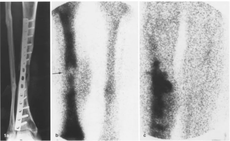
Fig. l a - e . True-positive result in a patient with infected pseudarthrosis and bone necrosis 3 years after trauma. Plate osteosynthesis had been performed 1 year prior to the current investigation, a Radiograph (anteroposterior) shows pseudarthrosis of the tibial diaphysis, b Late bone scan (anterior) shows hyperactivity of the complete tibia with a photopenic zone at the site of pseudarthrosis suggesting bone necrosis (black arrow), e Intense accumulation of 99mTc-MAB on IS (anterior) demonstrating the extent of the infection
anatomy. The degree of agreement between the two observers was measured by kappa statistics [18] calculating the index ~: (~: is 0.0 when there is just a chance agreement and 1.0 when there is per- fect agreement). Two of 26 studies were interpreted differently and had to be discussed with a third observer (C). After analysing these cases, agreement was achieved among all three observers and the final scintigraphic diagnosis was established. These data were the basis for calculating sensitivity, specificity and accuracy.
IS was considered positive when accumulation was observed in comparison to background and the contralateral side. Uptake was graded as absent, mild or strong. Normal late bone scan and positive IS were interpreted as indicating soft tissue infection. Os- teomyelitis was diagnosed if bone scan and IS were positive at the same anatomical location. Infected necrotic bone was considered in zones of decreased bone metabolism with positive IS.
Results
T w e n t y - f o u r patients with suspected osteomyelitis were examined. Thirteen patients presented with active osteo- myelitis, four had soft tissue infections and in the re-
maining seven surgery, histology, cultures r a d i o l o g y and/or clinical course failed to reveal evidence o f infec- tion. O f the 24 suspected lesions, 15 were located in the tibia, eight in the distal f e m u r and one in the diaphysis o f the humerus. L a b o r a t o r y results (ESR, C R R W B C count) s h o w e d elevated levels in six patients with active osteomyelitis, in four patients with soft tissue infection and in one patient without infection. The remaining pa- tients (seven with osteomyelitis, six without osteomyeli- tis) had n o r m a l laboratory results. In 9/13 patients the causative m i c r o - o r g a n i s m was Staphylococcus aureus, in 3/13 patients coagulase-negative Staphylococcus and in 1/13 patients diagnosis was c o n f i r m e d by histology without microbiology. D e l a y e d bone scan (3 h) revealed increase b o n e m e t a b o l i s m in 21/24 cases. In 3/21 cases areas o f p h o t o p e n i a o c c u r r e d beside zones o f increased tracer activity c o r r e s p o n d i n g to necrotic bone. Consider- ing the b o n e scan alone, sensitivity, specificity and accu- racy were calculated to be 92%, 18% and 58%, respec- tively. C o m b i n e d b o n e scan and IS had a sensitivity,
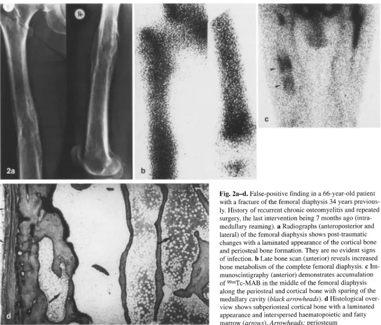
Fig. 2a-d. False-positive finding in a 66-year-old patient with a fracture of the femoral diaphysis 34 years previous- ly. History of recurrent chronic osteomyelitis and repeated surgery, the last intervention being 7 months ago (intra- medullary reaming), a Radiographs (anteroposterior and lateral) of the femoral diaphysis shows post-traumatic changes with a laminated appearance of the cortical bone and periosteal bone formation. They are no evident signs of infection, b Late bone scan (anterior) reveals increased bone metabolism of the complete femoral diaphysis, c Im- munoscintigraphy (anterior) demonstrates accumulation of 99mTc-MAB in the middle of the femoral diaphysis along the periosteal and cortical bone with sparing of the medullary cavity (black arrowheads), d Histological over- view shows subperiosteal cortical bone with a laminated appearance and interspersed haematopoietic and fatty marrow (arrows). Arrowheads: periosteum
Table 1. Individual patient data
No. Sex Age Interval No. of Interval Laboratory Anatomical Bone IS Final diagnosis Method of
(yrs) from operations from last parameters site Scan diagnosis
1 st surgery surgery verification
1 f 65 4 months 1 4 months 1" Humerus + ++ OM (Staph. Surgery TP
aureus)
2 f 83 8 months 3 4 months Normal Distal femur ++ ++ OM (coag. neg. Surgery TP
Staph.)
3 f 21 3 years 1 3 years Normal Distal femur ++ 0 OM Surgery/ FN
histology
4 m 74 4 months 1 4 months $ Distal femur ++ 0 No infection Surgery TN
5 f 27 10 years 3 8 years Normal Tibia ++ ++ No infection Surgery/ FP
histology
6 m 76 25 years 7 3 years Normal Tibia ++ + OM (Staph. Surgery TP
aureus, E. coli)
7 m 65 9 years 15 2 years 1" Tibia 0 ++ Soft tissue Clinical TN
infection course, MRI
8 m 66 34 years 5 7 months Normal Femur ++ ++ No infection Surgery/ FP
histology
9 m 40 3 years 5 2 years Normal Tibia ++ ++ OM (Staph. Surgery TP
aureus)
10 m 49 3 years 3 1 year Normal Tibia + + OM (Staph. Surgery TP
alAreblS)
11 m 59 1.5 years 2 6 months Normal Distal femur + 0 No infection Clinical TN course
12 m 43 1.5 years 4 1 year I" Tibia ++ ++ OM (Staph. Surgery TP
aureus)
13 m 35 5 years 3 2 years $ Tibia 0 ++ Soft tissue Clinical TN
infection course
14 f 21 2 months 1 2 months 1" Tibia ++ ++ Soft tissue Surgery TN
infection
15 f 58 42 years 1 42 years 1" Tibia + 0 No infection Surgery TN
16 m 42 17 years 5 8 years $ Tibia ++ + OM (Staph. Surgery TP
abtYeblS)
17 m 42 5 months 1 5 months Normal Tibia ++ ++ OM (coag. neg. Surgery TP
Staph. )
18 m 39 8 months 1 8 months 1" Tibia 0 0 OM (Staph. Surgery FN
aureus)
19 m 35 10 years 5 2 years Normal Tibia + 0 No infection Clinical TN
course
20 m 45 25 years 4 1 year Normal Tibia ++ + OM (coag. neg. Surgery TP
Staph.)
21 m 42 1.5 years 3 7 months Normal Distal femur ++ ++ Soft tissue Surgery/ FP infection histology
22 f 25 3 years 1 3 years $ Distal femur ++ ++ OM (Staph. Surgery TP
aureus
23 m 27 2 years 3 1.5 years "]" Distal femur ++ + OM (Staph. Surgery TP
aureus)
24 m 37 10 years 8 I year Normal Tibia + 0 No infection Clinical TN
course 0, No uptake; +, mild uptake;++, strong uptake; TP, true-positive; FN, false-negtive; TN, true-negative; FR false-positive
s p e c i f i c i t y a n d a c c u r a c y o f 8 4 % , 7 2 % a n d 7 9 % , r e s p e c - tively. O f t h e 24 s t u d i e s , 11 w e r e t r u e - p o s i t i v e ( T P ) a n d e i g h t , t r u e - n e g a t i v e ( T N ) . T h e s c i n t i g r a p h i c p a t t e r n t h a t o c c u r r e d in a c t i v e o s t e o m y e l i t i s w i t h i n f e c t e d p s e u d a r - t h r o s i s a n d n e c r o t i c b o n e is d e m o n s t r a t e d in F i g . 1. T w o s t u d i e s w e r e f a l s e - n e g a t i v e ( F N ) a n d t h r e e , f a l s e - p o s i t i v e ( F P ) . F i g u r e 2 i l l u s t r a t e s an F P f i n d i n g t h a t is d i s c u s s e d later. T h e r e w a s a s t r o n g a g r e e m e n t b e t w e e n t h e t w o o b - s e r v e r s A a n d B. T h e l~-value f o r i n t e r o b s e r v e r a g r e e - m e n t w a s 0 . 8 5 ( 9 5 % c o n f i d e n c e i n t e r v a l 0 . 7 4 - 0 . 9 9 ) . T h e i n d i v i d u a l p a t i e n t d a t a are s u m m a r i z e d in T a b l e 1.
Discussion
The diagnosis of chronic complicated osteomyelitis rep- resents a particular challenge for radiology and nuclear medicine. Low-grade and recurrent longstanding infec- tion may cause chronic pain, fistulae and bone necrosis with pathological fracture. The disease may lead to dis- ability and impair quality of life in a young patient pop- ulation. Frequently clinical and biochemical parameters are inadequate for making the diagnosis. Our series con- firms that laboratory parameters are inconclusive in the evaluation of chronic osteomyelitis. The assessment of disease activity, disease extension and the separation of bony from soft tissue infection influence the therapeutic management by the referring surgeon. Nuclear medicine offers very useful information in the investigation of bone infection. 99mTc-diphosphonate bone scan repre- sents the basic study and is very sensitive in detecting bone alterations. However, it is unable to distinguish be- tween infection and aseptic post-traumatic changes; thus, we calculated a specificity of 18%. Diphosphonate scan has to be followed by inflammatory scintigraphy, and radiolabelled autologous leucocyte scintigraphy and 99mTc-labelled MAB-IS are specific for the detection of infected foci. The clinical use of these methods is pre- dominantly determined by their potential availability and their practicability in routine clinical practice. Radiola- belled autologous white cells are still considered the gold standard in the nuclear imaging of infected sites. However, the examination requires time-consuming ex vivo blood handling techniques and special training, and entails risk of infection for both laboratory personal and patients.
99mTc-MAB binding granulocytes show a quite similar in vivo distribution to that of radiolabelled granulocytes and allow the detection of granulocyte accumulation in the appendicular skeleton [19, 20]. The advantage of 99mTc-MAB is the simplicity of the labelling process of Tc with the antibodies and the in vivo labelling of granu- locytes. The antibodies are directed against the myeloid- specific surface antigen NCA-95 expressed by granulocy- tes. A disadvantage of MAB is the induction of HAMA in 3%, which can lead to altered biodistribution [21, 22]. Al- lergic reactions or serious adverse reactions have not been reported so far. Problems in the detection of infection ap- pear in areas of active bone marrow such as the spine, the pelvis and the proximal femora because precursor cells of the granulopoietic system such as promyelocytes are also labelled. A physiological uptake in the bone marrow oc- curs and infection causes non-specific photopenic zones due to bone marrow destruction. All patients included in our study had suspected infections in the appendicular skeleton. Chronic infection of the pelvis and spine were excluded, as was hip prosthesis infection. Sensitivity, specificity and accuracy for proven infection were calcu- lated to be 84%, 72% and 79% respectively.
Currently there are four published series with small numbers of patients reporting the use of 99mTc-MAB in
chronic osteomyelitis [6, 14-16]. The reported sensitivi- ty (79%-100%) and specificity (64%-100%) show a wide range due to the different inclusion criteria for chronic osteomyelitis. A major part of the reported data has been based on patient groups with acute and sub- acute infections [6, 14, 15]. Furthermore, patients with septic hip arthroplasties [15] and even chronic infection of the axial skeleton [6, 14] have been included. We con- sidered only patients with suspicion of infection of the tubular bones at least 6 weeks after surgery or trauma, with the majority of patients having suffered from long- standing recurrent osteomyelitis. We therefore believe that the results of our study accurately reflect the ability to image infection with MAB in chronic post-traumatic osteomyelitis. The published studies reporting the use of 99mTc-HMPAO-labelled leucocytes for the detection of chronic osteomyelitis calculated values for sensitivity (83%-93%) and specificitiy (78%-100%) that suggest superiority over IS [7, 10, 23, 24]. Until now, however, no comparative study that proves this hypothesis has been performed in this field of application.
There are several possible reasons for false-negative studies. One is technical problems such as incomplete labelling of the MAB. In follow-up studies there may be induction of HAMA that have the potential to intercept the antibody and therefore hinder specific accumulation at infection sites [22]. In our study no HAMA response was observed and the laboratory titre controls were nor- mal. Another potential explanation is the inaccessibility of the infection site due to encapsulation. In osteomyeli- tis after intramedullary nailing an avascular zone in the central part of the diaphysis occurs due to hyperthermic injury and may be infected [25]. This infection between the nail and cortical bone may be encapsulated and thereby prevent the migration of the radiolabelled MAB- binding granulocytes. One of our two false-negative studies is explained by this phenomenon. A further pos- sible reason for equivocal findings is a small number of granulocytes at the infected site in chronic low-grade os- teomyelitis. The histology of one patient with positive microbiology for infection revealed a chronic inflamma- tion with predominantly lymphocytes and plasma cells and only some scattered granulocytes. IS did not show significant tracer accumulation and the study was finally considered to be false-negative.
On the other hand, non-purulent inflammation may sometimes include some polymorphonuclear cells which lead to a mild uptake on IS and cannot be distinguished from low-grade osteomyelitis. False-positive scans have been described in fractures, osteosarcoma, eosinophilic granuloma, Paget's disease and granulation tissue of all kinds [14, 26]. Sometimes it is very difficult to deter- mine whether infection is in bone or soft tissue. In one false-positive case, planar scintigraphy performed in two projections had been positive in bone scan and IS. Exact anatomical localization was impossible and the examina- tions were interpreted as positive for osteomyelitis. Sur- gery revealed a soft tissue infection without osseous in-
volvement. Increased bone metabolism may be ex- plained by the postoperative state and sometimes an overlying positive IS is of uncertain significance. This may lead to misinterpretation, and careful analysis of scintiscans and X-ray plain films for anatomical land- marks is necessary. In doubtful cases, single-photon emission tomography may be indicated.
Two patients presented with unexpected ectopic ac- tive bone marrow in the appendicular skeleton that gave rise to false-positive studies. In both cases histology re- vealed haematopoietically active bone marrow at the suspected locations. No histological signs of infection were found and microbiology was negative. The strongly positive IS was explained by the labelling of the promyelocytes with the 99mTc-MAB. One of the two pa- tients had a 10-year history of recurrent post-traumatic osteomyelitis of the tibial diaphysis with repeated surgi- cal procedures. The reason for this unexpected finding remains unclear. It is possible that various mediators such as cytokines, macrophage colony stimulating fac- tor, granulocyte colony stimulating factor or bone mor- phogenetic protein [27-29] are released by stromal ele- ments due to a chronic inflammatory stimulus and in- duce neogenesis of active bone marrow in the peripheral skeleton. Until now, however, there have been no reports of such a phenomenon, and further investigations are re- quired. The other patient had a 34-year history of chron- ic osteomyelitis at the femoral middiaphysis that had been treated several times by medullary nailing or intra- medullary reaming. Histology revealed islands of hae- matopoietically active bone marrow arranged in layers between the architecture of the remodelled cortical bone. One possible explanation for this case is that active bone marrow had been displaced from the proximal femur to the diaphyseal location during the intramedullary surgi- cal procedures and integrated into cortical bone in the course of endosteal remodelling.
Two studies were differently interpreted by the two observers (A, B) and definitive diagnosis was estab- lished after discussion with a third observer. Both cases had been interpreted as negative by one observer due to mild radionuclide accumulation on IS which was thought to demonstrate a non-purulent inflammation. Considering the results of all examinations, however, a high degree of agreement was found between observer A and B 0¢:-0.85), proving IS with MAB to be a repro- ducible method for the recognition of chronic osteomy- elitis.
Two recently published reports have presented en- couraging results using a 99mTc-antigranulocyte mono- clonal antibody fragment [10, 24]. The advantages over MAB are the shortened imaging time and a negligible H A M A response rate. The sensitivity, specificity and ac- curacy were comparable or even superior to those ob- tained with ~1 lin_oxin e or 99mTc-HMPAO labelled white blood cells. Further studies to assess the clinical value of this imaging method in chronic osteomyelitis are being planned.
In conclusion, this study confirms that IS is a very sensitive, accurate and reproducible method for evaluat- ing chronic complicated osteomyelitis. We found the specificity to be only 18% with bone scan alone whereas combined bone scan and IS yielded a specificity of 72%. One major problem is the differentiation between low- grade infection and aseptic inflammation; a further prob- lem derives from the ectopic active bone marrow that may occur in the appendicular skeleton due to chronic inflammation or former surgical procedures, resulting in false-positive studies, hnage interpretation should be performed with X-ray plain film since disturbed postop- erative anatomy may exist, and one should be aware of the patient's history and former surgical procedures. The majority of patients represent complicated cases with limitations on activities of daily life. Therefore preopera- tive assessment utilizing the modern imaging modalities of nuclear medicine (bone scan, IS) and radiology (CT, MRI) is required, with the modalities being used in a complementary fashion.
References
1. Sorsdahl OA, Goodhart GL, Williams HT, Hanna LJ, Rodri- quez J. Quantitative bone gallium scintigraphy in osteomyeli- tis. Skeletal Radiol 1993; 22: 239-242.
2. Al-sheikh W, Sfakianokis GN, Mnaymnehz W, et al. Subacute and chronic bone infection: diagnosis using l llIn, 67Ga and 99mTc-MDP bone scintigraphy and radiography. Radiology
1985; 155: 501-506.
3. Flivik G, Sloth M, Rydholm U, Herrlin K, Lidgren L. 99mTc- nanocolloid scintigraphy in orthopedic infections: a compari- son with rllIn-labeled leukocytes. J Nucl Med 1993; 34: 1646-1650.
4. Datz FL, Anderson CE, Ahluwalia R, et al. The efficiency of l i ~In-polyclonal IgG for the detection of infection and inflam- mation. J N u c I M e d 1994; 35: 74-83.
5. Ang ES, Sundram FX, Goh ASW. 99mTc-polyclonal IgG and 99mTc-nanocolloid scans in orthopedics: a comparison with conventional bone scan. Nucl Med Commun 1993; 14: 419-432.
6. Sciuk J, Brandau W, Vollet B, Stticker R, Erlemann R, Barten- stein R Peters PE, Schober O. Comparison of 99mTc potyclo- hal human immunoglobin and 99mTc monoclonal antibodies for imaging chronic osteomyelitis. First clinical results. Eur J Nucl Med 1991 ; 18: 401-407.
7. Esper EL, Dacquet V, Paillard J, Bascoulergue G, Talion MM, Fonroget J. 99mTc-HMPAO-labeled leukocyte scintigraphy in suspected chronic osteomyelitis related to an orthopedic de- vice: clinical usefulness. Nucl Med Commun 1992; 13: 799-805.
8. Roddie ME, Peters AM, Osman S, Danpure H J, Lavender JR Neirinckx RD. Osteomyelitis. NucI Med Commun 1988; 9: 713-717.
9. Schauwecker DS. Osteomyelitis: diagnosis with l l~in_labeled leucocytes. Radiology 1989; 171: 141-146.
10. Becker W, Bair J, Behr T, Repp R, Streckenbach H, Beck H, Gramatzki M, Winship MJ, Goldenberg DM, Wolf F. Detec- tion of soft tissue infection and osteomyelitis using a 99mTc-la- beled antigranulocyte monoclonal antibody fragment. J Nucl Med 1994; 35:1436 1443.
11. Seabold JE, Ferlic RJ, Mash JL, Nepola JV. Periarticular bone sites associated with traumatic injury: false positive findings with rain-labeled white blood cell and 99mTc-MDP scintigra- phy. Radiology 1993; 186: 845-849.
12. Seybold K, Frey LD, Locher J. Immunoscintigraphy of infec- tions using 123I and 99mTc-labeled monoclonal antibodies. Ad- vanced experiences in 230 patients. Angiology 1992; 43: 85-90.
13. Seybold K, Locher J. Infektdiagnostik mit monoklonalen An- tigranulozyten-Antik6rpern. Therapiewoche Schweiz 1994; 10:275-283.
14. Hotze AL, Briele B, Overbeck B, Kropp J, Grt~nwald F, Me- kkawy MA, von Smekal A, MOller F, Biersack HJ. 99mTc-la- beled antigranulocyte antibodies in suspected bone infections.
J Nucl Med 1992; 33:526-531.
15. Reuland P, Winkler KH, Heuchert T, Ruck R, Mfiller- Schauenburg W, Weller S, Feine U. Detection of infection in postoperative patients with 99mTc-labeled monoclonal antibod- ies against granulocytes. J Nucl Med 1991; 32: 2209-2214. 16. Scheidler J, Leinsinger G, Pfahler M, Kirsch CM. Diagnosis
of osteomyelitis. Accuracy and limitations of antigranulocyte antibody imaging compared to three-phase bone scan. Clin Nucl Med 1994; 19: 731-737.
17. Boubaker A, Bischof Delaloye A, Blanc CH, Dutoit M, Le- yvraz PF, Delaloye B. Immunoscintigraphy with antigranulo- cyte monoclonal antibodies for the diagnosis of septic loosen- ing of hip prostheses. Eur J Nucl Med 1995; 22:139 147. 18. Sachs L. Angewandte Statistik, 7th edn. Berlin Heidelberg
New York, Springer; 1991: 472-473.
19. Andres RY, Schubiger PA, Tiefenauer L, Seybold K, Locher JT, Mach JR Buchegger E Immunoscintigraphic localization of inflammatory lesions: concept, radiolablling and in-vitro testing of a granulocyte specific antibody. Eur J Nucl Med
1988; 13: 582-586.
20. Becker W, Borst U, Fischbach W, Pasurka B, Sch~ifer R, BiSrner W. Kinetic data of in-vivo labeled granulocytes in hu- mans with a murine 99mTc-labeled monoclonal antibody. Ear J NuclMed 1989; 15: 361-366.
21. Steinstraesser A, Oberhausen E. Granulocyte labelling kit BW 250/183. Result of the European multicenter trial. Nucl-Med
1996; 35: 1-11.
22. Hyams D, Reynolds JC, Carrasquillo JA, et al. The effect of circulating antimurine antibody on the pharmacokinetics and biodistribution of injected radiolabeled monoclonal antibody [abstract]. J Nucl Med 1986; 27: 922.
23. Ivancevic V, Dodig D, Livakovic M, Hancevic J, Ivancevic D. Comparison of three-phase bone scan, 99mTc-HMPAO leuko- cyte scan and 67gallium scan in chronic bone infection. Prog Clin Biol Res 1990; 355: 189-198.
24. Becker W, Palestro CJ, Winship J, Feld T, Pinsky CM, Wolf F, Goldenberg DM. Rapid imaging of infections with a monoclo- nal antibody fragment (LeukoScan). Clin Orthop 1996; 329: 263-272.
25. Ochsner PE, Brunazzi MG. Intramedultary reaming and soft tissue procedures in treatment of long bones. Orthopedics
1994; 17: 433440.
26. Oyen WJG, van Horn JR, Claessens AMJ, Sloof TJJH, van der Meer JWM, Corstens FHM. Diagnosis of bone, joint and joint prosthesis infection with 11 iin_labele d nonspecific human
immunoglobulin. Radiology 1992; 182: 195-199.
27. Groopman JE, Molina JM, Scadden DT. Hematogenic growth factors: biology and clinical applications. N Engl J Med 1989; 321: 1449-1459.
28. Moore MAS. Clinical implications of positive and negative hematopoietic stem regulators. Blood 1991; 78:1-19.
29. Kawai M, Hattori H, Yasue K, Mizutani H, Ueda M, Kaneda T, Hoshino T. Development of hemopoietic bone marrow within the ectopic bone induced by bone morphogenetic pro- tein. Blood Cells 1994; 20: 191-191.