Osteoporos Int (1998) 8:115-120
© 1998 European Foundation for Osteoporosis and the National Osteoporosis Foundation
usteoporosis
International
Original Article
Influence of Anthropometric Parameters and Biochemical Markers of
Bone Metabolism on Quantitative Ultrasound of Bone in the
Institutionalized Elderly
M . - A . K r i e g 1, J. C o r n u z 2, A. F. J a c q u e t 1, D. T h i 6 b a u d I a n d P. B u r c k h a r d t 1
1Department of Internal Medicine, University Hospital, Lausanne; and 2Institute of Social and Preventive Medicine, Lausanne, Switzerland
Abstract.
The assessment of bone quality by quantita- tive ultrasound (QUS), a transportable and relatively cheap method, shows some correlations with bone mineral density (BMD) as measured by dual-energy X-ray absorptiometry (DXA) and with fracture risk. To examine its correlation with bone metabolism in a population of institutionalized elderly people known to be at high risk for vitamin D deficiency and secondary hyperparathyroidism, QUS of the calcaneus and bio- chemical parameters were measured in 264 women aged 85 + 7 (SD) years and in 103 men aged 81 + 8 years living in 19 nursing homes. Vitamin D deficiency was frequent in this population: 41.9% of the women and 31.4% of the men had a serum 25-hydroxyvitamin (25OHD) level below the 2.5th percentile level of 3276 normal Swiss adults (6.2 /~g/1 or 15.5 retool/l). Hyperparathyroidism was less frequent: serum parathy- roid hormone (PTH) levels were above the 97.5th percentile level of normal adults (70 pg/1) in 18.9% of women and 9.8% of men. In women, QUS data correlated significantly with age (r = --0.297), body mass index (BMI) (r -- 0.403), calcium (r = 0.220), PTH (r = --0.296), 25OHD (r = 0.298) and alkaline phosphatase (AP) (r = --0.170) for broadband ultra- sound attenuation (BUA), and with age (r = - 0 . 1 9 5 ) , BMI (r = 0.208), PTH (r = - 0 . 1 7 4 ) , 25OHD (r = 0.140) and AP (r = --0.130) for speed of sound (SOS). In men, ultrasound data correlated with BMI (r = 0.326), calcium (r = 0.199), 25OHD (r = 0.258) and AP (r = --0.311) forCorrespondence and offprint requests to: P. Burckhardt, MD, Department of Internal Medicine University Hospital - CHUV, CH- 1011 Lausanne, Switzerland. Tel: +41/21/31 40870. Fax: 41/21/31 40871.
BUA, and with AP (r = --0.196) for SOS. In women, but not in men because of their smaller number, a multi- variate analysis was performed to examine relationships between age, BMI, biochemical markers and QUS. Age, BMI, PTH and phosphate explained 30% of the variance of BUA and 10% for SOS. In conclusion, QUS of bone evaluates characteristics of bone that are influenced, at least partially, by age, BMI and the secondary hyperparathyroidism due to vitamin D deficiency.
Keywords:
Institutionalized elderly; Secondary hyper- parathyroidism; Ultrasound of bone; Vitamin D defi- ciencyIntroduction
It has been shown that vitamin D deficiency is common in institutionalized elderly people [1-5]. This is explained by the decrease in endogenous production of vitamin D due to the lack of sun exposure, which is not balanced by an adequate vitamin D intake. The latter is lower than 200 IU/day in the institutionalized elderly [2,6] whereas the recommended intake is 400 IU/day [7]. This situation leads to secondary hyperparathyroidism with an increased risk of fractures, principally hip fractures. Controlled studies showed a significant decrease in non-vertebral fractures after correction of the secondary hyperparathyroidism with supplements of vitamin D and calcium [4,8].
Among the new techniques of bone assessment, quantitative ultrasound appears particularly promising. Recent studies showed that it was able to discriminate
116 M.-A. Krieg et at. subjects with osteoporosis from those without osteo-
porosis [9-12]. In estimating the risk of hip fracture, it gave a similar positive predictive value to dual-energy X-ray absorptiometry (DXA) [13,14].
Previous studies [15-17] showed that quantitative ultrasound of bone assessed parameters of bone quality other than density, such as three-dimensional bone structure for the broadband ultrasound attenuation (BUA) and bone elasticity for the speed of sound (SOS). But little is known about the relation between ultrasound data and the biochemical disturbances that are pathogenetic factors for osteoporosis, such as secondary hyperparathyroidism and vitamin D defi- ciency [4,8,18]. This study examined the potential relationships between the results of quantitative ultra- sound of the calcaneus and biochemical markers of bone metabolism in a population of elderly people known to be at high risk for vitamin D deficiency and secondary hyperparathyroidism [ 1-5].
Subjects and Methods
Three hundred and fl)rty-nine elderly persons living in
19 nursing homes around the University Hospital of l.ausanne were studied. The exclusion criteria were spccitic treatments or diseases affecting bone metabo- lism, including calcium and vitamin D supplements, primary hyperparathyroidism and neoplasm with hyper- calcemia, severe chronic diseases and poor vital prognosis (life expectancy less than 1 month). All subjects gave their written consent, and the study was approved by the ethics committee of the University of Lausanne.
All subjects were simultaneously assessed by quanti- tative ultrasound of their calcaneus and by a fasting serum analysis of bone metabolism parameters influ- enced by osteoporosis (albumin, calcium, phosphate, 25- hydroxyvitamin D, parathyroid hormone (PTH), alkaline phosphatase and creatinine).
Ultrasound of Bone
Ultrasound measurements were performed at the calcaneus with an Achilles ultrasound apparatus (Lunar, Madison, WI). Attenuation (broadband ultra- sound attenuation, BUA, expressed in dB/MHz) and speed (speed of sound, SOS, expressed in m/s) through the heel were measured. In a preliminary study [12], in which the reproducibility of the method was assessed, coefficients of variation were 1.8% for the BUA and 0.3% for the SOS. Controls with the manufacturer's phantom were performed throughout the study, as already reported [19]. They showed a good stability of the ultrasonic parameters.
Biochemical Analysis
Albumin, calcium, phosphate, creatinine and alkaline phosphatase concentrations were measured by standard laboratory methods. Serum 25-hydroxyvitamin D was measured by protein binding assay (Amsersham Life Science, Little Chalfont, Bucks, UK); the normal range for Swiss adults was: 6.2-42.0 #g/1 (Swiss adults older than 65 years: 4.3-40.5 #g/l) [20]. Serum intact parathyroid hormone (PTH) was measured by Allegro (Nichols Institute, San Juan Capristrano, CA); the normal range was 10-70 ng/1).
Statistical Analysis
For comparison between independent groups, Mann- Whitney U-tests were performed. To assess the relation- ship between ultrasound and both anthropometric parameters and biochemical markers of bone metabolism a bivariate analysis (Spearman correlation coefficients) was pertk~rmed first. Since these variables were likely to be correlated, and in order to find the amount of the variability of ultrasound explained by the parameters, we then performed a multiple regression analysis. Due to the relatively small number of men, this analysis was performed only in women. We included in the model all the variables from the bivariate analysis. The biochemical markers that were not normally distributed underwent natural logarithmic transformation to normal- ize the data. A stepwise procedure was used to identify all predictors that significantly added to the model's predictive power, using p value of 0.05 for both entry and stay. The absence of collinearity between selected independent variables was assessed using the variance inflation factor. The model assumptions of normality and constant variance were tested by residual diagnostics.
Results
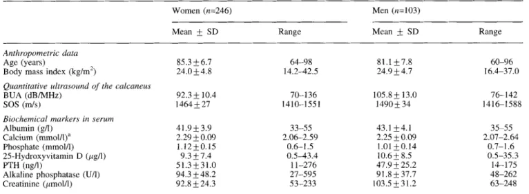
Anthropometric data of the population (246 women and 103 men), and the mean values of the quantitative ultrasound measurements of the calcaneus and of the biochemical markers, are shown in Table 1. The mean age was over 80 years in both sexes. Compared with the normal population, the mean values of 25-hydroxyvita- min D were low in both sexes, while the mean PTH and alkaline phosphatase values were slightly above the normal mean. The plasma levels of 25-hydroxyvitamin D were below the 2.5th percentile (6.2 #g/l, i.e. 15.5 nmol/1 in IU) of a large (n = 3276) Swiss adult general population [20] in 41.9% of the women and in 31.4% of the men. Compared with the general population aged > 6 5 years, 28.8% of the women and 21.6% of the men were below the 2.5th percentile (4.3
#g/1,
i.e. 10.7 mmol/1 in IU). Concerning the serum level of PTH, 18.9% of the women and 9.8% of the men were above the 97.5th percentile (70 pg/1) of normal adults.Factors Influencing QUS of Bone in the Elderly
Table 1. Anthropometric data, quantitative ultrasound of the calcaneus and biochemical markers
117 Women (n=246) Mean ,+ SD Range Men (n=103) Mean ,+ SD Range Anthropometric data Age (years) 85.3 _+ 6.7 64-98 81. l +_ 7.8 60-96
Body mass index (kg/m 2) 24.0_+4.8 14.2.42.5 24.9-+4.7 16.4-37.0
Quantitative ultrasound of the calcaneus
BUA (dB/MHz) 92.3 -t- 10.4 70-136 105.8 ,+ 13.0 76-142
SOS (m/s) 1464 -t- 27 1410-1551 1490 ,+ 34 1416-1588
Biochemical markers in serum
Albumin (g/l) 41.9 ,+ 3.9 33-55 43.1 -t- 4.1 35-55
Calcium (mmoltl)" 2.29 4- 0.09 2.06-2.59 2.25 _+ 0.09 2.07-2.64
Phosphate (mmol/1) 1.12 ,+ 0.15 0.6-1.5 1.01 -t- 0.14 0.7-1.6
25-Hydroxyvitamin D (#g/l) 9.3 _+ 7.4 0.5-43.4 10.6 ,+ 8.5 0.5-35.3
PTH (ng/l) 5 1.3 ,+ 31.0 11-276 47.9 ,+ 25.2 14-175
Alkaline phosphatase (U/l) 94.3 -+48.2 27-595 91.8 ,+ 37.7 48-262
Creatinine (/~mol/1) 92.8 -t- 24.3 53-233 103.5 -t-_ 31.2 63-248
BUA, broadband ultrasound attenuation; SOS, speed of sound; PTH, parathyroid hormone. ~Corrected for albumin.
e - v u.I +1 -1- I - C 0= E 10 25 9O 8O 7O 6O 5O 4O 3O 2O 10 0 0
I
50 75 90 97.5 percentiles of ~stitutionalized women 2 5 O H V i t D 5 10 15 20 25 30 35 40 45gg/I
I I
[ [ I I I percentiles of 2,5 5 25 50 75 95 97.5 Swiss adultsII
I
I
I
I
I
swiss adu,s
2.5 5 25 50 75 95 97.5 >65
y e a r sFig. 1. Comparison of mean parathyroid hormone (PTH) values ( ,+ SEM) and the 25-hydroxyvitamin D levels presented as groups of percentiles of a population of 246 institutionalized elderly women. For comparison, the percentiles of 25-hydroxyvitamin D of the normal Swiss adult population (general population, (n = 3276; people aged > 65 years, n = 215) are shown at the bottom. An increase in mean PTH is observed when 25-hydroxyvitamin D levels are below 7.6/~g/1 (19.0 nmol/1 in IU), corresponding to the 50th percentile for institutionalized elderly women but to the 5th percentile for healthy Swiss adults.
After excluding subjects with serum creatinine higher than 120 gmol/1 (n = 46), the mean values of calcium and PTH levels remained unchanged; calcium (-t- SD) values were 2.27 mmol/1 (_+ 0.09) and 2.27 mmol/1 (-t- 0.09), PTH (_+ SD) values were 51.1 pg/1 (-t- 31.3) and 49 pg/1 ( + 30.1) (NS) before and after this exclusion respectively.
Fig. 1 shows the mean PTH values as a function of the 25-hydroxyvitamin D levels for the women, presented by groups of percentiles. For comparison, the percentiles of 25-hydroxyvitamin D of the normal Swiss adult
population, where no sex differences were found, are also shown [20]. In these elderly women, PTH increased progressively when the 25-hydroxyvitamin D levels were below 7.6 #g/1 (19.0 nmol/1 in IU), which corresponds to the 50th percentile of this group, and to the 5th percentile of the normal Swiss adult population. No such regular increase of PTH could be shown in men.
In women, PTH was negatively correlated with 25- hydroxyvitamin D (-- 0.485, p < 0.001) and phosphate (r=--0.188, p < 0 . 0 1 ) and positively correlated with
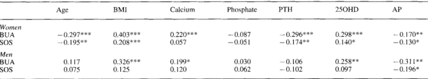
118 M.-A. Krieg et al. Table 2. Spearman rank correlation between quantitative ultrasound parameters and age, body mass index (BMI) and biochemical markers
Age BMI Calcium Phosphate PTH 25OHD AP
Wolrten BUA -- 0.297*** 0.403*** 0.220*** -- 0.087 -- 0.296*** 0.298*** -- 0.170"* SOS -0.195"* 0.208*** 0.057 --0.051 -0.174"* 0.140" -0.130" Men BUA 0.117 0.326*** 0.199" 0.030 -- 0.106 0.258** --0.311 ** SOS 0.075 0.125 0.120 0.062 --0.102 0.097 -0.196"
PTH, parathyroid hormone; 25OHD, 25-hydroxyvitamin D; AP, alkaline phosphatase. *p < (/.(/5, * *p < 0.02, ** *p < 0.01.
Table 3. Cocfticicnts of the multiple regression models ( _+ SEE) explaining the ultrasound parameters in w o m e n (n=246)
Intercept Age BMI PTH Phosphate R 2
B U A + 134 0.26 + 0.08 +0.88 ± 0.13 -- 6.38 _+ 1.18 - 14.70 + 3.93 0.3()
S()S + 1519 -- 0.62 +_ 0.25 + 1.12 ± 0.38 - 7.86 + 3.29 NS 0.10
alkaline phosphalase ( r = 0.206, p <0.01). 2 5 q t y d r o x y - vitamin D was correlated with alkaline phosphatase ( r = 0.285, p < 0 . 0 0 1 ) . In men, PTH was negatively correlated with 25-hydroxyvitamin D ( -- 0.317, /~<().01), phosphate ( - 0 . 2 6 5 , p < 0 . 0 1 ) and calcium ( r = - ( I . 3 3 9 , p <0.001).
The correlations between age, body mass index, biochemical markers and the ultrasound measurements by biwu'ialc analysis arc presented in Table 2. Significant prediclors of BUA were age, body mass index, calcium,
PTH, 25-hydroxyvitamin D and alkaline phosphatase in
women, and body mass index, calcium, 25-hydroxy- vitamin D and alkaline phosphatase in men. Age, body mass index, PTH, 25-hydroxyvitamin D and alkaline phosphatase were significant predictors of SOS in women, but with lower coefficients of correlation than for BUA, whereas in men, only alkaline phosphatase was a signilicanl predictor of SOS.
In Table 3, the multiple regression analysis in women showed that age, body mass index, P T H and phosphate were independent significant predictors of BUA. Because of their correlation with PTH, 25-hydroxyvita- min D and alkaline phosphatase were no longer independent predictors of BUA. According t o R 2, 30% of the variability of B U A was explained by this model. For SOS, only age, body mass index and P T H were independent significant predictors, with, according to R 2, only 10% of the variability in SOS being explained by this model.
Discussion
The aim of this study was to assess whether anthropo- metric parameters and biochemical markers of bone metabolism have a significant influence on quantitative ultrasound of bone in institutionalized elderly persons known to have a high percentage of vitamin D deficiency and secondary hyperparathyroidism. Indeed, the percen-
tagc of these metabolic disturbances was high, in agreement with the literature ll-5,21 ], and was higher in women than in men. As expected, P T H was negatively correlated with 25-hydroxyvitamin D. In addition, in women, PTH and 25-hydroxyvitamin D were both correlated with alkaline phosphatase (the latter nega- tively), confirming the known interdependency of these markers. These correlations with alkaline phosphatase were not found by Brazier et al. [21] who assessed institutionalized elderly with a serum level of 25- hydroxyvitamin D below 6 /~g/1 (15.6 mnol/l). In a subgroup analysis in women with a serum level of 25- hydroxyvitamin D below 6
itg/l,
we still found significant correlations. This discrepancy might be explained by the smaller number of subjects examined by Brazier et al. The correlation between P T H and 25- hydroxyvitamin D showed that the mean PTH level increased when the 25-hydroxyvitamin D level dropped below 7.6/tg/1 (19.0 nmol/1), and rose above 70 ng/1 (the upper normal value of our laboratory) at a 25- hydroxyvitamin D level of 3.4 bzg/1 (8.9 nmol/1). This is in partial agreement with the observations in an adult normal population by Chapuy et al. [22J who showed that P T H rose above 55 ng/1 (their upper normal limit) when the 25-hydroxyvitamin D level was below 4.6/tg/1 (11.3 nmol/l) [22],The ultrasound measurements were low when compared with other studies, mainly due to the advanced age of the subjects [19] (mean T-score for women as defined by the manufacturer: B U A = 57%, SOS = 47%; mean T-score for men: B U A = 75%, SOS = 61%). They were also found to be linked to the disturbances of bone metabolism related to older age: the bivariate analysis revealed that in addition to the already known negative influence of age and positive influence of B M I [19, 2 3 - 25] all major markers were correlated with BUA, i.e. 25-hydroxyvitamin D, alkaline phosphatase and to a lesser degree calcium, in both sexes, and P T H in women.
Factors Influencing QUS of Bone in the Elderly 119
This points to a rather non-specific reflection of bone mass and structure by BUA, as influenced by the various known pathogenic factors of osteoporosis in advanced age. The absence of correlation with P T H in men is probably explained by the smaller number of subjects. It might also be explained by the lower incidence of secondary hyperparathyroidism in men even though there is no reason to assume that bone metabolism in advanced age is sex dependent. In essence, men showed the same trend in correlation between markers and BUA, probably less significantly because of the smaller number.
SOS correlated in the women with all parameters in the same order as the correlations with BUA, but less significantly. In men all the correlations of BUA were less significant than in women. SOS was not significantly correlated with any of the biological parameters, except alkaline phosphatase. SOS is supposed to measure a quality of bone other than that measured by BUA, but its relation to the biochemical profile is the same as that of BUA, only less evidently.
The correlations are comparable to those found between biochemical markers and bone mineral density of the femoral neck as measured by DXA. Although Chapuy et al. [26] did not find any correlation between 25-hydroxyvitamin D or PTH and hip BMD, Ooms et al. [27] found a correlation coefficient between BMD of the femoral neck and P T H of - 0 . 2 7 0 ( p < 0 . 0 0 1 ) in 330 women aged +_ 80.3 years. Martinez et al. [28] found a correlation between 25-hydroxyvitamin D and B M D of the femoral neck with a correlation coefficient of 0.290 (,9 < 0.005) in 62 women aged more than 68 years. Szluc et al. [29] published data on the relationship between hip BMD and anthropometric parameters and biochemical markers in 98 elderly institutionalized women aged ± 81.4 years. The COl'relation coefficients were --0.41 to
--0.47 for age ( p < 0 . 0 0 1 ) , 0.39 to 0.51 for body weight ( p < 0 , 0 0 1 ) , - 0 . 2 7 to --0.33 for PTH ( p < 0 . 0 0 1 ) , --0.21 to - 0 . 3 3 for alkaline phosphatase ( p < 0 . 0 0 5 ) and --0.01 to --0.09 (NS) for 25-hydroxyvitamin D. This leads to the assumption that DXA and quantitative ultrasound reflect partially the same pathogenetic factors, although probably measuring different charac- teristics of bone. Age and body weight showed the highest correlations, followed by PTH. The absence of a significant correlation with 25-hydroxyvitamin D in this study can be explained by the relatively small number of subjects examined. From these studies it can be concluded that there is no essential difference between BUA of the heel and BMD of the hip as measured by DXA regarding their correlations with anthromometric and biochemical parameters.
In women, a multivariate regression analysis was performed to examine more specific relationships between biochemical markers and quantitative ultra- sound. This analysis explained 30% of the variance of BUA and 10% of the variance of SOS by the evaluated parameters.
The multivariate regression analysis showed that secondary hyperparathyroidism expressed by PTH level
was an independent predictor of BUA. However, neither 25-hydroxyvitamin D nor alkaline phosphatase was an independent predictor. This allows the conclusion that bone quality evaluated by B U A reflects the effect of the elevated P T H level rather than that of the lower 25-hydroxyvitamin D. Surprisingly, phosphate was an independent negative predictor of BUA, although it showed no significant correlation with any of the markers (except with PTH) or with BUA in the bivariate analysis. Phosphate levels were normal or low in this population, and a positive correlation with BUA could be expected since low phosphate levels reveal malnutri- tion and/or vitamin D deficiency, both potentially contributing to a low BUA. It can only be hypothesized that a relatively high serum phosphate level (there was no hyperphosphatemia) revealed a more advanced reduction in renal function and by this a tendency to secondary hyperparathyroidism, but no such correlations could be seen in the bivariate analysis.
In conclusion, the quantitative ultrasound parameter BUA, and to a much smaller degree SOS, evaluates the characteristics of bone that are influenced mainly by age, BMI and secondary hyperparathyroidism. In order to support these observations, further investigations are needed to examine the question of whether the reduction of secondary hyperparathyroidism leads to an improve- ment in BUA.
Part of this work was presented at the 17th Annual Meeting of the American Society of Bone and Mineral Research in Baltimore, MA, USA, September 1995.
References
1. Corless D, Gupta SP, Sattar DA, Switala S, Boucher BJ. Vitamin D status of residents of an old people's home and long-stay patients. Gerontology 1979;25:350-5.
2. Chapuy MC, Chapuy P, Meunier PJ. Calcium and vitamin D supplements: effects on calcium metabolism in elderly people. Am J Clin Nutr 1987;46:324-8.
3. Lips P, Wiersinga A, Van Ginkel FC, Jongen MJM, Netelenbos JC, Hackeng WHL, et al. The effect of vitamin D supplementa- tion on vitamin D status and parathyroid function in elderly subjects. J Clin Endocrinol Metab 1988;67:644-50.
4. Heikinheimo RJ, Inkovaara JA, Harju EJ, Haavisto MV, Kaarela RH, Kataja JM, et al. Annual injection of vitamin D and fractures of aged bones. Calcif Tissue Int 1992;51:105-10.
5. Deroisy R, Zheng XS, Pirenne H, Frederick I, Collette J, Reginster JY. 25-Hydroxyvitamin D deficiencies and secondary hyperparathyroidism in elderly women living at home or in nursing/retirement homes. Osteoporos Int 1996;6(Suppl 1):119. 6. Burckhardt P, Gerber S, Jacquet AF, Thi6baud D. Lack of fish is
the main cause of vitamin D deficiency in institutionalized elderly swiss subjects. J Bone Miner Res 1993;8(Suppl 1):$218. 7. Recommended Dietary Allowances. 9th ed. Food and Nutrition
Board, Nutritional Research Council. Washington, DC: National Academy of Sciences, 1980.
8. Chapuy MC, Arlot ME, Duboeuf F, Brun J, Crouzet B, Arnaud S, et al. Vitamin D 3 and calcium to prevent hip fractures in elderly women. N Engl J Med 1992;327:1637-42.
9. Lees B, Stevenson JC. Preliminary evaluation of a new ultrasound bone densitometer. Calcif Tissue Int 1993;53:149-52.
10. Yamasaki K, Kushida K, Ohmura A, Sano M, Inoue T. Ultrasound bone densitometry of the os calcis in Japanese women. Osteoporos Int 1994;4:220 5.
120
11. Schott AM, Weill-Engerer S, Hans D, Duboeuf F, Delmas PD, Meunier PJ. Ultrasound discriminates patients with hip fracture equally well as dual energy X-ray absorptiometry and indepen- dently of bone mineral density. J Bone Miner Res 1995;10:243-9. 12. Krieg MA, Thi6baud D, Landry M, Burckhardt P. Evaluation de la qualitE osseuse par ultrason quantitatif. Schweiz Med Wochenschr 1996;126:159-63.
13. Bauer DC,Gltier CC, Pressman AR, Vogt TM, Ensrud KE, Cauley JA, et al. Broadband ultrasonic attenuation (BUA) and the risk of fracture: a prospective study. J Bone Miner Res 1995;10(Suppl
I):S175.
14. Hans D, Dargent-Molina P, Schott AM, Sebert JL, Cormier C, Kotzki PO, et al. Ultrasonographic heel measurements to predict hip fracture in elderly women: the EPIDOS prospective study. Lancet 1996;348:511-4.
15. Ashman RB, Cowin SC, Van Buskirk WC, Rice JC. A continuous wave techique for measurement of elastic properties of cortical bone. J Biomech 1984; 17:349-6 [.
16. Gliiere CC, Wu Cy, Genant HK. Broadband ultrasound attenuation signals depend on trabecular orientation: an, in vitrol sludy. Osteoporos Int 1993;3:185 91.
17. Hans D, Arlot ME, Schott AM, Roux JP, Kotzki PO, Meunier PJ. Do ultrasound measurements on the us calcis reflect more the bone microarchitecture than the bone mass? A two-dimensional histomorphometric study. Bone 1995;16:295-300.
18. Wacrn E, Christiansen C, Gause-Nilsson 1, G. Lindstedt, Oden A, Riis B, Mellst]6m 1). Calcidiol (25-()ll vitamin D) is a predictor of bone mineral density and hip fracture. Osteoporos ln/
1996:6(Suppl I ): 127.
19. Krieg MA, Thidbaud D, Burckhardt P. Quantitative ultrasound of bone in institutionalized elderly women: a cross-sectional and longitudinal study. Osteoporos Int 1996;6:189-95.
20. Bumand B, Sloutskis D, Gianoli F, Cornuz J, Riggenbach M, Paccaud F, Burckhardt P. Serum 25-()H vitamin D: distribution and detcrnfinants in the Swiss population. Am J Clin Nutr 1992:56:537-42.
M.-A. Krieg et al. 21. Brazier M, Kamel S, Maamer M, Agbomson F, Elesper I, Garabedian M, et al. Markers of bone remodeling the elderly subject: effects of vitamin D insufficiency and its correction. J Bone Miner Res 1995;10:1753-61.
22. Chapuy MC, Preziosi P, Maamer M, Arnaud S, Galan P, Hercberg S, Meunier PJ. Prevalence of vitamin D insufficiency in an adult normal population. Osteoporos Int 1997;7: in press. 23. Schott AM, Hans D, Sornay-Rendu E, Dehnas PD, Meunier PJ.
Ultrasound measurements on os calcis: precision and age-related changes in a normal female population. Osteoporos Int 1993;3:249-54.
24. Van Daele PLA, Burger H, Algra D, Hofman A, Grobbe DE, Birkenh~iger JC, Pols HAP. Age-associated changes in ultrasound measurements of the calcaneus in men and women: the Rotterdam study. J Bone Miner Res 1994;9:1751-7.
25. Yamasaki K, Kushida K, Ohmura A, Sano M, Inoue T. Ultrasound bone densitometry of the os calcis in Japanese women. Osteoporos Iut 1994;4:220 5.
26. Chapuy MC, Schott AM, Garnero P, Hans D, Delmas PD, Meunier PJ, and the EPIDOS Study Group. Healthy elderly French women Jiving at home have secondary hyperparathyroid- ism and high bone turnover in winter. J Clin Endocrinol Metab 1996;81:1129-33.
27. Ooms ME, Lip P, Roos JC, van der Vijgh WJ, Popp-Snijders C, Bezemer PD, Bouter LM. Vitamin D status and sex hormone binding globulin: determinants of bone turnover and bone mineral density in elderly women. J Bone Miner Res ~995;10:1177 84. 28. Martmez ME, del Campo MT, Sanchez-Cabezudo M J, Garcia JA,
Sanchez-Calvin MT, Torrijos A, et al. Relations between calcidiol, serum levels and bone mineral density in postmeno- pausal women with low bone density. Calcif Tissue lnt
1994;55:253 6,
29. Szluc P, Arlot M, Chapuy MC, Duboeuf F, Meunier P J, Dehnas PD. Serum undercarboxylated osteocalcin correlates with hip bone mineral density in elderly women. J Bone Miner Res
1994;9:1591-5.
Received fi?r publication II Februao? 1997 Accepted in revised /orm 13 August 1997