1092-2172/04/$08.00⫹0 DOI: 10.1128/MMBR.68.4.692–744.2004
Copyright © 2004, American Society for Microbiology. All Rights Reserved.
Type V Protein Secretion Pathway: the Autotransporter Story
Ian R. Henderson,
1* Fernando Navarro-Garcia,
2Mickae
¨l Desvaux,
1Rachel C. Fernandez,
3and Dlawer Ala’Aldeen
4Division of Immunity and Infection, University of Birmingham, Birmingham,1and Division of Microbiology &
Infectious Diseases, University Hospital, Nottingham,4United Kingom; Department of Cell Biology,
CINVESTAV, Mexico City, Mexico2; and Department of Microbiology & Immunology,
University of British Columbia, Vancouver, British Columbia, Canada3
INTRODUCTION ...693
SECRETION BY NUMBERS: PROTEIN SECRETION MECHANISMS IN GRAM-NEGATIVE BACTERIA...693
Type I Protein Secretion ...694
Two-Step Type II Protein Secretion ...695
The Injectisome: Type III Protein Secretion...696
Type IV Protein and Nucleoprotein Secretion ...697
TYPE V PROTEIN SECRETION...697
Autotransporter Secretion Pathway (Type Va)...698
Inner membrane transport...699
Periplasmic transit ...701
-Domain ...702
Translocation of the passenger domain...702
Processing of autotransporters at the surface...704
Energy of transport ...705
Evolution and distribution of autotransporter proteins ...705
Two-Partner Secretion Pathway (Type Vb) ...705
Type Vc ...708
CLUSTER 1: SUBTILASE FAMILY OF AUTOTRANSPORTERS ...709
Ssp...709
PspA and PspB...709
Ssa1...709
SphB1 ...709
AspA/NalP ...710
CLUSTERS 2 AND 11: HELICOBACTER PYLORI AUTOTRANSPORTERS ...710
VacA, the Vacuolating Cytotoxin ...710
Molecular and structural features ...710
Vacuolating activity ...711
Other functions ...711
Epidemiology of H. pylori infection ...711
BabA, SabA, and AlpA ...712
CLUSTERS 3 AND 4: AIDA AUTOTRANSPORTER FAMILY...713
AIDA-I ...713
Antigen 43 ...714
IcsA ...716
TibA, ShdA, and MisL ...716
AutA and AutB ...718
CLUSTERS 5 AND 8: SERINE PROTEASE AUTOTRANSPORTERS ...718
IgA1 Proteases...718
H. influenzae Adhesion and Penetration Protein (Hap) ...720
N. meningitidis App and MspA Autotransporter Proteins...720
Serine Protease Autotransporters of the Enterobacteriaceae ...721
Tsh ...721
SepA ...722
EspC ...722
EspP...722
Pet ...723
* Corresponding author. Mailing address: Division of Immunity and Infection, The Medical School, University of Birmingham, Birming-ham B15 2TT, United Kingdom. Phone: 44 121 414 4368. Fax: 44 121 414 3599. E-mail: [email protected].
Pic ...723
SigA...725
Sat ...725
Vat ...725
Others ...725
CLUSTER 6: BORDETELLA AUTOTRANSPORTERS ...726
Identification of Autotransporters in the Bordetella Genomes ...726
Pertactin ...727
BrkA, TcfA, and Vag8...728
CLUSTER 7: CHLAMYDIAL AUTOTRANSPORTERS ...729
CLUSTER 9: RICKETTSIAL AUTOTRANSPORTERS ...730
CLUSTER 10: LIPASES AND ESTERASES ...730
EstA...730
ApeE...731
Lip-1...731
McaP...731
OTHER AUTOTRANSPORTER PROTEINS...731
Adhesins ...731 Sialidases ...732 FUTURE DIRECTIONS ...732 ACKNOWLEDGMENTS ...733 REFERENCES ...733 INTRODUCTION
Until the 1960s, protein secretion through biological mem-branes was thought to be a relatively rare phenomenon. Fur-thermore, this view was governed by the assumption that where a protein was secreted it crossed the membrane via its own dedicated and specific mechanism. In the intervening years this view has radically changed, with the growing realization that both prokaryotic and eukaryotic cells secrete a prodigious ar-ray of proteins and possess protein translocation pathways which are surprisingly conserved throughout the phylogenetic kingdoms.
A surprising amount of information regarding bacterial pro-tein secretion has been derived from the study of bacterial pathogenesis. Indeed, molecular and genetic investigations of bacteria have shown that pathogenic organisms may be differ-entiated from their nonpathogenic counterparts by the pres-ence of genes encoding specific virulpres-ence determinants and that those proteinaceous virulence determinants, in the form of adhesins, toxins, enzymes, and mediators of motility, are usually secreted to or beyond the bacterial cell surface (139). Thus, by decorating their cell surfaces with such proteins, pathogens can directly or indirectly increase their ability to survive and multiply in the host. However, a facet often ig-nored by those studying bacterial pathogenesis and protein secretion is that many nonpathogenic organisms also secrete proteins which are adaptive to their life-styles; e.g., saprophytic bacteria may secrete cellulases or other degradative enzymes. However, in both cases, secretion of these factors is governed in large part by the structure of the bacterial cell envelope.
Gram-positive bacteria produce a single plasma membrane, the cytoplasmic membrane, followed by a thick cell wall layer. In contrast, gram-negative bacteria produce a double-mem-brane system consisting of a cytoplasmic memdouble-mem-brane, also called the inner membrane, and an outer membrane which sandwich the peptidoglycan and periplasmic space between them. Pro-tein secretion across the inner membrane of both gram-posi-tive and gram-negagram-posi-tive organisms generally follows the same
routes, normally involving the Sec-dependent pathway (often referred to as the general secretory pathway [GSP]), although other routes have recently been identified, e.g., the Tat and signal recognition particle (SRP) pathways (96, 109, 116, 119, 208, 338, 419, 434). However, once across the inner membrane, the fate of the translocated proteins diverge. In gram-positive organisms, the proteins are either released into the extracel-lular environment or incorporated into the cell wall layer by virtue of one of several cell wall peptide-anchoring mecha-nisms (78, 319). In contrast, translocation across the gram-negative inner membrane results in release of the protein into the periplasmic space. Thus, proteins that are targeted for the cell surface or extracellular milieu must also cross the addi-tional barrier to secretion formed by the outer membrane. To achieve translocation of these proteins to the cell surface and beyond, gram-negative bacteria have evolved several dedicated secretion systems, some of which bypass the Sec-dependent system and integrate both inner and outer membrane transport in a temporally linked fashion (see below).
In this review, we comprehensively describe the autotrans-porter secretion pathway, which is present in gram-negative organisms including animal and plant pathogens. We examine the roles of these proteins in the context of bacterial patho-genesis and highlight areas for future research. Readers are referred to more concise reviews of the autotransporter secre-tion pathway (101, 102, 203) and the roles of these proteins in pathogenesis (201).
SECRETION BY NUMBERS: PROTEIN SECRETION MECHANISMS IN GRAM-NEGATIVE BACTERIA Recent progress in the molecular analysis of the protein secretion pathways of gram-negative bacteria has revealed the existence of at least five major mechanisms of protein secre-tion. These pathways are highly conserved throughout the gram-negative bacterial species and are functionally indepen-dent mechanisms with respect to outer membrane transloca-tion; commonalities exist in the inner membrane transport
steps of some systems. For better or worse, these pathways have been numbered type I, II, III, IV, and V. Several more extensive reviews of the gram-negative protein secretion sys-tems have recently been published (4, 32, 64, 66, 147, 156, 264, 274, 294, 377, 395, 428, 429). Figure 1 gives an overview of the Type I, II, III and IV secretion pathways.
At this juncture, it is worth noting that some confusion has existed in the literature regarding the nomenclature of the type IV and type V protein secretion pathways. Almost simulta-neously, the type IV nomenclature appeared in several articles referring to both the autotransporter secretion pathway and the family of pathways exemplified by Agrobacterium tumefa-ciens T-DNA secretion. However, this matter has recently been resolved, with a consensus reached on the nomenclature of the pathways (160, 202). Thus, the A. tumefaciens and similar se-cretion pathways have retained the type IV terminology and the pathway used by the autotransporter and similar proteins has been termed the type V secretion system.
Type I Protein Secretion
The prototypical example of the type I secretion system (TOSS) is that required for secretion of Escherichia coli alpha-hemolysin (HlyA) (156). HlyA is a lipid modified protein with a repetitive domain composed of 11 to 17 9-amino-acid repeats which bind calcium and are thought to interact with host cells. This interaction stimulates the insertion of HlyA into the plasma membrane of eukaryotic cells, causing pore formation and release of the cytoplasmic contents. Other well-studied members of this group include the metalloprotease of Erwinia chrysanthemi (32, 160), the leukotoxin of Pasteurella haemo-lytica (345), and the adenylate cyclase of Bordetella pertussis (272).
Secretion of HlyA, and similar effector molecules, occurs in a Sec-independent manner and in a continuous process across both the inner and outer membranes. Furthermore, proteins secreted by TOSSs are not processed during secretion and do FIG. 1. Schematic representation of the type I, II, III, and IV protein secretion systems. The type I pathway is exemplified by hemolysin A (HlyA) secretion in E. coli, the type III system is exemplified by Yop secretion in Yersinia, the type II system is exemplified by pullulanase secretion in Klebsiella oxytoca, and the type IV system is exemplified by the VirB system in A. tumefaciens. ATP hydrolysis by HlyB, YscN, SecA, and VirB11 is indicated. Secreted effector molecules are depicted as grey ovals. The type II and in some cases the type IV secretion systems utilize the cytoplasmic chaperone SecB, although the Tat export pathway has recently been implicated in the secretion of molecules via the type II pathway. Type III secretion also involves cytoplasmic chaperones (SycE); however, they do not interact with the Sec inner membrane translocon. The major structural proteins of each system are depicted in relation to their known or deduced position in the cell envelope. EM, extracellular milieu; OM, outer membrane; Peri, periplasm; IM, inner membrane; Cyto, cytoplasm.
not form distinct periplasmic intermediates. Rather, the TOSS consists of three proteins: a pore-forming outer membrane protein, a membrane fusion protein (MFP), and an inner membrane ATP-binding cassette (ABC) protein (32). These proteins are represented in E. coli by TolC, HlyD and HlyB, respectively. Evidence suggests that the MFP (HlyD) and the ABC transporter (HlyB) in E. coli interact before binding of the effector (156). The secretion process begins when a secre-tion signal located within the C-terminal end of the secreted effector molecule interacts with the ABC transporter protein (224). In general, this signal sequence is specific and is recog-nized only by the dedicated ABC transporter. Once binding of the effector molecule occurs, interaction of HlyD with the outer membrane protein TolC is triggered, allowing secretion of the effector molecule to he external milieu (450). However, this model is somewhat controversial, since investigation of the E. chrysanthemi TOSS indicates that the ABC transporter and MFP interact only after binding of the effector molecule and subsequently MFP interacts with the pore-forming outer mem-brane protein (282). Nevertheless, it is apparent that in both cases the bridging formed by MFP and the outer membrane protein collapses after export of the effector molecule (485). ATP hydrolysis by the ABC transporter provides the impetus to drive secretion of the effector molecule across the cell en-velope to the external milieu once bridging of all molecules has occurred. Hydrolysis of ATP is not required for interaction of the effector with the ABC protein or assembly of the TOSS complex (261, 262).
Recent data have indicated a possible structure for the TOSS complex. Resolution of the crystal structure demon-strated that TolC exists as a trimer and that the portion
span-ning the outer membrane formed a-barrel structure (Fig. 2)
(263). However, unlike the typical porins, each monomer of
TolC contributes four-strands to form a 12-strand
antipar-allel -barrel. In addition to forming the -barrel structure,
TolC possesses a novel protein structure: the␣-helical barrel.
This extends from the integral outer membrane-barrel
struc-ture into the periplasm (263). HlyD is also known to form a trimeric structure which projects from the inner membrane into the periplasm. Thus, it is easy to envisage a scenario where the monomers of HlyD and TolC, while forming their trimeric complexes, can extend to form a continuous channel across the inner and outer membranes at the appropriate time. Never-theless, further investigations are required to fully appreciate the roles of each protein in the TOSS.
Two-Step Type II Protein Secretion
The type II secretion pathway is often referred to as the main terminal branch (MTB) of the Sec-dependent GSP (428). The subunits of a type II secretion pathway identified in E. coli have been designated GspA to GspO (146). Unfortunately, this has served to confuse the general reader about the no-menclature of the type II secretion pathway and the GSP (100). Thus, it must be stressed here that type II secretion does not refer to the GSP in general but refers only to one of several branches, as described below. Recently, it has been suggested that the pathway be renamed the secreton-dependent pathway (516).
Secretion via the MTB is exemplified by pullulanase (PulA)
secretion from Klebsiella oxytoca (478, 57). PulA is a starch-hydrolyzing lipoprotein which forms micelles once it has been secreted into the external environment (478). Since the discov-ery of the MTB in K. oxytoca, the pathway has been identified in a variety of other bacterial species exporting a range of proteins with diverse functions including the cholera toxin of Vibrio cholerae, exotoxin A of P. aeruginosa, and several cell wall-degrading enzymes (428, 429). Furthermore, analyses of the MTBs and type IV fimbrial systems at the genetic and structural levels have shown sufficient similarity between the two systems to suggest that the fimbrial biogenesis pathway be classed as a type II secretion pathway (34).
Secretion of substrate proteins via the MTB occurs in a two-step Sec-dependent fashion. Thus, all proteins secreted via a type II secretion pathway are synthesized with an N-terminal FIG. 2. Structure of TolC. The structure of TolC was solved to 2.1-A˚ resolution. The functional TolC unit consists of three TolC mon-omers oligomerized into a regular structure embedded in the outer membrane and extending into the periplasm such that it forms a continuous solvent-accessible conduit. The portion of the trimeric mol-ecule that is embedded in the outer membrane adopts a 12-strand -barrel conformation consisting of four -strands per monomer. The molecule is 140 A˚ long, the-barrel is 40 A˚ long (spanning the outer membrane), and the remaining portion of the molecule adopts a 100 A˚ ␣-helical conformation spanning the periplasmic space. This ␣-helical domain consists of 12␣-helices, such that the ␣-helices extend from or connect to the periplasmic side of each-strand. It is the ␣-helical domain that is predicted to interact with the MFP. Each monomer is highlighted in a separate color. Adapted from reference 263 and the Protein Data Bank (30).
signal sequence which targets proteins for secretion to the Sec inner membrane secretion pathway (for in-depth reviews, see references 115; 245; and 509). The Sec machinery is composed of an ATPase, SecA, several integral inner membrane proteins (SecD, SecE, SecF, SecG, and SecY, and a signal peptidase (116, 338). SecB, a cytoplasmic chaperone, does not recognize the signal sequence but recognizes the mature part of the protein targeted for secretion and subsequently directs the protein to the Sec translocon (109). SecA provides the energy for transport through the translocon, and the signal peptidase cleaves the signal sequence, releasing the remainder of the protein into the periplasm. Evidence suggests that, once in the periplasm the remaining protein adopts a quasi-native state which is facilitated by certain chaperones such as disulfide bond isomerase, DsbA (486). These folding steps are necessary for translocation across the outer membrane.
Transport across the outer membrane requires an additional 12 to 16 accessory proteins, which are collectively referred to as the secreton (428, 429, 486). A consensus nomenclature has been defined by the letters A to O and S for homologous genes and their products (the Xcp proteins of P. aeruginosa are la-belled P to Z). The organization of the loci encoding the MTB components is relatively conserved, and most of the genes are transcribed from a single operon in which several of the genes are overlapping (428, 429). Evidence from both the Klebsiella Pul system and the Erwinia Out system for pectinase secretion suggests that protein C contributes to a species-specific inter-action defining the transport of specific substrate proteins (42, 290, 404, 405). Protein D belongs to a family of proteins termed secretins. These proteins are integral outer membrane proteins predicted to consist largely of transmembrane -strands and form a -barrel structure in the outer mem-brane, with 12 to 14 subunits forming a complex (34, 188, 257). Several possible configurations for the secretin complex have been proposed. The most popular of these include a complex where monomers of protein D oligomerize to form a ring with a central channel and a model reminiscent of the TolC
struc-ture, where each monomer contributes -strands to form a
single central channel (486). The C-terminal region is con-served among the secretins and is thought to be embedded in the outer membrane. It is interesting that homologues of the protein D secretin proteins have been found in the type III secretion pathways (see below) (486). Evidence suggests that protein E may act as a kinase regulating the secretion process by supplying energy to promote translocation and assembly of the pilin-like subunits, proteins G to K (430). No protein-protein interactions have been demonstrated for protein-protein F (488). Proteins G to K possess homology to the type IV pili and are thought to form a pseudopilus (112, 264). Furthermore, these proteins are N-terminally processed and methylated by a prepilin peptidase, protein O (36, 390). A lipoprotein, protein S, has been demonstrated to play a role in stabilization of the outer membrane component protein D (428). Several other accessory proteins, which are not present in all MTB pathways, have a demonstrated requirement for substrate secretion in certain MTB systems. Evidence suggests that this may be re-lated to the type of proteins being secreted or to substrate recognition by the MTB. In summary, the available evidence indicates that most if not all of the components of an MTB interact to form a multiprotein complex spanning both the
inner and outer membranes (Fig. 1). Many more comprehen-sive reviews are available which describe the structure and function of the components of the MTB (428, 429).
The Injectisome: Type III Protein Secretion
The archetypal type III secretion system (TTSS) was first identified in pathogenic Yersinia spp. for the secretion of Yop proteins (326). Since this discovery, TTSS have been identified in several mammalian and plant pathogens including Salmo-nella enterica, Shigella flexneri, E. coli, Ralstonia solancearum, Pseudomonas syringae, and Chlamydia trachomatis (214, 395). Recently, TTSS have been the subject of considerable interest because of their primary role in the virulence of these animal and plant pathogens. Interestingly, detailed investigations have shown that TTSS and the gram-negative flagellar export appa-ratus are homologous, leading some investigators to classify the flagellar biosynthesis pathway as a TTSS (37, 307). Thus, while the mechanism for secretion of effector molecules is highly conserved in the TTSS, the effector molecules them-selves are divergent and perform a variety of functions. Read-ers are referred to more in-depth reviews of the structure and function of TTSS, which cover the voluminous research avail-able (4, 37, 64, 214, 307, 395).
Genes encoding components required for TTSS are gener-ally located on a single plasmid or chromosomal locus. Com-parison of the sequences demonstrates that the genes at these loci are conserved among different species, suggesting that these loci are inherited as a distinct genetic unit and that a common mechanism exists for the recognition and secretion of effector molecules (4, 64, 294, 395).
Like the TOSS, the TTSS translocate their effector mole-cules across the inner and outer membranes in a Sec-indepen-dent fashion. However, it is important to note that the Sec translocon, as in the case of TOSS, is required for secretion of structural components across the inner membrane. Contro-versy exists about the mechanism of effector molecule recog-nition and targeting to the TTSS. One hypothesis suggests
that the signal resides in the 5⬘ mRNA, which may target the
ribosome-RNA complex to the TTSS, permitting temporal coupling of translation and secretion (11). A second proposal suggests that the N-terminal 20 amino acids serve as a binding site for cytoplasmic chaperones which specifically target the effector molecules to the TTSS (295). Notwithstanding the differences in these hypotheses, it is apparent that the region encoding the first 20 amino acids (either the untranslated mRNA or the first 20 amino acids of the polypeptide) is es-sential for secretion and the process is highly regulated (214). Once targeted to the cytoplasmic side of the TTSS, secretion of the effector molecule may occur without the formation of periplasmic intermediates, with secretion proceeding through a needle-like structure composed of approximately 20 different proteins (214, 269). Unfortunately, a description of the volu-minous evidence to support the roles of specific proteins in the formation of the TTSS complex is beyond the scope of this review. Based on the current evidence, the placement of spe-cific proteins in the TTSS complex is indicated in Fig. 1. How-ever, it is worth reiterating that the TTSS possesses a secretin-like component homologous to those found in the MTB systems (486). This secretin-like protein has been
demon-strated to form ring-shaped structures with large central pores, which facilitates secretion of effector molecules and stabilizes the needle-like complex formed by the other components of the TTSS (153). This complex spans both the inner and outer membranes, resembling a hypodermic needle. The formation of a needle-like structure is supported by the visualization of the S. enterica and S. flexneri TTSS by electron microscopy (269). These studies demonstrated a striking resemblance be-tween the TTSS and the flagellar basal body, and since the TTSS were shown to form long pilus-like structures, it was assumed, through analogy to the flagellar system, that the pilus structure allows protein transport across the bacterial outer membrane to the external milieu (214, 307). This was sup-ported by investigations showing structural and sequence sim-ilarities between the EspA pilus protein of the E. coli TTSS and the flagellar filament (89, 255).
Perhaps the most striking feature of TTSS is their ability to target effector proteins directly into eukaryotic cells. This phe-nomenon is triggered in some cases when the bacterium comes in contact with a eukaryotic cell; hence, secretion via the type III pathway has been termed “contact dependent” (545). The analogy between the TTSS and the hypodermic syringe, cou-pled with the ability of the systems to inject proteins directly into the host cytosol, has allowed the phrase “injectisome” to be coined for this protein secretion system. Nevertheless, in-vestigators should be reminded that not all TTSS are contact dependent and some effector molecules secreted by TTSS are released into the external environment (75).
Type IV Protein and Nucleoprotein Secretion The type IV protein secretion system (TFSS) is the least well understood gram-negative protein secretion pathway. The sys-tem is ancestrally related to the bacterial conjugation machin-ery, although some controversy exists over which system comes first on the evolutionary ladder. Nevertheless, it is clear that, like the TTSS, the unifying characteristic of the TFSS and the conjugative machinery is the ability to translocate protein mol-ecules intercellularly, i.e., from one bacterium to another or from a bacterium to a eukaryotic host cell. The prototypical TFSS is that of the A. tumefaciens nucleoprotein T-DNA trans-fer system. However, several other systems, which are required for the full virulence of pathogenic bacteria, have been de-scribed, including the B. pertussis Ptl (pertussis toxin) system (130), the Dot/Icm system of Legionella pneumophila (546), and those of Brucella suis (41), Bartonella henselae (443), and Helicobacter pylori (18). Representatives of the conjugal DNA transfer systems include the IncF, IncP, and IncW plasmid transfer systems (Tra) (277). Thus, despite being a relatively poorly understood protein secretion system, the TFSS has been studied over several generations of researchers through its analogies to the Tra systems.
As in other secretion pathways, the effector molecules se-creted by the TFSS have a wide variety of functions. Arguably, the best-characterized function is that of pertussis toxin, which
belongs to the A-B5toxin family (130, 481). Unlike the other
type IV systems, the pertussis toxin is secreted to the extracel-lular milieu rather than directly into a host cell (481). The B domain (subunits S2 to S5) interacts with the host cell glyco-protein receptors and mediates translocation of the A domain
(subunit S1) into the host cytosol. Once in the cytosol, the S1
subunit ribosylates the ␣ subunits of G-proteins, interfering
with signaling pathways (310). In contrast, the A. tumefaciens system secretes several effector molecules, VirD2, VirE2, and VirF, into the host cell cytosol (67, 212). VirD2 is secreted as a nucleoprotein complex; the protein remains covalently asso-ciated with a single-stranded copy of T-DNA (266). Once in the host cytosol, VirE2 interacts with the VirD2/T-DNA com-plex, mediating delivery of the T-DNA to the host cell nucleus and resulting in crown gall tumor formation. A role for the VirF protein has yet to be defined conclusively. Unlike the pertussis toxin, the Vir effector proteins are secreted separately through the TFSS.
Controversy exists over how the substrate molecules for TFSS pass through the inner membrane. For the A. tumefa-ciens protein-DNA complex, export is postulated to take place in a single continuous step from cytoplasm to the interior of the cell. In contrast, export of pertussis toxin, like the type II secretion pathway effector molecules, occurs in two steps; the toxin subunits are first translocated across the inner membrane via the Sec machinery and are subsequently targeted for export across the outer membrane (51, 52). Thus, one evolutionary branch of the TFSS appears to be a branch of the GSP whereas the division encompassing T-DNA transfer is not.
The process governing transfer across the outer membrane is best understood for the A. tumefaciens TFSS. The quantity of information to support the individual roles of separate proteins is too great to review here; however, it is worth mentioning the roles of a few major proteins. Transfer of T-DNA and assem-bly of the pilus requires VirA, VirB1 to VirB11, VirD1 to VirD4, VirE2, and VirG. VirB2 is the major pilus subunit, although it is not yet clear whether this pilus emanates from the inner or outer membrane. VirB4 and VirB11 are inner membrane proteins, and VirD4 is a cytoplasmic protein, all of which possess ATPase activity. It has been suggested that these proteins provide the energy for translocation of the effector molecules through the pilus structure in a fashion analogous to the TOSS and TTSS. The remaining proteins are variously associated with the inner and outer membranes and with each other; the proposed placement of each protein is indicated in Fig. 1. TFSS has been divided into two subclasses: type IVa corresponds to protein machinery containing VirB homo-logues of A. tumefaciens, and type IVb corresponds to func-tional secretion systems assembled from Tra homologues of the Incl Collb-P9 plasmid of Shigella flexneri (66). Several ex-cellent reviews have covered the functions of each subunit in more detail (66, 111, 143, 274, 277, 448).
TYPE V PROTEIN SECRETION
Perhaps the simplest protein secretion mechanisms are those which are included under the umbrella of type V secre-tion. This family of secreted proteins includes those secreted via the autotransporter system (type Va or AT-1), the two-partner secretion pathway (type Vb), and the recently de-scribed type Vc system (also temed AT-2) (99, 102). Proteins secreted via these pathways have similarities in their primary structures as well as striking similarities in their modes of biogenesis. Figure 3 provides a schematic overview of the type V secretion pathways.
Autotransporter Secretion Pathway (Type Va) Pohlner et al. (401) were the first to describe and propose a model for type V secretion, elegantly elucidating the relation-ship between the gene encoding the gonococcal immunoglob-ulin A1 (IgA1) protease and its extracellular product. Thus, the DNA sequence of a cloned fragment (4.6 kb) revealed a single gene coding for a 169-kDa precursor of IgA1 protease; the precursor contained three functional domains: the N-ter-minal leader (which is assumed to initiate the inner membrane transport of the precursor), the extracellular IgA1 protease, and a C-terminal “helper” domain (which is required for se-cretion across the outer membrane). In this seminal work, the authors proposed a model in which the helper serves as a pore for the secretion of the protease domain through the outer membrane. IgA1 protease acquires an active conformation as its extracellular transport proceeds and is released as a
pro-form from the membrane-bound helper by autoproteolysis. The soluble proform matures further into the functional
106-kDa IgA1 protease and a small stable␣-protein. It is possible
to find the active IgA1 protease in the culture medium from iga-transformed E. coli and S. enterica (185, 253), supporting the concept that the IgA1 protease precursor contains all the necessary determinants to direct the translocation of the pro-tease from the periplasm into the medium, without the partic-ipation of accessory proteins (401). Since no energy coupling or accessory factor seemed to be required for the translocation process, and unlike the type I to IV systems, the molecules targeted for secretion were strictly dedicated to their
co-valently linked cognate-domain, the proteins secreted in this
fashion received the name of autotransporters.
Since this initial description, many more proteins which fol-low this pathway of secretion have been identified among the FIG. 3. Schematic overview of the type V secretion systems. The secretion pathway of the autotransporter proteins (type Va) is depicted at the bottom left of the diagram, the two-partner system (type Vb) is depicted in the center of the diagram, and the type Vc or AT-2 family is depicted on the right. The four functional domains of the proteins are shown: the signal sequence, the passenger domain, the linker region, and the -domain. The autotransporter polyproteins are synthesized and generally exported through the cytoplasmic membrane via the Sec machinery. Interestingly, effector proteins with an unusual extended signal sequence, which purportedly mediates Srp-dependent export, are found in all three categories of type V secretion. Once through the inner membrane, the signal sequence is cleaved and the-domain inserts into the outer membrane in a biophysically favored-barrel structure that forms a pore in the outer membrane. After formation of the -barrel, the passenger domain inserts into the pore and is translocated to the bacterial cell surface, where it may or may not undergo further processing.
gram-negative bacteria (201). In all cases, the primary struc-ture of the autotransporter protein is reminiscent of the IgA1 protease, consisting of a modular structure composed of three domains (231). These domains have been given different names throughout the literature. In this review, we use a con-sistent nomenclature throughout; thus, the domains are termed the signal sequence, the passenger domain, and the translocation unit. The signal sequence (also called the signal peptide or leader sequence) is present at the N-terminal end of the protein and allows targeting of the protein to the inner membrane for its further export into the periplasm (203). The
next domain is the passenger domain (also called the
␣-do-main, N-passenger do␣-do-main, or N-domain), which confers the diverse effector functions of the various autotransporters. The last main domain, located at the C-terminal end of the protein,
is the translocation unit (also called the-domain, helper
main, C-domain, transporter domain, or autotransporter
do-main), consisting of a short linker region with an ␣-helical
secondary structure and a-core that adopts a -barrel tertiary
structure when embedded in the outer membrane (317, 364, 365, 475), facilitating translocation of the passenger domain through the outer membrane.
Inner membrane transport. Bioinformatic analysis of the
gonococcal IgA1 protease N terminus using SignalP (352) pre-dicts that this domain functions as a signal sequence, allowing targeting of the protein to the inner membrane. Similarly, the N-terminal regions of all other autotransporter proteins dis-play characteristics of the prototypical Sec-dependent signal sequence, for which SecB is presumed to act as the molecular chaperone (45, 203). These signal sequences possess (i) an n-domain with positively charged amino acids, (ii) an h-domain containing hydrophobic amino acids, and (iii) a c-domain with a consensus signal peptidase recognition site which often con-tains helix-breaking proline and glycine residues as well as
uncharged and short lateral-chain residues at positions⫺3 and
⫺1 that determine the site of signal sequence cleavage (315). However, some autotransporters, like AIDA-1, Pet, or Hbp, exhibit an unusually long signal sequence consisting of a C-terminus resembling a normal signal sequence with a hydro-phobic core and a consensus signal peptidase cleavage site but presenting a conserved extension of the n-domain which con-tributes most to the variation in overall length (203). In silico analyses of completed bacterial genomes has revealed the pres-ence of at least 80 proteins possessing these extended signal sequences across the breadth of the Proteobacteria (Fig. 4) (M. Desvaux and I. R. Henderson, unpublished observations). In-terestingly, these extended signal sequences appear to be as-sociated exclusively with proteins larger than 100 kDa. All of these atypical signal sequences, consisting of at least 42 amino acids, appear to consist of two charged domains (n1 and n2) and two hydrophobic domains (h1 and h2), in addition to the C-domain signal peptidase I recognition site (Fig. 4). While the n1 and h1 domains of these autotransporters are well con-served, the sequence of the n2 domain is variable and that of the following h2 domain even more so (203). The variability within the n2 and h2 domains is expected and consistent with Sec-dependent signal sequences, although, notably, the n2 do-main contains an unusual number of charged amino acids (49). In contrast, the conservation within the n1 and h1 domains is highly unusual and is characterized by the presence of
con-served aromatic amino acids in the n1 domain and a glutamate residue in the h1 domain (Fig. 4). Such sequence conservation is frequently indicative of the existence of a specialized biolog-ical function, suggesting that the extended n1-h1 portion of the signal sequence directs inner membrane export through a pathway different from the Sec system or that it recruits acces-sory proteins to work in tandem with the Sec pathway.
It was speculated that the features of those unusually long signal sequences might serve to recruit accessory proteins, dif-ferent from SecB, like the SRP, or to direct secretion via an alternative pathway such as the Tat pathway (203). In E. coli, protein export through the Sec translocon can use at least two pathways: (i) the pathway involving the molecular chaperone SecB, which keeps the preprotein in a translocation-competent state and targets it to SecA (413), and (ii) the SRP pathway, which permits a cotranslational translocation of the preprotein by targeting the ribosome-SRP complex directly to the trans-locon (208). Sijbrandi et al. (455) recently investigated the role of this signal sequence in Hbp. It was demonstrated that targeting and translocation through the inner membrane involved the targeting factor SRP and the Sec translocon. SecB was not required for the targeting of Hbp, but it could com-pensate for the absence of a functional SRP pathway to some extent. It was suggested that cotranslational translocation ver-sus posttranslational translocation might prevent degradation or premature folding of Hbp in the cytoplasm. This study presents the first example of an extracellular protein targeted by SRP; previously in gram-negative bacteria SRP was associ-ated only with insertion of proteins into the inner membrane (497). The authors speculated that the extended domain as a whole, i.e., n1-h1, and the hydrophobic region in particular might play an important role for the SRP recognition of the signal sequence. In fact, crystal structure analysis of the SRP has revealed an unusual RNA-protein interface that could interact with the hydrophobic core of the signal sequence (20). Controversially, the homologous EspP was found to be trans-located across the inner membrane via the classical Sec path-way (476). However, recent investigations have shown that the
truncated EspP signal sequence, i.e.,⌬Esp lacking n1-h1
do-mains, fused to proteins normally targeted to the Sec appara-tus by SecB leads to the rerouting of these proteins into the SRP pathway; therefore, the role of the extended region n1-h1 in secretion remains enigmatic (391). Furthermore, no evi-dence exists to support a role for this signal sequence in tar-geting to the Tat pathway (455; I. R. Henderson et al., unpub-lished observations) or for the involvement of other accessory factors.
Interestingly, several recent reports have described auto-transporter proteins, namely, the H. pylori AlpA, B. pertussis SphB1, and N. meningitidis AspA/NalP proteins (82, 358, 508), with typical lipoprotein signal sequences which are indicative of processing by signal peptidase II. While investigations of AlpA demonstrated that it was processed as a lipoprotein
when expressed in E. coli DH5␣, no such processing could be
demonstrated in wild-type H. pylori (358). An alternative hy-pothesis is that AlpA is processed further downstream by sig-nal peptidase I. Indeed, the protein may be acylated and pro-cessed by signal peptidase II and may subsequently undergo processing by signal peptidase I prior to insertion into the
Coutte et al. (83) demonstrated that SphB1, like AlpA, was processed as a lipoprotein in E. coli. However, such processing could not be demonstrated directly in the natural B. pertussis host. Nevertheless, growth in the presence of globomycin, a specific inhibitor of signal peptidase II, prevented localization of SphB1 in the outer membrane (83). Furthermore, the lo-calization of AspA/NalP (also termed NalP) was also inhibited by treatment with globomycin (508). Interestingly, the pres-ence of the lipoprotein signal sequpres-ence was found to be nec-essary for the functional localisation of SphB1 in the outer membrane, suggesting that it is required for anchoring SphB1 on the bacterial cell surface (83).
Periplasmic transit.After export through the inner
mem-brane, the autotransporter proprotein exists as a periplasmic intermediate. The original model of outer membrane secretion
predicts that the first of the-strands passes from the
peri-plasmic space to the external surface, thus leaving the passen-ger domain temporarily extending into the periplasm (203). Using a reporter single-chain antibody as a reporter passenger domain, the secretion process has been investigated (510). It was concluded that the folding of the passenger domain takes place in the periplasm before, or at least simultaneously with its own translocation through the outer membrane. However, a recent investigation by Oliver et al. (365) revealed the presence of an intramolecular chaperone in BrkA, spanned by residues 606 to 702. This region corresponds to PD002475 in the Pro-Dom database (76) and is found in many autotransporters but not in other proteins in the database. In the model derived from this study, the complete folding of the protein occurs on the surface of the bacteria (see the discussion of outer mem-brane transport below). This suggests that previous findings by Veiga et al. (510) may have been influenced by the study of a passenger domain unrelated to the autotransporters. Further-more, it is unlikely that large molecules, such as the passenger domains, can pass through a pore of 2 nm while fully folded, and therefore the molecules must be maintained in a translo-cation-competent unfolded state in the periplasm.
Thus, the status of the autotransporter proteins in the peri-plasm remains controversial. Of particular interest is how such large proteins can maintain an unfolded, or partially folded, state and resist degradation by periplasmic proteases. For IcsA, the soluble periplasmic form of the autotransporter
ap-pears transient (46); hence, it was suggested that the-barrel
inserts rapidly into the outer membrane or may interact with a general or autotransporter-specific periplasmic chaperone (46). It was proposed that the chaperone activity of DegP was involved (410). It is worth mentioning here that the formation of disulfide bonds in the passenger domain in the presence of the periplasmic disulfide bond-forming enzyme DsbA de-creases the efficiency of secretion of the passenger domain (46, 232, 499, 510). This suggests that the proprotein is accessible to periplasmic enzymes and that at least partial folding of the autotransporter may arise in the periplasm. However, it re-mains possible that the decrease in secretion efficiency is a result of the translocation of a nonnative passenger domain rather than the formation of disulfide bonds per se. Indeed, the IcsA protein forms disulfide bonds in the periplasm and is efficiently secreted via its native translocating unit (46). Nev-ertheless, the paucity of cysteine residues which contribute to disulfide bond formation is also a unifying characteristic of
FIG. 4. Structure and alignment of the extended signal sequences. The extended signal sequences belonging to autotransporter proteins from a wide ra nge of gram-negative bacteria are depicted. Blue shading indicates the positions of the positively charged n1 and n2 domains. The positions of the hydrophobic h1 and h2 domains are deno ted by yellow shading. The signal peptidase recognition sites are indicated by green shading. The n2, h2 and C-domains are characteristic of a typical signal sequence secreted via the Sec-dependent translocon in conjunction with the SecB chaperone. The in silico-predicted and/or empirically determined cleavage site between the signal sequence and the passenger domain is indicated. Conserved residues are highlighted.
autotransporter passenger domains; thus, the secretion of pro-teins with secondary structure appears to be an exception rather than a rule (231).
-Domain. -Barrels can have different topologies; the sim-plest of those theoretical topologies is the all-next-neighbor
connection between adjacent strands, in which each-strand
aligns in an antiparallel fashion to form a-sheet. The -barrel
architecture has been found in all integral outer membrane
proteins whose structures have been solved (97);␣-helical
bun-dles have been found only in the cytoplasmic membrane (257, 444). Generally, it is assumed that this differentiation origi-nates from the biogenesis of the outer membrane proteins, whose polypeptide chains have to cross the cytoplasmic
mem-brane, where, if they contained hydrophobic␣-helix-rich
re-gions, they would be stuck. As with all hitherto known integral outer membrane proteins, the C-terminal domain of
autotrans-porter proteins is predicted to consist of-pleated sheets in the
form of a-barrel (303).
Generally, the C-terminal domains of autotransporters
con-sist of 250 to 300 amino acid residues. The-domain of
auto-transporters are all homologous but extremely diverse in se-quence. Bioinformatic analyses of the C-terminal sequences of highly diverse autotransporters have permitted a consensus signature sequence to be proposed for this domain, which has been proven useful for the identification of new
autotransport-ers from databases (203). While the exact shape of the
-bar-rels of various autotransporters is still subject to speculation, some general features can be determined from bioinformatic analysis. Using the AMPHI algorithm (223) or the WHAT and AveHAS programs in combination (543, 544), it was predicted
that the-domains of most autotransporters exhibit 14
anti-parallel amphipathic strands consisting of 9 to 12 residues (303, 540).
In addition to the overall-sheet structure, the -domains
share a consensus amino acid motif at the extreme C terminus, which represents the final spanning segment (231, 303). The terminal amino acid is always phenylalanine or tryptophan, preceded by alternating hydrophilic (charged or polar) and hydrophobic residues, i.e., (Y/V/I/F/W)-X-(F/W). This motif is also found in other gram-negative outer membrane proteins; in the case of PhoE, an E. coli porin, substitution or deletion of the C-terminal phenylalanine has a drastic effect on protein folding and stabilization of the monomer, resulting in an inef-fective trimerization and outer membrane localization of the protein (95, 470). This observation has also been extended to the autotransporter proteins, since deletion of the final three amino acid residues of the H. influenzae Hap autotransporter, i.e., YSF, abolishes outer membrane localization of the protein (206). However, mutation of individual amino acids in the terminal three residues does not seem to affect its localization significantly.
By analogy to the mechanism of porin biogenesis (480), the current model proposes that the autotransporter proprotein spontaneously inserts into the outer membrane in a
biophysi-cally favoured-barrel conformation as it interacts with the
local nonpolar environment of the membrane (Fig. 3) (203).
The first and last -strands spontaneously form hydrogen
bonds in an antiparallel fashion to close the ring conformation, permitting the establishment of a molecular pore. The alter-nating hydrophobic side chains of amino acids are embedded
within the hydrophobic lipid bilayer, while the hydrophilic side chains project into an aqueous environment in the center of the barrel (507). However, investigations with other outer membrane proteins, like PhoE and OmpA, suggest that lipo-polysaccharide (LPS) and the periplasmic chaperone Skp are required for efficient assembly into the outer membrane (95, 148). Recently, Bulieris et al. (50) proposed the first assisted-folding pathway of an integral membrane protein by using the example of OmpA. In this model, once OmpA is translocated into the periplasm, it binds to three molecules of Skp, which maintains OmpA in an unfolded state. This OmpA-Skp com-plex interacts with two to seven LPS molecules to form a folding-competent intermediate that facilitates insertion and folding of OmpA into the outer membrane lipid bilayer. It was suggested that the cochaperone function of Skp and LPS could be involved in the folding of a large number of outer mem-brane proteins (50). However, it also appeared that deletion of the skp gene does not entirely eliminate the presence of outer membrane proteins in the outer membrane, indicating this Skp-LPS assisted-folding pathway was certainly not the only mechanism by which outer membrane proteins insert and fold into the outer membrane.
Recently, the involvement the surface antigen Omp85 chal-lenged the idea of a spontaneous assembly of outer membrane proteins into the outer membrane (517). Omp85 is a highly conserved protein since homologues are present in all the complete genome sequences of gram-negative bacteria. Inter-estingly, the gene is located in close proximity to the skp gene and to the lpxA and lpxB genes involved in LPS biogenesis. This protein is essential for cell viability and for outer membrane protein assembly in mitochondria and bacteria and demon-strates sequence similarity to Toc75 of the chloroplast protein import machinery, suggesting a common evolutionary origin for all systems (155, 518). It has been shown that, following Omp85 depletion, unassembled forms of various outer mem-brane proteins, including autotransporters, accumulated. Thus, Omp85 could possess a general function in the assembly of outer membrane proteins, although the molecular mechanisms underlying this function remain to be elucidated. However, Genevrois et al. challenged this view by demonstrating that Omp85 was in fact involved in lipid export from the inner to the outer membrane (154). In this study, the localization of some outer membrane proteins was not impaired upon Omp85 depletion but degradation products were observed. Interest-ingly, the isolation of an omp85 knockout mutant in an LPS-deficient mutant, which would have definitively demonstrated that Omp85 was involved only in the LPS transport, was im-possible, suggesting additional function(s) for Omp85. Thus, a concerted mechanism where Omp85 permits the phospho-lipid and LPS transport and assembly of the outer mem-brane as well as the assembly of outer memmem-brane proteins cannot be completely ruled out and necessitates further investigations (518).
Translocation of the passenger domain. Originally, it was
proposed that the secretion of the unfolded passenger domain occurs through the hydrophilic channel formed by the
mono-meric-barrel and that a globular conformation was not
com-patible with this mode of translocation (252–254). In support of this hypothesis, a recent study has demonstrated the
average conductance of 3.0 nS in 1 M KCl (449). However, recent investigations have challenged this long-held hypothe-sis. Several investigations suggested that the presence or ab-sence of DsbA, the major periplasmic disulfide bond-forming catalyst in E. coli, did not make any difference to the stability and targeting of the passenger domain to the outer membrane (46, 499, 510). These investigations questioned whether a structured polypeptide could be translocated through the
some-what narrow hydrophilic channel of a-barrel (355). Indeed,
an alternative model of secretion was proposed after in vitro investigations of the secretion of gonococcal IgA1 protease
using the -domain embedded in multilamellar liposomes.
This model suggests that the passenger domain is instead se-creted through an oligomeric ring-shaped structure consisting
of a minimum of six-barrels and forming a central
hydro-philic ⬃2-nm-diameter pore (512). Nevertheless, these
re-searchers also suggested that the oligomeric ring could consist
of as many as 10 monomers and that the exact structure of the oligomeric ring remained speculative. Such a structure would be analogous to the outer membrane complexes found in other secretion systems, e.g., secretin or fimbrial ushers (459, 487, 540). This study has allowed the proposal of an alternative mechanism for autotransporter secretion through a 2-nm pore depicted as large enough to tolerate the passage of certain protein domains in a folded state (Fig. 5) (512). The use of different immunoglobulin domains, with distinct and defined
folding properties, fused to the IgA1 protease-domain tends
to demonstrate that type Va secretion is compatible with fold-ed passenger domains containing disulfide bonds following the action of periplasmic or intramolecular chaperones prior to the outer membrane translocation (511). However, based on the available evidence from a variety of different systems, which each offer only a partial view of the translocation process, several other hypotheses have been proffered for the outer FIG. 5. Alternative mechanisms of autotransporter protein secretion. (A) Several different hypotheses have been proffered for the mechanism of autotransporter protein biogenesis. From left to right are depicted the traditional view of secretion originally proposed by Pohlner et al. (401), in which the passenger domain passes through the pore of the-barrel to the outside; a recently proposed structure where the passenger domain extends directly from the-barrel into the extracellular milieu; the secretion pathway proposed by Veiga et al. (512), in which a central channel is formed through which the passenger domains are secreted to the outside, and the pathway proposed by Hoiczyk et al. (210) for the type Vc autotransporter family, in which several molecules contribute-strands to make a large -barrel pore through which the proteins are secreted to the external side of the outer membrane. (B) Crystal structure of the AspA/NalP translocating unit to 2.6 A˚ . A side view and a stereo-top view are depicted. The protein forms a 12-strand-barrel structure characterized by short periplasmic turns and longer external loops. The barrel interior is highly hydrophilic due to the presence of charged amino acids. Within the barrel is embedded an␣-helical region, which is attached to the first transmembrane-strand such that the extreme N terminus of the protein, and to which it is presumed a native passenger domain would be attached, is located on the extracellular surface. Adapted from reference 366 and the Protein Data Bank (30).
membrane translocation step. These include scenarios where the export of the passenger domain occurs through a common channel shared by the different subunits assembled into a sin-gle complex and also where the passenger domain is not se-creted through any pore but, rather, extends directly from the translocating unit into the extracellular milieu, in a manner analogous to the YadA-like proteins (Fig. 5) (422, 474).
However, recent evidence has shed light on the translocation process. On the basis of the sequence alignments published by Yen et al. (540) the AspA/NalP (or NalP) autotransporter translocator domain was estimated to consist of a 32-kDa 308-amino-acid protein. An in vitro refolded version of this domain demonstrated the same heat modifiability observed with the native form of the protein (366). Pore activity was measured in planar lipid bilayers, and two pore sizes were determined, 0.15 and 1.3 nS. The same protein was crystallized, and the
struc-ture was determined to a 2.6-A˚ resolution. As predicted from
the data discussed above, the overall structure of AspA/NalP
was characterized by a-barrel and possessed a pore of 10 by
12.5 A˚ (366). Unlike the 14-strand structure predicted from
bioinformatic analyses, the-barrel consisted of only 12
anti-parallel-strands. The interior of the barrel was shown to be
highly hydrophilic, with charged amino acids forming patches axially along the barrel wall in a manner reminiscent of the protein-translocating outer membrane pore formed by TolC (see “Type I protein secretion” above). The N-terminally
lo-cated-strand is connected to an ␣-helical stretch of residues
which is located within the pore. The␣-helix interacts with the
interior of the barrel through salt bridges, hydrogen bonding, and van der Waals’ forces (366). It is presumed that
displace-ment of the␣-helix from the pore, which may occur due to the
detergent or salts used in the planar lipid bilayer experiments, would account for the 1.3-nS activity; the 0.15-nS activity would occur when the helical region is embedded in the pore (366). While these data strongly support the original model suggested by Pohlner et al. (401), it remains possible that the refolded protein does not adopt its native conformation. Alternatively, the passenger domain is secreted via a central pore, as
pro-posed by Veiga et al. (512), and subsequently the␣-helical
region inserts into the -barrel pore after secretion of the
passenger domain. This latter theory has some support from the different measurements obtained for the pore size in planar lipid bilayers (366).
Oliver et al. (364, 365) recently discovered an autochaper-one domain in BrkA. This well-conserved domain, found up-stream of the translocation unit, is present within the passen-ger domains of several autotransporters, like AIDA-I, IgA1 protease, Ag43, Hap, and IcsA, but was not detected in some autotransporters such as TcfA and Hia (365). It was demon-strated that this domain is important for the folding of the BrkA passenger domain, probably by triggering or initiating correct folding of the passenger domain. In BrkA, this domain could act as an intramolecular building block (305). It was speculated that this domain might function as a general chap-erone to scaffold the folding of any protein linked to it. This autochaperone domain is cleaved from the passenger domain of PrtS but not from BrkA, suggesting that the folding mech-anism(s) may differ depending on the autotransporter (365).
Assuming that the model described by Pohlner et al. (401) is correct, the passenger domain forms a hairpin structure and
appears to remain unfolded, or partially folded, as it travels through the channel. Indeed, the structure demonstrated for
the-barrel of AspA/NalP revealed a pore sufficiently wide
to accommodate two extended polypeptide chains passing through the pore simultaneously (366). Then, as the passenger
domain emerges from the -domain channel, folding is
trig-gered beginning vectorially from a C-terminal direction on the bacterial cell surface. Evidence that passenger folding can oc-cur on the bacterial surface has been provided by studies of PrtS autotransporter (362). Interestingly, the passenger do-mains of the autotransporters demonstrate a high degree of -helix structure, as predicted from the BetaWrap program (44), suggesting a structure analogous to pertactin (123) (see “Cluster 6: Bordetella autotransporters” below); thus, trans-location of the passenger domains may actually be assisted
by C-terminal winding of the-helix. However, it was shown
recently that while autotransporters lacking the autochaper-one domain could be translocated and displayed on the cell surface, they did not fold into their native conformation (365). Unfortunately, the influence of the autochaperone domain deletion on the efficiency and kinetics of the passenger domain secretion is unknown.
Processing of autotransporters at the surface.Once at the
bacterial surface, several alternative processing steps have been demonstrated for autotransporters. First, the passenger domain may be processed and released into the extracellular milieu, e.g., Pet and EspP (49, 124). In contrast, for some autotransporters like Ag43 (373), AIDA-I (24) and the pertac-tins (279, 287), once the passenger domain is cleaved, it re-mains in contact with the bacterial surface via a noncovalent
interaction with the -domain. However, the passenger
do-main is not necessarily cleaved but may also redo-main intact as a large protein with a membrane-bound C-terminal domain and an N-terminal domain extending into the external milieu, e.g., Hia or Hsr (367, 465). Nevertheless, the cleavage between the passenger domain and the translocation unit can occur either well upstream of the linker region or within the predicted ␣-helical region, like BrkA (364). In this latter case, BrkA remains steadfastly associated with the bacterial outer mem-brane, raising the possibility that the linker region could also act as an anchor for the passenger domain.
Controversy still exists over how cleavage of the passenger domain from the translocation unit occurs, especially whether cleavage is a result of a membrane-bound protease or an au-toproteolytic event. For IgA1, Hap, and, more recently, App, it has been demonstrated that the cleavage was the result of an autoprocessing event involving the integral serine protease ac-tive sites of the autotransporter, present in the passenger do-main (206, 401, 445). AIDA-I is also thought to be autopro-cessed, although no serine protease motif has been found (472). Passenger domains may also be processed at several sites and possibly by a variety of protease as observed with Hap, where the primary autocleavage site was mutated but the protein was still cleaved at alternative sites (206). Some auto-transporters have been shown to undergo processing even after deletion of their serine protease motif, suggesting the action of alternative proteases (22, 409). It has been demonstrated that in S. flexneri, IcsA (also called VirG) is cleaved by another membrane protease, IcsP (also called SopA) (117, 451).
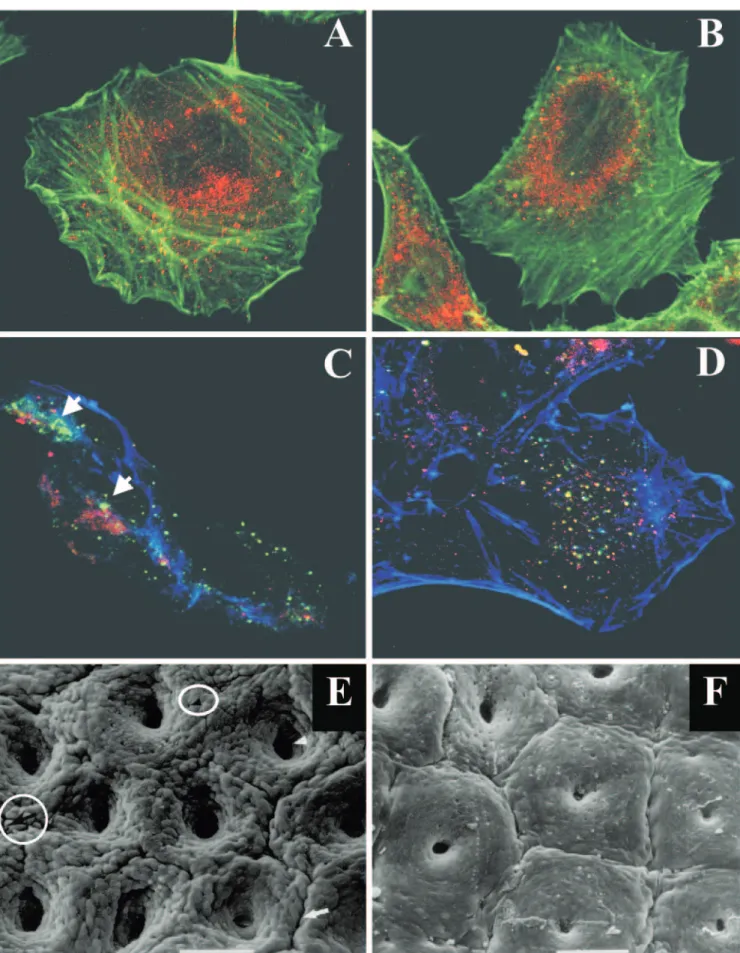
Interestingly, the IcsA autotransporter has the unusual char-acteristic of being localized to a single pole of the bacillus (166). The current model suggests that, on translocation into the outer membrane at one pole, IcsA, which is anchored in
the membrane by its -domain, diffuses laterally within the
outer membrane such that some of it drifts down the sides of the bacillus towards the septum (463). IcsP, which is localized in the outer membrane, slowly cleaves IcsA at all sites on the bacterial surface, but since the insertion of IcsA is occurring exclusively at one pole, it results in a unipolar distribution of IcsA (463). This study raises the question of the fate of the -barrel once cleavage of the passenger domain has occurred.
By analogy to other outer membrane proteins, the-barrel of
autotransporters might be a rather stable structure (444). Re-sults from cellular localization studies seem to corroborate the
persistence of the-barrel in the outer membrane (60).
How-ever, this question has not yet been satisfactorily addressed. One might speculate that accumulation of pore-forming struc-tures in the outer membrane would be lethal for the bacteria and that some kind of degradation or regulation process must therefore occur. However, the degradation mechanism(s) of periplasmic and outer membrane proteins remains elusive and requires further investigation, although it might involve periplasmic enzymes such as DegP and DegQ or outer mem-brane proteases such as OmpT.
Energy of transport.Comprehensive understanding of the
energy requirements used to translocate proteins across the bacterial outer membrane has lagged behind the wide-ranging studies investigating inner membrane transport (378). Since neither ATP nor GTP occurs in the periplasm, hydrolysis of these molecules cannot be the driving force for the transloca-tion of the passenger domain through the outer membrane. Moreover, there is still no evidence of the existence of a proton
motive force (⌬p) across the outer membrane. The presence of
P-loop nucleotide motifs in several members of the autotrans-porter family has been reported (203). Proteins bearing a P-loop motif are not always ATP- or GTP-binding proteins, and even proteins binding ATP or GTP do not always possess this motif (433, 522). Subsequently, it was suggested that since autotransporters can be correctly targeted and exported in nonnative gram-negative bacterial backgrounds, e.g., gonococ-cal IgA1 protease in E. coli, no additional secretion function is required and thus the energy source driving translocation across the outer membrane is derived from the correct folding of the passenger domain on the bacterial cell surface (254). From the study by Veiga et al. (510), where a nonnative single-chain antibody reporter passenger domain was used, it ap-peared unlikely that the folding of the passenger domain on the cell surface would provide the energy required for the translocation through the outer membrane. Unfortunately, this set of experiments used a nonnative passenger domain lacking the intramolecular chaperone domain. Thus, a common sort-ing mechanism assistsort-ing the assembly and export of autotrans-porter proteins and involving the autochaperone domain may exist. However, as mentioned above, BrkA lacking the au-tochaperone domain is still secreted, suggesting that other factors provide the necessary driving force for secretion. In conclusion, the energy dependence of the autotransporter se-cretion is still controversial and speculative and further inves-tigation is required to clarify this issue.
Evolution and distribution of autotransporter proteins. While the translocation units of autotransporters are highly homologous, the passenger domains are very diverse. To date, all characterized passenger protein domains have been impli-cated in virulence by displaying enzymatic activity (protease, peptidase, lipase, and esterase); mediating actin-promoted bacterial motility; acting as adhesins, immunomodulatory pro-teins, toxins or cytotoxins, or, as more recently discovered, permitting the maturation of other virulence proteins (82, 201, 203, 540). The autotransporters are present only in the Bacteria kingdom and are most prevalent in the phylum Proteobacteria, including the␣-, -, ␥-, andε-Proteobacteria classes (540). Like the TTSS, the only other phylum in which autotransporters have also been identified is the phylum Chla-mydiae, genera Chlamydia and Chlamydophila (198, 213). In most cases, multiple genes encoding autotransporters are found throughout the genome sequence of the microorganism, e.g., E. coli (530), Bordetella spp. (382), Yersinia pestis (98), Helicobacter pylori (313), Pseudomonas aeruginosa (306) and Neisseria (91). It has been suggested through genome sequence analysis that the passenger domain is not always covalently
linked to the-domain, since some open reading frames (ORFs)
code either only for a passenger domain or a-domain.
How-ever, these could simply be pseudogenes, since there is no experimental evidence that these proteins are expressed (540). It has been proposed that autotransporter proteins could have evolved by domain shuffling (91, 303). Functionally novel autotransporters could arise by linking a new active passenger
domain to a generic -barrel domain. Phylogenetic analysis
showed that horizontal transfer between distant organisms of
the portions of the gene encoding the autotransporter
-do-main was a rare evolutionary event; instead, close homologues appear to arise almost exclusively by speciation and late gene duplication events within a single organism. Such analyses have allowed Yen et al. to classify the autotransporters into 10 distinct phylogenetic clusters (540). However, reinvestigation of the phylogenetic relationship of the autotransporters reveals that the proteins can be grouped into at least 11 clusters (Fig. 6) (I. R. Henderson, unpublished data). However, it is clear that the functional passenger domains have spread by
horizon-tal transfer (91, 201). For example, while the-domains of the
IgA1 proteases and the serine protease autotransporters of the Enterobacteriaceae (SPATE) have different evolutionary lin-eages (cluster 8 and 5, respectively), the functional passenger domains are evolutionarily related and belong to the same clan of serine proteases (Table 1, Fig. 6). These data suggest that most autotransporters have arisen through fusion events
be-tween passenger domains and-domains.
Two-Partner Secretion Pathway (Type Vb)
In the bacterial two-partner secretion (TPS) pathway, as with the autotransporter secretion pathway, the passenger do-main possesses a signal sequence that directs translocation across the inner membrane (195). After its export into the periplasm, the passenger domain inserts into an outer
mem-brane pore formed by a-barrel. Once at the surface of the
bacterium, the passenger domain may undergo further proteo-lytic processing to achieve its physiological function (221). However, in contrast to the autotransporter pathway, where
the protein is produced as a single polypeptide, the passenger domain (also called the exoprotein) and the pore-forming -domain (also called the transporter domain) are translated as two separate proteins, respectively referred to as TpsA and TpsB family members (Fig. 3) (221). Compared to the
auto-transporter, the-barrel topology seems different, as indicated
by prediction of 19 amphipathic-strands in TpsB instead of
14 for the autotransporter-domain (176, 303, 540) and
dif-ferent conductance values in artificial bilayers (220, 259). Fur-thermore, TpsB proteins possess an additional level of com-plexity, as suggested by their involvement in passenger domain maturation into an active form (107, 221, 275, 439, 467). The genes for cognate exoprotein and transporter protein are gen-erally organized in an operon (221). The TPS pathway implies specific recognition events between the passenger domain and the transporter domain. In TpsA, a conserved N-proximal do-main, called the TPS dodo-main, interacts specifically with TpsB to initiate the outer membrane translocation (172, 219, 441, 498). Like the autotransporter pathway, the TPS pathway ap-pears to be dedicated to the secretion of very large proteins,
i.e.,⬎100 kDa (221). It has been suggested that the exoprotein
transits to the periplasm in an unfolded conformation and folds progressively at the cell surface as it is translocated through the transporter domain (175). Thus, the translocation
across both membranes seems coupled and the free energy of folding might be the driving force for the outer membrane translocation. Concomitantly with secretion, several passenger domains undergo further proteolytic processing (221). Inter-estingly, it has been demonstrated that the B. pertussis auto-transporter SphB1 acted as a specific protease responsible for the bacterial surface maturation of FhaB, the filamentous haemagglutinin (FHA) secreted by the TPS pathway (82).
Thus, despite certain differences, the TPS pathway shares fundamental features with the autotransporters, the most im-portant being the mode of secretion described above. More-over, examination of the signal sequences of TpsA family members reveals that several of these proteins possess unusual signal sequences which are remarkably conserved with the extended signal sequence of autotransporters (Fig. 4) (195, 221). It can be hypothesized that such exoproteins are trans-located through the inner membrane by the SRP-dependent pathway, as has been demonstrated for the Tsh/Hbp autotrans-porter (455). The third line of evidence supporting a connec-tion with the autotransporter secreconnec-tion pathway is derived from phylogenetic analyses, where it has been suggested that the shuffling of some passenger domains between the auto-transporter and TPS pathways had occurred frequently in the course of evolution (195), even though the translocating units FIG. 6. Phylogenetic tree of the autotransporter proteins. The CLUSTALX and TREE programs were used for multiple alignments and construction of a phylogenetic tree for the functionally characterized autotransporter proteins. The tree depicted in this figure is derived from analyses of the C-terminal translocating domains of the autotransporters. Proteins are clustered according to the pattern proposed by Yen et al. (540). A single additional cluster (cluster 11) is indicated.