Skin proteomics – analysis of the extracellular matrix in health and disease
Jörn Dengjela, Leena Bruckner-Tudermanb and Alexander Nyströmb
aDepartment of Biology, University of Fribourg, Fribourg, Switzerland; bDepartment of Dermatology, Faculty of Medicine, Medical Center - University of Freiburg, Freiburg, University of Freiburg, Freiburg, Germany Germany
ABSTRACT
Introduction: The skin protects the human body from external insults and regulates water and
temperature homeostasis. A highly developed extracellular matrix (ECM) supports the skin and instructs its cell functions. Reduced functionality of the ECM is often associated with skin diseases that cause physical impairment and also have implications on social interactions and quality of life of affected individuals.
Areas covered: With a focus on the skin ECM we discuss how mass spectrometry (MS)-based proteomic
approaches first contributed to establishing skin protein inventories and then facilitated elucidation of molecular functions and disease mechanisms.
Expert opinion: MS-based proteomic approaches have significantly contributed to our understanding of skin pathophysiology, but also revealed the challenges in assessing the skin ECM. The numerous posttranslational modifications of ECM proteins, like glycosylation, crosslinking, oxidation, and proteo-lytic maturation in disease settings can be difficult to tackle and remain understudied. Increased ease of handling of LC-MS/MS systems and automated/streamlined data analysis pipelines together with the accompanying increased usage of LC-MS/MS approaches will ensure that in the coming years MS-based proteomic approaches will continue to play a vital part in skin disease research. They will facilitate the elucidation of molecular disease mechanisms and, ultimately, identification of new druggable targets.
KEYWORDS
Skin fragility; wound healing; mass spectrometry
1. Introduction
The skin is the largest organ of the human body, with an area of ca. 1.7 m2 and a weight of ca. 3 kg. It consists of three different layers – epidermis, dermis, and subcutis – that have distinct tissue architectures and functions. Through tight cohesion to each other and through highly regulated interactions, the three layers form a barrier to the environment ensuring defense against microbial attacks and chemical and physical influences from the environment, protection against loss of water or pro-teins, and temperature control, which is essential for the proper functionality of the entire organism [1]. Loss of its protective functions is associated with numerous genetic and acquired skin diseases that manifest with a broad spectrum of symptoms. It may not be well known that about 20% of patients seeking help of a general practitioner have skin symptoms [2]. Therefore, not only the medical but also the socio-economic importance of understanding the physiological functions and interactions of skin cells and their derailment in disease states is huge.
Unbiased, i.e. hypothesis-free, global proteomic approaches have offered unprecedented opportunities to disclose mole-cular players in skin homeostasis and pathology. This review deals with MS-based proteomic analyses of the skin and skin cells, keratinocytes and fibroblasts, and covers historic publi-cations as well as recent work focusing on molecular mechan-isms. A special focus is the skin extracellular matrix (ECM). We highlight examples of how MS-based proteomic analyses have
generated important new information on skin diseases and delivered novel therapeutic cues, especially for skin fragility.
The ECM is essential to the skin. It aids the formation of an effective, protective barrier and provides toughness to shearing forces, whilst at the same time allowing sufficient elasticity to not restrict movement. The creation of an ECM scaffold to support a tissue encompassing such seemingly incompatible abilities is achieved through the close interactions of multiple, highly specia-lized substructures, which condition the skin with their unique properties. These substructures appear distinct in their ECM pro-teome and/or in their supramolecular arrangement and makeup four distinctive intertwined ECMs: the epidermal ECM, the dermal- epidermal junction zone, the papillary dermal ECM, and the reti-cular dermal matrix. Assembly of these ECMs requires highly coor-dinated cell-cell and cell-microenvironment communications, in time and in space [3]. Within each ECM considerable heterogeneity exists that provides important biological functions, for example, by creating stem cell-supporting microniches [4].
The complex structure of the skin ECM and certain traits of it make its detailed proteomic analysis challenging. The ECM is made up of proteins that often need to undergo extensive posttranslational modifications to become functional, includ-ing hydroxylation of selected proline and lysine residues (elas-tin and collagens), intramolecular cross-linking, N- and O-linked glycosylation (glycoproteins and proteoglycans, respectively) and extracellular proteolytic maturation. The
CONTACT Jörn Dengjel joern.dengjel@unifr.ch Department of Biology, University of Fribourg, Chemin du Musée 10, Fribourg, 1700, Fribourg 1700, Switzerland; Leena Bruckner-Tuderman leena.bruckner-tuderman@uniklinik-freiburg.de; Alexander Nyström alexander.nystroem@uniklinik-freiburg.de
Department of Dermatology, Faculty of Medicine, Medical Center - University of Freiburg, Hauptstr. 7, 79104 Freiburg, Germany
http://doc.rero.ch
Published in "Expert Review of Proteomics 17(5): 377–391, 2020"
which should be cited to refer to this work.
ECM backbone is composed of large proteins such as col-lagens, non-collagenous glycoproteins including laminins, fibrillins and fibronectin, and proteoglycans with repetitive domains that are closely or more distantly homologous to each other and other ECM proteins [5–9]. Many proteins have low solubility, which is further reduced through inter-molecular crosslinking by e.g. transglutaminases and lysyl oxi-dases [10]. However, for its proper organization and functionality the skin ECM is also dependent on smaller solu-ble proteins. A further challenge to achieve comprehensive coverage and quantitative assessment of the skin by proteo-mic approaches comes from the great range in protein abun-dances. To illustrate this, there is a more than 70,000-fold difference in abundance of collagen I and collagen VII in the skin – yet both proteins are essential for skin stability [11]. 2. The skin: structure, major cell types, and the organization of its ECM proteome
The main layers of the skin, the epidermis, the dermis, and the subcutis (Figure 1) act synergistically to form a functional organ.
However, they are distinctly disparate in terms of cellular composi-tion and ECM organizacomposi-tion. The following paragraphs will briefly summarize the cellular and extracellular proteome characteristics of the skin layers. The aim is to provide the readers with informa-tion to follow the subsequent discussion of the use of MS-based proteomic approaches for the understanding of ECM function in skin pathobiology. For a more detailed review of the skin ECM, we refer readers to our recent reviews on this subject [12–14].
2.1. The epidermis and epidermal keratinocytes
The outermost layer, the epidermis, is a multi-level cornified epithelium and a tightly packed cellular compartment with little intercellular space or ECM. The epidermis consists of 90% of keratinocytes, but contains also minor cell populations, such as melanocytes, Langerhans cells, and Merkel cells that have speci-fic functions in pigment production, first-line immune defense, and in touch sensation, respectively [1]. Keratinocytes secure epidermal integrity and the formation of the skin barrier. The basal keratinocytes in the lowermost epidermis, the stem cell compartment of the epidermis, sit on a basement membrane, and undergo vertical proliferation and differentiation cycles that ensure the renewal of the epidermis every 30 days. During this process, as they move from the basal layer of the epidermis toward its surface, the keratinocytes differentiate first into spi-nous cells and then to granular cells. Each of the differentiation stages exhibits a specific expression profile of intermediate fila-ment keratins and barrier proteins, such as loricrin or filaggrin. Finally, the terminally differentiated keratinocytes undergo pro-grammed cell death and contribute with their plasma mem-branes, cytoskeleton, structural proteins, extra- and intracellular lipids to the formation of the horny layer (stratum corneum) that is constantly being sloughed off [15]. Thus, the epidermal cells
Figure 1. Skin structure and ECM crosstalk. Schematic illustration of skin indicating tissue compartments and the different skin ECMs. In addition, selected key ECM proteins with their predominant location and main producing cells are shown with white arrows. Essential protein–protein interactions are shown with black arrows.
Article highlights
Ⴠ Skin functions are dependent on a highly complex extracellular matrix (ECM).
Ⴠ Both keratinocytes and skin fibroblasts contribute to the ECM.
Ⴠ MS-based proteomic approaches yield unprecedented insights into the composition and regulation of the proteome of skin and skin cells.
Ⴠ MS-based proteomic analyses of PTM and crosslinked ECM proteins are challenging and require special protocols
Ⴠ Current robust MS-based proteomic analyses help identify new drug-gable targets in skin diseases.
represent a dynamic system where basal keratinocytes replenish the cellular components of the entire epidermis and the building blocks for the skin barrier via a differentiation process [15].
Parallel to this protective function as a skin barrier, epidermal keratinocytes exert significant regulatory functions [16]. They instruct other epidermal cells, but also signal into the dermis and regulate cellular events there via paracrine mechanisms. For exam-ple, upon epidermal damage, ample interleukin 1 is secreted into the dermis where it induces inflammatory processes and subse-quent repair [16].
2.2. The epidermal ECM
The epidermis is an ECM-poor tissue compartment (Table 1); however, the scant ECM is essential for the formation and main-tenance of a functional epidermal microenvironment and finally contributes to producing the skin barrier. Activity of extracellular proteinases of the kallikrein family contributes to this process [17]. The extracellular microenvironment varies in its composi-tion from the inner to the outer layers of the epidermis. In the upper granular and horny layers, the environment is lipophilic; deposition of lipids contributes to the impermeability of the skin [18,19]. In extracellular spaces around the living epidermal layers composed of basal and spinous keratinocytes, the environment is hydrophilic with a notable presence of glycosaminoglycans (GAGs) such as hyaluronan and cell-surface, pericellular, and extracellular proteoglycans such as syndecan 1, perlecan, and versican (Figure 1) [19–22].
2.3. The dermal-epidermal junction zone
The dermal-epidermal junction zone (DEJZ) binds and separates the epidermis and the dermis. A firm anchorage of these layers is important for resistance of the skin to frictional challenges. Structurally, the DEJZ is a highly specialized, extended base-ment membrane zone that is often described in ultrastructural terms. As viewed in the electron microscope, it consists of several suprastructural units: hemidesmosomes at the inner plasma membrane of basal keratinocytes, the anchoring fila-ments, the bi-layered basement membrane proper (with an electron-lucid lamina lucida and an electron-dense lamina densa), and the anchoring fibrils that protrude from the lamina
densa into the uppermost dermis [23]. For practical reasons, the scientific community continues to use these ultrastructural terms, although it is unlikely that the functional supramolecular units in vivo will strictly correspond to the images seen in the electron microscope. Through specific protein–protein interac-tions the molecular components of these suprastructural units provide a cohesive ‘chain’ that extends from inside the basal keratinocytes into the dermal ECM and thus enforces dermal- epidermal anchorage. The molecular components of the DEJZ are introduced below and in Table 1.
2.4. The cell-surface and ECM proteome of the DEJZ
Under steady-state conditions, the main adhesion structures of the basal keratinocytes are the hemidesmosomes and the anchoring filaments. These contain a number of molecules, including cytoskeleton linker proteins, integrins, transmem-brane collagens, and ECM components that undergo highly specific protein–protein interactions. At the molecular level, the keratin cytoskeleton in basal keratinocytes is linked via the P1a isoform of plectin and BP230 to the intracellular tail of integrin α6β4, which, in turn, binds also the intracellular domain of transmembrane collagen XVII and tetraspanin CD151. This complex condenses into the hemidesmosome [24]. The extracellular domains of integrin α6β4 and collagen XVII support adhesion by interacting with laminin-332 in the basement membrane. Collagen XVII further promotes adhe-sion through binding to the basement membrane collagen IV in the lamina densa (Figure 1) [25]. The quantitatively minor transmembrane collagens XIII, XXII, and XXIII are also observed at the DEJZ and may support keratinocyte attachment to the epidermal basement membrane in certain contexts [26–29]. They exist in two forms that may have altered biological activity, a full-length transmembrane form, and a shed ecto-domain that is released by ADAM- (A disintegrin and metallo-proteinases) or furin-mediated shedding and incorporated in the pericellular ECM [30–32].
Keratinocytes utilize also other adhesive structures including integrin α3β1-based focal adhesions and integrin α3β1- and α6β4-containing tetraspanin-enriched microdomains [33]. For both integrins laminins are the main ligands. Laminins are tri-meric proteins composed of genetically distinct α, β, and γ chains
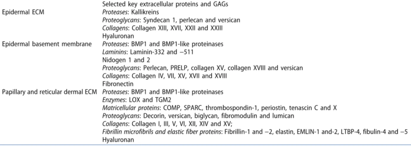
Table 1. The proteome and the glycome of the skin ECM.
Selected key extracellular proteins and GAGs Epidermal ECM Proteases: Kallikreins
Proteoglycans: Syndecan 1, perlecan and versican Collagens: Collagen XIII, XVII, XXII and XXIII Hyaluronan
Epidermal basement membrane Proteases: BMP1 and BMP1-like proteinases Laminins: Laminin-332 and −511 Nidogen 1 and 2
Proteoglycans: Perlecan, PRELP, collagen XV, collagen XVIII and versican Collagens: Collagen IV, VII, XV, XVII and XVIII
Fibronectin
Papillary and reticular dermal ECM Proteases: BMP1 and BMP1-like proteinases Enzymes: LOX and TGM2
Matricellular proteins: COMP, SPARC, thrombospondin-1, periostin, tenascin C and X Proteoglycans: Decorin, versican, biglycan, fibromodulin and lumican
Collagens: Collagen I, III, V, VI, XII, XIV and XV;
Fibrillin microfibrils and elastic fiber proteins: Fibrillin-1 and −2, elastin, EMLIN-1 and-2, LTBP-4, fibulin-4 and −5 Hyaluronan
and named according to the chain composition, i.e. laminin-332 is made of laminin α3, β3, and γ2 chains [34]. Laminin-332 and laminin-511 are the major laminin isoforms at the DEJZ and both are mainly contributed by keratinocytes [35]; however, they are not equally distributed within the epidermal basement mem-brane. Laminin-511 is enriched around hair follicles, whereas laminin-332 is more abundant under the interfollicular epidermis (Figure 1). The laminin-511:laminin-332 ratio has been shown to regulate stem cell activity [36].
There are key structural differences between laminin-511 and laminin-332. Laminin-511 has three N-terminal short-arms, which are the parts of the laminin chains that are not folded into an α-helical coiled-coil with other chains [34]. Through intermolecular interactions of the short-arms laminin-511 can form a laminin network [37]. Contrastingly, laminin-332’s short-arms are truncated and cannot participate in network formation [34]. However, although the usual gene product of the LAMA3 gene, the laminin α3 chain, is network incompe-tent, the gene may still potentially influence laminin network assembly through the expression of its alternative splice pro-duct, the LaNt α31 protein [38]. LaNt α31 is homologous to netrins and based on this has been proposed to regulate laminin network formation and/or stability and consequently basement membrane formation [38,39].
Subjacent to the hemidesmosomes, laminin-332, and the ectodomain of collagen XVII arrange into molecularly densely packed anchoring filaments [40], which connect to collagen VII-composed anchoring fibrils [41]. Within the basement membrane proper, perlecan, nidogen 1 and 2, and collagen VII link the laminin network to a collagen IV network. This consists mainly of collagen IV α1.α1.α2 but with a notable presence of collagen IV α5.α5.α6 [37,42,43] and provides stiff-ness and stability to the basement membrane [44]. Hybrid collagen/proteoglycans, collagen XV, and collagen XVIII, are also present at the DEJZ [45,46], but their functional contribu-tions need further elucidation. Both keratinocytes and dermal fibroblasts are capable of expressing these collagens; however, collagen XV appears to be primarily a fibroblast product [46– 48]. PRELP is an extracellular proteoglycan at the DEJZ [49]. It belongs to small leucine-rich proteoglycans (SLRPs) [22] and has been proposed to function as an anchor between perlecan and collagen-containing fibrils in the papillary dermal ECM (Figure 1) [49]. Cultured keratinocytes have been shown to express another SLRP, decorin, but the physiological relevance of this remains to be explored [48,50].
Proteolytic activity of the astacin-like proteinase BMP-1/ tolloid-like is essential for the assembly of a functional DEJZ, papillary, and reticular dermal ECM. These proteinases process proforms of multiple ECM components to mature functional molecules, including the SLRP biglycan, the N-terminal end of the laminin γ2 chain, and the C-terminal end of laminin α3 in laminin-332, procollagen VII, and the C-terminal ends of pro-collagens I and III [51–53]. In addition, the other members of the astacin-like proteinases – meprin α and β – have been shown to be involved in processing of the N- and C-terminal ends of fibrillar procollagens in the skin [54]. ADAMTS2, −3, and −13 are generally considered the main proteases involved in removal of the procollagen N-pro-termini [55]. Although often considered to be ECM degrading proteases, also matrix
metalloproteinases (MMPs) are involved in the assembly of the DEJZ through e.g. processing of laminin-332 [56].
In healthy homeostatic skin, the glycoprotein fibronectin is present around the DEJZ and in the papillary dermis [57]. It can be synthesized at substantial levels by both keratinocytes and fibroblasts [48,58]. After an injury it is dramatically increased by deposition of plasma fibronectin and later by fibronectin pro-duced by tissue-resident cells [59]. Fibronectin fills key roles during development and regeneration serving as an adhesive substrate for migrating cells but also as an important facilitator of fibrillar collagen and fibrillin microfibril deposition [60,61].
2.5. The dermis and dermal fibroblasts
The second important skin layer is the dermis (Figure 1). It provides tensile strength, elasticity, and resilience to the skin. Histologically the dermis can be divided into a thin superficial layer, the papillary dermis, and a thicker deeper layer, the reticular dermis. Although adnexal, vascular, lym-phatic and neural structures traverse the dermis, fibroblasts constitute the major cell population embedded in the der-mal connective tissue. These are ECM producing mesenchy-mal cells responsible for maintenance, repair, and regeneration of the dermis. Recent research data indicate that the superficial, middle, and deeper dermal layers harbor distinct fibroblast subpopulations [62], nevertheless defini-tive cell markers still remain elusive and functional charac-teristics that distinguish them are not fully understood. Quantitatively minor and changing cell populations in the dermis encompass immune cells. Upon need, they migrate into the skin and exert decisive functions in immune response, inflammation, and regenerative processes [63].
Apart from their structure-maintaining role, fibroblasts have regulatory functions. They can direct cell behavior in both the epidermis and the dermis, e.g. via paracrine signaling [64]. Intriguingly, also the dermal ECM itself has cell-instructive capabilities; it sequesters growth factors, cytokines, and other signaling molecules via specific binding to structural macro-molecules [12]. A major regulatory mechanism is the release of these soluble factors, e.g. via proteolysis of ECM proteins or conformational changes that eliminate or weaken binding sites. The soluble molecules then migrate into the tissue microenvironment and influence cell functions.
These processes and mechanisms play an important role in epidermal-dermal cross-talk. The skin layers are in constant exchange, and cells sense changes in other compartments. Typical examples are physiological situations like develop-ment, repair, and regeneration after injury [63]. Examples of pathological conditions with highly active cross-talk between the epidermis and the dermis include skin fragility syndromes, perturbed wound healing, or the interactions between epithe-lial cancers and their stroma [62,65].
2.6. The papillary and reticular dermal ECM
Composition and arrangement of the dermal ECM mirror that of the histological division into a papillary and a reticular layer with their specific tissue architectures and specialized ECMs (Table 1). The structural scaffold of the ECM is provided by
collagen-, elastin- and microfibrils-containing supramolecular fibril networks, with a variety of associated distinct ECM com-ponents. These are often quantitatively minor ECM molecules, but they confer substantial effects on the suprastructures and provide specific ligand binding capabilities to the fibril net-works. The interfibrillar space is filled with glycoproteins, pro-teoglycans, and ample hyaluronan with a very high water- binding capacity that provides the skin with its tautness [12].
The papillary ECM just below the DEJZ is an extension of this zone providing additional points of stability and securing epidermal attachment to the dermis. This structure has higher flexibility than the more rigid epidermal basement membrane enabling deformable joining of the basement membrane and the reticular ECM [12]. As for the epidermal basement mem-brane, both keratinocytes and fibroblasts can contribute to its proteome. Collagen VI, predominantly collagen VI α1.α2.α3, assembles into a tissue-stabilizing microfibrillar network below the basement membrane [66]. Collagen V also shows notable accumulation below the epidermal basement mem-brane [67], where it can provide further anchoring stability by interacting with collagens IV and VI (Figure 1) [68].
The matricellular protein COMP (cartilage oligomeric matrix protein) can also be involved in the assembly of the papillary ECM by binding the FACIT (Fibril Associated Collagens with Interrupted Triple helices) collagen XII and collagen XIV [69]. Both of these collagens are expressed by dermal fibroblasts and epidermal keratinocytes [47,48].
Fibrillin microfibrils connecting to the epidermal basement membrane-papillary ECM interface provide additional stability [70]. Although generally considered a fibroblastic product, inter-estingly, keratinocytes can also express fibrillin-1 and 2 and assemble microfibrils in vitro [71,72]. Fibrillin-1 is most abundant in adult tissue and fibrillin-2 is expressed during development and regeneration [73]. Additional molecules such as elastin microfibril interface-located proteins (EMILINs) 1 and 2 associate with dermal fibrillin microfibrils [74,75]. Collagen XVI is expressed by both keratinocytes and fibroblasts and co- localizes with fibrillin microfibrils at the epidermal basement membrane-papillary ECM interface, whilst also showing close proximity to collagen VII (Figure 1). Collectively, this suggests a role for it in supporting dermal-epidermal cohesion [76].
The matricellular protein tenascin-X interacts with many pro-teins in the papillary ECM, including collagen XII, XIV, and fibrillar collagens. The latter fill key architectural functions [77,78], for example, collagen I, III, and V are the principal components of dermal heterotypic collagen fibrils. Of these, collagen I is most abundant making up around 70%. Collagen III and V comprise around 20% and >5%, respectively, of all dermal collagens [79–82]. The collagen fibrils in the papillary ECM are generally thinner than those found in the reticular ECM. Collagen fibril diameter is in part determined by the ratio of collagen III and collagen V, the latter nucleating collagen fibril formation leading to thinner fibrils [83].
Additional key regulators of collagen fibrillogenesis are pro-teoglycans and matricellular proteins. Decorin, a dermatan sul-fate/chondroitin sulfate SLRP, and versican, a chondroitin sulfate proteoglycan, are the most abundant proteoglycans in the skin [84]. Decorin shows a higher abundance in the papillary ECM and versican in the reticular ECM [84,85]. The importance of these proteoglycans in the skin is reflected by skin fragility in decorin-
deficient mice [86] and in dermatan sulfate-deficient humans [87]. Despite being of lower abundance, also other proteoglycans are important regulators of collagen fibrillogenesis in the dermis. These include the SLRPs biglycan, lumican and fibromodulin [88]. Proteoglycans can be present without GAG substitution as just the protein core. Furthermore, although not a protein, the GAG hyaluronan, which is distributed throughout the skin, shows its highest abundance in the papillary dermis [89].
The matricellular protein SPARC (Secreted Protein, Acidic, Rich in Cysteine), also known as BM40 or osteonectin, has its highest abundance in the papillary dermis [90]. It interacts directly with collagen I, III, and V [91] and may regulate maturation of procollagens and fibrillogenesis in the skin [92]. Periostin, another matricellular protein in the papillary ECM [93] can regulate collagen fibrillogenesis by directly interacting with collagen I [94]. It also interacts with BMP-1 evoking proteolytic activation of lysyl oxidase and subse-quent collagen crosslinking [95]. During homeostasis the abundance of matricellular proteins is generally low in skin; however, after tissue damage or under pathological condi-tions they can be greatly increased and also involved in driving pathological processes such as fibrosis. In addition to the aforementioned matricellular proteins, these also include thrombospondin-1 and tenascin-c [12].
The deeper reticular dermal ECM is characterized by thicker collagen-containing fibrils and more prominent elastic fibers. Tropoelastin molecules are assembled and become cross-linked by lysyl oxidases to serve as the core of elastic fibers which associate with a multitude of proteins including fibril-lins, fibulin 4 and 5 and latent transforming growth factor beta-binding protein 4 (LTBP4) (Figure 1) [96]. MS-based pro-teomic analysis of papillary and reticular dermis separated through laser microdissection illustrates that the difference in appearance of the ECM organization in the papillary and reticular dermis is not caused by an altered abundance of major structural proteins, such as fibrillar collagens [97]. Rather the differences in organization are achieved by altera-tions in lesser abundant ECM proteins and their effects on supramolecular assembly of ECM structures.
It should be emphasized that in proteome analyses of whole skin also endothelial, muscle, adipose, and specialized adnexal basement membranes and ECMs add to the detected proteome. In some situations, also immune cells may contri-bute [98]. When and to what extent will need further investi-gations. Furthermore, the proteome, its arrangement, and post-translational modifications (PTMs) change dynamically during development and aging [99].
3. Quantitative mass spectrometry approaches to study the skin, its cells and ECM
3.1. A historical perspective – Mapping the proteome of skin and skin cells
At the beginning of this millennium with the first draft of the human genome [100,101], the golden era of MS-based pro-teomics began [102]. This was fueled by several parallel devel-opments. The sequenced genome allowed the establishment of comprehensive protein databases that alleviated the
identification of peptides and proteins by incomplete MS data; technical developments increased experimental robustness, instrument sensitivity, and sample throughput; the establish-ment of quantitative protocols allowed functional analyses addressing molecular mechanisms [103]. The proteomic ana-lyses of skin and skin-derived cells nicely mirror the overall trend of MS-based proteomic studies. Amongst others, this is reflected by the number of publications: whereas in the 1990 s on average less than six manuscripts per year were published addressing proteomic analyses of skin-derived samples, this number increased to more than 20 in the 2000 s. In the last three years more than 70 manuscripts per year presented data from MS-based experiments of skin specimens (using follow-ing PubMed search criteria: skin AND (keratinocyte OR fibro-blast) AND (proteomics OR ‘mass spectrometry’)). This illustrates that MS-based proteomics has become a standard technique to study scientific questions related to skin pathophysiology.
Proteomic characterizations of skin-derived samples started with the generation of inventories (Table 2) [104]. From the very beginning comparative analyses were performed, i.e., relatively quantifying proteins from the different cell or dis-ease states. Quantification was initially performed by densito-metric analyses of polyacrylamide gel electrophoresis (PAGE) bands or spots. MS analyses were used qualitatively to identify proteins of interest. A major focus was the characterization of the epidermis and keratinocytes, pioneered by Celis and col-leagues [105,106]. The underlying dermis and its major cell type, fibroblasts, were thought to generate merely the struc-tural scaffold on which keratinocytes would execute their vital functions, and received considerably less attention. Keratinocyte lysates, and later also fibroblast lysates, were analyzed by two-dimensional (2D)-PAGE coupled to tryptic peptide-mass-mapping by matrix-assisted laser desorption ionization (MALDI)-MS [106,107]. Unbiased analyses of whole skin using the same approach followed few years later, e.g. characterizing protein abundance changes related with age [108]. Using the mouse as a model system, whole skin as well as separated epidermis and dermis analyses were performed [109]. Again, using unbiased 2D-PAGE-based proteome quan-tifications, protein abundance differences due to heat stress were quantified. In general, at that time 10–20 proteins were identified by MS per study. Whereas results were often con-firmatory to existing knowledge, these studies were important steps in generating hypothesis-free, unbiased experimental workflows, which are the basis of current data-driven research in systems biology.
Around the turn of the millennium, 2D-PAGE in combination with peptide-mass-mapping by MALDI-MS got steadily replaced by peptide sequencing using a combination of liquid- chromatography (LC) and electrospray ionization (ESI)-MS/MS analysis [102]. This led to a dramatic increase in protein identifi-cations per MS study, from several tens of proteins to more than thousand. In one of the first studies, a comprehensive analysis of the plasma membrane proteome of human keratinocytes was performed identifying 1306 proteins [110]. Of these 866 carried Gene Ontology annotation of which ca. 60% were classified as an integral membrane or membrane-associated proteins, many of them being involved in cell adhesion nicely highlighting the physiological function of keratinocytes.
3.2. Quantitative approaches
In parallel to peptide sequence-based protein identification and the use of LC-MS/MS approaches as main MS-based proteomic workflow, relative quantification methods were developed for robust and accurate peptide-based protein quantifications [102]. Relative quantification approaches can be performed label-free or with the help of stable-isotope labeling allowing the relative com-parison of two or more experimental states. Labels can be intro-duced by different means as well as on different stages of experiments. In general, metabolic, chemical, and enzymatic label-ing approaches are discriminated, which label proteins or tryptic peptides [111]. Interestingly, skin-derived samples were among the first ones to be analyzed, or even used as showcase to highlight the benefits of the newly developed quantification approaches. This might either highlight the general interest in the skin as an important human organ, or the easy accessibility of skin as a model to study human organ function and cell-cell crosstalk.
In general, metabolic labeling approaches yield the most accurate quantitative data as biological samples can already be combined at the tissue or cell state. Unspecific losses due to subsequent sample handling will affect mixed samples to the same extent and thus will not influence quantification data. Various metabolites can be isotopically labeled to yield a labeled proteome. The most commonly used approaches employ 2D, 13 C, or 15 N labeled amino acids, Stable Isotope Labeling by Amino Acids in Cell Culture (SILAC) being the most used variant [112]. Labeled amino acids introduce rela-tively few, precisely defined labeled sites per peptide which aids bioinformatic data interpretation. In the same year in which the SILAC approach using leucine labeling of C2C12 and NIH 3T3 cells was published, human skin fibroblasts were labeled with serine, leucine or tyrosine variants, and
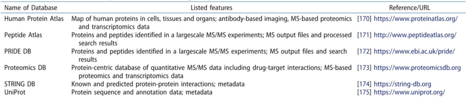
Table 2. Non-comprehensive table of proteomics databases listing skin proteins. DB: database.
Name of Database Listed features Reference/URL
Human Protein Atlas Map of human proteins in cells, tissues and organs; antibody-based imaging, MS-based proteomics and transcriptomics data
[170] https://www.proteinatlas.org/
Peptide Atlas Proteins and peptides identified in a largescale MS/MS experiments; MS output files and processed search results
[171] http://www.peptideatlas.org/
PRIDE DB Proteins and peptides identified in a largescale MS/MS experiments; MS output files and search results
[172] https://www.ebi.ac.uk/pride/
Proteomics DB Protein-centric database of quantitative MS/MS data including drug-target interactions; MS-based proteomics and transcriptomics data
[173] https://www.proteomicsdb.org
STRING DB Known and predicted protein-protein interactions; metadata [174] https://string-db.org
UniProt Protein sequence and annotation data; metadata [175] https://www.uniprot.org/
their response to radiation was analyzed by quantifying pro-tein abundance differences as well as by identifying changes in protein phosphorylation [113,114]. The latter study, which employed an isotope-label-based identification of a PTM, was ahead of its time. Whereas Chen and colleagues used the isotope label in a targeted approach to aid identification of phosphorylated Ser140 of histone H2AFX after radiation, the full potential of this approach was unleashed when isotopic labeling was employed to quantify site-specific phosphoryla-tion events on a global scale [115]. In the following years, SILAC-based quantification was employed, amongst others, to highlight that keratinocyte and fibroblast proteomes were relatively stable during cell culture preserving cell-type specific
in vivo characteristics [116], and that gender, age, and skin localization only have minimal influences on the keratinocyte proteome [117].
Next to metabolic labeling, chemical labeling was employed to follow skin proteome dynamics in an unbiased fashion. The most widely used chemical tags are isobaric tags for relative and abso-lute quantitation (iTRAQ) [118] and tandem mass tags (TMT) [119]. As the tags are isobaric, sample complexity is not increased and quantification is performed on the MS/MS level compared to MS- based quantification of metabolic labeling strategies. Compared to the latter, isobaric chemical tags allow a higher degree of multi-plexing [120]. Depending on the product up to 16 samples may be compared in a single analysis. iTRAQ labeling was used to study unconventional protein secretion of keratinocytes in response to UVB irradiation [121]. The authors could show that UVB-activated caspase-1 plays a general role in unconventional protein secretion. By comparing secretomes of cells treated with the caspase-1 inhibitor YVAD with those of non-treated cells, 77 proteins were identified that were likely secreted in a caspase-1 activity- dependent manner. iTRAQ labeling in combination with skin fibro-blasts was used to investigate possible side-effects of the HDAC inhibitor valproate in spinal muscular atrophy [122]. 2171 proteins were detected of which 1329 could be accurately quantified. While the majority of proteins appeared not affected by valproate treat-ment, collagen I, VI, and osteonectin were significantly reduced indicating potential side-effects on the ECM.
In the last years, label-free approaches integrating either MS or MS/MS signals as quantitative readout have gained popularity [103]. Due to newly developed data analysis algorithms and MS- based experiment designs, label-free quantifications yield robust accurate quantitative data, reducing costs of experiments, and increasing the dynamic range as well as the number of identifi-cations. Thus, two- to three-fold differences can now be accu-rately quantified [123]. In so-called data-dependent acquisition (DDA)-based approaches MS signal intensities are used for quan-tification whereas MS/MS spectra of selected single peptides are recorded for identification. In data-independent acquisition (DIA)-based methods such as sequential window acquisition of all theoretical mass spectra (SWATH-MS) [124] entire m/z ranges of precursor peptides are fragmented at the same time generat-ing comprehensive fragment ion maps and intensities of frag-ments that belong to a given peptide are used for quantification [103]. In skin research, SWATH-MS was used amongst others to study the response of keratinocytes to cannabidiol identifying the transcriptional repressor BACH1 as cannabidiol target [125],
to study the response of fibroblasts to redox stress characterizing collagen homeostasis as being redox sensitive [126], and to identify potential biomarkers for actinic keratosis, Bowen’s Disease, and cutaneous squamous cell carcinoma using formalin- fixed paraffin-embedded samples [127].
3.3. Analyses of PTMs
One of the unique features of MS-based proteomic approaches is the unbiased identification and quantification of PTMs. Dynamic regulation of PTMs is at the core of cellular signal transduction often resulting in changes of protein activities, and localizations being causal for observed cell phenotypes. Hence, from the very beginning, the characterization of PTMs has been a major focus of MS-based proteomic approaches [128]. As more than 200 different PTMs have been described, this review cannot be exhaustive. However, we would like to summarize the most relevant studies in skin biology.
As protein phosphorylation is a major determinant of protein activation and regulator of protein–protein interactions its analysis has received a lot of attention. MS-based proteomic approaches are the method of choice to study alterations in the phosphoryla-tion status of proteins giving rise to the research field of phospho-proteomics [129]. As for expression proteomic experiments, initial studies addressing protein phosphorylation in skin cells were coupled to 2D-PAGE [130]. Especially the effect of irradiation on skin cells was analyzed by phosphoproteomic approaches, high-lighting that skin fibroblasts respond differentially to low and high doses of ionizing radiation [131,132]. Later, phosphoproteomic approaches were coupled with elegant mechanistic studies. E.g. in mouse skin and mouse keratinocytes Polo-like Kinase 1 (Plk1) was identified to regulate keratinocyte planar cell polarity by phosphorylating the protein Celsr1, regulating its endosomal recruitment during mitosis [133]. Specific phosphorylation events were also shown to be important for skin cell differentiation: the kinase CSNK1a1 was shown to be important for keratinocyte progenitor maintenance by phosphorylating protein arginine methyltransferase 1 (PRMT1) [134]; receptor-interacting serine- threonine kinase 4 (Ripk4) was identified to phosphorylate the desmosome component plakophilin-1 (Pkp1) in mouse keratino-cyte differentiation [135]. Absence of either of the two proteins led to enhanced epidermal carcinogenesis.
Whereas protein phosphorylation is a highly dynamic, reversi-ble PTM being regulated by protein kinases and phosphatases, several (quasi) non-reversible PTMs were also shown to be impor-tant in skin biology and to be dysregulated in human diseases. Next to phosphorylation, protein oxidation is important in skin pathophysiology. UVB radiation, which induces amongst others an increase in reactive oxygen species (ROS), was shown to change protein abundances in keratinocytes and increase protein carbo-nylation [136]. In contrast, enzymatic protein oxidation plays an important role in physiological collagen maturation, hydroxypro-lines and hydroxylysines being important for protein stability and crosslinking. It was shown by unbiased proteomic analyses of fibroblast ECM that amino acid hydroxylation as well as protein crosslinking by transglutaminases are altered on a global scale in skin fragility probably reflecting alterations in ECM stability [47,137].
A truly irreversible PTM is non-digestive proteolytic processing of proteins generating new proteoforms with potential new phy-siological functions. Several MS-based proteomic approaches have been designed that focus on the characterization of proteolytically processed proteins, which can be summarized as ‘positional pro-teomics,’ ‘terminomics,’ or ‘degradomics’ approaches [138]. Skin function critically depends on a tightly controlled protease net-work as highlighted by the high number of proteases expressed by the skin and invading immune cells and their frequently found dysregulation in disease [139]. Thus, several terminomics studies aimed to identify protease substrates in skin or skin cells. Terminal Amine Isotopic Labeling of Substrates (TAILS) was employed to characterize type I procollagen I processing by metalloproteases meprin α and meprin β [140]. iTRAQ-based TAILS was used to identify amongst others integrin alpha 6 and dermokine as new MMP10 substrates in mouse keratinocytes [141]. As MMP10 has been implicated in wound healing being highly expressed in keratinocytes at wound edges, processing of these adhesion mole-cules by MMP10 might promote cell migration. MS-based proteo-mic approaches were also employed to characterize ectodomain shedding of transmembrane proteins such as collagen XVII, char-acterizing neoepitopes in bullous pemphigoid [142].
3.4. Proteomic analysis of the skin ECM
The term ECM often means different things to different people and the definition of the ECM is ever-changing and expanding [143]. Classically the ECM referred to the structural material that surrounds cells and that can be divided into three major protein classes: collagens, glycoproteins and proteoglycans [144]. For mass spectrometrists/analytical chemists, the ECM was considered all the proteins that are found outside of the cell. Hynes and Naba came up with a clear definition that has been accepted by the different research communities: the matrisome [143,145], which is based on a bioinformatics approach to define the complete repertoire of ECM proteins.
They discriminate the ‘core-matrisome’ and ‘matrisome- associated proteins.’ Core matrisome proteins (encoded by 278 genes in humans) have to contain minimally one out of 55 protein domains of known extracellular proteins and must not contain known intracellular or transmembrane domains, defining proteins that belong to the three ECM protein classes collagens, glycoproteins, and proteoglycans. Matrisome- associated proteins are made up of ECM-affiliated proteins, ECM regulators, and secreted factors (encoded by 778 genes in humans). Whereas we fully support the matrisome approach, we would like to point out that from a biological point of view important information might be missed, when MS-based proteomic data are filtered on matrisome proteins without taking other proteins into account that are identified in biochemically enriched ECM samples (Figure 2). E.g. cell surface receptors interacting with ECM proteins and important for cellular phenotypes are lost in filtered data. Also, secreted lysosomal proteins, like lysosomal cathepsins, are not listed in matrisome-associated proteins.
Gel-based approaches for MS sample preparation are gener-ally considered as old-fashioned, especigener-ally as they are not auto-matable and labor-intensive. Also, not all proteins are well soluble in SDS-based buffers, or well separable by PAGE, such as some membrane proteins or highly crosslinked ECM proteins found in corneocytes. Due to fixation in the gel matrix, protein recovery might also be limited; however, when sample amount is not a limiting factor, gel-based approaches still yield the most comprehensive coverage of ECM proteins according to our observations (Figure 2) (own unpublished data and [146]). Especially as crosslinked ECM proteins are often very stable and weakly soluble, gel-based approaches probably support pro-tease accessibility and increase peptide recovery. Thus, approaches similarly as outlined in Figure 2 were performed to study the confluence switch in human keratinocytes, identifying that cell confluence inhibits plasmin via SERPINE1 activation leading to increased perlecan deposition and cell adhesion
Figure 2. Bottom-up MS-based proteomic analyses of ECM and skin samples. (a) Analysis of ECM derived from primary skin cell cultures. Skin fibroblasts or keratinocytes are kept in culture for 6 days and ascorbate is added to the culture medium to support collagen hydroxylation. Cells are removed by addition of 0.5% Triton X-100 in 20 mM NH4OH for 30 s according to [176]. (b) Analysis of whole skin. After sacrifice, skin is shaved and punch biopsies are taken. Skin is macroscopically cleaned from adipose tissue and prepared as in [154]. Due to highly crosslinked ECM complexes, SDS-polyacrylamide gel-based digestion protocols [177] yield the most comprehensive coverage of ECM proteins (own-unpublished results).
[147]. Comparing the matrisomes of mouse skin fibroblasts expressing a constitutively active variant of the transcription factor NRF2 and WT cells, it was shown that NRF2 supports cellular senescence via deposition of a senescence-promoting matrisome. The alterations in the matrisome promoted acceler-ated wound healing, but also tumor growth [148].
4. Proteomic alterations in skin fragility diseases Illustrative examples of diseases affecting the skin ECM are genetic skin fragility disorders that manifest with blistering and easy breakability of the skin upon minor mechanical stress or shearing forces. These disorders are caused by pathogenic var-iants in genes encoding structural proteins of the epidermis or the DEJZ and are collectively called epidermolysis bullosa (EB). EB is a group of similar, yet distinct disorders, and mutations in more than different 20 genes are known to cause EB [149].
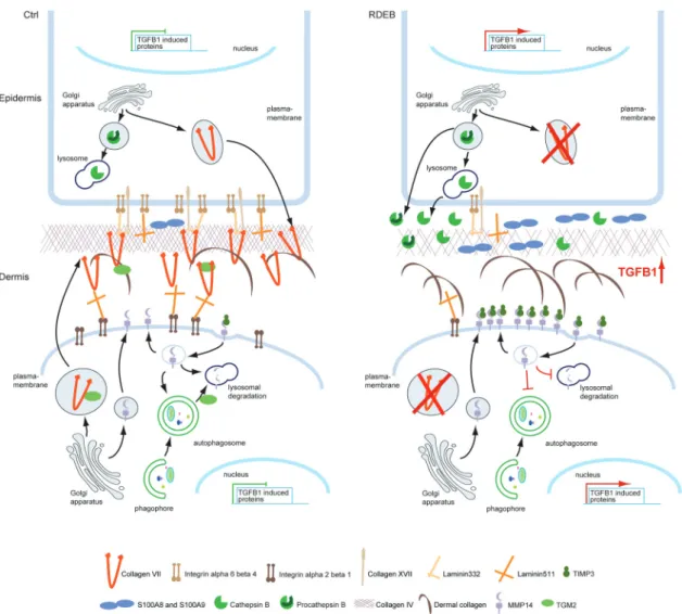
One EB subtype with a particularly severe and interesting phenotype is recessive dystrophic EB (RDEB). It is caused by mutations in the COL7A1 gene that encodes collagen VII, the major protein constituent of the anchoring fibrils [150] (Figure 3). Although RDEB is monogenic, i.e. mutations in a single gene
cause the disease, the consequences at tissue level are progres-sive and remarkably complex. Loss of collagen VII leads to lack of functional anchoring fibrils and to the separation of the epider-mis from the derepider-mis upon mechanical stress, clinically manifest-ing as skin blistermanifest-ing. But the loss of anchormanifest-ing fibrils also destabilizes the dermal tissue architecture, and the blisters heal with scarring [151]. Repeated cycles of blistering and scarring become associated with generalized soft tissue fibrosis with advancing course of the disease. Although the clinical picture has been long known, the disease mechanisms remained elusive for a long time. Recent global proteomic analyses have delivered novel cues on molecular and cellular events contributing to the phenotype and on promising therapeutic targets.
A collagen VII hypomorphic mouse model of RDEB led to the identification of the fact that collagen VII-deficiency causes dysregulation of TGF-beta 1 signaling [152]. However, it was not clear how profound the alterations of the proteome in general, and the ECM in particular, are due to the loss of a single protein. In several disease proteomic studies, we could show that loss of collagen VII affects both, skin fibro-blasts and keratinocytes, and has numerous cell intrinsic and extrinsic implications. The full spectrum of consequences of
Figure 3. Dysregulation of the proteome due to loss of collagen VII in RDEB. Loss of collagen VII affects both, intra- and extracellular proteomes of skin fibroblasts and keratinocytes. Increased TGF-beta 1 signaling appears to be a driver of the disease. Fibrosis and inflammation are increased, and lysosomal capacity is decreased in RDEB. However, increased extracellular activity of lysosomal hydrolases such as cathepsin B might contribute to blister formation. Interestingly, matrix metalloproteases, e.g. MMP14 are less active indicating a disturbance of the entire protease web.
collagen VII loss was gained from a number of omics analyses of skin cells and whole skin tissue, and the biological valida-tion of the observed changes in vitro and in vivo.
Combinatorial omics showed that in keratinocytes loss of collagen VII leads to the downregulation of mRNA levels of several collagen VII binding partners, indicating that this loss is sensed by cells and that it affects gene transcription [48]. Furthermore, keratinocytes appear to actively contribute to the inflammation observed in RDEB individuals, amongst others by secreting S100A8 and S100A9 proteins, which together form calprotectin, a biomarker for acute and chronic inflammation [153]. Interestingly, also protein homeostasis appeared altered leading to the enhanced secretion of active lysosomal proteases from keratinocytes [48]. In fibroblasts, loss of collagen VII nega-tively affects the abundance and deposition of several basement membrane components such as collagen IV and laminin 511. This suggests that in vivo loss of collagen will likely weaken the suprastructure of the basement membrane and amplify the skin fragility phenotype caused by a lack of functional anchoring fibrils (Figure 3) [47]. Due to the concomitant loss of collagen VII- binding partners, among them transglutaminase II (TGM2), der-mal collagens may have fewer crosslinks negatively affecting dermis integrity [137].
Disease proteomics approaches not only contributed to understanding the molecular consequences of lack of collagen VII in RDEB, but also helped outline mechanisms of pharma-cological interference with TGF-beta 1 signaling in a pre- clinical study employing the RDEB mouse model [154]. The small-molecule angiotensin II type 1 receptor antagonist losar-tan, known to negatively interfere with TGF-beta 1 signaling, caused a reduction of proteins involved in complement acti-vation and immune and inflammatory responses in the RDEB mouse, indicating that by limiting tissue inflammation and fibrosis losartan ameliorates the disease phenotype [154]. These findings led to the initiation of a phase I/II clinical study testing the effects of losartan treatment in children with RDEB (EudraNr. 2015–003670-32), which is currently ongoing.
In line with the fact that the transcriptome of RDEB fibro-blasts resembles that of cancer-associated fibrofibro-blasts [155] (Figure 3), proteomic analysis of paraffin-embedded human tissue identified similarities between the microenvironment of RDEB-associated cutaneous squamous cell carcinoma (cSCC) and sporadic metastasizing non-RDEB-cSCC, indicating that alterations in the ECM contribute to cancer invasion in RDEB [156]. Taken together, disease proteomic analyses sig-nificantly contributed to outlining the complex and wide- spread molecular changes underlying loss of collagen VII and contributing to RDEB pathology.
Another relevant example in which disease proteomic approaches generated new knowledge is perturbed wound heal-ing and chronic wounds. These may occur in rare diseases, such as RDEB [151], but are also prevalent in Western life-style diseases such as diabetes and obesity, as well as with aging. Normal wound healing proceeds in a highly orchestrated manner in three phases, i) inflammation; ii) re-epithelialization of the epi-dermis; and iii) tissue remodeling of the regenerated dermis [63]. In slow-healing, chronic wounds these phases do not follow each
other in a precisely regulated manner, but are retarded and/or derailed. The reasons are not fully understood. Several investiga-tions have addressed re-epithelialization and epidermal regenera-tion [157], but only recently did disease proteomic approaches help identify changes in the dermis and define novel cell subpo-pulations that contribute to poor wound closure [158].
MS-based proteomics have been used since the early 2000s as an analytic tool for studying wound healing [159]. Despite the obvious necessity for wound healing of a timely and well- coordinated deposition and arrangement of the ECM, few studies have directly by means of proteomics interrogated the ECM in physiological and pathophysiological wounds. The insights toward specific changes in the ECM that have come from general proteomic analyses of diseased skin are scarce and often limited to the description of altered abun-dance in individual proteins. Collagen VI was shown to be increased in human scleroderma skin [160] and the SLRP asporin in human keloids. In one of the few studies with a more direct focus on the ECM in normal and pathologically healing skin Eming et al. compared wound exudates from healing leg wounds and chronic venous leg ulcers [161]. Healing wounds showed an increased abundance of collagen I, collagen III, perlecan, COMP, lumican, and fibulin. Contrastingly, exudates from chronic wounds contained more transitional ECM proteins, fibronectin, and vitronectin. Chronic ulcers also contained a unique abundance of olfacto-medin-4, an ECM protein with reported immunosuppressive activities [161,162]. In addition, chronic wounds showed an imbalance in proteolytic activities due to the dysregulated balance of proteases and protease inhibitors [161].
Analyses of wound exudates have also brought some insights to the general proteolytic microenvironment in wounds. This has been achieved by analyzing neo-N-termini performing TAILS-proteomics of wound exudates collected from acute porcine skin wounds or negative pressure-treated traumatic porcine and human wounds [163,164] and led to the identification of biomarker candidates for specific stages in wound healing progression [164].
A challenge with analyzing the ECM from whole extracts of healthy and diseased skin is that aspects of differential orga-nization and spatial distribution cannot be resolved. Toward this end, analyses of microdissected skin have been per-formed; this would allow a description of the spatial abun-dance of proteins [97]. Imaging MS may be a more useful approach to provide such information and one study showed that increased abundance of collagen XII was associated with wound beds of chronic wounds [165]. Such approaches have also been combined with tissue decellularization to improve the resolution of low abundant ECM proteins [166]. The obvious drawback with this is that more soluble ECM compo-nents can be lost and also the detergents and protocols used for decellularization will impact the ECM arrangement, thus the resulting data do not represent ECM in a native state.
Analyses of cultured fibroblasts can provide highly relevant information of pathomechanisms in skin diseases. This is due to that the proteome of cultured human fibroblasts remains rather stable over prolonged time in culture [116]. Cultured fibroblasts generally show some level of plasticity but also
maintain organ-specific differences [167]. An advantage of using cultured cells is that the separation of deposited ECM and cells can be effectively and quickly achieved, limiting concerns of ECM degradation and reduced resolution due to contamination of cellular proteins.
Along the same lines, we recently leveraged the high-level proteome stability of culture fibroblasts to provide new insights into the pathomechanism of chronic wounds. By performing MS- based proteomic profiling of cultured dermal fibroblasts isolated from unwounded control skin, from acute healing operation wounds and from chronic wounds, combined with biological validation, we could disclose that chronic wound fibroblasts represent a specific cellular phenotype [168]. This phenotype is associated with an inability to heal wounds due to reduced motility, increased contractility, and senescence-like lysosomal dysfunction. Together, these changes make chronic wound- associated fibroblasts (cWAFs) an active contributor to wound healing failures and their targeting to eliminate or reverse their phenotype could be an attractive approach for the treatment of chronic wounds [168].
5. Expert opinion
MS-based proteomic approaches have significantly contribu-ted to our understanding of skin pathophysiology. Whereas initial studies were performed to establish comprehensive inventories of skin and skin cell proteomes, subsequent quan-titative approaches paved the way to functional studies addressing molecular mechanisms underlying skin disease. As inventories appear to be complete and as technical advancements allow the detection of a product of basically each protein-coding-gene, the analysis of the functions of specific proteoforms will receive more and more attention. ECM proteins appear – again – to be particularly challenging as their PTMs are not easy to analyze by MS. Thus, potential alterations in the glycosylation patterns of ECM proteins in disease settings remain truly understudied. Also, changes in protein crosslinking and oxidations are not well understood on a system level.
The necessity to study protein functions is nicely illustrated by studies in RDEB. Whereas both skin fibroblasts and kerati-nocytes exhibit alterations in their degradative proteome, fibroblasts expressing increased levels of MMP14 and kerati-nocytes of cathepsin B, only the latter appears to be active and might contribute to blister formation in RDEB. Approaches such as TAILS, directly addressing altered protease activities on a global scale promise to shed more light on skin disease mechanisms, potentially revealing new druggable targets to be tested in pre-clinical and clinical settings.
The increased sensitivity of modern LC-MS/MS systems allows the analysis of less and less cells. For skin research, this opens the opportunity to couple cell perturbations in microfluidic devices to MS-based analyses. Especially in the field of wound healing this holds great promise. E.g. the effects of surface coatings, surface tension, and rigidity on cell migration and adhesion can now be tested in screen-type of experiments [169]. By analyzing the proteomes of respective cells mechanisms actively support-ing wound healsupport-ing can be discerned. On the translational level
this knowledge facilitates the design of novel, biologically active wound dressings.
Taken together, the increased ease of handling of LC-MS /MS systems and automated/streamlined data analyses pipe-lines together with increased distribution and usage of LC-MS /MS approaches will ensure that also in the coming years MS- based proteomic approaches will play a vital part in skin disease research. As in other fields, these approaches are likely moving into the clinic supporting new developments to be used in diagnostic and prognostic settings.
Funding
This work was supported by the Swiss National Science Foundation (SNSF) and the canton of Fribourg (JD), the German Research Foundation (DFG) through DE 1757/3-2 (JD), NY90/2-1, NY90/3-2, NY90/5-1 and SFB850 project B11 (AN), and a research grant from the dystrophic epidermolysis bullosa research association (DEBRA) Nyström-Bruckner-Tuderman 1 (LBT, AN).
Declaration of interest
The authors have no relevant affiliations or financial involvement with any organization or entity with a financial interest in or financial conflict with the subject matter or materials discussed in the manuscript. This includes employment, consultancies, honoraria, stock ownership or options, expert testimony, grants or patents received or pending, or royalties.
Reviewer disclosures
Peer reviewers on this manuscript have no relevant financial or other relationships to disclose.
References
Papers of special note have been highlighted as either of interest (•) or of considerable interest (••) to readers.
1. Vandergriff TW. Chapter 1. Anatomy and physiology. In: Dermatology. 4th ed. 2008. Elsevier, ISBN: 9780702062759.
2. Schaefer I, Rustenbach SJ, Zimmer L, et al. Prevalence of skin diseases in a cohort of 48,665 employees in Germany. DRM.
2008;217(2):169–172.
3. Iozzo RV, Gubbiotti MA. Extracellular matrix: the driving force of mammalian diseases. Matrix Biol. 2018;71-72:1–9.
4. Watt FM, Fujiwara H. Cell-extracellular matrix interactions in normal and diseased skin. Cold Spring Harb Perspect Biol. 2011;3(4). DOI:10.1101/cshperspect.a005124
5. Hynes RO. The evolution of metazoan extracellular matrix. J Cell Biol. 2012;196(6):671–679.
6. Kivirikko KI. Collagens and their abnormalities in a wide spectrum of diseases. Ann Med. 1993;25(2):113–126.
7. Rappu P, Salo AM, Myllyharju J, et al. Role of prolyl hydroxylation in the molecular interactions of collagens. Essays Biochem. 2019;63 (3):325–335.
8. Schmelzer CEH, Nagel MBM, Dziomba S, et al. Prolyl hydroxylation in elastin is not random. Biochim Biophys Acta. 2016;1860 (10):2169–2177.
9. Vadon-Le Goff S, Hulmes DJS, Moali C. BMP-1/tolloid-like protei-nases synchronize matrix assembly with growth factor activation to promote morphogenesis and tissue remodeling. Matrix Biol.
2015;44-46:14–23.
10. Schuppan D, Ashfaq-Khan M, Yang AT, et al. Liver fibrosis: direct antifibrotic agents and targeted therapies. Matrix Biol. 2018;68- 69:435–451.
11. Bruckner-Tuderman L, Schnyder UW, Winterhalter KH, et al. Tissue form of type VII collagen from human skin and dermal fibroblasts in culture. Eur J Biochem. 1987;165(3):607–611.
12. Nyström A, Bruckner-Tuderman L. Matrix molecules and skin biology. Semin Cell Dev Biol. 2019;89:136–146.
13. Nystrom A, Bernasconi R, Bornert O. Therapies for genetic extra-cellular matrix diseases of the skin. Matrix Biol. 2018;71-72:330–347. 14. Nystrom A, Bornert O, Kuhl T. Cell therapy for basement
membrane-linked diseases. Matrix Biol. 2017;57-58:124–139. 15. Lechler T. Chapter 5, Growth and differentiation of the epidermis.
In: Kang, Amagai, Bruckner, Enk, Margolis, McMichael, Orringer, editors. Fitzpatrick´s dermatology, 9th, 2019. 2 Bände (xxvii, 3949 Seiten). ISBN10 0071837795.
16. Murphy JE, Robert C, Kupper TS. Interleukin-1 and cutaneous inflammation: a crucial link between innate and acquired immunity. J Invest Dermatol. 2000;114(3):602–608.
17. Nauroy P, Nyström A. Kallikreins: essential epidermal messengers for regulation of the skin microenvironment during homeostasis, repair and disease. Matrix Bio Plus. 2019;100019. DOI: 10.1016/j. mbplus.2019.100019
18. Abdayem R, Formanek F, Minondo AM, et al. Cell surface glycans in the human stratum corneum: distribution and depth-related changes. Exp Dermatol. 2016;25(11):865–871.
19. Sandjeu Y, Haftek M. Desmosealin and other components of the epidermal extracellular matrix. J Physiol Pharmacol. 2009;60(Suppl 4):23–30.
20. Dos Santos M, Michopoulou A, André-Frei V, et al. Perlecan expres-sion influences the keratin 15-positive cell population fate in the epidermis of aging skin. Aging (Albany NY). 2016;8(4):751–768. 21. Gallo R, Kim C, Kokenyesi R, et al. Syndecans-1 and −4 are induced
during wound repair of neonatal but not fetal skin. J Invest Dermatol. 1996;107(5):676–683.
22. Iozzo RV, Schaefer L. Proteoglycan form and function: A comprehensive nomenclature of proteoglycans. Matrix Biol. 2015;42:11–55.
23. Tidman MJ, Eady RA. Ultrastructural morphometry of normal human dermal-epidermal junction. The influence of age, sex, and body region on laminar and nonlaminar components. J Invest Dermatol. 1984;83(6):448–453.
24. Walko G, Castañón MJ, Wiche G. Molecular architecture and function of the hemidesmosome. Cell Tissue Res. 2015;360 (3):529–544.
25. Nishie W, Kiritsi D, Nyström A, et al. Dynamic interactions of epidermal collagen XVII with the extracellular matrix: laminin 332 as a major binding partner. Am J Pathol. 2011;179 (2):829–837.
26. Koch M, Veit G, Stricker S, et al. Expression of type XXIII collagen mRNA and protein. J Biol Chem. 2006;281(30):21546–21557. 27. Peltonen S, Hentula M, Hägg P, et al. A novel component of
epidermal cell–matrix and cell–cell contacts: transmembrane pro-tein type XIII collagen. J Invest Dermatol. 1999;113(4):635–642. 28. Veit G, Zwolanek D, Eckes B, et al. Collagen XXIII, novel ligand for
integrin alpha2beta1 in the epidermis. J Biol Chem. 2011;286 (31):27804–27813.
29. Watanabe M, Natsuga K, Nishie W, et al. Type XVII collagen coordi-nates proliferation in the interfollicular epidermis. Elife. 2017;6. DOI:10.7554/eLife.26635.
30. Franzke C-W, Bruckner-Tuderman L, Blobel CP. Shedding of col-lagen XVII/BP180 in skin depends on both ADAM10 and ADAM9. J Biol Chem. 2009;284(35):23386–23396.
31. Jacków J, Löffek S, Nyström A, et al. Collagen XVII shedding sup-presses re-epithelialization by directing keratinocyte migration and dampening mTOR signaling. J Invest Dermatol. 2016;136 (5):1031–1041.
32. Veit G, Zimina EP, Franzke C-W, et al. Shedding of collagen XXIII is mediated by furin and depends on the plasma membrane microenvironment. J Biol Chem. 2007;282(37):27424–27435., 33. Te Molder L, Juksar J, Harkes R, et al. Tetraspanin CD151 and
integrin alpha3beta1 contribute to the stabilization of integrin alpha6beta4-containing cell-matrix adhesions. J Cell Sci. 2019;132 (19). DOI:10.1242/jcs.235366.
34. Hohenester E. Structural biology of laminins. Essays Biochem.
2019;63(3):285–295.
35. Wegner J, Loser K, Apsite G, et al. Laminin α5 in the keratinocyte basement membrane is required for epidermal-dermal intercom-munication. Matrix Biol. 2016;56:24–41.
36. Morgner J, Ghatak S, Jakobi T, et al. Integrin-linked kinase regulates the niche of quiescent epidermal stem cells. Nat Commun. 2015;6. DOI:10.1038/ncomms9198.
37. Has C, Nyström A. Epidermal basement membrane in health and disease. Curr Top Membr. 2015;76:117–170.
38. Hamill KJ, Langbein L, Jones JCR, et al. Identification of a novel family of laminin N-terminal alternate splice isoforms: structural and func-tional characterization. J Biol Chem. 2009;284(51):35588–35596. 39. Barrera V, Troughton LD, Iorio V, et al. Differential distribution of
laminin N-terminus α31 across the ocular surface: implications for corneal wound repair. Invest Ophthalmol Vis Sci. 2018;59 (10):4082–4093.
40. Walko G, Castanon MJ, Wiche G. Molecular architecture and func-tion of the hemidesmosome. Cell Tissue Res. 2015;360(3):529–544. 41. Has C, Nyström A, Saeidian AH, et al. Epidermolysis bullosa: mole-cular pathology of connective tissue components in the cutaneous basement membrane zone. Matrix Biol. 2018;71-72:313–329. 42. Behrens DT, Villone D, Koch M, et al. The epidermal basement
membrane is a composite of separate laminin- or collagen IV-containing networks connected by aggregated perlecan, but not by nidogens. J Biol Chem. 2012;287(22):18700–18709. 43. Ninomiya Y, Kagawa M, Iyama K, et al. Differential expression of two
basement membrane collagen genes, COL4A6 and COL4A5, demon-strated by immunofluorescence staining using peptide-specific monoclonal antibodies. J Cell Biol. 1995;130(5):1219–1229.
44. Bhave G, Colon S, Ferrell N. The sulfilimine cross-link of collagen IV contributes to kidney tubular basement membrane stiffness. Am J Physiol Renal Physiol. 2017;313(3):F596–F602.
45. Fukushige T, Kanekura T, Ohuchi E, et al. Immunohistochemical studies comparing the localization of type XV collagen in normal human skin and skin tumors with that of type IV collagen. J Dermatol. 2005;32(2):74–83.
46. Karppinen S-M, Honkanen H-K, Heljasvaara R, et al. Collagens XV and XVIII show different expression and localisation in cutaneous squamous cell carcinoma: type XV appears in tumor stroma, while XVIII becomes upregulated in tumor cells and lost from microvessels. Exp Dermatol. 2016;25(5):348–354.
47. Küttner V, Mack C, Rigbolt KTG, et al., Global remodelling of cellular microenvironment due to loss of collagen VII. Mol Syst Biol. 2013;9 (1): 657.
• Comprehensive proteomic mapping of DEB cells highlighting multifaceted pertubations due to loss of collagen VII.
48. Thriene K, Grüning BA, Bornert O, et al. Combinatorial omics analysis reveals perturbed lysosomal homeostasis in collagen VII-deficient keratinocytes. Mol Cell Proteomics. 2018;17(4):565–579.
49. Bengtsson E, Mörgelin M, Sasaki T, et al. The leucine-rich repeat protein PRELP binds perlecan and collagens and may function as a basement membrane anchor. J Biol Chem. 2002;277(17):15061–15068.
50. Velez-DelValle C, Marsch-Moreno M, Castro-Muñozledo F, et al. Decorin gene expression and its regulation in human keratinocytes. Biochem Biophys Res Commun. 2011;411(1):168–174.
51. Muir AM, Massoudi D, Nguyen N, et al. BMP1-like proteinases are essential to the structure and wound healing of skin. Matrix Biol.
2016;56:114–131.
52. Rattenholl A, Pappano WN, Koch M, et al. Proteinases of the bone morphogenetic protein-1 family convert procollagen VII to mature anchoring fibril collagen. J Biol Chem. 2002;277(29):26372–26378. 53. Veitch DP, Nokelainen P, McGowan KA, et al. Mammalian tolloid
metalloproteinase, and not matrix metalloprotease 2 or membrane type 1 metalloprotease, processes laminin-5 in keratinocytes and skin. J Biol Chem. 2003;278(18):15661–15668.
54. Broder C, Arnold P, Vadon-Le Goff S, et al. Metalloproteases meprin α and meprin β are C- and N-procollagen proteinases important for collagen assembly and tensile strength. Proc Natl Acad Sci USA.
2013;110(35):14219–14224.