HAL Id: hal-02277009
https://hal.archives-ouvertes.fr/hal-02277009
Submitted on 4 Sep 2019
HAL is a multi-disciplinary open access
archive for the deposit and dissemination of
sci-entific research documents, whether they are
pub-lished or not. The documents may come from
teaching and research institutions in France or
abroad, or from public or private research centers.
L’archive ouverte pluridisciplinaire HAL, est
destinée au dépôt et à la diffusion de documents
scientifiques de niveau recherche, publiés ou non,
émanant des établissements d’enseignement et de
recherche français ou étrangers, des laboratoires
publics ou privés.
Kernel selection in statistical femur modeling
Alireza Asvadi, Guillaume Dardenne, Aziliz Guezou-Philippe, Asma Salhi,
Bhushan Borotikar, Jocelyne Troccaz, Valérie Burdin
To cite this version:
Alireza Asvadi, Guillaume Dardenne, Aziliz Guezou-Philippe, Asma Salhi, Bhushan Borotikar, et al..
Kernel selection in statistical femur modeling. Surgetica’2019, Haigron, Simon and Jannin, Jun 2019,
Rennes, France. �hal-02277009�
A. ASVADI et al. Surgetica 2019 Rennes, France – 17 - 18 June
Kernel selection in statistical
femur modeling
Alireza ASVADI
1, Guillaume DARDENNE
1, Aziliz GUEZOU-PHILIPPE
1, Asma
SALHI
2, Bhushan BOROTIKAR
1, Jocelyne TROCCAZ
3and Val ´erie BURDIN
21
Univ of Western Brittany, LaTIM INSERM U1101, Brest, France
2
IMT Atlantique, Mines Telecom Institute, LaTIM INSERM U1101, Brest, France
3Univ Grenoble Alpes, CNRS, Grenoble INP, TIMC-IMAG, F-38000 Grenoble, France
W
e aim to contribute to the development,analysis, and assessment of the statisti-cal femur model when combined with a set of different analytical kernel functions. Re-ported results demonstrate the superior perfor-mance of data-driven femur model (computed from a few femur examples) when combined with an anisotropic kernel. These femur models have great potential for surgery applications.
1
Introduction
The topic of this paper is the construction of the femoral Statistical Shape Model (SSM) from a limited number of data for surgical applications to build a patient-specific femur model and to plan implant placement. Most of the current models are based on deep learning and trained on big data. However, in medical image analysis despite the large anatomical variations in size and shape, sometimes only a few medical data are available. In this context, statistical models have demonstrated to be promising [1, 2]. The main goal of SSMs is to build a flexible shape model using the statistics computed from a set of shape instances. Many variants of SSMs exist in the literature [3]. Among them, the Point Distribution Model (PDM) [4] is the most known. Considering a set of aligned and in correspondence shapes, PDMs model the shapes as a normal distribution of point variations. Gaussian Process Morphable Models (GPMMs) [5] are the generalization of PDMs which model shapes by deformations from a reference shape. GPMMs offer more flexibility in defining covariance (also known as ‘kernel’) function than in PDMs. This paper describes the impact of using different kernel functions in GPMM-based modeling of the morphological variation of femurs. Performances on a femur dataset are presented.
This work was partly supported by the Investissements d’Avenir programme (Labex CAMI) under reference ANR-11-LABX-0004. Contact: [email protected]
2
Materials and methods
2.1
The Femur Dataset
Our dataset was generated using cadaveric femurs which were scanned at the University Hospital of Brest (CHRU Brest). It consists of a set of 13 femur meshes (about 116k points per mesh) and their corresponding land-marks (6 anatomical landland-marks per femur, similar to Albrecht et al. [6]). The dataset was shuffled and parti-tioned into two groups: 9 meshes as training and 4 as the test set. To perform this study Scalismo framework [7] was used.
2.2
Shape Modeling Pipeline
Figure 1 shows the global femur modeling pipeline. The first step consists in preprocessing to make the meshes in our training dataset rigidly aligned using landmarks and scaled using bounding box information of femurs. Next step is establishing the dense corre-spondences among the meshes. This step is crucial and involves: Firstly, the rigid registration to build the unbiased reference mesh using IMCP algorithm [8] followed by the screened poisson surface reconstruction algorithm [9]. The reference mesh was decimated to about 5k points. Secondly, the non-rigid registration which involves the construction of the ‘deformable fe-mur model’ using the unbiased reference mesh with smooth Gaussian deformation assumption. The Gaus-sian deformation kernel parameters were considered as s:100 and `:100. The ‘deformable femur model’ was fitted to each of the aligned and scaled meshes using non-rigid ICP algorithm based on Gaussian Process (GP) regression. The fitting Root Mean Square Error (RMSE) in this step was computed as 1.22 ± 0.09 mm. The fits (i.e., the set of fitted ‘deformable femur model’ to the meshes in the training set) were considered as the ‘incorrespondence mesh set’. It consists of the aligned, scaled and incorrespondence mesh data. It was used to
A. ASVADI et al. Surgetica 2019 Rennes, France – 17 - 18 June Training Set 10 Femur Meshes Landmarks Test Set 5 Femur Meshes Landmarks SSM Building Pipeline Incorrespondence mesh set GPMM-based SSM Algorithm Combining DD and AK
Establishing Dense Correspondences
Unbiased reference mesh
Preprocessing
(Rigid Alignment & Scaling)
Fitting (Non-rigid ICP using GP) Incorrespondence mesh set Dataset Analytical Kernel (AK) Data-Driven (DD) SSM Establishing Dense Correspondences IMCP Algorithm Combined SSM (DD+AK) Aligned and scaled set Rigid Registration Non-rigid Registration Training set Aligned and
scaled set Building Deformable
Femur Model
Deformable femur model
Figure 1: The shape modeling pipeline.
Table 1: Comparison of employed kernel functions.
Kernel func. Parameters
Data-driven Computed from 9 samples
Gaussian s:50 `:50
Multiscale s1:50 `1:200, s2:200 `2:50
Anisotropic sx:50 `x:50, sy:50 `y:50, sz:200 `z:200
data-driven (DD) DD+anisotropic
Figure 2: Six dominant modes of variation of the data-driven model and its combination with the anisotropic kernel. Represented in red and green are the variation of the mean femur by three times standard deviations along eigenmodes.
build the ‘data-driven model’ using GPMM-based SSM algorithm [5]. The next step was the combination of ‘data-driven model’ with the defined analytical kernel functions (see Table 1). The Gaussian kernel was de-fined by k(x, x0) = s e−(x−x0)
2
/`2, where s is the scale
and l indicates the length-scale (the influence radius of the kernel). The multiscale kernel was constructed by the summation of 2 Gaussians. The anisotropic ker-nel was defined as having more variation in the length direction of the femur. The kernels were low-ranked using the 10 prominent basis functions.
3
Experiments and results
Experiments were carried out for a comparative study to evaluate the model performance when combined with the specified kernels. The evaluation is performed by
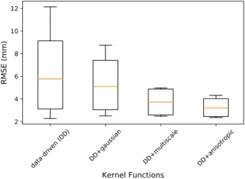
data-driven (DD) DD+gaussian DD+multiscale DD+anisotropic
Kernel Functions
2 4 6 8 10 12RMSE (mm)
Figure 3: Evaluation of different kernel functions.
fitting the SSMs to mesh data in the test set and then computing RMSE of the fitted femur models to the test set. Figure 2 shows some modes of the data-driven SSM and its combination with the anisotropic kernel. Results for comparing the SSMs can be seen in Fig. 3. The RMSE for the data-driven SSM and its combination with the Gaussian, multiscale and anisotropic kernels were computed as 6.48 ± 4.53, 5.36 ± 2.98, 3.73 ± 1.36 and 3.27 ± 1.00 mm, respectively.
4
Discussion and conclusion
The current study developed SSMs for femur bone mod-eling based on GPMMs. Out of different analytical kernels (Gaussian, multiscale and anisotropic kernels) and keeping the 10 most prominent basis functions, it has been found that the combination of our data-driven model with the anisotropic kernel more accurately en-codes the patterns of variability of femurs in our dataset. Specifically, we observed that the modes of data-driven SSM when combined with the anisotropic kernel corre-sponds well with the actual deformation of the femur bone (this can be confirmed by seeing how the dom-inant modes vary in Figure 2). Exploration of other customized kernels or transferring the knowledge of fe-mur variations from a larger dataset can be considered for future works.
A. ASVADI et al. Surgetica 2019 Rennes, France – 17 - 18 June
References
[1] Mutsvangwa, Tinashe, et al. (2015). An automated statistical shape model developmental pipeline: ap-plication to the human scapula and humerus. IEEE Transactions on Biomedical Engineering,
62.4:1098-1107.
[2] Salhi, Asma, et al. (2017). Comparing statistical shape model-based mesh fitting methods: towards patient-specific muscle modeling. In: Proc. RITS. [3] Heimann, Tobias, and Hans-Peter Meinzer. (2009). Statistical shape models for 3D medical image segmentation: a review. Medical image analysis, 13.4:543-563.
[4] Cootes, Timothy F., et al. (1992). Training models of shape from sets of examples. In: Proc. BMVC, 9-18.
[5] Luthi, Marcel, et al. (2018). Gaussian process morphable models. IEEE transactions on pattern analysis and machine intelligence, 40.8:1860-1873. [6] Albrecht, Thomas, et al. (2013). Posterior shape
models. Medical image analysis, 17.8:959-973. [7] Scalismo (2016). scalismo - scalable image
anal-ysis and shape modeling. available online at:
http://github.com/unibas-gravis/scalismo
[8] Jacq, Jean-Jos´e, et al. (2008). Performing accurate
joint kinematics from 3-D in vivo image sequences through consensus-driven simultaneous registra-tion. IEEE Transactions on Biomedical Engineer-ing, 55.5:1620-1633.
[9] Kazhdan, Michael, and Hugues Hoppe. (2013). Screened poisson surface reconstruction. ACM Transactions on Graphics, 32.3:29.