HAL Id: hal-00641695
https://hal.archives-ouvertes.fr/hal-00641695
Submitted on 30 May 2020
HAL is a multi-disciplinary open access
archive for the deposit and dissemination of
sci-entific research documents, whether they are
pub-lished or not. The documents may come from
teaching and research institutions in France or
abroad, or from public or private research centers.
L’archive ouverte pluridisciplinaire HAL, est
destinée au dépôt et à la diffusion de documents
scientifiques de niveau recherche, publiés ou non,
émanant des établissements d’enseignement et de
recherche français ou étrangers, des laboratoires
publics ou privés.
Copyright
Binding of Toxoplasma gondii
glycosylphosphatidylinositols to galectin-3 is required for
their recognition by macrophages
Françoise Debierre-Grockiego, Sebastian Niehus, Bernadette Coddeville,
Elisabeth Elass, F. Poirier, R. Weingart, R.R. Schmidt, Joel Mazurier, Yann
Guerardel, Ralf Schwartz
To cite this version:
Françoise Debierre-Grockiego, Sebastian Niehus, Bernadette Coddeville, Elisabeth Elass, F. Poirier,
et al.. Binding of Toxoplasma gondii glycosylphosphatidylinositols to galectin-3 is required for their
recognition by macrophages. Journal of Biological Chemistry, American Society for Biochemistry and
Molecular Biology, 2010, 285 (43), pp.32744-32750. �10.1074/jbc.M110.137588�. �hal-00641695�
Binding of Toxoplasma gondii Glycosylphosphatidylinositols
to Galectin-3 Is Required for Their Recognition by
Macrophages
*
Received for publication, April 23, 2010, and in revised form, July 27, 2010Published, JBC Papers in Press, August 20, 2010, DOI 10.1074/jbc.M110.137588
Franc¸oise Debierre-Grockiego‡§1, Sebastian Niehus‡2, Bernadette Coddeville¶, Elisabeth Elass¶, Franc¸oise Poirier储, Ralf Weingart**3, Richard R. Schmidt**, Joe¨l Mazurier¶, Yann Gue´rardel¶, and Ralph T. Schwarz‡¶
From the‡Institut fu¨r Virologie, AG Parasitologie, Philipps University, Marburg D-35043, Germany,§Unite´ Mixte de Recherche Universite´-Institut National de la Recherche Agronomique 0483 Immunologie Parasitaire, Vaccinologie et Biothe´rapies Anti-infectieuses, Unite´ de Formation de Recherche Sciences Pharmaceutiques, 31 Avenue Monge, F-37200 Tours, France, the ¶CNRS-Unite´ Mixte de Recherche 8576, Unit of Structural and Functional Glycobiology, Institut Fe´de´ratif de Recherche 147, Universite´ Lille Nord de France, F-59000 Lille, France, the储Laboratoire de Ge´ne´tique et De´veloppement des Mammife`res, Institut Jacques Monod, Paris F-75013, France, and the **Fachbereich Chemie, University of Konstanz, Konstanz D-78457, Germany
We showed that the production of tumor necrosis factor (TNF)␣by macrophages in response to Toxoplasma gondii gly-cosylphosphatidylinositols (GPIs) requires the expression of both Toll-like receptors TLR2 and TLR4, but not of their co-receptor CD14. Galectin-3 is a-galactoside-binding protein with immune-regulatory effects, which associates with TLR2. We demonstrate here by using the surface plasmon resonance method that the GPIs of T. gondii bind to human galectin-3 with strong affinity and in a dose-dependent manner. The use of a synthetic glycan and of the lipid moiety cleaved from the GPIs shows that both parts are involved in the interaction with galec-tin-3. GPIs of T. gondii also bind to galectin-1 but with a lower affinity and only through the lipid moiety. At the cellular level, the production of TNF-␣induced by T. gondii GPIs in macro-phages depends on the expression of galectin-3 but not of galec-tin-1. This study is the first identification of a galectin-3 ligand of T. gondii origin, and galectin-3 might be a co-receptor pre-senting the GPIs to the TLRs on macrophages.
Glycosylphosphatidylinositol (GPI)4-anchored proteins dominate the surface of the Toxoplasma gondii tachyzoite (1, 2). We have shown that T. gondii GPIs, as well as their glycan and lipid moieties, induce the production of tumor necrosis factor (TNF)-␣ in macrophages through the activation of the transcription factor NF-B (3). Toll-like receptors (TLRs)
rec-ognize microbial components leading to cytokine production and antimicrobial responses through NF-B activation (4). We have shown that both TLR2 and TLR4 are involved in the NF- B-dependent signaling cascade leading to production of TNF-␣ by macrophages exposed to T. gondii GPIs (5). The membrane protein CD14 is a co-receptor that associates with TLR4 to form a receptor complex for bacterial lipopolysaccharide (LPS) (6). CD14 is involved in the recognition of lipoproteins from Mycobacterium tuberculosisby TLR2 (7). In addition, CD14 is necessary for the TLR2-dependent response of macrophages to GPIs of Trypanosoma cruzi (8). In contrast, CD14 is not required for the signaling leading to production of TNF-␣ by macrophages in response to the GPIs of T. gondii (5). The asso-ciation of galectin-3 with TLR2 was demonstrated on PMA-differentiated THP-1 human macrophages infected with Can-dida albicans (9), and immunofluorescence microscopy showed the co-expression of galectin-3 with TLR4 on bone marrow-derived macrophages (10). Galectin-3 is a lectin spe-cific for -galactosides, composed of a C-terminal carbohy-drate-recognition domain (CRD) and an N-terminal domain (ND) (11).
The galectin-3-dependent recruitment of effector cells may be an important mechanism of resistance to parasite infection. Indeed, high levels of galectin-3 were found in the granulomas surrounding eggs and worms during Schistosoma mansoni infection (12). In addition, galectin-3 co-localizes with GalNAc1–4GlcNAc expressed on the surface of eggs and can mediate GalNAc1–4GlcNAc recognition and phagocytosis by macrophages. This implicates GalNAc1–4GlcNAc as a molecular parasite pattern for galectin-3-mediated immune recognition. In galectin-3⫺/⫺mice infected with S. mansoni, the size of the granulomas surrounding eggs was significantly decreased compared with infected wild type mice (13). Galec-tin-3 deficiency affects the number of splenic T and B cells in S. mansoni-infected mice, whereas it does not modulate the num-ber of dendritic cells. T. gondii infection of wild type mice leads to an up-regulation of galectin-3 expression in various tissues (14). T. gondii-infected galectin-3⫺/⫺ mice develop reduced inflammatory responses in all organs, except for the lungs, and exhibit a higher parasite burden in the lungs and the brain (14).
*This work was supported by the Deutsche Forschungsgemeinschaft (R. T. S.), the Universite´ des Sciences et Technologies de Lille 1 (Y. G.), the Institut Fe´de´ratif de Recherche 147, the Centre National de la Recherche Scientifique (Unite´ Mixte de Recherche CNRS no. 8576, Director Dr. J. C. Michalski), the Groupement des Entreprises Françaises dans la Lutte Con-tre le Cancer and Association pour la Recherche Sur le Cancer Grant 1113 (to F. P.), and the PROCOPE of Deutscher Akademischer Austausch Dienst/ Egide (R. T. S. and Y. G.).
1To whom correspondence should be addressed. Fax: 33 2 47 36 72 52; E-mail: francoise.debierre@univ-tours.fr.
2Present address: Unite´ Propre de Recherche 9022 CNRS, Institut de Biologie Mole´culaire et Cellulaire, 15 rue Rene´ Descartes, 67084 Strasbourg, France. 3Present address: Alta Pharma Deutschland GmbH, Byk-Gulden-Strasse 2,
D-78467 Konstanz, Germany.
4The abbreviations used are: GPI, glycosylphosphatidylinositol; CRD, carbo-hydrate recognition domain; ND, N-terminal domain; RU, resonance units; SPR, surface plasmon resonance; TLR, Toll-like receptor.
at INRA Institut National de la Recherche Agronomique on June 14, 2018
http://www.jbc.org/
Comparable survival rates were observed in galectin-3⫺/⫺and galectin-3⫹/⫹mice orally infected with T. gondii. In contrast, higher mortality was observed in galectin-3⫺/⫺ mice after intraperitoneal infection with T. gondii associated with a defi-cient influx of neutrophils and macrophages into the peritoneal cavity. Furthermore, a recent study indicates that galectin-3 has an important modulator function by interfering in the life span and activation of neutrophils early after their infection with T. gondii(15). In the present work, we show that the GPIs of T. gondii are ligands of galectin-3 and that the production of TNF-␣ induced by GPIs in macrophages requires the expres-sion of galectin-3.
EXPERIMENTAL PROCEDURES
Extraction and Purification of GPIs—T. gondii tachyzoites (strain RH) were grown in Vero cells (free of Mycoplasma) and released from host cells with the help of glass beads in the Mixer Mill homogenizer (Retsch, Haan) and purified by glass wool filtration (16). GPIs were extracted from tachyzoites as described previously (17). Briefly, glycolipids were extracted with chloroform-methanol-water (10:10:3, by volume) and par-titioned between water and water-saturated n-butyl alcohol. GPIs present in n-butyl alcohol were precipitated under a stream of nitrogen. The six different T. gondii GPIs (18) were then separated by thin layer chromatography (TLC), with [3H]glucosamine-labeled T. gondii GPIs used as tracers. For this, a part of the tachyzoite culture was metabolically labeled in glucose-free DMEM containing 20 mMsodium pyruvate and 0.5 mCi of [3H]glucosamine hydrochloride (Hartmann Ana-lytic GmbH, Braunschweig) for 6 h at 37 °C in 5% CO2 atmo-sphere. Labeled GPIs were extracted as above. TLC was performed with the solvent n-hexane-chloroform-methanol-water-acetic acid (3:10:10:2:1, by volume). Chromatograms were scanned for radioactivity (Berthold LB 2842 linear ana-lyzer), and areas corresponding to individual T. gondii GPIs were scraped off and reextracted with chloroform-methanol-water (10:10:3, by volume) by sonication. GPIs were separated from residual silica gel and recovered in the n-butyl alcohol phase after n-butyl alcohol/water partition. GPI quantification was based on monosaccharide composition of each GPI, deter-mined by gas chromatography (GC) on a GC Trace gas chro-matograph (Thermo Fisher Scientific) equipped with a 25-m⫻ 0.25-mm ECTM⫺1 capillary column, 0.25-m film thickness (Alltech, Templemars, France) after methanolysis (0.5N HCl-methanol for 24 h at 80 °C), N-reacetylation, and trimethylsily-lation. Diacylglycerols were obtained by the cleavage of TLC-purified GPIs using 1 unit of Bacillus cereus phosphatidyl-inositol phospholipase C (Sigma) in 100l of 0.1MTris-HCl (pH 7.4), 0.1% sodium deoxycholate overnight at 37 °C. The enzyme was inactivated by treatment for 3 min at 100 °C. Dia-cylglycerols were recovered in the butanol phase after partition between water and water-saturated n-butyl alcohol. The absence of endotoxin in purified T. gondii GPIs was checked with the Limulus Amebocyte Lysate kit QCL-100 (Bio-Whit-taker, Walkersville, MD). GPIa was chemically synthesized according to Pekari et al. (19). This molecule has no lipid moi-ety and corresponds to the glycan part of GPI III of T. gondii.
Direct Binding Assays by SPR—Binding analyses were per-formed on a BIAcore 3000 instrument (BIAcore Ab; Uppsala, Sweden). Human recombinant galectin-3 and galectin-1 (carri-er-free; R&D Systems, Minneapolis, MN) were covalently cou-pled to a CM5 (carboxymethylated) sensor chip following the manufacturer’s instructions (standard amine immobilization kit; BIAcore). Immobilized bovine serum albumin (BSA) was used as control for nonspecific binding. Amounts of 9,000 – 12,000 resonance units (RU) and 2,500 – 4,000 RU of galectin-3 and galectin-1, respectively, were immobilized under these conditions, where 1 RU corresponds to a concentration of 1 pg/mm2of immobilized protein (20). T. gondii GPIs, GPIa, or diacylglycerols diluted in PBS by sonication were injected using the multichannel mode. In some experiments, the GPI mole-cules were injected with 15 mMor 30 mMlactose. After injection of 65-l samples at 10 l/min and at 25 °C, the formed com-plexes were allowed to dissociate in PBS. Regeneration of the sensor chip surface was achieved by repeated pulses of 90% ethylene glycol and 10 mMNaOH, at a flow rate of 30l/min. Laminin, a well known substrate, was injected to check that the regeneration process did not modify the structure and the activity of the galectins. Control flow-cells were obtained by injection of the respective analyte solutions on a chip submitted to the activation-deactivation coupling protocol and on a chip with immobilized BSA. Data were analyzed using the BIAcore 3.0 evaluation software. Results represented as sensorgrams are expressed in RU, which are proportional to the quantity of ana-lytes bound to immobilized galectin-3 or galectin-1. Because binding equilibrium was not reached during the time of injec-tion, the equilibrium response values (Requ) were calculated from the association phases obtained with increasing concen-trations of analytes. The measured association (kon) and disso-ciation (koff) rate constants allowed the determination of the equilibrium dissociation constant KD, through the ratio koff/ kon. KD was also determined from Scatchard plot analysis by linearization of the saturation binding curve Requ/[GPI] versus [GPI], with Requthe response units at equilibrium. The slope corresponds to⫺1/KD.
Production of TNF-␣ by Primary Macrophages from Galec-tin-deficient Mice—The experimental procedures using mice were performed according to the institutional animal care and use guidelines from the Monod Institute, Paris Diderot Univer-sity. Thioglycollate-elicited peritoneal macrophages were obtained from wild type (129Sv), galectin-1⫺/⫺, galectin-3⫺/⫺, and galectin-1/3⫺/⫺mice by peritoneal washing with serum-free medium (Panserin 401; PAN Biotech GmbH, Aidenbach). Macrophages were incubated for 24 h with the medium alone or with the different GPIs of T. gondii at 2Mresuspended in the medium by sonication. The amount of TNF-␣ in culture supernatants was determined by using a specific sandwich ELISA (BD Biosciences).
Statistics—The unpaired Student’s t test was adopted for sta-tistical evaluation. p⬍ 0.05 was considered significant.
RESULTS
T. gondii GPIs, the Glycan GPIa, and Diacyglycerols Bind to Human Galectin-3—SPR was used to test whether human galectin-3 may interact with GPIs, the main glycolipids of T.
Recognition of T. gondii GPIs by Galectin-3
at INRA Institut National de la Recherche Agronomique on June 14, 2018
http://www.jbc.org/
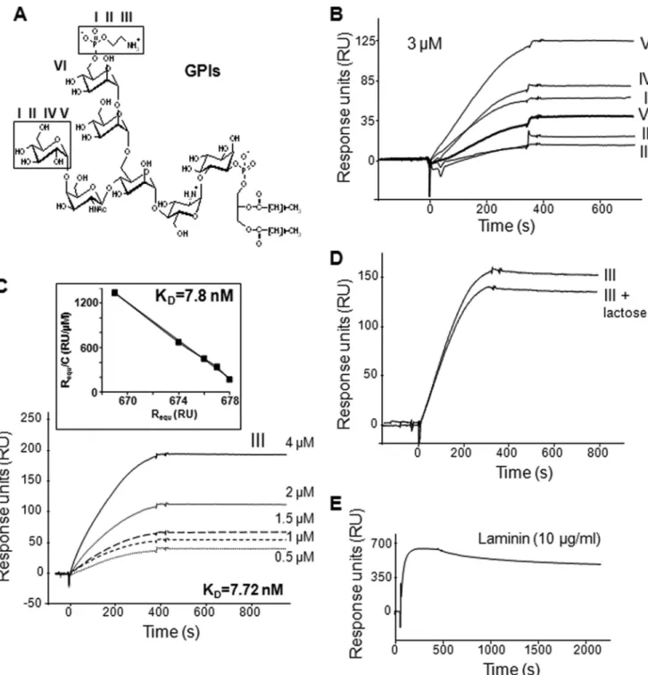
gondii. The six different GPIs of T. gondii (structures in Fig. 1A) were injected separately over immobilized galectin-3. The molecular association and dissociation phases were illustrated by sensorgrams obtained after subtraction of the nonspecific binding to a sensor chip control cell. As shown in Fig. 1B, all T. gondiiGPIs bound to galectin-3, and the very slow dissociation that occurred in PBS suggests the formation of highly stable complexes. The results presented here show that the different
GPIs bound to galectin-3 with variable amounts (RU). How-ever, repeated experiments with other galectin-3-immobilized sensorchips indicated that these differences between GPIs were not significant, suggesting that the binding is independent of the variation between the six GPI structures. Injection of increasing concentrations of GPI permitted us to calculate the association and dissociation constants. The binding of GPI to galectin-3 is dose-dependent as illustrated with GPI III in Fig.
FIGURE 1. GPIs of T. gondii associate with galectin-3. A, structures of T. gondii GPIs: GPIs I and II, (EtN-P)-Man␣1–2Man␣1–6(Glc␣1–4GalNAc1–4)Man␣1– 4GlcN␣-Ino-P-DAG; GPI III, (EtN-P)-Man␣1–2Man␣1–6(GalNAc1–4)Man␣1–4GlcN␣-Ino-P-DAG; GPIs IV and V, Man␣1–2Man␣1–6(Glc␣1–4GalNAc1– 4)Man␣1–4GlcN␣-Ino-P-DAG; and GPI VI, Man␣1–2Man␣1–6(GalNAc1–4)Man␣1–4GlcN␣-Ino-P-DAG. GPIs I and II, and GPIs IV and V differ in their diacylg-lycerol composition with palmitic and stearic acids as predominant fatty acids. EtN, ethanolamine, P, phosphate; Man, mannose; Glc, glucose; GalNAc, acetylgalactosamine; GlcN, glucosamine; Ino, inositol; DAG, diacylglycerol. B–E, SPR analysis of the interaction of immobilized galectin-3 with the six different
T. gondii GPIs (I–VI) at 3M(B); different concentrations of T. gondii GPI III with Scatchard plot analysis of the sensorgram data shown in the inset (slope⫽ ⫺127.4) (C); GPI III at 3Min the presence of 30 mMlactose (D); or laminin at 10g/ml (E). Results are expressed as RU (proportional to the quantity of molecules) after the subtraction of the nonspecific binding. Sensorgrams are representative of independent experiments with similar results.
at INRA Institut National de la Recherche Agronomique on June 14, 2018
http://www.jbc.org/
1C. The equilibrium dissociation constant KDcalculated from the ratio of the kinetic rate constants (dissociation rate constant koff ⫽ 1.75 ⫻ 10⫺6s⫺1/association rate constant kon ⫽ 227
M⫺1䡠s⫺1) was 7.72 nM. The KDdetermined by Scatchard plot analysis was consistent with the kinetically determined value (KD⫽ 7.8 nM; Fig. 1C, inset). To understand which part of galec-tin-3 is involved in its association with T. gondii GPIs, an excess of lactose that specifically blocks the CRD of galectin-3, was added. However, 30 mMlactose induced a very low reduction (12%) in the binding of GPI III at 3M(Fig. 1D). This suggests that the ND is also involved in the association of galectin-3 with T. gondiiGPIs. A saline buffer (pH 7.2) containing 1MNaCl was inefficient to regenerate the sensor chip, and alternated pulses of ethylene glycol (90%) and NaOH (10 mM) were required to release bound GPIs, suggesting the involvement of hydropho-bic interactions. This treatment did not alter the structure of galectin-3 because its well known ligand laminin (at 10g/ml) was able to associate with galectin-3 after several regeneration processes (Fig. 1E).
All GPIs are constituted by a glycan moiety and a lipid moi-ety, and we then investigated which part is involved in the inter-action with galectin-3. To this end, a chemically synthesized molecule, GPIa, corresponding to the glycan moiety of GPI III and lacking the lipid moiety (structure in Fig. 2A) was tested in
SPR assay. This molecule was also able to interact with galectin-3 but with a much lower affinity than the whole GPI (KD⫽ 576M; see also the Scatchard plot analysis in the inset KD⫽ 526M), illustrated by a complete dissociation at the end of the GPIa injection (Fig. 2B). Because of its glycan structure GPIa might interact with the CRD of galectin-3. This hypothesis was confirmed by the addition of an excess of lactose (15 mM) that strongly reduced (80%) the binding of GPIa at 200 M to galectin-3 (Fig. 2C). This inhibition assay gives good evidence that GPIa interacts with the CRD of galec-tin-3. The discrepancy between the binding of the whole GPI and the binding of GPIa suggests that the lipid moiety of the entire GPI also participates in the interac-tion with galectin-3. To check this hypothesis, the diacylglycerol was isolated by the cleavage of the T. gondiiGPI III with a phosphatidyli-nositol-specific phospholipase C and tested in SPR assay. The diacyl-glycerol alone was able to bind to galectin-3 (Fig. 2D) with an inter-mediate affinity between those of GPIa and of the entire GPI (KD⫽ 0.16M). These results suggest that the glycan moiety and the lipid moi-ety act in synergy in the interaction with galectin-3. Because lactose only partially inhibited the binding of the whole GPIs, the ND is certainly involved in the interaction with the lipid moiety.
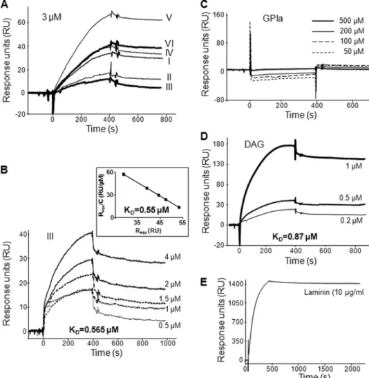
T. gondii GPIs and Their Diacyglycerols, but Not GPIa, Bind to Human Galectin-1—To understand further the specificity and the characteristics of the GPI binding to human galectin-3, we have tested the interaction of GPIs to human galectin-1, a-galactoside-binding protein related to galectin-3 but lacking a ND. As for galectin-3, galectin-1 interacted with the GPIs of T. gondii without significant dif-ference between the six structures, but the dissociation was more rapid, suggesting that GPI association with galectin-1 is less stable than with galectin-3 (Fig. 3A). A lower affinity for galectin-1 than for galectin-3 was obtained with the injection of increased concentrations of the GPI III (Fig. 3B), with a calculated KDof 0.565M(koff⫽ 3.24 ⫻ 10⫺4s⫺1/kon⫽ 573 M⫺1䡠s⫺1), correlated with the value of 0.550 M obtained with the Scatchard plot analysis (Fig. 3B, inset). Because galectin-1 is only constituted by a CRD, the synthetic glycan GPIa should bind to galectin-1 to same extent as the entire GPIs. Surprisingly, GPIa was unable to interact with galectin-1 (Fig. 3C). In contrast, the GPI diacylglycerols sus-pected to be recognized by the ND of galectin-3 interacted with
FIGURE 2. Both glycan and lipid moieties participate in the association of T. gondii GPIs with galectin-3.
A, structure of GPIa, the chemically synthesized glycan of T. gondii GPI III. B–D, SPR analysis of the interaction of
immobilized galectin-3 with GPIa at different concentrations (B) (Scatchard plot shown in the inset, slope⫽ ⫺0.0017); GPIa at 200Min the presence of 15 mMlactose (C); or diacylglycerols (DAG, general structure in the
inset) isolated from GPI III at different concentrations (D). Results are expressed as RU (proportional to the
quantity of molecules) after the subtraction of the nonspecific binding. Sensorgrams are representative of independent experiments with similar results.
Recognition of T. gondii GPIs by Galectin-3
at INRA Institut National de la Recherche Agronomique on June 14, 2018
http://www.jbc.org/
galectin-1 with a KDvalue similar to that of the whole GPIs (Fig. 3D). As with galectin-3, the regeneration of the sensor chip was achieved only after using of ethylene glycol and NaOH. How-ever, this treatment did not degrade or modify the conforma-tion of galectin-1 because laminin (10g/ml) associated with
galectin-1 after several regeneration processes (Fig. 3E). Altogether, these results indicate that the recog-nition of T. gondii GPIs is differ-ent and specific for galectin-3 and galectin-1.
Galectin-3 Is Required for the Pro-duction of TNF-␣ by GPI-stimu-lated Macrophages—Because the affinity of T. gondii GPIs is higher for galectin-3 than for galectin-1, galectin-3 may be specifically involved in the TNF-␣ produc-tion by macrophages in response to T. gondii GPIs. To study this point, peritoneal macrophages from galectin-3⫺/⫺, galectin-1⫺/⫺, galectin-1/3⫺/⫺, and control (129Sv) mice were incubated with individual GPIs at 2 M, and TNF-␣ secretion was measured in supernatants. According to our previous work (5), wild type macrophages were stimulated by the six T. gondii GPIs to produce higher amounts of TNF-␣ than in the presence of medium alone (Fig. 4). This induction of TNF-␣ by T. gondii GPIs was not inhibited in the absence of galectin-1 (galec-tin-1⫺/⫺). The reduction observed for the GPI II in the experiment shown here is not significant (p⫽ 0.069). On the contrary, macro-phages lacking galectin-3 (galec-tin-3⫺/⫺) were not stimulated by the GPIs, and the levels of TNF-␣ were similar to those measured in the supernatant of cul-tures with medium alone. Lack of TNF-␣ induction was also obtained with double null mutant macrophages (galectin-1/ 3⫺/⫺) and it was certainly due to the absence of galectin-3 and not of galectin-1. These data suggest that galectin-3 is involved in the recognition of T. gondii GPIs at the surface of the macrophages.
DISCUSSION
Galectin-3 has more extended carbohydrate-recognition sites than galectin-1. This structural difference allows galec-tin-3 to bind to a wider range of oligosaccharide structures, including structures containing mannose (21). We identified the GPIs of T. gondii as ligands of galectin-3. KDvalues of galec-tin-3 for diacylglycerols and entire GPIs at theMlevel and the nMlevel, respectively, were in the range of KDvalues of galec-tin-3 for other ligands, for example 3.6 – 6.1Mfor (Gal1–3)n lactose, and 1.4Mfor biantennary N-linked oligosaccharide (22–24). Six different GPIs are synthesized by the parasite. GPIs III and VI present a GalNAc1–4 branched on the first man-nose of the evolutionary conserved core glycan, whereas GPIs I,
FIGURE 3. GPIs of T. gondii associate with galectin-1 through their lipid moiety. SPR analysis of the inter-action of immobilized galectin-1 with (A) the six different T. gondii GPIs (I-VI) at 3M; (B) GPI III at different concentrations (Scatchard plot shown in the inset, slope⫽ ⫺1.77); (C) GPIa at different concentrations; (D) diacylglycerols isolated from GPI III at different concentrations; or (E) laminin at 10g/ml. Results are expressed as RU (proportional to the quantity of molecules) after the subtraction of the nonspecific binding. Sensorgrams are representative of independent experiments with similar results.
FIGURE 4. T. gondii GPIs require galectin-3 but not galectin-1 to induce TNF-␣ production in macrophages. Peritoneal macrophages from wild type (129Sv), galectin-1⫺/⫺, galectin-3⫺/⫺, and galectin-1/3⫺/⫺mice were incubated for 24 h with medium alone or with the six different GPIs (I–VI) at 2 M. Supernatants were assayed for TNF-␣ production using a sandwich ELISA. Values represent the means⫾ S.D. of triplicate samples.
at INRA Institut National de la Recherche Agronomique on June 14, 2018
http://www.jbc.org/
II, IV, and V have an additional glucose residue linked with an ␣1–4 bond to the GalNAc. This glucose residue does not influ-ence the recognition of the GPIs by galectin-3, because no dif-ference was observed in the amounts of GPIs interacting with galectin-3 in the SPR assay. Binding of GalNAc1–4-contain-ing glycans is not common to all galectin family members. Indeed, galectin-3 but not galectin-1 binds GalNAc 1-4GlcNAc-glycans of S. mansoni (12). Galectin-3 can accommo-date the O-2 N-acetyl moiety of the GalNAc residue, whereas a bulky histidine (at position 52) in galectin-1 may prevent GalNAc binding. Binding of galectin-3 to GalNAc1–4GlcNAc was completely prevented in the presence of lactose, supporting involvement of the CRD in this interaction (12). These results are in agreement with our observation that the synthetic glycan GPIa does not bind to galectin-1 but binds to the CRD of galec-tin-3, as evidenced by the lactose inhibitory effect. Galectin-3 recognizes the polygalactose structure, (Gal1–3)n, found on lipophosphoglycan of Leishmania major (25). Galectin-1 was not shown to have significant affinity for L. major, suggesting that the polygalactose epitope binds to the galectin-3-unique extended binding pocket, which is not present in galectin-1 (25). The polygalactose is not expressed by Leishmania donovani. Thus, galectin-3 can distinguish between the two related parasites. In contrast, GPIs are expressed on both virulent (RH) and nonvirulent (PTG) strains of T. gondii, and the slight differences we have found in their structure5did not affect the interaction with galectin-3 (KD⫽ 0.2 nM, not shown).
Galectin-1 binds to Davanat (composed of 1–4-linked D-mannopyranosyl units and D-galactopyranosyl residues attached via␣1–6 linkage) with an apparent equilibrium disso-ciation constant of 10M, compared with 260Mfor lactose (26). The-galactoside-binding domain remains accessible in the galectin-1䡠Davanat complex because lactose can still bind with no apparent loss in affinity. Galectin-1 acts as a receptor for a protein, the tissue plasminogen activator. The interaction of tissue plasminogen activator with galectin-1 is higher than with galectin-3 (KD9.1Mand 17 mM, respectively) (27). This illustrates that galectins interact not only with-galactosides. Here, we show that the diacylglycerols of T. gondii GPIs interact strongly with galectin-1, which lacks a ND. Because the CRD of both galectins has affinities for different structures, further investigations are needed to conclude whether the lipid moiety of GPIs interacts with the CRD or the ND of galectin-3. We have shown previously that the diacylglycerols cleaved from the T. gondiiGPIs are able to stimulate macrophages to produce TNF-␣ (3) via signaling of both TLR2 and TLR4, whereas only the TLR2 signaling seems to be triggered by GPIa or the whole GPIs (5). The three-dimensional conformation of the com-plexes galectin-3䡠diacylglycerol and galectin-3䡠GPI (or GPIa) might determine their recognition or not by TLR4. Free GPIs are present in the extracellular medium in their entire form, probably secreted by the parasites.6We assume that
macro-phage enzymes like phospholipases, cleave the GPI lipid moi-ety, allowing its interaction with galectin-3 and the TLR signaling.
Galectin-3 may play a major role in controlling cytokine pro-duction. LPS markedly increased amounts of inflammatory cytokines in galectin-3⫺/⫺cells (10). S. mansoni- and T. gondii-infected galectin-3⫺/⫺ mice mount a higher Th1-polarized immune response and a decreased inflammation (14). Because its absence was associated with the production of higher levels of Th1 cytokines when these cells are stimulated, it suggests that galectin-3 may act in the regulatory loops controlling Th1 cytokine production. It was shown that galectin-3⫺/⫺ macro-phages produce lower amounts of TNF-␣ after incubation with Candida albicans, compared with galectin-3⫹/⫹macrophages (9). We showed in the present study that galectin-3⫺/⫺ macro-phages are unable to produce TNF-␣ in response to T. gondii GPIs. Cells other than macrophages might be responsible for the higher production of inflammatory Th1 cytokines observed in galectin-3-deficient animals. Our previous study indicates that the T. gondii GPI-induced TNF-␣ production involves both TLR2 and TLR4 in macrophages (5). CD14 is the well known associated molecule for the recognition of LPS, the his-torical ligand of TLR4. On the contrary, CD14 is not required for the production of TNF-␣ by mouse macrophages in response to the GPIs of T. gondii (5). We hypothesize that galec-tin-3 is a TLR-associated protein involved in the cell response to the GPIs of T. gondii. It is possible that extracellular galec-tin-3 molecules interact with N-glycans present in TLRs (28). It has been demonstrated that the surface expression of TLR2 is enhanced on galectin-3⫺/⫺macrophages (29). This up-regula-tion cannot lead to an increased producup-regula-tion of TNF-␣ in response to the GPIs because the expression of galectin-3 is absolutely needed. Our results show that galectin-1 is not involved in the TNF-␣ production by macrophages in response to GPI stimulation. Exogenous galectin-1 was found to inhibit LPS-induced NF-B activation macrophages (30). We suggest that galectin-1 blocks␣-2,3-sialyl residues linked to -galacto-side glycans on TLR4 receptors, and so Neu1 sialidase can no longer hydrolyze these sialic acids. In contrast, the GPI-induced TNF-␣ production was sometimes higher in galectin-1⫺/⫺ compared with wild type macrophages (Fig. 4), suggesting that galectin-1 may partially neutralize the TLR4 signaling in response to T. gondii GPIs.
Different studies indicate that galectin-1 is involved in the recognition of protozoan parasites. Endogenous galectin-1 expression is up-regulated in vitro and in vivo by T. cruzi, and the expression of galectin-1 is up-regulated in cardiac tissue from patients with chronic Chagas disease (31). Because the adhesion of T. cruzi to heart muscle cells is mediated by pro-tein-carbohydrate interactions, an overexpression of galectin-1 on heart tissue could facilitate parasite invasion toward this vulnerable infection site (32). Galectin-1 acts as a receptor for the protozoa Trichomonas vaginalis by interacting with para-site lipophosphoglycans at the surface of human cervical epi-thelial cells (32). Accordingly, galectin-1 may be involved in the attachment of T. gondii to the host cell, supporting by this way the invasion process.
5S. Niehus, T. K. Smith, N. Azzouz, M. A. Compos, J. F. Dubremetz, D. Sibley, R. T. Schwarz, R. T. Gazzinelli, and F. Debierre-Grockiego, unpublished observations.
6F. Debierre-Grockiego and R. T. Schwarz, unpublished observations.
Recognition of T. gondii GPIs by Galectin-3
at INRA Institut National de la Recherche Agronomique on June 14, 2018
http://www.jbc.org/
Acknowledgments—We are grateful to Ulrike Bieker, Elfriede Klein, and Jo¨rg Schmidt (Institut fu¨r Virologie, AG Parasitologie, Philipps Universität, Marburg, Germany) for assistance in parasite culture.
REFERENCES
1. Black, M. W., and Boothroyd, J. C. (2000) Microbiol. Mol. Biol. Rev. 64, 607– 623
2. Lekutis, C., Ferguson, D. J., Grigg, M. E., Camps, M., and Boothroyd, J. C. (2001) Int. J. Parasitol. 31, 1285–1292
3. Debierre-Grockiego, F., Azzouz, N., Schmidt, J., Dubremetz, J. F., Geyer, H., Geyer, R., Weingart, R., Schmidt, R. R., and Schwarz, R. T. (2003) J. Biol.
Chem. 278,32987–32993
4. Medzhitov, R., Preston-Hurlburt, P., and Janeway, C. A., Jr. (1997) Nature 388,394 –397
5. Debierre-Grockiego, F., Campos, M. A., Azzouz, N., Schmidt, J., Bieker, U., Resende, M. G., Mansur, D. S., Weingart, R., Schmidt, R. R., Golen-bock, D. T., Gazzinelli, R. T., and Schwarz, R. T. (2007) J. Immunol. 179, 1129 –1137
6. Yang, Z., Khemlani, L. S., Dean, D. F., Carter, C. D., Slauson, D. O., and Bochsler, P. N. (1994) J. Leukocyte Biol. 55, 483– 488
7. Drage, M. G., Pecora, N. D., Hise, A. G., Febbraio, M., Silverstein, R. L., Golenbock, D. T., Boom, W. H., and Harding, C. V. (2009) Cell. Immunol. 258,29 –37
8. Campos, M. A., Almeida, I. C., Takeuchi, O., Akira, S., Valente, E. P., Proco´pio, D. O., Travassos, L. R., Smith, J. A., Golenbock, D. T., and Gazzinelli, R. T. (2001) J. Immunol. 167, 416 – 423
9. Jouault, T., El Abed-El Behi, M., Martinez-Esparza, M., Breuilh, L., Trinel, P. A., Chamaillard, M., Trottein, F., and Poulain, D. (2006) J. Immunol. 177,4679 – 4687
10. Li, Y., Komai-Koma, M., Gilchrist, D. S., Hsu, D. K., Liu, F. T., Springall, T., and Xu, D. (2008) J. Immunol. 181, 2781–2789
11. Hsu, D. K., Zuberi, R. I., and Liu, F. T. (1992) J. Biol. Chem. 267, 14167–14174
12. van den Berg, T. K., Honing, H., Franke, N., van Remoortere, A., Schiphorst, W. E., Liu, F. T., Deelder, A. M., Cummings, R. D., Hokke, C. H., and van Die, I. (2004) J. Immunol. 173, 1902–1907
13. Breuilh, L., Vanhoutte, F., Fontaine, J., van Stijn, C. M., Tillie-Leblond, I., Capron, M., Faveeuw, C., Jouault, T., van Die, I., Gosset, P., and Trottein, F. (2007) Infect. Immun. 75, 5148 –5157
14. Bernardes, E. S., Silva, N. M., Ruas, L. P., Mineo, J. R., Loyola, A. M., Hsu, D. K., Liu, F. T., Chammas, R., and Roque-Barreira, M. C. (2006) Am. J.
Pathol. 168,1910 –1920
15. Alves, C. M., Silva, D. A., Azzolini, A. E., Marzocchi-Machado, C. M., Carvalho, J. V., Pajuaba, A. C., Lucisano-Valim, Y. M., Chammas, R., Liu, F. T., Roque-Barreira, M. C., and Mineo, J. R. (2010) Immunobiology 215, 475– 485
16. Grimwood, B. G., Hechemy, K., and Stevens, R. W. (1979) Exp. Parasitol. 48,282–286
17. Debierre-Grockiego, F., Rabi, K., Schmidt, J., Geyer, H., Geyer, R., and Schwarz, R. T. (2007) Infect. Immun. 75, 2886 –2893
18. Striepen, B., Zinecker, C. F., Damm, J. B., Melgers, P. A., Gerwig, G. J., Koolen, M., Vliegenthart, J. F., Dubremetz, J. F., and Schwarz, R. T. (1997)
J. Mol. Biol. 266,797– 813
19. Pekari, K., Tailler, D., Weingart, R., and Schmidt, R. R. (2001) J. Org. Chem. 66,7432–7442
20. Barboni, E., Coade, S., and Fiori, A. (2005) FEBS Lett. 579, 6749 – 6755 21. Seetharaman, J., Kanigsberg, A., Slaaby, R., Leffler, H., Barondes, S. H., and
Rini, J. M. (1998) J. Biol. Chem. 273, 13047–13052
22. Pelletier, I., Hashidate, T., Urashima, T., Nishi, N., Nakamura, T., Futai, M., Arata, Y., Kasai, K., Hirashima, M., Hirabayashi, J., and Sato, S. (2003)
J. Biol. Chem. 278,22223–22230
23. Sato, S., Ouellet, N., Pelletier, I., Simard, M., Rancourt, A., and Bergeron, M. G. (2002) J. Immunol. 168, 1813–1822
24. Hirabayashi, J., Hashidate, T., Arata, Y., Nishi, N., Nakamura, T., Hirashima, M., Urashima, T., Oka, T., Futai, M., Muller, W. E., Yagi, F., and Kasai, K. (2002) Biochim. Biophys. Acta 1572, 232–254
25. Pelletier, I., and Sato, S. (2002) J. Biol. Chem. 277, 17663–17670 26. Miller, M. C., Klyosov, A., and Mayo, K. H. (2009) Glycobiology 19,
1034 –1045
27. Roda, O., Ortiz-Zapater, E., Martínez-Bosch, N., Gutie´rrez-Gallego, R., Vila-Perello´, M., Ampurdane´s, C., Gabius, H. J., Andre´, S., Andreu, D., Real, F. X., and Navarro, P. (2009) Gastroenterology 136, 1379 –1390, e1– e5
28. Weber, K. B., Shroyer, K. R., Heinz, D. E., Nawaz, S., Said, M. S., and Haugen, B. R. (2004) Am. J. Clin. Pathol. 122, 524 –531
29. Ferraz, L. C., Bernardes, E. S., Oliveira, A. F., Ruas, L. P., Fermino, M. L., Soares, S. G., Loyola, A. M., Oliver, C., Jamur, M. C., Hsu, D. K., Liu, F. T., Chammas, R., and Roque-Barreira, M. C. (2008) Eur. J. Immunol. 38, 2762–2775
30. Amith, S. R., Jayanth, P., Franchuk, S., Finlay, T., Seyrantepe, V., Beyaert, R., Pshezhetsky, A. V., and Szewczuk, M. R. (2010) Cell. Signal. 22, 314 –324
31. Giordanengo, L., Gea, S., Barbieri, G., and Rabinovich, G. A. (2001) Clin.
Exp. Immunol. 124,266 –273
32. Okumura, C. Y., Baum, L. G., and Johnson, P. J. (2008) Cell. Microbiol. 10, 2078 –2090
at INRA Institut National de la Recherche Agronomique on June 14, 2018
http://www.jbc.org/
Guérardel and Ralph T. Schwarz
Elass, Françoise Poirier, Ralf Weingart, Richard R. Schmidt, Joël Mazurier, Yann
Françoise Debierre-Grockiego, Sebastian Niehus, Bernadette Coddeville, Elisabeth
Required for Their Recognition by Macrophages
Glycosylphosphatidylinositols to Galectin-3 Is
Toxoplasma gondii
Binding of
doi: 10.1074/jbc.M110.137588 originally published online August 20, 2010 2010, 285:32744-32750.
J. Biol. Chem.
10.1074/jbc.M110.137588
Access the most updated version of this article at doi: Alerts:
When a correction for this article is posted
•
When this article is cited
•
to choose from all of JBC's e-mail alerts
Click here
http://www.jbc.org/content/285/43/32744.full.html#ref-list-1
This article cites 32 references, 15 of which can be accessed free at at INRA Institut National de la Recherche Agronomique on June 14, 2018
http://www.jbc.org/