HAL Id: hal-01543043
https://hal.sorbonne-universite.fr/hal-01543043
Submitted on 20 Jun 2017
HAL is a multi-disciplinary open access
archive for the deposit and dissemination of
sci-entific research documents, whether they are
pub-lished or not. The documents may come from
teaching and research institutions in France or
abroad, or from public or private research centers.
L’archive ouverte pluridisciplinaire HAL, est
destinée au dépôt et à la diffusion de documents
scientifiques de niveau recherche, publiés ou non,
émanant des établissements d’enseignement et de
recherche français ou étrangers, des laboratoires
publics ou privés.
Distributed under a Creative Commons Attribution| 4.0 International License
Kiran Nistala, Hemlata Varsani, Helmut Wittkowski, Thomas Vogl, Petra
Krol, Vanita Shah, Kamel Mamchaoui, Paul A Brogan, Johannes Roth, Lucy
R Wedderburn
To cite this version:
Kiran Nistala, Hemlata Varsani, Helmut Wittkowski, Thomas Vogl, Petra Krol, et al.. Myeloid
related protein induces muscle derived inflammatory mediators in juvenile dermatomyositis. Arthritis
Research and Therapy, BioMed Central, 2013, 15 (5), pp.R131. �10.1186/ar4311�. �hal-01543043�
R E S E A R C H A R T I C L E
Open Access
Myeloid related protein induces muscle derived
inflammatory mediators in juvenile
dermatomyositis
Kiran Nistala
1†, Hemlata Varsani
1†, Helmut Wittkowski
3, Thomas Vogl
2, Petra Krol
4, Vanita Shah
1,
Kamel Mamchaoui
5, Paul A Brogan
1, Johannes Roth
2and Lucy R Wedderburn
1*Abstract
Introduction: The aetiopathogenesis of juvenile dermatomyositis (JDM) remains poorly understood. In particular the contribution of monocytes or macrophages, which are frequently observed to be an infiltrate within muscle tissue very early in the disease process, is unknown. We hypothesised that these cells secrete the pro-inflammatory myeloid related protein (MRP) 8/14 which may then contribute to muscle pathology in JDM.
Methods: In this study of 56 JDM patients, serum MRP8/14 levels were compared with clinical measures of disease activity. Muscle biopsies taken early in disease were assessed by immunohistochemistry to determine the frequency and identity of MRP-expressing cells. The effects of MRP stimulation and endoplasmic reticulum (ER) stress on muscle were tested in vitro. Serum or supernatant levels of cytokines were analyzed by multiplex immunoassay. Results: Serum MRP8/14 correlated with physician’s global assessment of disease activity in JDM (R = 0.65, p = 0.0003) and muscle strength/endurance, childhood myositis assessment score (CMAS, R =−0.55, p = 0.004). MRP8/14 was widely expressed by CD68+ macrophages in JDM muscle tissue. When cultured with human myoblasts, MRP8 led to the secretion of MCP-1 and IL-6, which was enhanced by ER stress. Both inflammatory mediators were detected in significantly higher levels in the serum of JDM patients compared to healthy controls. Conclusions: This study is the first to identify serum MRP8/14 as a potential biomarker for disease activity in JDM. We propose that tissue infiltrating macrophages secreting MRP8/14 may contribute to myositis, by driving the local production of cytokines directly from muscle.
Introduction
Juvenile dermatomyositis (JDM) is a rare inflammatory disease of childhood affecting skin and muscle, fre-quently resulting in calcinosis, and sometimes with po-tentially life threatening complications including gut vasculitis and interstitial lung disease [1]. Research into the disease pathogenesis has identified abnormalities of the immune system including circulating autoantibodies targeting nuclear antigens [2], dysregulation of T helper cell subsets [3] and a prominent interferon alpha (IFNα) gene signature in the peripheral blood of JDM pa-tients [4]. In JDM muscle, one of the earliest detectable
changes is increased expression of Class I major his-tocompatibility complex (MHC) on skeletal muscle [5]. Class I MHC can be upregulated non-specifically in res-ponse to muscle injury [6], but in JDM is itself thought to perpetuate the disease process [7]. In animal models, forced over-expression of Class I protein leads to muscle fibre damage and a marked myeloid infiltrate into the muscle [8]. One of the mechanisms of cellular injury, known as endoplasmic reticulum (ER) stress, results from accumulation of Class I protein within the ER which then activates multiple pathways, including the unfolded protein response (UPR) culminating in muscle cell death. Gene expression profiling from patients with adult myositis, as well as animal models, confirm that the UPR is up-regulated in myositis [7,9]. Despite clear evidence that ER stress contributes to muscle cell death, it remains unclear if myocytes directly promote
* Correspondence:l.wedderburn@ucl.ac.uk
†Equal contributors 1
Rheumatology Unit, UCL Institute of Child Health, 30 Guilford Street, London WC1N 1EH, UK
Full list of author information is available at the end of the article
© 2013 Nistala et al.; licensee BioMed Central Ltd. This is an Open Access article distributed under the terms of the Creative Commons Attribution License (http://creativecommons.org/licenses/by/2.0), which permits unrestricted use, distribution, and reproduction in any medium, provided the original work is properly cited.
the inflammatory process and in particular the mye-loid cell infiltrate frequently seen early in myositis, or whether this is merely a secondary response to tissue necrosis.
Once recruited, myeloid cells may potentiate the loss of muscle tissue by secreting a potent inflammatory he-terodimeric protein, myeloid related protein (MRP) 8/14 (S100A8/A9) [10] which signals in a Toll-like receptor (TLR) 4 dependent manner [11] to induce apoptosis of skeletal muscle. MRP plays a pathogenic role in other childhood rheumatic diseases, including arthritis and vasculitis, and in these conditions it is a valuable bio-marker of disease severity [12,13]. In JDM, the clinical and serological markers of skin and muscle disease cur-rently available lack sensitivity [14]. In this study we ex-amined the role of MRP8/14 as a biomarker of myositis and the mechanisms by which MRP8/14 may contribute to muscle disease. Our results show a close correlation between MRP8/14 levels in JDM serum and disease activity scores. MRP8/14 was expressed by CD68+ mac-rophages within JDM muscle and led to release of in-flammatory mediators from muscle, an effect that was accentuated by ER stress. Our study identifies a novel pathway by which macrophage-muscle crosstalk can per-petuate inflammatory myositis.
Methods
Patient and control study groups
All patients were recruited to the Juvenile Dermatomyo-sitis National Cohort Biomarker Study and Repository for Idiopathic Inflammatory Myopathies (hereafter called the JDM Cohort study), a UK-wide multicentre cohort study and biobank for JDM through the UK JDM Re-search Group (JDRG) [15]. The JDM cohort study had ethical approval from the North Yorkshire Multi-Centre Research Ethics Committee and was also approved by the Steering Committee of the UK JDM Cohort Study. A total of 56 patients who fulfilled the Bohan and Peter criteria for definite or probable JDM [16] were included in this study. Eighteen children (6 boys, 12 girls, mean age 5.3 years), who had a muscle biopsy taken as part of the investigation of diverse symptoms, such as clumsi-ness and falls, were used as controls. As previously re-ported [17], none of the controls received a diagnosis of myositis or myopathy and the biopsies were all reviewed by two expert histopathologists and confirmed to show normal muscle histology for age and, therefore, consi-dered to represent suitable control samples. Ethical ap-proval was obtained from the National Hospital for Neurology and Neurosurgery and the Institute Of Neu-rology Joint research ethics committee to use control muscle biopsies and serum from healthy controls. All patients (or their carers) provided consent to participate in the study.
Laboratory and clinical measurements
Serum levels of the muscle enzymes lactate dehydroge-nase (LDH) and creatine kidehydroge-nase (CK) were analysed by the Great Ormond Street Hospital clinical biochemistry department. Erythrocyte sedimentation rate (ESR) was measured by Great Ormond Street Hospital haematol-ogy department. Standardised outcome variables for the assessment of JDM, physician determined disease activity visual analogue score (VAS), childhood health assessment questionnaire (CHAQ), childhood myositis assessment score (CMAS) and parental global disease activity VAS [18,19] were collected prospectively as part of the UK JDM Cohort Study, as described [15].
Measurement of MRP8/14, MCP-1 and IL-6 concentrations
Serum was extracted within three hours of collection of peripheral blood and stored at −80°C. Serum concen-trations of MRP8/14 were determined by sandwich enzyme-linked immunosorbent assay (ELISA) as previ-ously described [13]. Monocyte chemoattractant protein-1 (MCP-1) levels were measured in serum using multiplex immunoassay (Meso Scale Discovery MSD technology, Meso Scale Diagnostics, USA). MCP-1 was measured in cell culture supernatants by ELISA (eBioscience, Hatfield, UK). IL-6, and in some experiments IL-1β, IL-10, IL-17, TNF-α and IFN-γ, measurements were performed using multiplex immunoassay [20].
Immunohistochemistry and immunofluorescence staining
Immunohistochemistry and immunofluorescence stain-ing were performed on biopsies from quadriceps (vastus lateralis) which were snap frozen within one hour and stored at −80°C. Staining was performed on acetone fixed 7μm cryostat sections. Immunohistochemistry was performed as described [21] using anti-human monoclo-nal antibodies to CD68 (KP1), CD3 (UCHT1), MHC class l heavy chain (W6/32; Novacastra, Milton Keynes, UK) and MRP8/14 (27E10 [22]). Degree of CD3 and CD68 infiltration was scored as 0, 1 or 2 as described [21]: briefly <4 cells in ×20 field scored 0, >4 cells in ×20 and/or 1 cluster scored 1, >20 cells in ×20 and/or >2 clus-ters in whole biopsy scored 2. For double immunofluo-rescence staining the following anti-human monoclonal antibodies were used: CD68 (KP1; Novocastra,), CD14 (TUK4), CD15 (C3D-1; DakoCytomation, Cambridgeshire, UK), CD163 (RM3/1 [23]) and anti-human polyclonal MRP14 [10]. Sections were incubated with primary antibodies at 4°C overnight followed by 30 minutes at room temperature with secondary antibodies sheep anti-mouse fluorescein isothiocyanate (FITC) (Sigma, Dorset, UK) and goat anti-rabbit alexa 568 (Life Technologies, Paisley, UK). Sections were mounted with Vectashield mounting medium with diamidino-2-phenylindole (DAPI) (Vector, Peterborough, UK) and analysed by fluorescent
microscopy. For each patient, cells positive for lineage markers and MRP14 were counted in ten 10× fields. The mean percentage MRP14 cells expressing specific lineage markers were determined.
Cell culture and stimulation
LHCNM2, a human skeletal myoblast cell line, was de-rived from the pectoralis major muscle of a 41-year-old male Caucasian heart-transplant donor, in accordance with local ethical legislation [24]. Myoblasts were grown in (Dulbecco’s) modified Eagle’s medium ((D)MEM) containing 4.5 mg/ml glucose + MEM 199 (at a ratio of 4:1), supplemented with 20% foetal calf serum (FCS), 100 IU penicillin, 100 μg streptomycin (GIBCO/In-vitrogen, Paisley, UK). Myoblasts were cultured at 5 × 10⁴/ml in six-well plates at 37°C in 5% C02 and the
medium was refreshed when cells reached 60% con-fluence. To test the effects of TLR4 agonists, myo-blasts were stimulated with lipopolysaccharide (LPS) or recombinant human MRP8, MRP14 or MRP8/14 [25] or left in culture medium alone. In some experi-ments myoblasts were pre-incubated for four hours with thapsigargin, an ER calcium-ATPase inhibitor which induces ER stress [26]. Thapsigargin was used at 0.05 μM, LPS at 100 ng/ml and MRP proteins at 5 μg/ml. Supernatants and cells were harvested as a time course over a 24-hour period. Supernatants were stored at −80°C for cytokine and chemokine measure-ments and cells were lysed in TRizol reagent (Invitrogen) for RNA extraction.
RNA isolation, cDNA synthesis and PCR
RNA from both myoblast cells and muscle tissue was isolated with TRIzol according to the manufacturer’s in-structions. cDNA was synthesised using superscript II RT and random hexamers (Invitrogen). TLR4 expres-sion on muscle tissue was analysed using Quantitect TLR4 primers (Qiagen, Crawley, UK). As a read out of ER stress, expression of x-box binding protein (XBP)-1 splice variants were analysed by PCR using the following primers: forward CGGAAGCCAAGGGGAATGAA, re-verse CCCAACAGGATATCAGACTCTGA, at an an-nealing temperature of 62°C for 35 cycles. Primers to detect HPRT mRNA (Qiagen) were used as a house-keeping control for all PCR experiments. PCR products were visualised by 1.5% agarose gel electrophoresis. MCP-1 mRNA levels were quantified using real time PCR, [27] and HPRT gene expression was used for normalisation. cDNA was amplified using SYBR green (Bio-Rad Hertfordshire, UK) and thermo cycler rotor-gene 6000 Corbert (Qiagen). Data were analysed using Rotor-gene 6000 software (Qiagen).
Statistical analysis
Serum MRP levels, muscle enzymes and ESR were com-pared with clinical measures of disease activity using Spearman’s Rank. Where parametrically distributed, data were compared using 1 way or 2 way analysis of variance (ANOVA) and if appropriate paired t-tests. For non-parametric results, unpaired data were compared using the Mann–Whitney test. Differences in CD3/CD68 fre-quency were analysed by the Chi-Square test for trend. Results of P <0.05 were considered statistically signi-ficant. Analyses were carried out in SPSS version 18 (IBM, New York, USA).
Results
Demographics
The JDM patients in this study were representative of the whole JDM Cohort Study [15] and had evidence of moderately severe disease activity at the time of sam-pling with a median CMAS 33, physician’s global assess-ment 4.2, and CHAQ 1.3 and were analysed early in the course of their disease (median disease duration six months, Table 1 and Additional file 1: Table S1).
Disease activity measures in JDM correlate with serum levels of MRP8/14
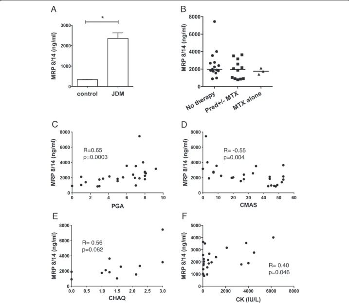
Serum levels of the heterodimer MRP8/14 were signifi-cantly higher in JDM patients compared to age-matched controls but did not vary according to drug treatment (Figure 1A, B). Importantly, MRP8/14 levels strongly correlated with validated clinical measures of disease ac-tivity (Figure 1B-E), including physician global assess-ment of disease activity (PGA, R = 0.65,P = 0.0003) and strength/stamina (CMAS, R = −0.55, P = 0.004), and only to a limited extent with disability assessment (CHAQ, R = 0.56, P = 0.062) and the muscle enzyme, CK (R = 0.4,P = 0.046).
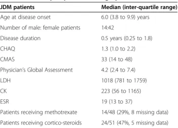
Table 1 Summary of patient demographics
JDM patients Median (inter-quartile range) Age at disease onset 6.0 (3.8 to 9.9) years
Number of male: female patients 14:42
Disease duration 0.5 years (0.25 to 1.8) CHAQ 1.3 (1.0 to 2.2) CMAS 33 (14 to 48) Physician’s Global Assessment 4.2 (2.4 to 7.4) LDH 1018 (781 to 1759) CK 223 (56 to 1165) ESR 19 (13 to 37)
Patients receiving methotrexate 14/48 (29%, 8 missing data) Patients receiving cortico-steroids 24/51 (47%, 5 missing data)
CK, creatine kinase; CHAQ, childhood health assessment questionnaire; CMAS, childhood myositis assessment score; ESR, erythrocyte sedimentation rate; LDH, lactate dehydrogenase.
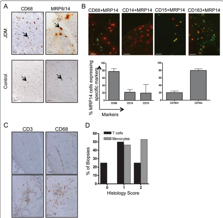
Identification of MRP8/14 expressing cells in inflamed muscle
To test if infiltrating myeloid cells in myositis secrete MRP proteins, biopsies from 46 JDM patients were stained for MRP14 or the MRP8/14 heterodimer and compared to muscle biopsies from 14 age-matched con-trols. In JDM muscle the staining patterns observed sug-gested that these cells secrete MRP8/14 (Figure 2A, top right panel). The staining pattern of MRP8/14 in control samples in contrast was quite distinct (Figure 2A, bot-tom panel). Thus, control biopsies showed only very occasional myeloid cells, which although they stained
positive for MRP8/14, showed no MRP8/14 protein secretion around the cells. To characterise MRP8/14-se-creting cells in JDM biopsies, cells were analysed by dual colour immunofluorescence using polyclonal rabbit anti-bodies against MRP14 co-stained together with markers specific for cells of the myeloid lineage CD14, CD15 and CD68 as well as the scavenger receptor CD163 (Figure 2B). By immunohistochemistry, antibody to MRP14 reliably identified the same cells as those identified by antibody to MRP8/14 heterodimer (data not shown). The majority of MRP14 was secreted by CD68 positive cells (Figure 2B, left hand bar graph). The expression of
B
A
C
D
F
E
R=0.65 p=0.0003 R= -0.55 p=0.004 R= 0.56 p=0.062 R= 0.40 p=0.046Figure 1 Disease activity measures in juvenile dermatomyositis (JDM) correlate with serum MRP8/14 levels. (A) Mean MRP8/14 concentrations were measured by ELISA in serum from 32 JDM patients and 32 paediatric controls, * P <0.05. (B) Serum MRP8/14 concentrations in JDM patients according to treatment status, n = 15 untreated, n = 12 on prednisolone+/−methotrexate, n = 3 methotrexate alone.
(C) Correlation of serum MRP8/14 with physician global assessment (PGA), n = 26, (D) childhood myositis assessment score (CMAS), n = 26, (E) childhood health assessment questionnaire (CHAQ), n = 12 and (F) creatine kinase (CK), n = 25. Spearman’s Rank was used to assess correlations between variables.
the scavenger receptor CD163 on tissue macrophages has been described as a marker of anti-inflammatory or ‘alter-nately activated’ macrophages [28] and the proportion of CD163+ macrophages in muscle has been shown to increase after exercise [29]. Double staining in the JDM
biopsies with MRP14 and CD163 showed that the majority of MRP8/14 secreting myeloid cells within the inflamed muscle were CD163- (Figure 2B right hand bar graph).
Examining muscle biopsies for myeloid sub-popu-lations, we frequently observed a heavy infiltration of
Figure 2 CD68 positive myeloid cells in muscle tissue from JDM patients express MRP14 and MRP8/14. (A) Immunohistochemical staining for CD68 (left) and MRP 8/14 (right) on muscle tissue from JDM patient (top panel) and controls (bottom panel). Positive cells stain brown as indicated by arrows. Bar represents 50 microns. (B) Two-colour immunofluorescence staining for infiltrating cells, lineage markers (green), MRP14 (red), double positive cells show as yellow. Bar represents 50 microns. Bar graph illustrates %MRP14+ cells expressing specific lineage markers, based on counting 10 fields at x10 magnification for each marker, bars and lines represent median and IQR respectively. (C) Immunohistochemical staining for CD3 and CD68 from JDM patients with a predominant macrophage infiltration, histological score of 0 for T cells and 2 for monocytes (upper panel), and mixed T cell-macrophage infiltrate, scores 2 for T-cells and 2 for macrophage (lower panel). Bar represents 100 microns. (D) Percentage of patients with histology scores 0, 1 and 2 for CD68 (macrophage) and CD3 (T cell) frequency in JDM muscle biopsies (n = 28, Chi square test for trend, P < 0.001). JDM, juvenile dermatomyositis; MRP, myeloid-related protein.
macrophages, often without a T cell aggregate (Figure 2C, top panels). In contrast biopsies with large numbers of T cells invariably had a significant macrophage infiltrate (Figure 2C, bottom panels). This observation raised the possibility that T cell recruitment could depend on the early macrophage infiltrate, which establishes the appro-priate chemokine milieu to attract lymphocytes from the circulation. To quantify the relative abundance of mac-rophages and T cells in JDM muscle, we used our biopsy score tool [21] which includes a semi quantitative assess-ment of cell infiltration (macrophage infiltration and T cell infiltration, each scored as 0, 1, or 2). Analysis of these biopsies (n = 28) showed that muscle sections scored significantly higher for macrophage infiltration, than for T cells (P <0.001, Chi square test for trend, Figure 2D); indeed, 25% of these early biopsies scored 0 for T cell infiltrate. Interestingly, none of the biopsies assessed had T cell infiltration in the absence of a ma-crophage population, which suggests that myeloid cells
may be more important in the early tissue inflammation of JDM than previously recognised.
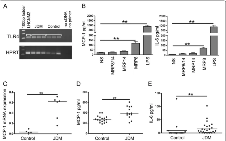
MRP8 drives the production of MCP-1 and IL-6 from inflamed muscle
Myoblasts are known to respond to pathogen associated molecular patterns (PAMPs), including bacterial LPS, by secreting inflammatory cytokines and chemokines [30]. Since MRP8 stimulates TLR4, similar to LPS, we hy-pothesised that tissue macrophages secreting MRP8/14 may propagate leukocyte recruitment in JDM by indu-cing skeletal muscle-derived cytokines. To test this hy-pothesis we first confirmed expression of TLR4 on human skeletal muscle from patients, controls and cul-tured human myoblasts (Figure 3A). The effects of MRP homo- and heterodimers on skeletal muscle were tested by culturing LHCNM2 cells in vitro in the presence of LPS or MRP proteins. LPS induced high levels of IL-6 and the chemokine MCP-1 from muscle cells, whilst
Figure 3 MRP8 drives production of the chemokine MCP-1 from human skeletal muscle. (A) Expression of TLR4 mRNA (upper panel) by RT-PCR in human skeletal myoblasts (LHCNM2) and muscle tissue from JDM and controls. HPRT mRNA was amplified as a house keeping gene (lower panel). (B) MCP-1 and IL-6 production by LHCNM2 cells stimulated for 24 hours with 5μg/ml MRP8, MRP14, MRP8/14 or 100 ng/ml LPS or cultured in medium alone (NS, non stimulated); MCP-1 and IL-6 were measured in cell culture supernatants by ELISA and multiplex immunoassay, respectively; n = 6, bars and lines represent mean and SEM, respectively. Paired t -test was used for comparisons. (C) Quantitative PCR analysis of MCP-1 expression (relative units) in quadriceps tissue from JDM patients (n = 6) compared to age matched controls (n = 4) normalised to HPRT expression. (D) MCP-1 protein concentration in serum from 14 JDM patients compared to 16 aged matched controls, measured by multiplex immunoassay. (E) Serum IL-6 in JDM patients (n = 27) and controls (n = 16), measured by multiplex immunoassay. Lines represent medians. Mann Whitney test was used for unpaired comparisons, *P <0.05, **P <0.001. JDM, juvenile dermatomyositis; LPS, lipopolysaccharide; MCP-1, monocyte chemoattractant protein-1; SEM, standard error of the mean; TLR4, Toll-like receptor 4.
MRP8 led to more modest but still significant increases compared to unstimulated controls (Figure 3B). IL-1β, IL-10, IL-17, TNF-α and IFN-γ were not detected in muscle supernatants after stimulation. We have pre-viously shown that MRP8 is the active component in TLR4 ligation [11]: again in this muscle system, MRP14 and MRP8/14 did not significantly upregulate MCP-1 or IL-6 from myoblasts. As MRP8 and MRP14 are present at increased levels in the muscles of JDM patients (Figure 2A), we asked if this would lead to a correspon-ding increase in tissue levels of MCP-1. Examining JDM muscle, we found significantly higher mRNA expression of MCP-1 when compared to control muscle biopsies (0.3 versus 0.02 relative units,P = 0.0095, Figure 3C). Analysis of JDM serum also showed significantly higher levels of MCP-1, as well as IL-6 (Figure 3D and E, respectively), than samples taken from healthy controls.
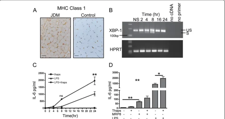
XBP-1 and MRP8 interplay in muscle inflammation
Class I MHC over-expression in skeletal muscle (Figure 4A), which is an early pathological change in JDM, is known to be associated with ER stress, an
established mechanism in the pathogenesis of myositis [7,9,31]. Recent reports have suggested that XBP-1, a key transcription factor involved in ER stress [32], may act via an alternative pathway in monocytes and augment the se-cretion of TLR induced cytokines [33]. It is, therefore, im-portant to understand how the interplay between ER stress and TLR stimulation, in the form of MRP8, would influence the secretion of inflammatory mediators from skeletal muscle. LHCNM2 cells were treated with thapsi-gargin, to induce a stress response which was confirmed by detecting expression of the active XBP-1 splice product (26 bp smaller), seen maximally between four to eight hours (Figure 4B). As previously shown, XBP-1 splicing is regulated by a negative feedback loop [34], and 16 hours after exposure to thapsigargin, only the unspliced variant was detected. To test whether TLR ligation in muscle cells synergised with ER stress, LHCNM2 cells were cultured in vitro in the presence of the TLR4 agonist LPS, either alone or after pre-incubation with thapsigargin. ER stress clearly synergised with LPS to significantly increase IL-6 secretion from myoblasts when compared to stimulation with LPS alone (Figure 4C). MRP8, although acting in a
Figure 4 Effect of MRP8 stimulation in combination with ER stress on IL-6 production by muscle cells. (A) MHC class 1 expression on muscle tissue from JDM patient and age- matched control. Bar represents 50 microns. (B) Human skeletal muscle cells LHCNM2 cultured for 2 to 24 hours in 0.05μM thapsigargin (Thaps), mRNA expression of XBP-1 was determined at indicated times; HPRT mRNA was measured as a control. RT-PCR products were analysed on 1.5% agarose gel and stained with ethidium bromide to visualise spliced (S), 142 bp and un-spliced (US), 168 bp XBP-1 transcripts. (C) Mean IL-6 production by LHCNM2 myoblasts following culture in thapsigargin, stimulated with LPS alone or with LPS following four hours pre-incubation with thapsigargin. Bars represent SEM, data analysed using two-way ANOVA. (D) IL-6 concentration in supernatants from LHCNM2 cells cultured with or without thapsigargin, stimulated with MRP8 or LPS alone, or MRP8 and LPS after four hours pre-incubation with thapsigargin. Bars and lines represent mean and SEM. Data analysed by 1 way ANOVA and paired t test *P <0.05, **P <0.001. ANOVA, analysis of variance; JDM, juvenile dermatomyositis; LPS, lipopolysaccharide; MRP, myeloid related protein; SEM, standard error of the mean; XBP-1, x-box binding protein 1.
TLR4 dependent manner, is less potent than LPS in acti-vating downstream signalling cascades, including NF-κB [11]. Consequently, stimulation with MRP8 resulted in lower IL-6 levels when compared to LPS (Figure 4D), but there was still a trend for ER stress to augment the action of MRP8 (mean IL-6 114.2 pg/ml) when compared to MRP8 alone (72.8 pg/ml, P = 0.06). ER stress did not upregulate MCP-1 production following stimulation with either LPS or MRP8 (data not shown).
Discussion
The assessment of disease activity in JDM, and the dis-tinction of disease flare from deconditioning or muscle atrophy, is still largely dependent on clinical evaluation. In this study, MRP8/14 has been identified as a novel biomarker of disease activity in JDM, and found to be superior to existing serological correlates of disease. By exploring the effects of MRP8 and MRP14 on skeletal musclein vitro and correlating results with ex vivo JDM samples, we have identified a pro-inflammatory role of MRP8 for myositic muscle that we propose contributes to the enrichment of key inflammatory mediators MCP-1 and IL-6, seen in JDM muscle and serum, which then con-tribute to further recruitment of inflammatory cells to muscle. MRP8 has been shown to be the active compo-nent stimulating TLR4 in murine models of inflammation whereas MRP14 seems to have a regulatory function in the MRP8/14 complex. The exact mechanisms activating the MRP8/14 heterodimer in vivo are currently not clear but co-stimulation may be required [35].
MRP8/14 has been shown to be a highly sensitive marker of disease activity in a range of rheumatic disor-ders, including arthritis, vasculitis and autoinflammatory disease [12,13]. As a biomarker, MRP8/14 has many characteristics that make it suitable for both clinical and research use; it is easily detected in serum even at low levels, is already in clinical use to detect gut inflamma-tion [36] and is stable in clinical serum samples even when transported at room temperature. Most recently, we have successfully used MRP8/14 to identify patients with juvenile arthritis who are likely to remain in remis-sion following withdrawal of immunosuppresremis-sion by methotrexate [37]. This proof of concept study confirms a role for MRP8/14 in the detection of sub-clinical dis-ease activity in rheumatic disorders and could poten-tially apply to JDM to assist with the withdrawal of immunosuppression.
Other biomarkers have been identified in JDM, in-cluding the IFNα gene signature and IFN induced che-mokines [4,38-40]. IFNα is produced by plasmacytoid dendritic cells (pDC), and induces transcriptional acti-vators which bind downstream response elements in promoter sequences (IFN-stimulated response elements, ISRE), enhancing the transcription of many immune
related genes including the chemokine MCP-1 [41]. Our results suggest that MCP-1 can also be produced by a MRP-dependent pathway, by muscle fibres themselves in JDM. It is, therefore, of interest, that in a recent report MCP-1, an IFNα associated chemokine correlated better with disease activity than the IFNα gene signature itself [4]. This may suggest that IFNα-independent production of MCP-1, including muscle derived MCP-1, may play a role in juvenile myositis.
It is striking that JDM biopsies that were taken re-latively early in disease (median disease duration six months), already show a major infiltration of mono-cytes/macrophages, often in the absence of cells from the adaptive immune system. Previous studies have not clearly defined the role of such infiltrating myeloid cells in muscle cell damage and inflammation during myo-sitis. Our results clearly show that MRP8 and MRP14 are secreted by CD68+ infiltrating cells and that these are largely CD163- suggesting that they are indeed pro-inflammatory in phenotype. We propose that the local secretion of MRP proteins by these cells has several downstream effects, including the stimulation of muscle to produce MCP-1 and IL-6. MCP-1 secreted by skeletal muscle, in response to MRP, may then play an important role in propagating the inflammatory infiltrate. Cells recruited into DM muscle, including monocytes and memory T cells, express high levels of CCR2, the sole re-ceptor for MCP-1 [42,43]. MRP8 and MRP14 may be important in linking an initial innate immune response with a later adaptive one by recruiting CCR2+ memory T cells and supporting the differentiation of local B cells into plasma cells as this is dependent on signalling through CCR2 [44]. As further evidence of the impor-tance of MCP-1 in myositis, animal models have impli-cated this chemokine in a transgenic model of selective over expression of self MHC Class l in skeletal muscle, MHC over expression induced MCP-1 production [8] and, in a model of viral induced myositis, blockade of MCP-1 significantly attenuated muscle inflammation [45].
Our results demonstrating that MRP8 induced IL-6 and MCP-1 secretion by myoblasts add to a growing body of evidence that suggests that muscle itself contri-butes to the inflammatory process [8,46]. To test how muscle derived cytokine secretion would be altered by non-immune insults to the muscle, known to occur in DM [7-9], we adopted a thapsigargin induced model of muscle ER stress. Using this system we have now identi-fied ER stress as a mechanism for priming myoblasts to secrete IL-6 in response to a second signal, such as macro-phage derived MRP8 binding TLR4. This is pertinent to myositis as IL-6 is known to correlate with disease activity in idiopathic inflammatory myositis (IIM) and IL-6 block-ade attenuates muscle inflammation in some mouse models [47]. Given that IL-6 is produced by a range of
immune cells, including macrophages, B and T cells, it is difficult to discern the exact contribution made by skeletal muscle towards the enrichment of this cytokine in JDM serum. Nevertheless, MCP-1 and IL-6 appear to be tightly co-regulated in JDM serum [4] and, given that both are expressed by inflamed muscle, it is possible that the close correlation between serum levels and muscle disease ac-tivity [4,40] is explained by the muscle itself being a key source of these cytokines in JDM.
One limitation of our study is that we are unable to ex-clude the action of MRP8 on other receptors apart from TLR4, such as the for advanced glycation end products (RAGE) [48]. However, data from the murine system would suggest that TLR4 is the dominant receptor for MRP8 [35].
This study contributes novel insights into the possible roles of macrophage derived MRP8 and MRP14 in dri-ving production of chemokines and cytokines by muscle cells. These data emphasise the importance of skeletal muscle as an organ with the potential for immune func-tions and demonstrate how cross talk between muscle and the innate immune system can be instrumental in sustaining further adaptive responses as well as on-going inflammation in autoimmune muscle disease.
Conclusions
Our results highlight the relationship between serum MRP8/14 levels and disease activity in JDM, and iden-tify a novel mechanism by which macrophage derived MRP8/14 may directly activate muscle cells and, thereby, perpetuate inflammatory myositis. Further prospective studies are required to test the role of MRP8/14 as a puta-tive biomarker for disease activity in JDM.
Additional file
Additional file 1: Patient demographics and clinical scores. Abbreviations
ANOVA:Analysis of variance; bp: Base pair; CHAQ: Childhood health assessment questionnaire; CK: Creatine kinase; CMAS: Childhood myositis assessment score; ELISA: Enzyme-linked immunosorbent assay; ER: Endoplasmic reticulum; ESR: Erythrocyte sedimentation rate; IFNα: Interferon alpha; JDM: Juvenile dermatomyositis; LDH: Lactate dehydrogenase; LPS: Lipopolysaccharide; MCP-1: Monocyte chemoattractant protein-1; MHC: Major histocompatibility complex; MRP: Myeloid related protein; PCR: Polymerase chain reaction; TLR: Toll-like receptor; UPR: Unfolded protein response; VAS: Visual analogue score; xbp: x-box binding protein.
Competing interests
The authors declare that they have no competing interests. Authors’ contributions
HV carried out immunohistochemistry, myoblast culture and PCR. HW, TV and JR assayed MRP and provided technical support. VS, PK and PB assayed MCP-1. KM provided the myoblast cell line. KN, KM and HV analysed data and performed statistical analyses. HV, KN, KM, JR and LW conceived of the study, designed experiments and participated in its design and coordination and helped to draft the manuscript. All authors read and approved the final manuscript.
Acknowledgements
The Juvenile Dermatomyositis Research Group would like to thank all of the patients and their families who contributed to the Juvenile Dermatomyositis Cohort Study. We thank all local research coordinators and principal investigators who have made this research possible. The members who contributed are as follows: Dr Kate Armon and Mr Joe Ellis-Gage (Norfolk and Norwich University Hospitals), Dr Liza McCann, Mr Ian Roberts, Dr Eileen Baildam and Ms Louise Hanna (The Royal Liverpool Children’s Hospital, Alder Hey, Liverpool), Dr Phil Riley and Ms Ann McGovern (Royal Manchester Children’s Hospital, Manchester), Dr Clive Ryder and Mrs. Janis Scott (Birmingham Children’s Hospital, Birmingham), Dr Sue Wyatt, Mrs Gillian Jackson, Dr Tania Amin, Mark Wood and Vanessa Van Rooyen (Leeds General Infirmary, Leeds), Dr Joyce Davidson, Dr Janet Gardner-Medwin, Dr Neil Martin and Ms Sue Ferguson (The Royal Hospital for Sick Children, Yorkhill, Glasgow), Dr Mark Friswell, Professor Helen Foster, Mrs Alison Swift, Dr Sharmila Jandial, Ms Vicky Stevenson and Ms Debbie Wade (Great North Children’s Hospital, Newcastle), Dr Helen Venning, Mrs Elizabeth Stretton and Ms Mary Jordan (Queens Medical Centre, Nottingham), Professor Lucy Wedderburn, Dr Clarissa Pilkington, Dr N. Hasson, Mrs Sue Maillard, Ms Elizabeth Halkon, Ms Virginia Brown, Ms Audrey Juggins, Dr Sally Smith, Mrs Sian Lunt, Ms Elli Enayat, Mrs Hemlata Varsani, Miss Laura Beard and Miss Katie Arnold (Great Ormond Street Hospital, London), Dr Kevin Murray (Princess Margaret Hospital, Perth, Western Australia) Dr John Ioannou (University College London Hospital).
We thank Dr Vincent Mouly, Institut de Myologie, Paris for advice on muscle cell lines and Dr Jane Goodall, Cambridge University for providing XBP-1 primer sequences.
The JDM Cohort Study and this work have been supported by generous grants from the Wellcome Trust UK (085860), Action Medical Research UK, (SP4252), and The Henry Smith Charity. The JDM Cohort study has been adopted onto the Comprehensive Research Network through the Medicines for Children Research Network (www.mcrn.org.uk). LW is supported in part by the Great Ormond Street Hospital Children’s Charity. KN is a Wellcome Trust Intermediate Clinical Fellow (097259). TV and JR were supported by grants from the Interdisciplinary Centre for Clinical Research at the University of Muenster (Vo2/014/09 to T.V. as well as grant Ro2/004/10 to J.R) and the Deutsche Forschungsgemeinschaft (DFG project RO 1190/9-1).
Author details
1
Rheumatology Unit, UCL Institute of Child Health, 30 Guilford Street, London WC1N 1EH, UK.2Institute of Immunology and Interdisciplinary
Centre for Clinical Research IZKF, University of Muenster, Muenster, Germany.
3Department of General Pediatrics, University Children’s Hospital Muenster,
Muenster, Germany.4Department of Pediatrics and Adolescent Medicine, First Faculty of Medicine, Charles University in Prague and General University Hospital in Prague, Ke Karlovu 2, 12000 Prague 2, Czech Republic.5Thérapie des maladies du muscle strié, Institut de Myologie, UM76, UPMC Université Paris 6/U974 - Inserm/UMR7215– CNRS, 47, bld de l’hôpital - G.H. Pitié-Salpétrière - Bâtiment Babinski, 75651 Paris, cedex 13, France.
Received: 10 February 2013 Accepted: 2 September 2013 Published: 23 September 2013
References
1. Wedderburn LR, Rider LG: Juvenile dermatomyositis: new developments in pathogenesis, assessment and treatment. Best Pract Res Clin Rheumatol 2009, 23:665–678.
2. Gunawardena H, Wedderburn LR, North J, Betteridge Z, Dunphy J, Chinoy H, Davidson JE, Cooper RG, McHugh NJ: Clinical associations of autoantibodies to a p155/140 kDa doublet protein in juvenile dermatomyositis. Rheumatology (Oxford) 2008, 47:324–328.
3. Morita R, Schmitt N, Bentebibel SE, Ranganathan R, Bourdery L, Zurawski G, Foucat E, Dullaers M, Oh S, Sabzghabaei N, Lavecchio EM, Punaro M, Pascual V, Banchereau J, Ueno H: Human blood CXCR5(+)CD4(+) T cells are counterparts of T follicular cells and contain specific subsets that differentially support antibody secretion. Immunity 2011, 34:108–121.
4. Bilgic H, Ytterberg SR, Amin S, McNallan KT, Wilson JC, Koeuth T, Ellingson S, Newman B, Bauer JW, Peterson EJ, Baechler EC, Reed AM: Interleukin-6 and type I interferon-regulated genes and chemokines mark disease activity in dermatomyositis. Arthritis Rheum 2009, 60:3436–3446.
5. Li CK, Varsani H, Holton JL, Gao B, Woo P, Wedderburn LR: MHC Class I overexpression on muscles in early juvenile dermatomyositis. J Rheumatol 2004, 31:605–609.
6. Bartoccioni E, Gallucci S, Scuderi F, Ricci E, Servidei S, Broccolini A, Tonali P: MHC class I, MHC class II and intercellular adhesion molecule-1 (ICAM-1) expression in inflammatory myopathies. Clin Exp Immunol 1994, 95:166–172.
7. Nagaraju K, Casciola-Rosen L, Lundberg I, Rawat R, Cutting S, Thapliyal R, Chang J, Dwivedi S, Mitsak M, Chen YW, Plotz P, Rosen A, Hoffman E, Raben N: Activation of the endoplasmic reticulum stress response in autoimmune myositis: potential role in muscle fiber damage and dysfunction. Arthritis Rheum 2005, 52:1824–1835.
8. Nagaraju K, Raben N, Loeffler L, Parker T, Rochon PJ, Lee E, Danning C, Wada R, Thompson C, Bahtiyar G, Craft J, Hooft Van Huijsduijnen R, Plotz P: Conditional up-regulation of MHC class I in skeletal muscle leads to self-sustaining autoimmune myositis and myositis-specific autoantibodies. Proc Natl Acad Sci USA 2000, 97:9209–9214.
9. Li CK, Knopp P, Moncrieffe H, Singh B, Shah S, Nagaraju K, Varsani H, Gao B, Wedderburn LR: Overexpression of MHC class I heavy chain protein in young skeletal muscle leads to severe myositis: implications for juvenile myositis. Am J Pathol 2009, 175:1030–1040.
10. Seeliger S, Vogl T, Engels IH, Schroder JM, Sorg C, Sunderkotter C, Roth J: Expression of calcium-binding proteins MRP8 and MRP14 in inflammatory muscle diseases. Am J Pathol 2003, 163:947–956.
11. Loser K, Vogl T, Voskort M, Lueken A, Kupas V, Nacken W, Klenner L, Kuhn A, Foell D, Sorokin L, Luger TA, Roth J, Beissert S: The Toll-like receptor 4 ligands Mrp8 and Mrp14 are crucial in the development of autoreactive CD8+ T cells. Nat Med 2010, 16:713–717.
12. Hirono K, Foell D, Xing Y, Miyagawa-Tomita S, Ye F, Ahlmann M, Vogl T, Futatani T, Rui C, Yu X, Watanabe K, Wanatabe S, Tsubata S, Uese K, Hashimoto I, Ichida F, Nakazawa M, Roth J, Miyawaki T: Expression of myeloid-related protein-8 and−14 in patients with acute Kawasaki disease. J Am Coll Cardiol 2006, 48:1257–1264.
13. Frosch M, Strey A, Vogl T, Wulffraat NM, Kuis W, Sunderkotter C, Harms E, Sorg C, Roth J: Myeloid-related proteins 8 and 14 are specifically secreted during interaction of phagocytes and activated endothelium and are useful markers for monitoring disease activity in pauciarticular-onset juvenile rheumatoid arthritis. Arthritis Rheum 2000, 43:628–637. 14. Pilkington CA, Wedderburn LR: Paediatric idiopathic inflammatory muscle
disease: recognition and management. Drugs 2005, 65:1355–1365. 15. Martin N, Krol P, Smith S, Murray K, Pilkington CA, Davidson JE, Wedderburn
LR: A national registry for juvenile dermatomyositis and other paediatric idiopathic inflammatory myopathies: 10 years’ experience; the Juvenile Dermatomyositis National (UK and Ireland) Cohort Biomarker Study and Repository for Idiopathic Inflammatory Myopathies. Rheumatology (Oxford) 2011, 50:137–145.
16. Bohan A, Peter JB: Polymyositis and dermatomyositis (second of two parts). N Engl J Med 1975, 292:403–407.
17. Varsani H, Newton KR, Li CK, Harding B, Holton JL, Wedderburn LR: Quantification of normal range of inflammatory changes in
morphologically normal pediatric muscle. Muscle Nerve 2008, 37:259–261. 18. Lovell DJ, Lindsley CB, Rennebohm RM, Ballinger SH, Bowyer SL, Giannini
EH, Hicks JE, Levinson JE, Mier R, Pachman LM, Passo MH, Perez MD, Reed AM, Schikler KN, Smith M, Zemel LS, Rider LG: Development of validated disease activity and damage indices for the juvenile idiopathic inflammatory myopathies. II. The Childhood Myositis Assessment Scale (CMAS): a quantitative tool for the evaluation of muscle function. The Juvenile Dermatomyositis Disease Activity Collaborative Study Group. Arthritis Rheum 1999, 42:2213–2219.
19. Rider LG, Giannini EH, Harris-Love M, Joe G, Isenberg D, Pilkington C, Lachenbruch PA, Miller FW: Defining clinical improvement in adult and juvenile myositis. J Rheumatol 2003, 30:603–617.
20. de Jager W, Prakken BJ, Bijlsma JW, Kuis W, Rijkers GT: Improved multiplex immunoassay performance in human plasma and synovial fluid following removal of interfering heterophilic antibodies. J Immunol Methods 2005, 300:124–135.
21. Wedderburn LR, Varsani H, Li CK, Newton KR, Amato AA, Banwell B, Bove KE, Corse AM, Emslie-Smith A, Harding B, Hoogendijk J, Lundberg IE, Marie S, Minetti C, Nennesmo I, Rushing EJ, Sewry C, Charman SC, Pilkington CA, Holton JL, UK Juvenile Dermatomyositis Research Group: International consensus on a proposed score system for muscle biopsy evaluation in
patients with juvenile dermatomyositis: a tool for potential use in clinical trials. Arthritis Rheum 2007, 57:1192–1201.
22. Zwadlo G, Schlegel R, Sorg C: A monoclonal antibody to a subset of human monocytes found only in the peripheral blood and inflammatory tissues. J Immunol 1986, 137:512–518.
23. Roth J, Goebeler M, Erpenstein U, Sorg C: Differential regulation of the macrophage-specific surface antigen RM3/1 by cyclosporine, azathioprine, and dexamethasone. Transplantation 1994, 57:127–133. 24. Zhu CH, Mouly V, Cooper RN, Mamchaoui K, Bigot A, Shay JW, Di Santo JP,
Butler-Browne GS, Wright WE: Cellular senescence in human myoblasts is overcome by human telomerase reverse transcriptase and cyclin-dependent kinase 4: consequences in aging muscle and therapeutic strategies for muscular dystrophies. Aging Cell 2007, 6:515–523. 25. Vogl T, Leukert N, Barczyk K, Strupat K, Roth J: Biophysical characterization
of S100A8 and S100A9 in the absence and presence of bivalent cations. Biochim Biophys Acta 2006, 1763:1298–1306.
26. Price BD, Mannheim-Rodman LA, Calderwood SK: Brefeldin A, thapsigargin, and AIF4- stimulate the accumulation of GRP78 mRNA in a
cycloheximide dependent manner, whilst induction by hypoxia is independent of protein synthesis. J Cell Physiol 1992, 152:545–552. 27. Liprandi A, Bartoli C, Figarella-Branger D, Pellissier JF, Lepidi H: Local
expression of monocyte chemoattractant protein-1 (MCP-1) in idiopathic inflammatory myopathies. Acta Neuropathol 1999, 97:642–648.
28. Komohara Y, Hirahara J, Horikawa T, Kawamura K, Kiyota E, Sakashita N, Araki N, Takeya M: AM-3K, an anti-macrophage antibody, recognizes CD163, a molecule associated with an anti-inflammatory macrophage phenotype. J Histochem Cytochem 2006, 54:763–771.
29. Przybyla B, Gurley C, Harvey JF, Bearden E, Kortebein P, Evans WJ, Sullivan DH, Peterson CA, Dennis RA: Aging alters macrophage properties in human skeletal muscle both at rest and in response to acute resistance exercise. Exp Gerontol 2006, 41:320–327.
30. Nagaraju K, Raben N, Merritt G, Loeffler L, Kirk K, Plotz P: A variety of cytokines and immunologically relevant surface molecules are expressed by normal human skeletal muscle cells under proinflammatory stimuli. Clin Exp Immunol 1998, 113:407–414.
31. Vattemi G, Engel WK, McFerrin J, Askanas V: Endoplasmic reticulum stress and unfolded protein response in inclusion body myositis muscle. Am J Pathol 2004, 164:1–7.
32. Hirota M, Kitagaki M, Itagaki H, Aiba S: Quantitative measurement of spliced XBP1 mRNA as an indicator of endoplasmic reticulum stress. J Toxicol Sci 2006, 31:149–156.
33. Martinon F, Chen X, Lee AH, Glimcher LH: TLR activation of the transcription factor XBP1 regulates innate immune responses in macrophages. Nat Immunol 2010, 11:411–418.
34. Ozcan U, Cao Q, Yilmaz E, Lee AH, Iwakoshi NN, Ozdelen E, Tuncman G, Gorgun C, Glimcher LH, Hotamisligil GS: Endoplasmic reticulum stress links obesity, insulin action, and type 2 diabetes. Science 2004, 306:457–461. 35. Vogl T, Tenbrock K, Ludwig S, Leukert N, Ehrhardt C, van Zoelen MA,
Nacken W, Foell D, van der Poll T, Sorg C, Roth J: Mrp8 and Mrp14 are endogenous activators of Toll-like receptor 4, promoting lethal, endotoxin-induced shock. Nat Med 2007, 13:1042–1049.
36. Tibble J, Teahon K, Thjodleifsson B, Roseth A, Sigthorsson G, Bridger S, Foster R, Sherwood R, Fagerhol M, Bjarnason I: A simple method for assessing intestinal inflammation in Crohn’s disease. Gut 2000, 47:506–513.
37. Foell D, Wulffraat N, Wedderburn LR, Wittkowski H, Frosch M, Gerss J, Stanevicha V, Mihaylova D, Ferriani V, Tsakalidou FK, Foeldvari I, Cuttica R, Gonzalez B, Ravelli A, Khubchandani R, Oliveira S, Armbrust W, Garay S, Vojinovic J, Norambuena X, Gamir ML, García-Consuegra J, Lepore L, Susic G, Corona F, Dolezalova P, Pistorio A, Martini A, Ruperto N, Roth J, Paediatric Rheumatology International Trials Organization (PRINTO): Methotrexate withdrawal at 6 vs 12 months in juvenile idiopathic arthritis in remission: a randomized clinical trial. JAMA 2010, 303:1266–1273.
38. Salajegheh M, Kong SW, Pinkus JL, Walsh RJ, Liao A, Nazareno R, Amato AA, Krastins B, Morehouse C, Higgs BW, Jallal B, Yao Y, Sarracino DA, Parker KC, Greenberg SA: Interferon-stimulated gene 15 (ISG15) conjugates proteins in dermatomyositis muscle with perifascicular atrophy. Ann Neurol 2010, 67:53–63.
39. Tezak Z, Hoffman EP, Lutz JL, Fedczyna TO, Stephan D, Bremer EG, Krasnoselska-Riz I, Kumar A, Pachman LM: Gene expression profiling in DQA1*0501+ children with untreated dermatomyositis: a novel model of pathogenesis. J Immunol 2002, 168:4154–4163.
40. Reed AM, Peterson E, Bilgic H, Ytterberg SR, Amin S, Hein MS, Crowson CS, Ernste F, Gillespie EB: Changes in novel biomarkers of disease activity in juvenile and adult dermatomyositis are sensitive biomarkers of disease course. Arthritis Rheum 2012, 64:4078–4086.
41. Banchereau J, Pascual V: Type I interferon in systemic lupus erythematosus and other autoimmune diseases. Immunity 2006, 25:383–392.
42. Charo IF, Myers SJ, Herman A, Franci C, Connolly AJ, Coughlin SR: Molecular cloning and functional expression of two monocyte chemoattractant protein 1 receptors reveals alternative splicing of the carboxyl-terminal tails. Proc Natl Acad Sci USA 1994, 91:2752–2756.
43. De Paepe B, De Bleecker JL: Beta-chemokine receptor expression in idiopathic inflammatory myopathies. Muscle Nerve 2005, 31:621–627. 44. Delogu A, Schebesta A, Sun Q, Aschenbrenner K, Perlot T, Busslinger M:
Gene repression by Pax5 in B cells is essential for blood cell homeostasis and is reversed in plasma cells. Immunity 2006, 24:269–281.
45. Rulli NE, Rolph MS, Srikiatkhachorn A, Anantapreecha S, Guglielmotti A, Mahalingam S: Protection from arthritis and myositis in a mouse model of acute chikungunya virus disease by bindarit, an inhibitor of monocyte chemotactic protein-1 synthesis. J Infect Dis 2011, 204:1026–1030. 46. Tournadre A, Lenief V, Eljaafari A, Miossec P: Immature muscle precursors
are a source of interferon-beta in myositis: role of Toll-like receptor 3 activation and contribution to HLA class I up-regulation. Arthritis Rheum 2012, 64:533–541.
47. Okiyama N, Sugihara T, Iwakura Y, Yokozeki H, Miyasaka N, Kohsaka H: Therapeutic effects of interleukin-6 blockade in a murine model of polymyositis that does not require interleukin-17A. Arthritis Rheum 2009, 60:2505–2512.
48. Hofmann MA, Drury S, Fu C, Qu W, Taguchi A, Lu Y, Avila C, Kambham N, Bierhaus A, Nawroth P, Neurath MF, Slattery T, Beach D, McClary J, Nagashima M, Morser J, Stern D, Schmidt AM: RAGE mediates a novel proinflammatory axis: a central cell surface receptor for S100/calgranulin polypeptides. Cell 1999, 97:889–901.
doi:10.1186/ar4311
Cite this article as: Nistala et al.: Myeloid related protein induces muscle derived inflammatory mediators in juvenile dermatomyositis. Arthritis Research & Therapy 2013 15:R131.
Submit your next manuscript to BioMed Central and take full advantage of:
• Convenient online submission
• Thorough peer review
• No space constraints or color figure charges
• Immediate publication on acceptance
• Inclusion in PubMed, CAS, Scopus and Google Scholar
• Research which is freely available for redistribution
Submit your manuscript at www.biomedcentral.com/submit