HAL Id: hal-02149348
https://hal.sorbonne-universite.fr/hal-02149348
Submitted on 6 Jun 2019
HAL is a multi-disciplinary open access
archive for the deposit and dissemination of
sci-entific research documents, whether they are
pub-lished or not. The documents may come from
teaching and research institutions in France or
abroad, or from public or private research centers.
L’archive ouverte pluridisciplinaire HAL, est
destinée au dépôt et à la diffusion de documents
scientifiques de niveau recherche, publiés ou non,
émanant des établissements d’enseignement et de
recherche français ou étrangers, des laboratoires
publics ou privés.
promotes sprouting angiogenesis
Gaël Genet, Kevin Boyé, Thomas Mathivet, Roxana Ola, Feng Zhang,
Alexandre Dubrac, Jinyu Li, Nafiisha Genet, Luiz Henrique Geraldo, Lorena
Benedetti, et al.
To cite this version:
Gaël Genet, Kevin Boyé, Thomas Mathivet, Roxana Ola, Feng Zhang, et al.. Endophilin-A2 dependent
VEGFR2 endocytosis promotes sprouting angiogenesis. Nature Communications, Nature Publishing
Group, 2019, 10 (1), �10.1038/s41467-019-10359-x�. �hal-02149348�
Endophilin-A2 dependent VEGFR2 endocytosis
promotes sprouting angiogenesis
Gael Genet
1
, Kevin Boyé
1
, Thomas Mathivet
2
, Roxana Ola
1,7
, Feng Zhang
1
, Alexandre Dubrac
1
, Jinyu Li
1
,
Na
fiisha Genet
1
, Luiz Henrique Geraldo
2
, Lorena Benedetti
3
, Steffen Künzel
1
, Laurence Pibouin-Fragner
2
,
Jean-Leon Thomas
1,4,5
& Anne Eichmann
1,2,6
Endothelial cell migration, proliferation and survival are triggered by VEGF-A activation of
VEGFR2. However, how these cell behaviors are regulated individually is still unknown. Here
we identify Endophilin-A2 (ENDOA2), a BAR-domain protein that orchestrates
CLATHRIN-independent internalization, as a critical mediator of endothelial cell migration and sprouting
angiogenesis. We show that EndoA2 knockout mice exhibit postnatal angiogenesis defects
and impaired front-rear polarization of sprouting tip cells. ENDOA2 de
ficiency reduces
VEGFR2 internalization and inhibits downstream activation of the signaling effector PAK but
not ERK, thereby affecting front-rear polarity and migration but not proliferation or survival.
Mechanistically, VEGFR2 is directed towards ENDOA2-mediated endocytosis by the
SLIT2-ROBO pathway via SLIT-SLIT2-ROBO-GAP1 bridging of ENDOA2 and SLIT2-ROBO1. Blocking
ENDOA2-mediated endothelial cell migration attenuates pathological angiogenesis in oxygen-induced
retinopathy models. This work identi
fies a specific endocytic pathway controlling a subset of
VEGFR2 mediated responses that could be targeted to prevent excessive sprouting
angio-genesis in pathological conditions.
https://doi.org/10.1038/s41467-019-10359-x
OPEN
1Cardiovascular Research Center, Department of Internal Medicine, Yale University School of Medicine, New Haven, CT 06511, USA.2Inserm U970, Paris
Cardiovascular Research Center, Paris 75015, France.3Department of Neuroscience and Cell Biology, School of Medicine, Yale University School of Medicine, New Haven, CT 06511, USA.4Department of Neurology, Yale University School of Medicine, New Haven, CT 06511, USA.5Sorbonne Universités, UPMC Université Paris 06, Institut National de la Santé et de la Recherche Médicale U1127, Centre National de la Recherche Scientifique, AP-HP, Institut du Cerveau et de la Moelle Epinière, Hôpital Pitié-Salpêtrière, Paris, France.6Department of Cellular and Molecular Physiology, Yale University School of Medicine, New
Haven, CT 06511, USA.7Present address: Functional Genomics, Proteomics and Experimental Pathology Department, Prof. Dr. I. Chiricuta Oncology Institute, Cluj-Napoca, Romania, Department of Basic, Preventive and Clinical Science, University of Transylvania, Brasov, Romania. Correspondence and requests for materials should be addressed to A.E. (email:[email protected])
123456789
V
ascular endothelial growth factor A (VEGF) is a secreted
polypeptide that is critical for vascular development,
angiogenesis and arteriogenesis
1–4. VEGF exerts its action
by binding to the receptor tyrosine kinase VEGF receptor 2
(VEGFR2; also known as KDR or FLK1)
5, which is expressed
mainly in endothelial cells (ECs), but also in some neuronal cell
populations
6. VEGF binding to VEGFR2 triggers receptor
dimerization and phosphorylation of tyrosine residues in the
cytoplasmic kinase domain, in turn activating various
intracel-lular cascades, including PI3K/AKT, MAPK, SRC, and PAK
signaling, to mediate survival, proliferation, and migration
7–9. A
major challenge in the
field is to determine how these cascades
instruct specific EC behaviors.
Endocytosis and subsequent receptor signaling from
endoso-mal compartments have emerged as major determinants of
sig-naling output
10,11. Endosomes are distributed to various
intracellular locations via microtubules. The sorting and
traf-ficking processes of these small vesicles provide time for
protein–protein interactions and assembly of signaling
com-plexes
12–14. Thus, the knowledge of events involved in receptor
endocytosis and trafficking is essential to understand the
reg-ulation of its activity.
Receptor signaling is initiated by endocytic uptake into the cell.
Until now, the major known endocytic route for VEGFR2 was
thought to be via the CLATHRIN-mediated endocytic (CME)
pathway
10,15,16. CLATHRIN-mediated VEGFR2 endocytosis is
facilitated by various extracellular cues, including the guidance
receptors EPHRIN-B2 and NEUROPILIN-1 (NRP1), which both
promote CME uptake and intracellular VEGFR2 trafficking
17–22.
However, several studies reported that VEGF-induced
inter-nalization or signaling persisted upon inhibition of CME
23–26,
suggesting that the receptor might also be internalized through
CLATHRIN-independent endocytic routes.
Fast endophilin-mediated endocytosis (FEME) is a newly
dis-covered CLATHRIN-independent endocytosis pathway
27–31.
ENDOPHILIN proteins are cytoplasmic Bin/Amphiphysin/Rvs
(BAR)-domain-containing proteins involved in the formation
and scission of endocytic vesicles
32–34. FEME mediates
inter-nalization of Shiga and Cholera toxins, which are well known to
enter cells independently of CME
29. FEME is triggered upon
activation of specific receptors by their cognate ligands
29.
Receptor activation leads to the conversion of phospholipid
molecules PI(4,5)P
2to PI(3,4)P
2, which induces the recruitment
of ENDOPHILIN to the plasma membrane and the formation of
endocytic vesicles named ENDOPHILIN positive assemblies
(EPA)
27. FEME occurs preferentially at the lamellipodia of
migrating cells and mediates ligand-dependent uptake of several
G protein-coupled receptors and receptor tyrosine kinases
in vitro
27. Whether FEME contributes to vascular development
in vivo remained unknown.
Here we show that ENDOA2 selectively regulates EC migration
during postnatal and pathological angiogenesis by controlling
CME-independent VEGFR2 endocytosis and activation of
downstream PAK, but not ERK signaling. Targeting of VEGFR2
toward ENDOA2 is controlled by the SLIT-ROBO guidance
pathway, which has been previously identified as a regulator of
polarized endothelial migration
9,35. Altogether, this work reveals
an endocytic mechanism regulating sprouting angiogenesis,
opening avenues to selectively target this cell behavior.
Results
ENDOA2 regulates angiogenesis in the postnatal mouse retina.
Gene expression analysis of ENDOA1, 2, and 3 isoforms
(SH3GL2, SH3GL1, and SH3GL3, respectively) in purified ECs
from various sources showed that ENDOA2 was the only isoform
expressed in this cell type (Supplementary Fig. 1a–c).
Immu-nostaining of retinas from P5 wild-type mice showed that
ENDOA2 labeled the endothelium, with lower expression in the
neuronal retinal layers and in perivascular mural cells and
astrocytes (Fig.
1
a, Supplementary Fig. 1d–f). Furthermore,
measurement of mRNA levels in retinal non-ECs revealed that in
addition to low levels of EndoA2, these cells also expressed equal
levels of EndoA1 and EndoA3 (Supplementary Fig. 1g). These
data suggested a unique role of ENDOA2 in ECs, while it may
function redundantly with ENDOA1 and A3 in non-ECs, as
previously shown in neurons
36,37.
To test the function of ENDOA2 in vascular development, we
used Sh3gl1
−/−mice (hereafter named EndoA2
−/−)
36, which
were viable and recovered at expected Mendelian ratios at birth
(Supplementary Fig. 2a). EndoA2
−/−mice did not show any
detectable EndoA2 expression in retinas or mouse lung
endothelial cells (mLECs) (Fig.
1
a, Supplementary Fig. 2b), and
EndoA2 gene deletion did not affect EndoA1 or EndoA3 gene
expression (Supplementary Fig. 2c, d).
We analyzed the embryonic hindbrain and the postnatal
mouse retina vasculature, two convenient models that allow
detecting even subtle effects on angiogenesis
38. Vessel
morphol-ogy at embryonic day 11.5 was similar between wild-type and
EndoA2
−/−littermates (Supplementary Fig. 2e–g). In contrast,
EndoA2 deletion significantly reduced vascular radial expansion,
vessel density and branching in the postnatal retina (Fig.
1
b, c).
EndoA2 deletion did not affect the neuronal layers beneath the
retinal vasculature, nor astrocyte, pericyte or smooth muscle
coverage (Supplementary Fig. 3). Production and localization of
growth factors such as VEGF and SLIT2 were similar between
EndoA2
−/−mice and wild-type littermates (Supplementary
Fig. 4). Overall, these data support a selective function for
ENDOA2 in postnatal ECs.
EndoA2
−/−retinas displayed lower overall numbers of
ERG1,2,3 positive ECs but similar endothelial EdU incorporation
and cell size (Supplementary Fig. 5). Vessel stability was not
affected, as shown by similar area of collagen IV-positive,
IB4-negative empty basement membrane sleeves between wild-type
and EndoA2
−/−littermates (Supplementary Fig. 6a). However,
the numbers of angiogenic sprouts at the vascular front of
EndoA2
−/−mutant mice were severely decreased in comparison
to wild-type littermates (Fig.
1
b). Acquisition of tip cell front-rear
polarity is a key regulator of sprouting; therefore, we analyzed
front-rear polarity by staining P5 retinas with IB4, ERG1,2,3 and
an anti-GM130 antibody to label the Golgi apparatus. In
wild-type littermates, around 45% of tip cells had their Golgi
positioned toward the leading edge, while around 40% of
EndoA2
−/−tip cells had their Golgi positioned behind the
nucleus away from the migration front (Fig.
1
d, e), demonstrating
impaired front-rear polarization in EndoA2
−/−tip cells.
Apico-basal polarity of ECs in the stalk position appeared normal as
shown by podocalyxin staining of the luminal membrane
(Supplementary Fig. 6b). Sprouting defects in EndoA2
−/−mice
persisted until P12 and impaired formation of the deeper retinal
vasculature layer (Supplementary Fig. 6c). Together, these data
show that ENDOA2 is required for postnatal sprouting
angiogenesis by regulating tip cell polarization and endothelial
migration, although we cannot exclude that reduced cell cycling
may contribute to the lower EC numbers in ENDOA2 deficient
retinas.
ENDOA2 controls CLATHRIN-independent VEGFR2
inter-nalization. Since tip cell migration is controlled by VEGFR2
3–7,
and ENDOA2 has been implicated in VEGF endocytosis
27, we
internalization. HUVECs express ENDOA2 around the nucleus,
at the plasma membrane and in intracellular punctae called EPAs
carrying internalized receptors
27, and ENDOA2 siRNA silencing
abolished ENDOA2 expression (Supplementary Fig. 7a, b). Cell
surface biotinylation assay showed that ENDOA2 siRNA
decreased VEGFR2 internalization induced by VEGF by about
50% (Fig.
2
a, b). Next, we used an antibody feeding assay where
HUVECs or mLECs were incubated with an antibody binding to
a
b
c
e
EndoA2+/+ EndoA2–/– ENDOA2 ENDOA2 IB4 EndoA2+/+ EndoA2–/– IB4 IB4d
IB4 GM130 ERG1,2,3 EndoA2+/+ EndoA2–/– Tip cell Front Middle Rear Golgi 120° 120° Nucleus 0.4 0.5 0.6 0.7 0.8 Vascular progression (d/D)**
EndoA2+/+ EndoA2–/– EndoA2+/+ EndoA2–/– EndoA2+/+ EndoA2–/– EndoA2+/+ EndoA2–/– 200 400 600 800 1000 Branchpoints per mm 2***
0 5 10 15 20 25 Sprouts per mm***
10 15 20 25 30 35Vascular density (AU)
*
0
Front Middle Rear
20 40 60 % of golgi position EndoA2+/+ EndoA2–/–
**
ns***
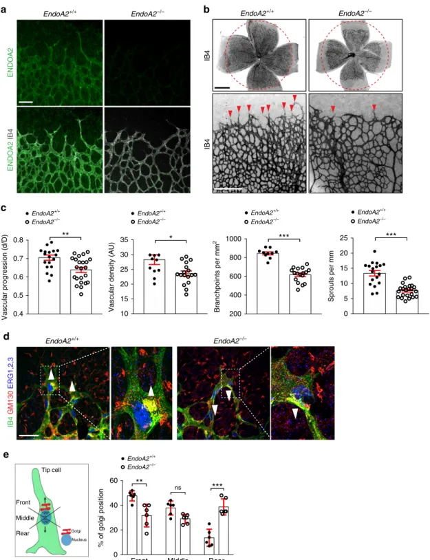
Fig. 1 ENDOA2 regulates postnatal mouse retina angiogenesis. a ENDOA2 and IB4 staining in retinalflatmounts from P5 mice. Note enrichment of ENDOA2 in IB4+ wild-type vessels, and absence of ENDOA2 expression in EndoA2−/−mice.b IB4 stained P5 retinalflatmounts of EndoA2+/+and EndoA2−/−mice. The dashed circles outline vascular coverage in wild-type retina. Red arrowheads show tip cell sprouting.c Quantification of vascular progression (d: vascular coverage length; D: retinal petal length), vessel density, number of branchpoints and vessel sprouts (N = 10–23 retinas/group, t-test: *P < 0.05, **P < 0.01, ***P < 0.001). d IB4, GM130, and ERG1/2/3 labeling shows Golgi orientation (white arrowheads) of P5 tip cells in EndoA2+/+ and EndoA2−/−retinas. Boxed areas are magnified in the right panels. e Schematic of Golgi positions in tip cells and quantification of the Golgi position in retinas shown ind (N = 6 retinas per group, at least 50 tip cells per retina were quantified; two-way ANOVA: ns P > 0.05, **P < 0.01, ***P < 0.001). Error bars represent mean ± s.e.m. Scale bars:a 100μm, b (upper panel) 1 mm, b (lower panel) 300 μm, d 50 μm
the extracellular domain of VEGFR2 prior to VEGF stimulation,
then stripped,
fixed, and labeled with a secondary antibody.
Again, ENDOA2 knockdown decreased VEGFR2 internalization
in both EC types (Fig.
2
c, d). No VEGFR2 internalization was
detected after PBS treatment and antibody specificity was
vali-dated using VEGFR2 (KDR) siRNA in HUVECs (Supplementary
Fig. 7c, d). To further characterize VEGFR2 endocytosis and
trafficking via ENDOA2, we used super-resolution structured
illumination microscopy (SIM)
39, which allows quantification of
the proximity between two proteins (0–200 nm) (Supplementary
Fig. 8). SIM imaging revealed that VEGF stimulation induced
formation of EPAs underneath the plasma membrane of HUVEC
lamellipodia (Fig.
2
e, f). VEGF stimulation promoted overlap
between VEGFR2 and ENDOA2 at the leading edge of the cell
a
b
e
g
c
mLEC HUVEC VEGF 30 ′ (1.5 nM) siCtrl siENDOA2 EndoA2–/– EndoA2+/+Internalized VEGFR2 Dapi
0 5 15 30 siENDOA2 VEGF 1.5 nM 0 5 15 30 VEGFR2 VEGFR2 Internalized Internalized Surf. Surf. 250 kDa 250 kDa ACTIN 42 kDa
h
i
j
d
f
(min) 0 20 40 60 80 100 Number of E P As per 100 0 μ m 2***
VEGF Unstimulated 0 2 4 6 8 VEGFR2 /ENDOA2overlap (fold change
) Unstimulated VEGF
***
0.0 0.5 1.0 1.5Internalized VEGFR2 intensity (fold change
) siCtrl siENDOA2
*
0.0 0.5 1.0 1.5 2.0 EndoA2+/+ EndoA2–/–*
ENDOA2 Input 42 kDa siCtrl ENDOA2 Unstimulated ENDOA2 VEGFR2 overlapping pixels VEGF 2 ′ 30 ′′ (1.5 nM) VEGFR2 CLATHRIN overlapping pixels VEGFR2 ENDOA2 overlapping pixels ENDOA2 CLATHRIN overlapping pixels VEGF 2 ′ 30 (1.5 nM) VEGFR2 ENDOA2 CLATHRINk
0 20 40 60 80 100 % Overla p VEGFR2-ENDOA2 VEGFR2-CLATHRIN ns VEGF 2′30′′ 0 5 15 30 0.0 0.2 0.4 0.6 0.8 1.0 Internalized VEGFR 2 (normalized to surface ) siCtrl siENDOA2*
ns*
**
(min)(Fig.
2
e, f), indicating that VEGF targeted VEGFR2 into
ENDOA2 positive vesicles. SIM analysis showed that after VEGF
stimulation, VEGFR2 overlapped with either CLATHRIN
HEAVY CHAIN (CHC) (48.02 ± 7%, n
= 8 different cells) or
ENDOA2 (51.98 ± 7%, n
= 8 different cells) (Fig.
2
g–k). The
overlap between VEGFR2 and both ENDOA2 and CHC
fluor-escent signals within a same complex was negligible (2.9±0.89%,
n
= 8 different cells) (Fig.
2
j, k), indicating that VEGF induces
VEGFR2 internalization through
distinct CLATHRIN or
ENDOA2-mediated endocytosis pathways. In support of this
idea, ENDOA2 silencing did not affect CLATHRIN-mediated
VEGFR2 internalization following VEGF stimulation
(Supple-mentary Fig. 9). These data suggest that ENDOA2 mediates
CLATHRIN-independent VEGFR2 endocytosis in ECs.
ENDOA2 promotes VEGF-induced endothelial migration.
VEGF signaling through VEGFR2 activates EC proliferation,
survival, and migration, leading us to examine VEGF responses in
ENDOA2 knockdown HUVECs and EndoA2
−/−mLECs. As seen
in vivo in EndoA2 deficient retinas, ENDOA2 silencing failed to
affect VEGF-induced cell proliferation or cell death in vitro
(Fig.
3
a, b). However, ENDOA2 silencing modified the
mor-phology of HUVECs by increasing cellular area and promoting
cell spreading (Supplementary Fig. 10a, b). ENDOA2 silenced
HUVECs exhibited more F-actin stress
fibers and increased
phospho-myosin light chain 2 staining (pMLC2) (Supplementary
Fig. 10a, c) which are common features of cells harboring
migration defects
40–42. ENDOA2 knockdown indeed inhibited
VEGF-induced cell migration in a scratch wound assay (Fig.
3
c,
d) and impaired VEGF-induced Golgi polarization toward the
leading edge (Fig.
3
e, f). At the molecular level, ENDOA2 siRNA
inhibited phosphorylation of the VEGFR2 Y1214 site as well as
PAK and p38 activation in response to VEGF, but did not affect
VEGFR2 Y1175 phosphorylation and downstream ERK
activa-tion (Fig.
3
g, h and Supplementary Fig. 10d, e). Similarly, reduced
pPAK but not ERK activation in response to VEGF was seen in
mLECs from EndoA2
−/−mice when compared with wild-type
littermates (Fig.
3
i, j). ERK activation in response to VEGF occurs
downstream of CLATHRIN-mediated VEGFR2 endocytosis
15,
further supporting that ENDOA2 affects a subset of
CLATHRIN-independent VEGF downstream signaling events leading to
polarized EC migration.
SLIT2/ROBO1
targets
VEGFR2
to
ENDOA2-dependent
endocytosis.
We
next
determined
mechanisms
directing
VEGFR2 toward the ENDOA2-mediated endocytosis pathway.
We had previously observed that treatment of HUVECs with the
SLIT2 ligand induced VEGFR2 internalization in a ROBO1 and
ROBO2 dependent manner
35. To test if SLIT2 was involved in
ENDOA2-mediated
VEGFR2
endocytosis,
we
stimulated
HUVECs with recombinant SLIT2 protein followed by cell
sur-face biotinylation or antibody feeding assays to assess VEGFR2
internalization. In both assays, we found that SLIT2 promoted
VEGFR2 internalization, and that both ROBO1/ROBO2 and
ENDOA2 siRNAs inhibited this process (Fig.
4
a–c, e). Likewise,
mLECs from EndoA2
−/−mice exhibited impaired SLIT2-induced
VEGFR2 internalization (Fig.
4
d, f). SLIT2 stimulation promoted
EPA formation at the lamellipodia (Fig.
4
g, h) and stimulated
overlap between VEGFR2 and ENDOA2 at the leading edge of
the cell (Fig.
4
g, i). Triple staining with antibodies against
ENDOA2, VEGFR2 and CHC showed that SLIT2 promoted
VEGFR2 endocytosis preferentially via the ENDOA2 pathway
(74.25±4.9%) compared with CME (25.27±4.9%) (Fig.
4
j–n).
Thus, SLIT2-ROBO1/2 signaling promoted ENDOA2-mediated
VEGFR2 endocytosis.
We next tested if ROBO receptors also influenced
VEGF-driven
VEGFR2
endocytosis.
ROBO1
was
co-immunoprecipitated with VEGFR2 after SLIT2 and VEGF
stimulation (Fig.
5
a). SIM imaging revealed that VEGFR2,
ROBO1, and ENDOA2 were clustered in close proximity at the
lamellipodia in HUVEC stimulated with VEGF (Fig.
5
b). These
results suggest protein interaction and potential function of
ROBO1 in ENDOA2-mediated endocytosis of VEGFR2.
Con-sistent with the hypothesis, SIM analysis showed that ROBO1/2
siRNA treatment in HUVECs abolished VEGFR2 targeting to
ENDOA2 vesicles induced by VEGF (Fig.
5
c, d). Consequently,
ROBO1/2 silenced cells exhibited reduced VEGFR2
internaliza-tion after VEGF treatment (Fig.
5
e–g). Like EndoA2
−/−mice,
retinal vascular tip cells from Robo1
−/−Robo2
fl/flCDH5
ERT235mice exhibited impaired front-rear polarity (Fig.
5
h). These
results show that ROBO1/2 guides VEGFR2 toward the
ENDOA2-mediated internalization pathway in response to ligand
activation.
srGAP1 mediates ROBO1–ENDOA2 interaction. To
under-stand the molecular mechanism linking ROBO1, ENDOA2, and
VEGFR2 we investigated whether SLIT ROBO GTPase-activating
protein (srGAP) might constitute a physical linker between
ROBO1 and ENDOA2. srGAP molecules can bind
ENDOPHI-LINS through their SH3 domain
43and to the ROBO1 CC3
domain via their C-terminal region
44. HUVECs express srGAP1
and srGAP2 (Supplementary Fig. 11a). SiRNA mediated
knock-down of srGAP1 but not srGAP2 impaired VEGF-induced PAK
activation and EC migration in response to VEGF and SLIT2
(Supplementary
Fig.
11b–e). ENDOA2 could be
co-Fig. 2 ENDOA2 mediates CLATHRIN-independent VEGFR2 internalization. a Cell surface biotinylation assay of VEGFR2 internalization in response to VEGF in Control siRNA (siCtrl) and ENDOA2 siRNA silenced HUVECs. VEGFR2, ENDOA2, and ACTIN expression in the total cell lysate are shown (input). Surf: surface expression of VEGFR2 before ligand stimulation and stripping.b Quantification of internalized VEGFR2 normalized to VEGFR2 surface expression (N = 7 independent experiments; two-way ANOVA: ns P > 0.05, *P < 0.05, **P < 00.1). c Antibody feeding assay of VEGFR2 internalization in response to VEGF (1.5 nM, 30 min) in Ctrl and ENDOA2 siRNA silenced HUVECs or mLECs isolated from EndoA2+/+and EndoA2−/−mice.d Quantification of internalized VEGFR2fluorescence in c (N = 4 independent experiments, at least 103cells analyzed per experiment; Mann–Whitney U test: *P < 0.05).e SIM images of HUVEC lamellipodia stained for ENDOA2 and VEGFR2 before and after VEGF stimulation (1.5 nM for 2′30″). f Left panel shows quantification of EPA number at the lamellipodia (N = 16–17 cells/group analyzed from three independent experiments; t-test and Mann–Whitney U test: ***P < 0.001). Right panel shows quantification of pixel overlap between VEGFR2 and ENDOA2 fluorescent signals (N = 8–10 cells/group analyzed from three independent experiments; Mann–Whitney U test: ***P < 0.001). g SIM image of HUVEC lamellipodia stained for ENDOA2, VEGFR2 and CLATHRIN heavy chain after VEGF stimulation (1.5 nM for 2′30″). h Overlapping pixels between VEGFR2/ENDOA2, i overlapping pixels between VEGFR2/ CLATHRIN, andj overlapping pixels between ENDOA2/CLATHRIN from the image presented in g are shown in white. Boxed areas are magnified to highlight VEGFR2/ENDOA2, VEGFR2/CLATHRIN, or ENDOA2/CLATHRIN overlaps (white).k Quantification of overlap between VEGFR2/ENDOA2 and VEGFR2 /CLATHRINfluorescent signals (N = 8–10 cells per group analyzed from three independent experiments; Mann–Whitney U test: ns P > 0.05). Error bars represent mean ± s.e.m. Scale bars:c 20μm, e 2 μm, g 1 μm
a
d
b
e
c
f
g
h
i
ENDOA2 PAK VEGF 1.5 nM – – – + + + – – – + + + EndoA2+/+ EndoA2–/– 0 5 10 15 0 5 10 15 siCtrl siENDOA2 VEGF 1.5 nM pY1175 VEGFR2 pY1214 VEGFR2 VEGFR2 pThr402 PAK PAK pERK1/2 ERK ENDOA2 120° 120° pThr402 PAK VE-CADHERIN pERK1/2 ERKj
250 kDa 250 kDa 250 kDa 68 kDa 63 kDa 68 kDa 44 kDa 42 kDa 44 kDa 42 kDa 42 kDa 63 kDa 63 kDa 44 kDa 42 kDa 44 kDa 42 kDa 115 kDa 42 kDa (min) (15 min) 0 h 18 h siENDOA2 siCtrl siCtrl siENDOA2 0.50 0.75 1.00 1.25 1.50Cell proliferation (fold increase)
PBS VEGF
**
**
ns 0 20 40 60 80 Cleaved caspase 3 + cells/10 3 cells siCtrl siENDOA2 ns 0 20 40 60 80 100 Wound closure (%) siCtrl siENDOA2**
20 40 60 80 % of golgi orientedtoward the leading edge
siENDOA2 siCtrl
*
0 5 10 15 0.0 0.2 0.4 0.6 0.8 1.0 1.2 pThr402 PAK /PAK siCtrl siENDOA2 ns**
***
**
0 5 10 15 0.0 0.5 1.0 1.5 pERK1/2/ERK siCtrl siENDOA2 ns ns ns ns 0 5 10 15 0.0 0.5 1.0 1.5 2.0 pY1175/ VEGFR2 siCtrl ns ns ns ns siENDOA2 0 5 10 15 0.0 0.5 1.0 1.5 2.0 2.5 pY1214/ VEGFR2 siCtrl siENDOA2 ns**
*
ns 0 15 0 1 2 3 pERK1/2/ERK EndoA 2+/+ EndoA 2 -/-ns ns 0 15 0.0 0.5 1.0 1.5 pThr402 PAK/PAK EndoA2+/+ EndoA2–/–**
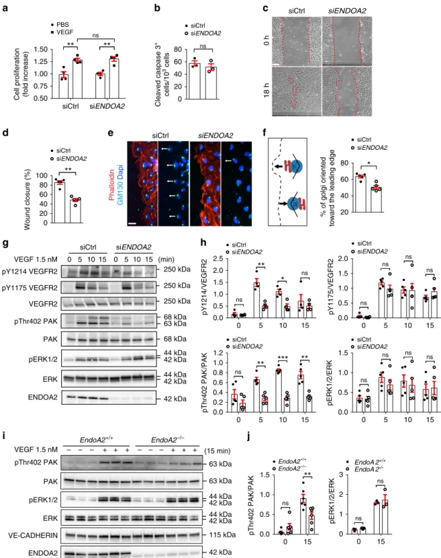
ns siCtrl siENDOA2 Phalloidin GM130 DapiFig. 3 ENDOA2 controls endothelial cell migration and polarity. a VEGF-induced cell proliferation (6 nM, 48 h stimulation) assessed by XCelligence system in siControl (Ctrl) and siENDOA2 silenced HUVECs (N = 4 independent experiments; two-way ANOVA: ns P > 0.05, **P < 0.01). b Cleaved caspase-3 staining of HUVECs cultured in 0.5% FBS for 24 h (N = 3 independent experiments; Mann–Whitney U test: ns P > 0.05). c HUVEC scratch wound migration in response to VEGF (3 nM).d Quantification of wound closure shown in c (N = 5 independent experiments; Mann–Whitney U test: **P < 0.01). e Phalloidin, Dapi, and GM130 Golgi labeling at the scratch wound edge to assess Golgi polarization in front of the nucleus (arrows indicate direction of migration) in response to VEGF (3 nM, 2 h).f Quantification of Golgi orientation (N = 5 independent experiments; Mann–Whitney U test: *P < 0.05). g Western-blot analysis of phosphorylation of the indicated proteins in response to VEGF (1.5 nM) in HUVECs. h Quantification of phosphorylation normalized to total protein levels (N = 4–5 independent experiments; two-way ANOVA: *P < 0.05, **P < 0.01, ***P < 0.001, ns P > 0.05). i Western-blot analysis of VEGF-induced (1.5 nM) PAK and ERK phosphorylation in mLEC from EndoA2+/+and EndoA2−/−mice. Each lane represents one mouse. j Quantification of phosphorylation normalized to total proteins (N = 3–5 mice; two-way ANOVA: ns P > 0.05, **P < 0.01). Error bars represent mean ± s.e.m. Scale bars:c 200μm, e 25 μm
immunoprecipitated with srGAP1 as well as ROBO1 and
VEGFR2 in cells cultured in serum-containing medium (Fig.
6
a),
demonstrating interaction between these molecules. Moreover,
confocal and SIM imaging showed colocalization between
VEGFR2, ENDOA2, and srGAP1 at the leading edge of HUVECs
treated with VEGF or SLIT2 (Fig.
6
b and Supplementary
Fig. 11f). siRNA silencing of srGAP1 abolished both VEGF- and
SLIT2-induced targeting of VEGFR2 to ENDOA2 positive
vesi-cles (Fig.
6
c, d) and impaired VEGF- and SLIT2-induced
VEGFR2 internalization (Fig.
6
e, f). Next, we reconstituted
ROBO1 and ROBO2 siRNA treated HUVECS with adenoviral
vectors encoding siRNA resistant rat full-length ROBO1-GFP
(ROBO1WT) or truncated version lacking the CC3 domain GFP
tagged (ROBO1ΔCC3) that binds srGAPs. Immunoprecipitation
with GFP followed by immunoblotting with srGAP1 antibody
showed that srGAP1 binding was strongly impaired in cells
expressing ROBO1ΔCC3 (Fig.
6
g), confirming that the CC3
domain is required for srGAP1 binding to ROBO1. Interestingly,
VEGFR2 binding to ROBO1 was also reduced in cells expressing
ROBO1ΔCC3 (Fig.
6
g) demonstrating that srGAP1 promotes
250 kDa 250 kDa
a
c
b
d
e
Internalized VEGFR2 Dapi
SLIT2 30
′ (6 nM)
siCtrl siROBO1/2 siENDOA2
HUVEC
Internalized VEGFR2 Dapi
SLIT2 30 ′ (6 nM) mLEC 0 5 15 30 siCtrl siROBO1/2 SLIT2 6 nM 0 5 15 VEGFR2 30 0 5 15 30 siENDOA2 Internalized Internalized
Surf. Surf. Surf.
Internalized
g
Unstimulated SLIT2 2 ′30 ″ (6 nM)ENDOA2 ENDOA2 VEGFR2
overlapping pixels EndoA2+/+ EndoA2–/– VEGFR2 ACTIN 250 kDa
f
h
i
j
SLIT2 2 ′30 ″ (6 nM)k
l
m
(min) 0.0 0.5 1.0 1.5 2.0Internalized VEGFR2 intensity (fold change)
EndoA2+/+ EndoA2–/–
*
0 20 40 60 80 100Number of EPAs per 1000
μ m 2 Unstimulated SLIT2
***
0 2 4 6 8 10 VEGFR2 /ENDOA2overlap (fold change)
Unstimulated SLIT2
***
ROBO1 ENDOA2 Input 42 kDa 42 kDa VEGFR2 ENDOA2 overlapping pixels VEGFR2 CLATHRIN overlapping pixels ENDOA2 CLATHRIN overlapping pixels VEGFR2 ENDOA2 CLATHRINn
0 20 40 60 80 100 % Overlap VEGFR2-ENDOA2 VEGFR2-CLATHRIN**
SLIT2 2′30″ 0 5 15 30 0.0 0.2 0.4 0.6 0.8 1.0Internalized VEGFR2 (normalized to surface)
siCtrl siROBO1/2 siENDOA2
**
ns ns*
***
**
**
**
(min) 0.0 0.5 1.0 1.5Internalized VEGFR2 intensity (fold change)
siCtrl siROBO1/2 siENDOA2
**
**
ROBO1-VEGFR2 complex formation. We next performed
scratch wound assays with reconstituted cells and tested
front-rear polarity in response to VEGF and SLIT2. Expression of
ROBO1WT in ROBO1/2 knockdown cells rescued front-rear
polarity in response to both VEGF and SLIT2, whereas
ROBO1ΔCC3 failed to rescue polarity (Fig.
6
h). These results
suggest that the physical interaction between srGAP1 and
ROBO1 is required for VEGF and SLIT2-induced
ENDOA2-mediated endocytosis, cell polarity and migration.
Blocking ENDOA2 inhibits pathological angiogenesis. To
determine the function of ENDOA2 in pathological
neovascu-larization, we subjected EndoA2
−/−mice to oxygen-induced
retinopathy
45(Fig.
7
a), which leads to pathological sprouting and
formation of abnormal vascular tufts that are prone to bleeding,
mimicking vision-threatening defects in infants with retinopathy
of prematurity. After hyperoxia exposure, P12 pups developed
vaso-obliteration leading to the formation of a capillary-free area
in the center of the retina. After return to room air, hypoxia in the
avascular area triggered re-growth of normal vessel sprouts form
centrally located veins and the remaining capillaries in the
per-iphery and neovascular tufts. Compared with EndoA2
+/+litter-mates, EndoA2
−/−mice showed decreased revascularization,
measured by a significant increase of the retina avascular area, as
well as decreased sprouting from veins and neovascular tuft
formation (Fig.
7
b, c). Thus, blockade of ENDOA2 attenuated
pathological ocular neovascularization.
Discussion
The data reveal ENDOA2 as a regulator of polarized endothelial
migration during sprouting angiogenesis (Fig.
8
). Among the
three ENDOPHILIN-A isoforms, only ENDOA2 was expressed
in ECs and required for developmental and pathological
angio-genesis. ENDOA2 promoted directional migration by regulating
VEGFR2 internalization and downstream signaling to PAK, but
not ERK. This result shows that distinct VEGFR2 uptake
path-ways can control cellular behaviors by promoting activation of
select downstream signaling pathways, and reveal that VEGFR2
endocytosis via ENDOA2 promotes polarized endothelial
migration.
The best studied VEGFR2 internalization route is via
CME
15,16, but VEGFR2 can be internalized by macropinocytosis
or caveolea as well
46–49. How VEGFR2 uptake via these distinct
routes is regulated is poorly understood. CLATHRIN dependent
VEGFR2 uptake requires the guidance molecule EPHRINB2, as
shown in mice carrying endothelial Efnb2 deletion which exhibit
lack of VEGFR2 uptake and signaling
18–20. Another guidance
molecule NRP1 regulates VEGFR2 endosomal trafficking, and
mice lacking endothelial Nrp1 exhibit arteriogenesis defects
because of impaired VEGF-induced ERK activation
17. Our results
identify the SLIT2-ROBO1 guidance pathway as a critical
med-iator of ENDOA2-mediated VEGFR2 uptake and subsequent
polarized endothelial cell migration. Previous studies had shown
that ENDOPHILINs are a component of CME
36,50. In line with
these
findings, we find that ENDOA2 and CLATHRIN show a
small overlap in unstimulated and ligand-stimulated cells.
How-ever, following VEGF stimulation, VEGFR2 segregates into
dis-tinct endosomes that are either positive for CLATHRIN or for
ENDOA2, suggesting largely distinct endocytic pathways. In
support of this idea, ENDOA2 knockdown failed to affect
CLATHRIN-mediated VEGFR2 endocytosis and ERK activation
by
VEGF,
which
is
known
to
depend
on
CME
17.
SLIT2 stimulation preferentially drives ENDOA2 but not
CLATHRIN-mediated uptake of ROBO1 and VEGFR2, thereby
promoting PAK but not ERK activation in migrating ECs.
Together, these
findings suggest that axon guidance receptor
signaling pathways in ECs function, at least in part, by guiding
endocytosis of critical surface receptors such as VEGFR2.
Fur-thermore, the data suggest that ENDOA2 and
CLATHRIN-mediated VEGFR2 endocytosis are two largely independent and
parallel internalization routes that trigger different downstream
signaling pathways and control specific cell behaviors.
SLIT2 and VEGF treatment of ECs promoted complex
for-mation between ROBO1 and VEGFR2 via srGAP1, leading to
internalization via the ENDOA2 pathway. This process is likely
facilitated by direct binding of srGAP1 to ROBO1 and ENDOA2,
although the molecular details remain to be established.
Asso-ciation of srGAP1 and ENDOA2 was recently shown in rat brain
lysates
31, confirming data obtained here in ECs. Our study
pro-vides a mechanistic understanding for how SLIT2/ROBO1
function controls sprouting angiogenesis. We had previously
shown that postnatal deletion of ROBO1/2 and the downstream
NCK1/2 adaptors in ECs induced selective defects in front-rear
polarity and angiogenic sprouting
9,35. While ROBO1 was the
predominant SLIT2-binding ROBO expressed in ECs, its deletion
caused upregulation of ROBO2, which was normally expressed at
very low levels, hence combined ROBO1 and 2 deletion was
required to prevent SLIT2 signaling and reveal angiogenic
sprouting defects in mice
35. In vitro, deletion of ROBO1 and 2
abolished SLIT2-induced EC front-rear polarity and migration, as
expected,
but
also
affected
VEGF-induced
polarity
and
migration
9,35, raising the question how lack of ROBO function
affected VEGF signaling mechanistically. The data shown here
reveal ENDOA2-mediated endocytosis as a major pathway for
ROBO-VEGFR2 interaction. We propose that ROBO1 acts as a
Fig. 4 SLIT2 induces ENDOA2-dependent VEGFR2 internalization. a Cell surface biotinylation assay of VEGFR2 internalization in response to SLIT2 (6 nM) in Control (Ctrl), ROBO1/2 and ENDOA2 siRNA silenced HUVECs. VEGFR2, ROBO1, ENDOA2, and ACTIN expression from the total cell lysate are shown as loading controls (input). Surf: surface expression.b Quantification of internalized VEGFR2 normalized to VEGFR2 surface expression before stimulation (N = 7 independent experiments; two-way ANOVA test: *P < 0.05, **P < 0.01, ***P < 0.001, ns P > 0.05). c, d Antibody feeding assay to measure VEGFR2 internalization in response to SLIT2 (6 nM) in Ctrl, ROBO1/2, and ENDOA2 siRNA silenced HUVECs (c) and mLECs from EndoA2+/+and EndoA2−/−mice (d). e, f Quantification of internalized VEGFR2 fluorescent intensity shown in c and d, respectively (N = 4 independent experiment, at least 103cellsanalyzed per experiment;e one-way ANOVA and f Mann–Whitney U test: *P < 0.05). g SIM images of HUVEC lamellipodia stained for ENDOA2 before and after SLIT2 stimulation (6 nM for 2′30″). h SLIT2 effects on EPA formation at the cell migration front (N = 16–17 cells per group analyzed from three independent experiments; t-test: ***P < 0.001). i SLIT2 increases the pixel overlap between VEGFR2 and ENDOA2 fluorescent signals (N = 8–10 cells per group analyzed from three independent experiments; Mann–Whitney U test: ***P < 0.001). j SIM images of HUVEC lamellipodia stained for ENDOA2, VEGFR2, and CLATHRIN heavy chain after SLIT2 stimulation (6 nM for 2′30″). k–m Overlapping pixels between VEGFR2/ENDOA2 (k), VEGFR2/ CLATHRIN (l), and ENDOA2/CLATHRIN (m) from the image presented in j are shown in white. Boxed areas are magnified to highlight VEGFR2/ENDOA2, VEGFR2/CLATHRIN, or ENDOA2/CLATHRIN overlap (white).n Quantification of overlap between VEGFR2/ENDOA2 and VEGFR2/CLATHRIN fluorescent signals (N = 8–10 cells per group analyzed from three independent experiments; Mann–Whitney U test: **P < 0.01) (right panel). Error bars represent mean ± s.e.m. Scale bars:c and d 20μm, g 2μm, j 1μm
42 kDa
b
c
a
g
e
Internalized VEGFR2 Dapi
VEGF 30 ′ (1.5 nM) siCtrl siROBO1/2 VEGF 1.5 nM VEGFR2 ROBO1 IP VEGFR2
h
SLIT2 6 nM VEGFR2 ROBO1 Input 250 kDa 250 kDa 250 kDa 250 kDa VEGF 2 ′30 ′′ (1.5 nM)ENDOA2 VEGFR2 overlapping pixels
250 kDa 250 kDa 250 kDa VEGF 1.5 nM 0 15 30 VEGFR2 siCtrl siROBO1/2 0 5 15 VEGFR2 Surf. Surf. Internalized Internalized 30 5 ROBO1 (min)
VEGFR2 ENDOA2 ROBO1
VEGF 2 ′30 ′′ (1.5 nM)
d
f
– 0.0 0.5 1.0 1.5 2.0 2.5 VEGFR2 /E NDOA2overlap (fold change
)
siROBO1/2 siCtrl
***
Front Middle Rear 0 20 40 60 80 % of golgi positio n Robo1–/– Robo2f/f
Robo1–/– Robo2ΔEC
*
ns***
0.0 0.2 0.4 0.6 0.8 1.0Internalized VEGFR2 intensity (fold change
) siCtrl siROBO1/2
*
ACTIN Input siCtrl siROBO1/2ENDOA2 VEGFR2 overlapping pixels
0 5 15 30 0.0 0.2 0.4 0.6 Internalized VEGFR 2 (normalized to surface ) siCtrl siROBO1/2
**
ns**
**
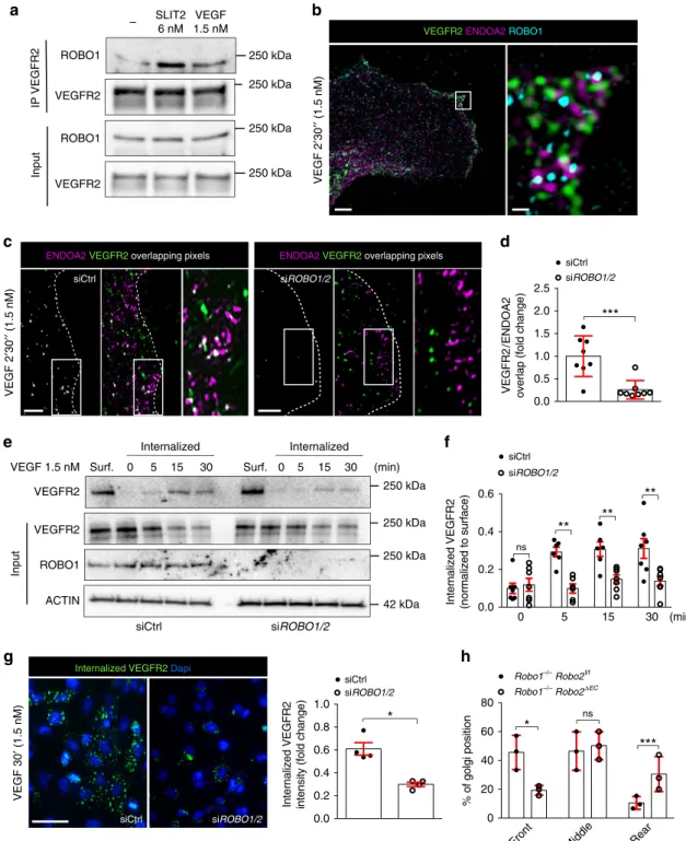
(min)Fig. 5 ROBO1 promotes VEGF-induced ENDOA2-mediated VEGFR2 endocytosis. a VEGFR2 immunoprecipitation in HUVEC after VEGF (1.5 nM for 2′30″) or SLIT2 (3 nM for 2′30″) stimulation, and western blot for ROBO1 and VEGFR2. ROBO1 and VEGFR2 expression from the total cell lysate are shown as loading controls (input).b SIM image of HUVECs stained for VEGFR2, ENDOA2, and ROBO1 after VEGF stimulation (1.5 nM for 2′30″). Right panel is a higher magnification of the boxed area in the left panel. c SIM images of the lamellipodia of Ctrl and ROBO1/2 siRNA treated HUVEC stained for ENDOA2 and VEGFR2 after VEGF stimulation (1.5 nM for 2′30″). Overlapping pixels between VEGFR2/ENDOA2 fluorescent signals are shown in white. Boxed areas are magnified to highlight VEGFR2/ENDOA2 proximity. d Quantification of pixel overlap between VEGFR2 and ENDOA2 fluorescent signals (N = 8 cells per group analyzed from three independent experiments; Mann–Whitney U test: ***P < 0.01). e Western-blot analysis of VEGF-induced VEGFR2 internalization (3 nM) from cell surface biotinylation assay in Ctrl or ROBO1/2 siRNA silenced HUVECs. VEGFR2, ROBO1, and ACTIN expression from the total cell lysate are shown as loading controls (input). Surf: surface expression.f Quantification of internalized VEGFR2 normalized to VEGFR2 surface expression before stimulation. (N = 7 independent experiments; two-way ANOVA: ns P > 0.05, **P < 0.01). g Antibody feeding assay to assess VEGFR2 internalization in response to VEGF (3 nM) in Ctrl and ROBO1/2 siRNA silenced HUVECs. Quantification of internalized VEGFR2 fluorescent intensity (right panel) (N = 4 independent experiments, at least 103cells analyzed per experiment; Mann–Whitney U test: *P < 0.05). h Golgi orientation in tip cells from
Robo1,2+/+and Robo1,2−/−P5 retinas. (N = 3 retinas per group, at least 50 tip cells per retina were quantified, two-way ANOVA: *P < 0.05, ***P < 0.001, ns P > 0.05). Error bars represent mean ± s.e.m. Scale bars: b left panel 5 μm, b right panel 1 μm, c 2 μm, g 20 μm
molecular cell surface tag driving VEGFR2 toward ENDOA2
(Fig.
8
). SLIT2 activation of ROBO1 was sufficient to initiate
VEGFR2 internalization via ENDOA2, however, in the absence of
VEGF ligand, the internalized VEGFR2 remained inactive. This
explains why SLIT2 pre-treatment of ECs in vitro can reduce
VEGF signaling
51,52, because less surface receptor is available for
ligand binding and signaling. In the combined presence of VEGF
and
SLIT2,
VEGFR2
signaling
toward
PAK/p38
was
enhanced
35,53–55, likely via enhanced ENDOA2-mediated
endo-cytosis. Conversely, absence of ROBO1 impaired SLIT2 and
VEGF-driven ENDOA2-mediated endocytosis of VEGFR2,
thereby impairing migration. Further studies are required to
reveal the exact mechanism promoting the selective effects of
ENDOA2-mediated endocytosis on cell polarity and migration,
but the results reveal the EPA as an environment conducive for
PAK/p38
activation,
providing
a
framework
for
future
investigations.
Our data reveal similar tip cell polarity defects in EndoA2 and
Robo1
−/−Robo2
fl/flCDH5
ERT2retina vessels. Interestingly, in
renal glomeruli, both ROBO2 and ENDOA2 interact with
NEPHRIN, a basement membrane protein regulating podocyte
structure and the SLIT-diaphragm organization
50,56,57. Mice
130 kDa 250 kDa
g
h
c
– AdROBO1WT -GFP AdGFP AdROBO1 ΔCC3-GFP siROBO1/2 srGAP1 VEGFR2 srGAP1 (input)a
b
e
130 kDa GFP IP GFP 250 kDaf
Internalized VEGFR2 Dapi
sisrGAP1 SLIT2 30 siCtrl sisrGAP1
′ (6 nM)
VEGF 30
′ (1.5 nM)
siCtrl
Internalized VEGFR2 Dapi
VEGF 2 ′30 ″ (1.5 nM) SLIT2 2 ′30 ″ (6 nM)
ENDOA2 VEGFR2 overlapping pixels ENDOA2 VEGFR2 overlapping pixels
siCtrl siCtrl sisrGAP1 0.0 0.5 1.0 1.5 2.0 VEGFR2 /E NDOA2
overlap (fold change
) siCtrl sisrGAP1
**
0.0 0.5 1.0 1.5 2.0 VEGFR2 /ENDOA2overlap (fold change
)
*
sisrGAP1 130 kDa VEGFR2 ROBO1 srGAP1 ENDOA2 IP:ENDOA2 – + Input 250 kDa 250 kDa 42 kDaVEGFR2 ENDOA2 srGAP1
VEGF 2 ′30 ″ (1.5 nM) SLIT2 2 ′30 ″ (6 nM)
d
VEGFR2 ENDOA2 srGAP1
20 40 60 80 100
*
ns*
SLIT2 40 60 80 100 % of golgi orientedtowards the leading edg
e siCtrl + AdGFP siROBO1/2 + AdGFP siROBO1/2 + AdROBO1WT siROBO1/2 + AdROBO1ΔCC3
**
ns**
VEGF 0.0 0.5 1.0 1.5Internalized VEGFR2 intensity (fold change)
siCtrl sisrGAP1
**
VEGF**
SLIT2carrying deletions of Robo2 or EndoA1-3 failed to establish a
normal glomerular
filtration barrier and exhibited severe
protei-nuria due to abnormal podocyte end-feet process formation.
Thus, ENDOPHILINS may interact with the ROBO pathway in
additional cell types besides ECs. Another interesting cell type to
be considered are commissural neurons, that respond to SLIT
midline guidance signals via ROBO1 and 2
58. While a previous
study had suggested CME as an endocytic route for ROBOs in
commissural axons
59, FEME has not been investigated yet.
The biological function of ENDOPHILINS is so far poorly
explored. ENDOA1 was shown to interact with EGFR in cultured
brain ECs to control cell permeability via cell junction
proteins
60,61; however, we have been unable to detect ENDOA1
expression in ECs, and single cell RNA sequencing also failed to
detect EndoA1 or A3 in adult brain ECs
62. In contrast to ECs,
EndoA1 and EndoA3 are abundantly expressed in neurons
33,37,63and combined deletion of all EndoA isoform was required to
induce neurological defects
36. In line with these
findings, we show
that the single deletion of EndoA2 did not affect EndoA1 or A3
gene expression in ECs, or neuron number and organization in
the retina. Our data reveal ENDOA2 driven endocytosis as a
target to prevent pathological angiogenesis in intraocular
neo-vascular diseases such as retinopathy of prematurity, which is
characterized by excessive angiogenesis promoting vascular leak
and edema, hemorrhage and retinal detachment compromising
vision
64. Specific targeting of sprouting angiogenesis could
be central to therapeutic strategies in such pathologies, where
anti-angiogenic approaches with VEGF blockers may produce
unwanted side-effects on photoreceptor survival
65,66.
Methods
Reagents and antibodies. Recombinant proteins: SLIT2 (5444-SL, R&D Systems), VEGF-A165 (293-VE, R&D Systems). Antibodies: Endophilin-A2 (1/200, sc-365704, Santa Cruz), CLATHRIN (1/400, 4796P, Cell Signaling), anti-Robo1 (MAB7118, R&D Systems), anti-GM130 (1/500, 610823, BD), anti-NG2 (1/200, AB5320, Millipore), anti-Desmin (1/200, AT3844, NovusBio), anti-VECadherin (1/ 200, Santa Cruz, Sc6458), anti-Collagen IV (1/300, AB769, Millipore), anti-ERG1/ Fig. 6 ROBO1 guides ENDOA2-mediated endocytosis via srGAP1. a ENDOA2 immunoprecipitation in HUVECs cultured in full medium and western-blot analysis of the indicated proteins. VEGFR2, ROBO1, srGAP1, and ENDOA2 expression from the total cell lysate are shown as loading controls (input). IP ENDOA2 (−): cell lysate incubated with beads alone; IP ENDOA2 (+): cell lysate incubated with anti-ENDOA2 antibody + beads. b Confocal images of HUVECs stained for VEGFR2, ENDOA2, and srGAP1 after 2′30″ VEGF (1.5 nM) or SLIT2 (3 nM) stimulation. Boxed areas are magnified to highlight VEGFR2/ENDOA2/srGAP1 pixel overlap.c SIM images of the lamellipodia of Ctrl and srGAP1 siRNA treated HUVECs stained for ENDOA2 and VEGFR2 after VEGF or SLIT2 stimulation (1.5 or 3 nM for 2′30″). Overlapping pixels between VEGFR2/ENDOA2 fluorescent signals are shown in white. Boxed areas are magnified to highlight VEGFR2/ENDOA2 pixel overlap. d Quantification of pixel overlap between VEGFR2 and ENDOA2 (N = 10 cells per group, three independent experiments; Mann–Whitney U test: *P < 0.05, **P < 0.01). e Antibody feeding assay to assess VEGFR2 internalization in response to VEGF or SLIT2 (3 or 6 nM) in Ctrl and srGAP1 siRNA silenced HUVECs. f quantification of internalized VEGFR2 fluorescent intensity (N = 3–6 independent experiment, at least 103cells analyzed per experiment; one-way ANOVA: **P < 0.01). g GFP immunoprecipitation of ROBO1/2 silenced cells transduced
with the indicated constructs and western blot analysis of the indicated proteins. IP GFP (−): cell lysate from ROBO1/2 silenced cells transduced with ROBO1WT-GFP construct incubated with beads alone.h Golgi orientation of ROBO1/2 silenced HUVECs transduced with the indicated constructs (N = 3 experiments, one-way ANOVA: ns P > 0.05, *P < 0.05, **P < 0.01). Error bars represent mean ± s.e.m. Scale bars: b 2 μm, c 2 μm, e 20 μm
a
c
b
P0 P7 P12 P17
Room air 75% O2 Room air Vaso-obliteration Neo-angiogenesis EndoA2 +/+ EndoA2 –/–
Retina Sprouting Tufts
P17 P17 0 20 40 60 80 Sprouting area (%)
**
15 20 25 30 35 Avascular area (%) EndoA2–/– EndoA2+/+**
0 10 20 30 40 50 Tuft area (%)*
Fig. 7 ENDOA2 promotes pathological angiogenesis. a Schematic of the experimental strategy to assess neoangiogenesis after oxygen-induced retinopathy (OIR).b Left panels show retinalflatmounts after OIR. Insets show avascular area measured for quantification. Middle panels show higher magnification of vessels sprouting from veins. Right panels show higher magnification of neovascular tufts. c Avascular area, sprouting and neovascular tuft quantifications of retinas shown inb (N = 6 retinas per group; Mann–Whitney U test: *P < 0.05, **P < 0.01). Error bars represent mean ± s.e.m. Scale bars: b left panel 1 mm,b middle and right panels 100μm
2/3 (1/100, SC353, Santa Cruz), anti-α-smooth muscle actin CY3 (1/200, C6198, Sigma), anti cleaved caspase-3 (9661S, Cell Signaling), anti-Podocalyxin (1/200, AF1556, R&D systems), Dapi (1/100, D1306, Life Technologies), anti-pVEGFR2 1175 (1/500, 2478, Cell Signaling), anti-pVEGFR2 1214 (1/250, 2477, Cell Sig-naling), anti-p44/42 MAP kinase (1/1000, phospho-ERK, 9106, Cell SigSig-naling), anti-p44/42 MAP kinase (1/1000, total ERK, 9102, Cell Signaling), anti-pPAK1 (Thr423)/PAK2 (Thr402) (1/500, 2601S, Cell Signaling), anti-PAK1/2/3 (1/500, 2604, Cell Signaling), anti-srGAP1 (1/200, ab76926, Abcam), anti-p38 MAPK (1/ 500, 8690T, Cell Signaling), anti-phosp38 MAPK (1/500, 4511T, Cell Signaling), anti-pMLC2 (1/200, 3671S, Cell Signaling), anti-VEGFR2 (1/500, 9698, Cell Sig-naling), anti-actin (1/2000, A1978, Sigma), anti-Calretinin (1/200, MAB1568, Millipore), anti-endomucin (1/200, HM1108, Hycult), anti-GFAP (1/200, ZO334, Dako). Appropriate secondary antibodies were conjugated to horseradish perox-idase (Vector Laboratories) orfluorescently labeled (Life Technologies). IsolectinB4 (I21411), Dapi (D1306), and phalloidin568were purchased from Life Technologies. Animals. EndoA2−/−mice were a gift from De Camilli Lab36. Robo1−/−Robo2fl/ flCDH5ERT2mice were previously described35. Mice were maintained under stan-dard specific pathogen-free conditions. All animal procedures were reviewed and approved by the Institutional Animal Care Use Committee of Yale University and comply with all ethical regulation.
Retina immunostaining and analysis. The eyes of P5 pups were prefixed in 4% PFA for 20 min at room temperature (RT). The retinas were dissected out and blocked during 30 min at RT in blocking buffer (1% fetal bovine serum, 3% BSA, 0.5% Triton X-100, 0.01% Na deoxycholate, 0.02% Na azide in PBS at pH 7.4). The retinas were incubated with antibodies in blocking buffer overnight. After washing with Pblec (1 mM MgCl2, 1 mM CaCl2, 0.1 mM MnCl2, 1% Triton X-100 in PBS), the retinas were incubated with IsolectinB4 and the corresponding secondary antibody in Pblec for 2 h at RT. Then the retinas were mounted influorescent mounting medium (DAKO). We performed the proliferation analysis using Click-iT EdU Alexa Fluor 647 Imaging kit (Life Technologies). P5 pups were injected with 300μg of EdU (5 mg/ml) and euthanized 4 h later. EdU staining was done according to the manufacturer’s protocol. For soluble Flt1 binding, after dissection, retinas were blocked and permeabilzed in TNBT (100 mM Tris pH 7.4, 150 mM
NaCl, 0.5% Triton X-100, 0.5% of TSA Blocking blocking reagent (FP1012, Perkin Elmer). Then, retinas were incubated in 1μg/ml of recombinant mouse soluble Flt-1 FC chimera (47Flt-1-FFlt-1-Flt-100, R&D) diluted in TNBT for 2 h at room temperature, rinsed three times in TNT (100 mM Tris pH 7.4, 150 mM NaCl, 0.5% Triton X-100),fixed with 4% PFA for 2 min at room temperature and incubated with anti-human IgG secondary antibodies diluted in TNBT overnight at 4 °C. Confocal pictures of retinas were acquired using a Leica SP5 confocal microscope with a Leica spectral detection system (Leica 15 SP detector) and the Leica application suite advancedfluorescence (LAS-AF) software. Quantification of retinal vascular development was done using the Image J software.
For immunostaining on sections, eyes were collected at P5 andfixed in 4% PFA for 1 h at room temperature. A hole was made in the cornea, and the eyes were incubated for 1 h in 10% sucrose (VWR, 27478.296) in 0.12 M phosphate buffer and then overnight at 4 °C in 30% sucrose in 0.12 M phosphate buffer. Eyes were then embedded and frozen in 0.12 M phosphate buffer containing 7.5% gelatin (Sigma, 62500) and 10% sucrose. Twenty-micrometer sections were cut with a cryostat (Leica, CM3050S). These sections were blocked in PBS containing 0.2% gelatin (VWR) and 0.25% Triton X-20 (PBS-GT) for 1 h and incubated overnight at room temperature with primary antibodies diluted in PBS-GT. Then the sections were incubated with secondary antibodies diluted in PBS-GT and 10μg/ml Dapi. Embryo whole-mount immunostaining. Embryos were harvested andfixed in 4% paraformaldehyde (PFA) in PBS for 2 h at room temperature, washed with PBS and incubated for 1 h in blocking solution (1% Triton X-100, 3% bovine serum albumin (BSA, Sigma-Aldrich) and 3% heat-inactivated bovine serum (Fisher) at room temperature. Embryos were incubated with the primary antibody overnight (1:100 Endomucin (Hycult Biotech) in blocking solution. Samples were washed and incubated in secondary antibody (1:200 Goat anti-Rat AlexaFluor 488 (Invitrogen) in blocking solution) overnight. Hindbrain dissections and immunostaining were done as previously described67.
Oxygen-induced retinopathy. OIR was performed as described9,35. Briefly, the breeding mother and P7 pups of both genders were placed in 75% O2until P12. The pups were then exposed to room air for an additional 5 days until P17. Eyes were collected at P17, retinas were stained with IB4. Avascular area and vascular tufts were quantified9,35using image J.
siRNA transfection. EndophinA2 siRNA was purchased from Invitrogen (SH3GL1SS109705, 81165385; siRNAs (FlexiTube siRNA)). ROBO1 siRNA (SMARTpool: ON-TARGETplus ROBO1 siRNA L-011381-00-0005), ROBO2 siRNA (SMARTpool: ON-TARGETplus ROBO2 siRNA L-023273-01-0005), and the matching negative controls (ON-TARGETplus Non-Targeting Pool D-001810-10-05) were purchased from Dharmacon. We transfected HUVECs with 25 pmol siRNA per 6-well plate with 2.5μl RNAiMax (Invitrogen) according to the manufacturer’s instructions. Cells were used for experiments 72 h after transfection.
Cell culture. HUVECs were obtained from the Yale University Vascular Biology and Therapeutics Core Facility and cultured in EGM2-Bullet kit medium (CC-3156 & CC-4176, Lonza). We starved HUVECs overnight in EBM-2 supplemented with 0.1%, 0.5%, or 1% FBS before SLIT2 or VEGF treatment.
Murine endothelial cell isolation. We harvested mouse lungs between P15 and P21, minced them and incubated them in 5 mL Dulbecco’s modified Eagle’s medium containing 2 mg/mL collagenase I (Invitrogen) for 45 min at 37 °C with shaking every 15 min followed byfiltering through a 40-μm nylon mesh (BD Falcon). The cells were then centrifuged at 1000 × g for 5 min at 4 °C, resuspended in buffer 1 (0.1% bovine serum albumin, 2 mM EDTA, pH 7.4, in PBS), and incubated with anti-rat immunoglobulin G-coated magnetic beads (Invitrogen) precoupled with rat anti-mouse platelet/endothelial cell adhesion molecule-1 (PECAM-1; MEC13.3, BD Pharmingen, 553370) for 30 min at 4 °C in an overhead shaker. Beads were separated from the solution with a magnetic particle con-centrator (Dynal MPC-S, Invitrogen). The beads were washedfive times with buffer 1 and centrifuged for 5 min at 1000 × g, and the supernatant was removed. The purified endothelial cells were then cultured in ECGM-2 (Promocell). For western-blot analysis, lung endothelial cells (2 × 10⁵) were seeded in 60-mm dishes and cultured for 24 h in ECGM-2 at 37 °C and 5% CO2.
Western blot. Cells were lysed in lysis buffer including phosphatase and protease inhibitors (Thermo Scientific, 78420, 1862209). Equal amounts of proteins were separated on 4–15% Criterion precast gel (#567-1084, Bio-Rad) and transferred on nitrocellulose membrane (Bio-Rad). Western blots were developed with chemilu-minescence HRP substrate (Millipore, WBKLS0500) on a Luminescent image analyzer, ImageQuant LAS 4000 mini (Ge Healthcare). See Supplementary Figs. 12 and 13 for the uncropped immunoblots.
Immunoprecipitation. Cell lysates were prepared in 50 mM Tris-HCl at pH 7.4, 50 mM NaCl, 0.5% Triton X-100, phosphatase and protease inhibitors, centrifuged
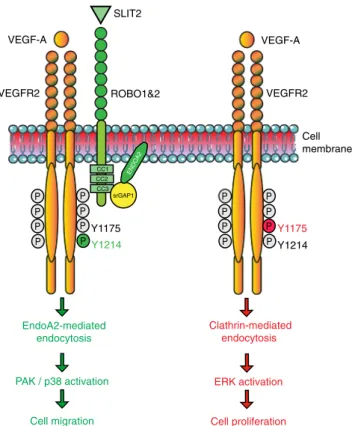
P P P P P P P P P P P P P P P P VEGF-A VEGFR2 VEGFR2 ROBO1&2 Clathrin-mediated endocytosis ERK activation Cell proliferation EndoA2-mediated endocytosis PAK / p38 activation Cell migration Y1175 Y1214 CC2 CC1 CC3 srGAP1 ENDOA2 Y1175 Y1214 Cell membrane SLIT2 VEGF-A
Fig. 8 Working model for ENDOA2-mediated VEGFR2 endocytosis. ENDOA2-mediated VEGFR2 endocytosis controls EC migration
independently of the CLATHRIN-mediated VEGFR2 endocytosis pathway. SLIT2-ROBO1 targets VEGFR2 to the ENDOA2-dependent internalization pathway via srGAP1, leading to downstream PAK/p38 activation and cell migration
at 16,000 × g for 20 min. Protein concentration was quantified using Bradford assay (Pierce). In total, 500μg of protein from cell lysate were incubated overnight at 4 °C with 10μg/ml of anti-ENDOA2 (Santa Cruz) or anti-GFP antibodies (BD Phar-mingen), andfinally incubated with protein A/G magnetic beads (88802, Thermo Scientific) for 2 h at 4 °C. The immunocomplexes were washed three times in lysis buffer and resuspended in 2X Laemmli’s sample buffer. For western-blot analysis, 50μg of protein was loaded for each condition.
Cell immunostaining. Cells were plated on gelatin coated glass bottom dishes. Growing cells werefixed for 10 min with 4% paraformaldehyde (PFA) and per-meabilized with 0.1% Triton X‐100, for 10 min prior to overnight incubation with primary antibody and then secondary antibody conjugated withfluorophore. Samples were mounted with ProLong Gold (Invitrogen).
SIM. Images were acquired using a U-PLANAPO 603/1.42 PSF, oil immersion objective lens (Olympus, Center Valley, PA) and CoolSNAP HQ2 CCD cameras with a pixel size of 0.080 mm (Photometrics, Tucson, AZ) on the OMX version 3 system (Applied Precision) equipped with 488, 561, and 642 nm solid-state lasers (Coherent and MPB communications). Samples were illuminated by a coherent scrambled laser light source that had passed through a diffraction grating to generate the structured illumination by interference of light orders in the image plane to create a 3D sinusoidal pattern, with lateral stripes ~0.270 nm apart. The pattern was shifted laterally throughfive phases and through three angular rota-tions of 60 for each z section, separated by 0.125 nm. Exposure times were typically between 50 and 200 ms, and the power of each laser was adjusted to achieve optimal intensities of between 2000 and 4000 counts in a raw image of 16-bit dynamic range, at the lowest possible laser power to minimize photo bleaching. Raw images were processed and reconstructed to reveal structures with 100–125 nm resolution68. The channels were then aligned in x and y, and rotationally using predetermined shifts as measured using a target lens and the Soft-worx alignment tool (Applied Precision). RG2B Colocalization plugin for Image J was used to isolate colocalized pixel data with automatic selection threshold values and express the data as the average of the corresponding red and green channels.
Biotinylation. HUVECs were grown to confluence and starved overnight in EBM2 with 0.5% FBS. Cells were rinsed, incubated with EZ-Link Sulfo-NHS-SS-Biotin (0.25 mg/ml, Thermo Scientific) at 4 °C for 1 h in PBS and rinsed with 50 mM glycine in PBS to stop the reaction. A portion of the cells were harvested and used to determine total biotinylated cell surface protein. The remaining cells were rinsed once with cold media+1% BSA, stimulated with EBM2 containing VEGF (25 ng/ ml or 1.5 mM) or SLIT2 (1μg/ml or 6 mM) at 37 °C for different times and then rinsed and incubated twice for 20 min each time on ice with the membrane-nonpermeable reducing agent GSH (45 mM, Sigma) in 75 mM NaCl, 75 mM NaOH, 1 mM EDTA, 1% BSA. GSH was quenched by incubating twice for 5 min each time with iodoacetamide (5 mg/ml) in PBS. Cell lysates were prepared using NP-40 lysis buffer (Roche). In total, 200μg of protein from the cell lysate was immunoprecipitated with 50μl of NeutrAvidin beads (Invitrogen) at 4 °C over-night, after which the beads were rinsed and resuspended in Laemmli SDS sample buffer. Samples were analyzed by SDS–PAGE followed by western blotting with anti-VEGFR2 antibody. The remaining cell lysates after bead incubation were used to blot VEGFR2, ROBO1, ENDOA2, and ACTIN loading controls.
Antibody feeding assay. HUVECs were grown to confluence and starved overnight in EBM2 with 0.5% FBS. Cells were rinsed, incubated with anti-VEGFR2 antibody (AF357, R&D) at 4 °C for 20 min in EBM2 containing 0.5% FBS and rinsed with cold PBS. Cells were then incubated with EBM2 containing VEGF (25 ng/ml or 1.5 mM) or SLIT2 (1μg/ml or 6 mM) at 37 °C for 30 min. Cells were rinsed twice for 2 min with cold PBS pH 2.5. Afterfixation with 4% PFA for 10 min at room temperature and permeabilization with 0.1% triton/PBS, cells were incubated with anti-Goat alexa488antibody in 1% BSA/PBS for 1 h at room temperature.
Scratch wound migration. We grew confluent monolayer of HUVECs in 6-well plates. Twenty-four hours after siRNA transfection, we starved the cells for 18 h in EBM-2 medium with 1% FBS. We created a horizontal wound in the confluent monolayer using a sterile 200-μl pipette tip. Next, we incubated the cells in EBM-2 supplemented with VEGF-A (50 ng/ml) or SLIT2 (1μg/ml) at 37 °C for 18 h. Pictures of scratch wounds were taken just before stimulation (time 0) and after 18 h. We calculated the extent of cell migration using ImageJ software.
Cell proliferation assay. The xCELLigence RTCA DP analyzer was used to measure proliferation of control and ENDOA2 knockout HUVEC (10,000 cells/ well) in response to VEGF-A (100 ng/ml, 6 nM). The plate was monitored every 15 min for 48 h.
Apoptosis analysis. The in vitro apoptosis analysis was performed using cleaved caspase-3 staining of confluent HUVEC monolayers. Twenty-four hours after siRNA transfection, the confluent cells were starved during 24 h (EBM2, 0.5% FBS).
Microarray. Data were extracted from a previous study69.
qPCR. RNAs from HUVEC or from MLECs were purified using RNeasy-kit (Qiagen). One microgram RNA was reverse transcribed using IScript cDNA Synthesis Kit (Bio-Rad) and quantitative PCR were assayed (15 ng cDNA) using SYBR Green Supermix (Bio-Rad) and the corresponding primers: mouse EndoA1 (forward: (CGGATGAGCCTAGAGTTTGC; reverse: GCTGATCCATTTGGA-CACCT); mouse EndoA2 (forward: TCCTTCGGCACCACTTATT; reverse: CGGTGTTCAGCATAGTCAGC); mouse EndoA3 (forward: GGCTCAA-GAAGCAGTTCCAC; reverse: GTGGATGTCACCAGCAAG); mouse Vegfa (QT00160769, Qiagen); mouse Slit2 (QT00163828); mouse Actb (QT01136772); Human ENDOA1 (QT00012796, Qiagen); human ENDOA2 (QT00016415, Qia-gen); human ENDOA3 (QT00041027, QiaQia-gen); human ACTB (QT01680476, Qiagen). The data werefirst normalized to actin level in each sample, and the relative expression levels of different genes were calculated by the comparative Ct method70.
Statistical analysis. For continuous variables, data are presented as mean ± SEM. Between-group comparisons used the Mann–Whitney U test or t-test depending on the sample size for continuous variables. In cases more than two groups are compared one-way or two-way ANOVA test were performed as appropriate, fol-lowed by Turkey’s multiple comparison or Bonferroni multiple comparison tests, respectively, if P < 0.05. A value of P < 0.05 was considered statistically significant. All the analyses were performed using Prism 7.0 software (GraphPad). Reporting summary. Further information on research design is available in the Nature Research Reporting Summary linked to this article.
Data availability
All data that support thefindings of this study are available within the article and its Supplementary Information File and from the corresponding author upon reasonable request.
Received: 4 June 2018 Accepted: 30 April 2019
References
1. Potente, M., Gerhardt, H. & Carmeliet, P. Basic and therapeutic aspects of angiogenesis. Cell 146, 873–887 (2011).
2. Herbert, S. P. & Stainier, D. Y. Molecular control of endothelial cell behaviour during blood vessel morphogenesis. Nat. Rev. Mol. Cell Biol. 12, 551–564 (2011).
3. Eichmann, A. & Simons, M. VEGF signaling inside vascular endothelial cells and beyond. Curr. Opin. Cell Biol. 24, 188–193 (2012).
4. Simons, M., Gordon, E. & Claesson-Welsh, L. Mechanisms and regulation of endothelial VEGF receptor signalling. Nat. Rev. Mol. Cell Biol. 17, 611–625 (2016).
5. Koch, S. & Claesson-Welsh, L. Signal transduction by vascular endothelial growth factor receptors. Cold Spring Harb. Perspect. Med. 2, a006502 (2012). 6. Okabe, K. et al. Neurons limit angiogenesis by titrating VEGF in retina. Cell
159, 584–596 (2014).
7. Ruan, G. X. & Kazlauskas, A. VEGF-A engages at least three tyrosine kinases to activate PI3K/Akt. Cell Cycle 11, 2047–2048 (2012).
8. Sun, Z. et al. VEGFR2 induces c-Src signaling and vascular permeability in vivo via the adaptor protein TSAd. J. Exp. Med. 209, 1363–1377 (2012). 9. Dubrac, A. et al. Targeting NCK-mediated endothelial cell front-rear polarity
inhibits neovascularization. Circulation 133, 409–421 (2016). 10. Simons, M. An inside view: VEGF receptor trafficking and signaling.
Physiology 27, 213–222 (2012).
11. Miaczynska, M., Pelkmans, L. & Zerial, M. Not just a sink: endosomes in control of signal transduction. Curr. Opin. Cell Biol. 16, 400–406 (2004). 12. Platta, H. W. & Stenmark, H. Endocytosis and signaling. Curr. Opin. Cell Biol.
23, 393–403 (2011).
13. Sorkin, A. & Von Zastrow, M. Signal transduction and endocytosis: close encounters of many kinds. Nat. Rev. Mol. Cell Biol. 3, 600–614 (2002). 14. Sorkin, A. & von Zastrow, M. Endocytosis and signalling: intertwining
molecular networks. Nat. Rev. Mol. Cell Biol. 10, 609–622 (2009). 15. Lampugnani, M. G., Orsenigo, F., Gagliani, M. C., Tacchetti, C. & Dejana, E.
Vascular endothelial cadherin controls VEGFR-2 internalization and signaling from intracellular compartments. J. Cell. Biol. 174, 593–604 (2006). 16. Ewan, L. C. et al. Intrinsic tyrosine kinase activity is required for vascular
endothelial growth factor receptor 2 ubiquitination, sorting and degradation in endothelial cells. Traffic 7, 1270–1282 (2006).