DOI 10.1007/s00402-012-1553-0
A R T H R O S C O P Y A N D S P O R T S M E D I C I N E
Viscoelastic adaptation of tendon graft material to compression:
biomechanical quanti
Wcation of graft preconditioning
Dominik C. Meyer · Jess G. Snedeker · Robert A. Weinert-Aplin · Mazda Farshad
Received: 18 December 2011 / Published online: 6 June 2012 © Springer-Verlag 2012
Abstract
Purpose The tensile viscoelastic behaviour of tendon tis-sue is of central biomechanical importance and well exam-ined. However, the viscoelastic tendon adaptation to external compression, such as when a tendon graft is Wxated with an interference screw, has not been investigated before. Here, we quantify this adaptive behaviour in order to develop a new method to mechanically precondition ten-don grafts and to better understand volumetric changes of tendinous tissue. The hypothesis of this study was that under compressive loads, tendon grafts will undergo a tem-porary volumetric (and therefore diametric) reduction, due to the extrusion of water from the tendon.
Methods Compressive testing was performed on a mate-rial testing machine and load applied through the use of a custom-made mould, with a semi-circular cross section to accommodate the tendon graft. The eVects of diVerent com-pressive forces on the length, diameter and weight of ten-don grafts were measured by calipers and a weighing scale, respectively. Further, diVerent strain rates (1 vs. 10 mm/ min) (n = 6, per rate), compression method (steady com-pression vs. creep) (n = 15 for each method) and diVerent compression durations (1, 5, 10 min) (n = 5 for each dura-tion) were tested to identify the most eVective combination to reduce graft size by preserving its macroscopic structure. Results The eVect of compression on volume reduction (75 % of initial volume and weight) reached a plateau at 6,000 N on an 8-mm tendon bundle. Length thereby increased by approximately 10 %. Both steady compression
and creeping were able to reduce dimensions of the graft; however, creeping was more eVective. There was no diVer-ence in eVect with diVerent durations for compression (p > 0.05) in both methods.
Conclusion The viscoelastic behaviour of hamstring ten-don grafts under pressure allows preconditioning of the grafts for reduction of volume and diameter and therefore to drill a smaller bone tunnel, retaining more of the original bone. At the same time, the collagen content of the trans-plant is preserved and a tight Wt of the transtrans-plant in the bone tunnel achieved.
Keywords Viscoelastic tendon adaptation · Compression · Interference screw
Introduction
The tensile elastic and viscoelastic properties of tendons are of great importance for the function of the musculotendinous unit and have been extensively investigated [7, 15–18]. When used as an auto- or allograft, however, tendon material will not only be subjected to tensile, but also to compressive force, such as in a tunnel Wxated with an interference screw, within the loop of a suture stitch or if bent around a bone edge [11]. The behaviour of tendon tissue under compressive load has been described before [5, 10], however it has not yet been quantiWed in detail. As we could observe in an earlier study that tendon tends to slowly adapt to external compres-sive force [11], we speculated that under pressure, water would be pressed out from tendon tissue and tendon volume lost. We further speculated that it might be useful to exploit this eVect before graft implantation by compressing the ten-don transplants to a desired shape and to anticipate later loss of volume and undesired loss of geometric Wt [6, 12]. D. C. Meyer · J. G. Snedeker · R. A. Weinert-Aplin ·
M. Farshad (&)
Department of Orthopedics, University of Zürich, Balgrist, Forchstrasse 340, 8008 Zürich, Switzerland e-mail: [email protected]
The hypothesis of this study was that under compressive loads, bundled tendon grafts will undergo a temporary vol-umetric (and therefore diametric) reduction, due to the extrusion of water from the tendon.
Therefore, the aim of this study was to quantify the vis-coelastic and volumetric adaptation of tendon tissue under various pressurizing conditions and to translate the Wndings into a practical surgical method for tendon compression by the example of tendon bundles, such as those used for reconstruction of cruciate ligaments of the knee. The Wnal clinical relevance is to achieve a smaller drill hole for the same graft and tighter Wt with better ingrowth, character-ized by less or no tunnel widening.
Methods
Fresh Xexor digitorum tendons of the calf and adult sheep extensor digitorum tendons were used (Table1). Due to better availability, further experiments with need of large amount of tendons were performed with calf Xexor digito-rum tendons. Preliminary experiments were performed to assure the interspecies similarity with regard to behaviour of the tendon material under compression [3].
Tendons were harvested directly after slaughter and additional tissue was removed. The number of folds of ten-don required to obtain grafts of 8- to 9-mm diameter and 30-mm length was determined by measurement with a cali-per and veriWed by use of a pull-through block, with 7-, 8- and 9-mm holes as a rough check of graft diameter. The
diameter was measured at the ends and mid-length of the tendon, and the mean of the three values was considered to be a representative diameter of the graft. The tendon was laid out in a zigzag pattern, with Vicryl® USP 0 at the ends of the tendon and Ethibond® sutures (Ethicon; John-son&Johnson, Norderstedt Germany) were placed at the folds of the tendon. Once all the sutures were in place, the folds of the tendon were lightly pushed together, so that there were no sections of the graft protruding out. With the bundles still under tension, No. 0 Vicryl sutures were used to pierce the tendon about 1/4 of the way along the graft and loop the suture around the entire graft. This was repeated at halfway and 3/4 of the length of the graft to Wnish the bundling process. Finished grafts were measured with a caliper (with a tolerance of 0.05 mm) to determine the Wnal length and diameter and weighed on a weighing scale (with a tolerance of 0.005 g), before being stored in Ringer’s lactate-soaked gauze.
In order to characterize the behaviour of tendon in com-pression, an aluminium custom mould was manufactured, which consisted of a loading head and a mould which would accommodate a circular section of material in com-pression (Fig.1). All mechanical testing was performed on a universal testing machine (Zwick 1456, Zwick GmbH, Ulm, Germany) with a 20-kN load cell.
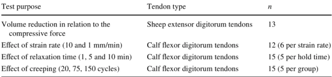
Compression was applied by displacement-controlled motion of the load head. Compression of samples was done by initially applying a 5-N preload at a rate of 10 mm/min, at which point the sample could be shifted in the mould, to ensure that the entire sample was compressed by the load Table 1 Material used for
diVerent test settings Test purpose Tendon type n
Volume reduction in relation to the compressive force
Sheep extensor digitorum tendons 13
EVect of strain rate (10 and 1 mm/min) Calf Xexor digitorum tendons 12 (6 per strain rate) EVect of relaxation time (1, 5 and 10 min) Calf Xexor digitorum tendons 15 (5 per hold time) EVect of creeping (20, 75, 150 cycles) Calf Xexor digitorum tendons 15 (5 per group)
Fig. 1 An aluminium custom mould with a loading head and a mould (a), which could accommodate a circular section of material in com-pression, was designed to suit a mechanical testing machine (Zwick 1456, Zwick GmbH, Ulm, Germany) for load- and/or strain-controlled
compression of the grafts. A compressed tendon graft (b, lower graft) showed a smaller diameter and increased total length compared to an untreated graft (b, upper graft)
head. Then, a 100-N nominal load was applied to allow for the bundles to reorganize within the graft and to allow for a more steady compression of the graft up to peak load, as the slippage of bundles across their surfaces would lead to an unsteady loading proWle.
Volume reduction in relation to the compressive force Experiments for characterization of the eVect of compres-sion on volume were performed on 13 sheep extensor digi-torum tendons, which were bundled to become a bundle with a diameter of 8 mm and a length of 30 mm. To deter-mine the geometric changes due to compression at diVerent peak loads in steps of 2,000 N, sample weight, length, vol-ume and diameter were measured before and after compres-sion. Two or three grafts were tested at each compressive load for repeatability. However, to determine the reduction in diameter, the weight loss of the tendon and the com-pressed graft length were used as a secondary measure to determine the compressed graft diameter.
Strain rate
Compression tests to determine an appropriate strain rate were performed at 10 and 1 mm/min, to determine how sta-ble the tendon was under the diVerent rates of loading. Six grafts were tested per group.
Relaxation tests
Graft dimensions were taken immediately before and after loading. Grafts were preloaded in the above-explained manner. Load was then increased to 6,000 N at 1 mm/min and held at a Wxed displacement for the desired time (1, 5 or 10 min), after which samples were unloaded. Five grafts were tested at each hold time.
EVective creep tests
Creep testing was also performed, as this was thought to be the most eYcient way of squeezing all of the water out of the tendon, leaving only an elastic material. Graft dimen-sions were taken immediately before and after loading. Grafts were preloaded in the above-explained manner. The creep test required a loading protocol that performed an eVective creep test. This entailed loading up to 6,020 N at 1 mm/min and holding for 1 s before decreasing the load to 5,990 N for 1 s. Cycles between these two forces were per-formed until the desired duration (1, 5 or 10 min) had been achieved, after which grafts were unloaded completely. It should be noted that approximately 20, 75 and 150 cycles corresponded to 1, 5 and 10 min of load-controlled loading, respectively. Five grafts were tested at each hold time.
Statistical analysis
A standard statistical software (PRISM Version 5.01 for Windows; Graphpad) was used for all analyses. Values were documented as mean with their 95 % conWdence inter-val [mean (lower 95 % CI, upper 95 % CI)]. Where appli-cable, data were tested for normal distribution using the Kolmogorov–Smirnov test and either two-tailed Student’s t test or Mann–Whitney test was employed for intergroup comparison for normal and not normal distributed data, respectively.
For intragroup comparison of the eVect of compression on the tissue, the Wilcoxon matched pairs test was used. SigniWcance was set at a level of p < 0.05.
Results
Volume reduction in relation to the compressive force The volume decreased with increasing compression force (Fig.2) and reached a plateau regarding the cross-sectional area at about 6,000 N (75 % of initial volume). The decrease of volume was concordant to the decrease in weight (Fig.2), with a decrease in area and diameter both being constituent parts of the volume.
Strain rates
At a strain rate of 10 mm/min of loading up to 6 kN, sig-niWcant rupturing occurred in four out of four samples, disrupting the transplant partially in the mid-substance. Total disruption did not occur. At a rate of 1 mm/min, no rupturing was observed in any of the cases, with only mild (less than 3 mm) extrusion of tendon from the end of the bundle (and not at the fold of the graft) in two out of four cases.
Fig. 2 The normalized volume and the weight of the bundled tendon grafts decreased with increasing compression force
Relaxation tests
The average initial bundle diameters were reduced from 8.3 mm (95 % CI 8.0, 8.7), 8.9 mm (95 % CI 7.8, 10) and 8.7 mm (95 % CI 7.9, 9.5) to 7.4 mm (95 % CI 6.9, 7.9) (p = 0.0625), 7.8 mm (95 % CI 6.9, 8.7) (p = 0.0625) and 7.8 mm (95 % CI 7.2, 8.3) (p = 0.0625) after 1, 5 and 10 min of compression, respectively. Diametric reductions were found to be reasonably constant for relaxation periods between 1 and 10 min (p = 0.25).
Creep tests
The average initial diameters of the bundles used in creep testing were 9.3 mm (95 % CI 0.9, 0.6), 8.3 mm(95 % CI 7.7, 8.9) and 9.3 mm (95 % CI 8.4, 10.2) for 1, 5 and 10 min, respectively, and was reduced to 8.1 mm (95 % CI 7.8, 8.4) (p < 0.05), 7.1 mm (95 % CI 6.3, 7.9) (p = 0.0625) and 7.8 mm (95 % CI 6.9, 8.7) (p < 0.05), respectively, after creep testing. There seemed to be no signiWcant gains in terms of diametric reduction for a creep period of 10 min compared to 1 min (p = 0.5). Comparison of the relaxation and creep tests (using values measured directly) showed slightly increased diametric reduction that is possible through the creep approach (Fig.3).
Discussion
Tendons are designed to transmit tensile muscle force and react to load in a viscoelastic manner. This has been exten-sively investigated [7, 14–18] and can in part be attributed to the signiWcant water content, which may exceed 60 % [1] of the total weight. However, tendons also play an impor-tant role as grafts for various surgical procedures, particu-larly for cruciate and collateral ligament repair such as in the knee, elbow and ankle surgery [13]. In almost all of
these reconstructive procedures, and also in common ten-don repair, there is the requirement of mechanically holding the graft ends, which is almost invariably achieved by applying a partially compressive force on the tendon tissue [2, 4]. Common Wxation techniques include the use of
inter-ference screws or sling-shaped suture stitches, creating mechanical hold mostly through pressure-induced friction of the screw or suture against the collagen Wbres. However, in a recent study, it has been found that tendon grafts may slowly lose this mechanical Wxation due to viscoelastic adaptations in response to the pressure Wt created [11]. The water is pressed out from the tendon, which reduces its vol-ume and, consequently, the mechanical Wxation of the graft [11].
Even though many operative procedures rely on the compressive resistance of tendon tissue [2, 4], there is no report to our knowledge characterizing the behaviour of tendon tissue under compressive load. The hypothesis of this study was that under compressive loads, bundled ten-don grafts will undergo a temporary volumetric (and there-fore diametric) reduction, due to the extrusion of water from the tendon.
It was therefore the purpose of this study to understand and quantify the compressive viscoelastic behaviour of hamstring tendon grafts under pressure and consequently to develop a new method for tendon preconditioning to reduce the graft volume before implantation using a dedicated compression device. The motivation is to prevent later vis-coelastic loss of pressure and to achieve a tighter Wt of the transplant in a smaller bone tunnel and to potentially pre-vent tunnel widening [12] in case of ACL-replacement.
Limitations of the applied methods are that due to lim-ited availability and large variability of fresh human tendon material, mainly bovine tendons of very similar appearance to human hamstrings were used, which, however, are con-sidered to be comparable to human hamstrings [3]. Due to the large number of diVerent experiments performed, we considered it important not to inXuence the results addition-ally with a variability of the tendon quality of unknown extent. In future tests, it will be of interest to study the in Xu-ence of tendon quality on the behaviour under pressure, and on postoperative swelling in the body.
This study has shown that using external mechanical pressure, the tendon volume may be reduced by 10–30 % through extrusion of water. If we assume a mean water con-tent of 60 % [1] in a normal tendon, this represents an extrusion of 17–50 % of the total tendon water. Interest-ingly, there was little diVerence in the volume of extrudable water before mechanical damage to the tendon tissue occurred, regardless of the technique. We speculate that the extruded liquid represents extracellular water, which may be removed relatively easily, while the intracellular water may not be so readily displaced. Even though total volume Fig. 3 Comparison of the single compression relaxation and creep
tests showed the slightly increased diameter reduction and increased gain in length that is possible through the creep approach
was lost, we could document an approximately 5–10 % gain in length of the transplant, which we attributed to an indirect pressure-induced longitudinal elongation and stretching.
These Wndings seem important in various ways:
Firstly, this method oVers a simple approach to decrease the graft volume and to facilitate the handling of a tendon transplant through a process, which anticipates an eVect that inevitably occurs, if not during then after implantation. It has been previously shown that modiWcation of the ten-don graft surface, namely embossing a screw thread and TCP coating had a large inXuence on the biomechanical performance of interference screw Wxation and resulted in less bone damage inXicted during insertion to a smaller tun-nel diameter, while simultaneously achieving superior pull-out strength [5].
Secondly, tendon (or collagenous tissue in general) is under compressive load in virtually any surgical procedure, which utilizes sutures or tendon Wxation implants. The here described Wndings help to better understand and quantify the reaction of the tissue.
Thirdly, the water content of tendon tissue is variable in vivo and inXuenced by gender, training, inXammation and other factors [8]. Particularly after transplantation, the vol-ume may not only be decreased, but also be increased by 40 % as a reaction to tissue transformation and healing [9]. In future studies, it may be of interest to investigate the vol-umetric behaviour of compressed transplanted tendons in vivo, or possible treatment options applying pressure to collagenous tissue in vivo.
Fourthly, the observed stretching, which is otherwise attempted through direct pull on the transplant with sutures, may be beneWcial for the desired pre-tension of the trans-plant after imtrans-plantation.
A secondary observation from this study is that the vis-coelastic nature of the tendon graft is highly susceptible to the rate of loading. Theoretically, the same volume of extracellular Xuid can be extruded from a graft under the same loads. However, it was noted that the graft requires time to adapt to the compressive loading and that even low loads can lead to plastic deformation and rupturing of the graft bundles. This failure of the graft was found to occur at both high (approximately, 9,000 N) and low (approxi-mately, 1,000 N) loads, highlighting the eVect that high strain rates alone can have on viscoelastic materials, such as a tendon graft. Future studies could investigate the eYcacy and repeatability of higher compressive strain rates, as higher rates would result in a shorter precondition-ing time, which would be desirable in any surgical settprecondition-ing.
Alternative procedures to decrease the water content may include the use of chemicals or heat; however in our opinion, the above-described technique appears to cause minimal damage to the tissue and anticipates physical
con-ditions to which the transplant will be subjected anyway during an implantation procedure, such as interference screw Wxation. Additionally, the potentially beneWcial eVect of gain in length will most likely not occur through drying or chemical treatment.
Conclusion
In summary, the volume of an 8-mm diameter and 3-cm long tendon graft may be reduced by up to 30 % by apply-ing external pressure of 6,000 N within few minutes through extrusion of water. Simultaneously, the tendon may be pre-stretched by 10 % of its length. This Wnding has to our knowledge not been described before and may be considered and exploited in surgical procedures where external pressure occurs on a tendon or transplant. From a practical perspective, a compressed tendon graft may allow drilling a smaller bone tunnel, retaining more of the origi-nal bone. At the same time, the collagen content of the transplant is preserved and a tight Wt of the transplant in the bone tunnel achieved.
ConXict of interest None.
References
1. Audia M (1958) Sodium and chloride spaces and water content of tendon in normal and depleted rats. Am J Physiol 195:702–704 2. Dargel J, Koebke J, Bruggemann GP et al (2009) Tension
degra-dation of anterior cruciate ligament grafts with dynamic Xexion– extension loading: a biomechanical model in porcine knees. Arthroscopy 25:1115–1125
3. Donahue TL, Gregersen C, Hull ML et al (2001) Comparison of viscoelastic, structural, and material properties of double-looped anterior cruciate ligament grafts made from bovine digital exten-sor and human hamstring tendons. J Biomech Eng 123:162–169 4. Fabbriciani C, Mulas PD, Ziranu F et al (2005) Mechanical
analy-sis of Wxation methods for anterior cruciate ligament reconstruc-tion with hamstring tendon graft. An experimental study in sheep knees. Knee 12:135–138
5. Farshad M, Weinert-Aplin RA, Stalder M et al (2012) Embossing of a screw thread and TCP granules enhances the Wxation strength of compressed ACL grafts with interference screws. Knee Surg Sports Traumatol Arthrosc 20:268–274
6. Herbort M, Weimann A, Zantop T et al (2007) Initial Wxation strength of a new hybrid technique for femoral ACL graft Wxation: the bone wedge technique. Arch Orthop Trauma Surg 127:769–775 7. Howard ME, Cawley PW, Losse GM et al (1996) Bone-patellar tendon–bone grafts for anterior cruciate ligament reconstruction: the eVects of graft pretensioning. Arthroscopy 12:287–292 8. Lemoine JK, Lee JD, Trappe TA (2009) Impact of sex and chronic
resistance training on human patellar tendon dry mass, collagen content, and collagen cross-linking. Am J Physiol Regul Integr Comp Physiol 296:R119–R124
9. McFarland EG, Morrey BF, An KN et al (1986) The relationship of vascularity and water content to tensile strength in a patellar ten-don replacement of the anterior cruciate in dogs. Am J Sports Med 14:436–448
10. Meyer DC, Farshad M, Snedeker JG (2011) Method and device for modelling tendinous tissue into a desired shape. Switzerland Patent 11. Meyer DC, Stalder M, Koch PP et al (2011) Contact pressure on ACL hamstring grafts in the bone tunnel with interference screw Wxation—dynamic adaptation under load. Knee [Epup ahead of print]
12. Nebelung W, Becker R, Merkel M et al (1998) Bone tunnel enlargement after anterior cruciate ligament reconstruction with semitendinosus tendon using endobutton Wxation on the femoral side. Arthroscopy 14:810–815
13. O’Donoghue DH (1963) A method for replacement of the anterior cruciate ligament of the knee. J Bone Joint Surg Am 45:905–924 14. Olabisi RM, Best TM, Hurschler C et al (2010) The biomechanical
eVects of limb lengthening and botulinum toxin type A on rabbit tendon. J Biomech 43:3177–3182
15. Schatzmann L, Brunner P, Staubli HU (1998) EVect of cyclic preconditioning on the tensile properties of human quadriceps ten-dons and patellar ligaments. Knee Surg Sports Traumatol Arthrosc 6(Suppl 1):S56–S61
16. Screen HR (2008) Investigating load relaxation mechanics in ten-don. J Mech Behav Biomed Mater 1:51–58
17. Taylor DC, Brooks DE, Ryan JB (1997) Viscoelastic characteris-tics of muscle: passive stretching versus muscular contractions. Med Sci Sports Exerc 29:1619–1624
18. Taylor DC, Dalton JD Jr, Seaber AV et al (1990) Viscoelastic properties of muscle-tendon units. The biomechanical eVects of stretching. Am J Sports Med 18:300–309