Ministry of Higher Education and Scientific Research
Djilali LIABES University of Sidi-Bel-Abbes
Faculty of Life and Natural Sciences
Department of Biology
Submitted for the degree of
Doctorate in Sciences
Biological Sciences
Option: Biology of Reproduction and development Presented by:
Mrs. Amina BENABBOU
Defended on: ……….
Board of Examiners:
Mr. KANDOUCI Baderdine Abdelkrim (Professor) Djilali LIABES University of Sidi-Bel-Abbes; Chairman Mr. KHALED Meghit Boumediene (Professor) Djilali LIABES University of Sidi-Bel-Abbes; Supervisor Mrs. BENDAHMANE SALMI Malika (Professor) Djilali LIABES University of Sidi-Bel-Abbes; Examiner Mrs. AIT HAMADOUCHE Nadia (Professor) Mohamed BOUDIAF University of Oran; Examiner Mr. KAHLOULA Khaled (Professor) Moulay Tahar University of Saida; Examiner Mr. BENARBA Bachir (Associate Professor) Mustapha STAMBOULI University of Mascara, Examiner
Academic Year: 2017-2018
Thesis
Evaluation of the Efficiency of Combined and Separated Antioxidant
Supplementation of Vitamin C and E on Semen Parameters in
Page | II
Preface
he current PhD thesis aims to examine the effectiveness of supplementation with vitamin C and E, on the infertility of male diabetes patients, by conducting experiments on male Wistar rats. The study is based on the assessment of semen parameters, blood hormonal levels, and histological investigations of testicular sections. The manuscript is structured as follows:
- The first section describes the theoretical considerations on male infertility (Chapter 1), Oxidative Stress, Mechanisms and Treatment (Chapter 2), and Diabetes Mellitus and Male Infertility (Chapter 3);
- The second part, covers the study design and the protocol carried out according to the preliminary hypothesis and the objectives of the study (chapter 4);
- The third section deals with the results of the study (Chapter 5);
- And the conclusion that draws all the results and prospects for future research. The wording of this manuscript follows the latest version of ISO-690 (International Organization for Standardization) for bibliographic referencing in the preparation of documents, content, form and structure, published in 2010.
The research work in this thesis involved many people, of which I am indebted, and I want to thank them all for their help and support.
First and foremost, I have to thank “Allah” for providing me blessings, courage, strength, and patience to achieve this modest work.
I express my deepest thanks to my supervisor, Professor KHALED Méghit Boumédiène, researcher lecturer at Djillali Liabès University of Sidi-Bel-Abbes, for the competent help, for his encouragement to accomplish a work begun a long time ago. His critical eye was very valuable for structuring the work and improving the quality of its different sections.
Page | III I would like to thank Pr. KANDOUCI Baderdine Abdelkrim, researcher lecturer at Djillali Liabès University of Sidi-Bel-Abbes for accepting to be the chairman of the jury. To Pr. BENDAHMANE Malika, researcher lecturer at Djillali Liabès University of Sidi-Bel-Abbes. To Pr. AIT HAMADOUCHE Nadia, researcher lecturer at Ben Bella University of Oran 1, to Pr. KAHLOULA Khaled, researcher lecturer at Moulay Taher University of Saïda, and to Dr. BENARBA Bachir, researcher lecturer at Mustapha Stambouli University of Mascara, for accepting to examine this modest work.
A special thanks to Dr. Ali Saeed ALCHALABI from University of Mousol (Iraq), for his help and assistance during the experimental period and particularly for his help in data analysis.
Special recognition and thanks go to the engineers of developmental biology laboratory, Ms. LALOUT, and microbiology laboratory, Mrs. MERAZI and Mrs. HADDAD. Special thanks to Mrs. HABBAR and Mrs. CHAMA.
Special thanks to my parents and my husband for their assistance, understanding and patience.
Page | IV
Abstract
Background and Aims: Male reproductive functions could be affected at multiple levels due to diabetes mellitus. Studies have shown that antioxidant vitamins have direct impact on improving male reproductive capacity. Thus, the aim of the current study was to assess the efficiency of supplementation with vitamin C and E in the case of whether diabetes mellitus could be the cause of infertility or if infertile men suffer from diabetes by inducing diabetes on male Wistar rats.
Material and methods: Adult male Wistar rats were divided into five groups of six animals each: a normoglycemic control and other four groups were given a single dose of streptozotocin (40 mg/kg). These groups were divided into: hyperglycemic control, hyperglycemic +250 mg/kg/day of vitamin C, hyperglycemic +250 mg/kg/day of vitamin E, and hyperglycemic + 250 mg/kg/day of vitamin C + 250 mg/kg/day of vitamin E using gavage during 30 consecutive days. The normoglycemic and the hyperglycemic control groups received vehicle (water and corn oil). The first day after the treatment, the rats were anesthetized and sacrificed to assess body and reproductive organ weights, plasma hormone levels, and semen quality.
Results: Compared to normoglycemic animals, hyperglycemic rats showed a significant decrease in body weight (211 ± 8.7gr; p<0.05) and epididymis (1.35 ± 0.10 gr; p<0.05). Whereas, the testis weight (3.10 ± 0.14 gr) and sperm count (2.72 ± 4.61 x 10⁶) were
maintained. Furthermore, a decrease of LH and testosterone levels (0.10 ± 0.00, and 0.25 ± 0.12) respectively, were observed too. There was a significant increase (p<0.05) in the final body weight (309 ± 8.77 gr; 268 ± 5.00 gr; 275 ± 3.65 gr), hormonal blood levels of : testosterone (12.53 ± 0.68 ng/mL; 1.46 ± 0.22 ng/mL; 8.61 ± 2.46 ng/mL); LH (0.13 ± 0.01 ng/mL; 0.11 ± 0.00 ng/mL ; 0.13 ± 0.00 ng/mL); and FSH (0.13 ± 0.01 ng/mL ; 0.11 ± 0.00 ng/mL, 0.13 ± 0.00 ng/mL), among hyperglycemic vitamin C, vitamin E, and vitamins C+E supplemented groups, respectively, as compared to the normoglycemic, and thebhyperglycemic control groups. In addition, a significative increase (p<0.05) in the epididymis weight (1.69 ± 0.05 gr; 1.61 ± 0.06 gr; 1.69 ± 0.15 gr), and sperm count (9.53 ± 0.73 x10⁶; 6.89 ± 0.53 x10 6; 6.75 ± 0.68 x10⁶) among hyperglycemic vitamins C, E and C+E,
respectively groups, were observed too. Microscopic examination of the vitamins supplemented rats’ testes, revealed normal histological structure of most seminiferous tubules with normal spermatogenic series. The mean Johnsen score value was low among diabetic
Page | V rats group (5.17 ± 0.30) and was significantly improved in hyperglycemic supplemeted vitamins C (8.17 ± 0.40), E (8.17 ± 0.30), and C+E (8.33 ± 0.49) groups.
Conclusion: Diabetes mellitus induced controversial effects on reproductive capacity. This issue does not affect the efficiency of vitamin C and E supplementation, that could exhibit beneficial effects in preventing the histological changes in the rats testes as well as their serum testosterone levels.
Keywords: Diabetes mellitus, antioxidant vitamins, infertility, semen quality, supplmentation.
Page | VI
Résumé
Objectifs : Les fonctions reproductrices masculines pourraient être affectées à plusieurs niveaux en raison du diabète sucré. Des études ont montré que les vitamines antioxydantes ont un impact direct sur l'amélioration de la capacité reproductrice masculine. Ainsi, l'objectif de ce travail était d'évaluer l'efficacité d’une supplémentation en vitamines C et E dans le cas où le diabète sucré est la cause de l'infertilité ou bien, si les hommes infertiles souffrent de diabète, en induisant le diabète sur les rats Wistar mâles.
Matériel et méthodes : Des rats Wistar mâles adultes ont été divisés en 5 lots de 6 animaux chacun : un témoin normoglycémique et les quatre autres groupes ont reçu une dose unique de streptozotocine (40 mg/kg). Ces groupes étaient divisés en : témoin hyperglycémique, hyperglycémique + 250 mg / kg / jour de vitamine C, hyperglycémique + 250 mg / kg / jour de vitamine E et hyperglycémique + 250 mg / kg / jour de vitamine C + 250 mg / kg / jour de vitamine E, par gavage pendant 30 jours consécutifs. Les lots témoins normoglycémiques et hyperglycémiques ont reçu le véhicule (eau et huile de maïs). A la fin de la période du traitement, les rats ont été anesthésiés et sacrifiés pour évaluer les poids corporels et d'organes reproducteurs. Les taux d'hormones plasmatiques, la qualité du sperme et l’étude des coupes histologiques des testicules, ont aussi été évalués.
Résultats : Comparés aux animaux normoglycémiques, les rats diabétiques ont montré une réduction significative du poids corporel (211± 8,70 gr p<0,05) et de l'épididyme (1,35 ± 0,10 gr p<0,05). En revanche, le poids des testicules (3,10 ± 0,14 gr) et le nombre de spermatozoïdes (2,72 ± 4,61 x10⁶) ont été maintenus. Il a été également observé une réduction
des niveaux de LH et de testostérone (0,10 ± 0,00 ng/mL et 0,25 ± 0,12 ng/mL) respectivement. En outre, une augmentation significative (p<0,05) du poids corporel final (309 ± 8,77 gr ; 268 ± 5,00 gr ; 275 ± 3,65 gr) ; des niveaux hormonaux du testostérone (12,53 ± 0,68 ng/mL ; 1,46 ± 0,22 ng/mL ; 8,61 ± 2,46 ng/mL), de LH (0,13 ± 0,01 ng/mL ; 0,11 ± 0,00 ng/mL ; 0,13 ± 0,00 ng/mL) et du FSH (0,13 ± 0,01 ng/mL ; 0,11 ± 0,00 ng/mL, 0,13 ± 0,00 ng/mL) a été observée. Le poids de l'épididyme (1,69 ± 0,05 gr ; 1,61 ± 0,06 gr ; 1,69 ± 0,15 gr) et le nombre des spermatozoïdes (9,53 ± 0,73 x10⁶ ; 6,89 ± 0,53 x10 6 ; 6,75 ± 0,68
x106) ont aussi connu une augmentation significative, dans les lots hyperglycémiques
Page | VII hyperglycémiques témoins. L'examen microscopique des testicules de rats, ayant reçu des vitamines, a révélé une structure histologique normale de la plupart des tubules séminifères avec des séries spermatogéniques normales. La valeur moyenne du score de Johnsen a été réduite chez les rats hyperglycémiques (5,17 ± 0,30) et elle s'est significativement améliorée dans le groupe des rats hyperglycémiques supplémentés en vitamines C (8,17 ± 0,40), E (8,17 ± 0,30) et C+E (8,33 ± 0,49).
Conclusion : Le diabète sucré induit des effets indésirables sur la capacité de reproduction masculine qui n'affecte pas l'efficacité du traitement avec la vitamine C et E. Ces derniers ont un effet bénéfique dans la prévention des changements histologiques dans les testicules des rats ainsi que leurs niveaux sériques de testostérone.
Mots-clés : Diabète sucré, vitamines antioxydantes, infertilité, qualité du sperme, supplementation.
Page | VIII
ــ ّخلم
ـــــــــــ
صــ
ّيركسلاّءادّببسبّةددعتمّتأيوتسمّىلعّروكذلاّىدلّةيلسأنتلاّفـئأظولاّرث
أتت
.
ّن
اّتأساردلاّترهظ
اّدقو
ّروكذللّةيلسأنتلاّةردقلاّنيسحتّىلعّرشأبمّريث
أتّأهلّةدسك
للّةدأضملاّتأنيمأتيفلا
. ّ
ّهذهّنمّفدهلاّنأكّ،اذهلو
نيمأتيفبّجلعلاّةءأفـكّمييقتّةساردلا
ج
و
ـه
ّىنأعّاذإّو
اّمقعلاّيفّببستملاّوهّيركسلاّضرمّنأكّاذإّأمّةلأحّيف
ناذرجّروكذّىلعّيركسلاّضرمّثادحإّقيرطّنعّيركسلاّضرمّنمّ،ةفيعضّةيبأجنإّةردقّووذّ،لأجرلا
.رأتسيو
ّ
ّىلإّرأتسيوّناذرجّروكذّم ِّ سق
5
ّ
ّنمّةنوكتمّتأعومجم
6
ّ
تأناويح
ّ:
ّ،ةميلسلاّةرطيسلا
تيطع
او
ّ
ّتأعومجملا
ا
ّنيسوتوزوتبرتسلاّنمّةدحاوّةعرجّىرخ
لاّعبر
ل
(
04
ّ
غم
/
غك
ّ.)
ىلإّتأعومجملاّهذهّميسقتّمت
ّ:
ّةبأصمّةرطيسلا
ّيركسلاّةعومجمّ،يركسلأب
ّ+
054
ّ
غم
/
غك
/
ّيركسلاّةعومجمّ،جّنيمأتيفّنمّموي
ّ+
054
ّ
غم
/
غك
/
ّنمّموي
نيمأتيف
ّيركسلاّةعومجموّ،ه
ّ+
054
ّ
غم
/
غك
/
جّنيمأتيفّنمّموي
ّ+
054
ّ
مّموي/غك/عم
ـهّنيمأتيفّن
ّ..
ّقيرطّنع
ّللخّمفلا
04
ّ
ّ.ةرمتسمّةفصبّأموي
ّتأبكرملاّيركسلأبّةبأصملاّةرطيسلاوّةميلسلاّةرطيسلاّةعومجمّءأطعإّمت
ّةيرأيعملا
(
ةرذلاّتيزوّءأملا
. (
ّءأضع
لاوّمسجلاّنازو
اّسأيقلّأهتيحضتوّناذرجلاّريدختّمتّ،ّجلعلاّةيأهنّدعب
وتسيتلاّنومرهّتأيوتسموّ،ةيلسأنتلا
ّـلاوّنوريتس
LH
ّـلاوّ،
FSH
ّ
يونملاّلئأسلاّةيعونوّ،
.ّ
ّ
جئأتنلا
:ّ
تأناويحّعمّةنرأقمّ،يركسلاّءادبّةبأصملاّناذرجلاّترهظ
ا
ّمسجلاّنزوّيفّضأفخناّ،ةرطيسلا
(
0...00
ّ
ّ±
0..4
ّ
مارغ
ّ)
ّخبربلاو
(
..05
ّ
ّ±
4..4
ّ
مارغ
. (
ّ
ح
ّةيصخلاّنزوّىلعّظأفحلاّمتّثي
(
0..4
ّ
ّ±
4..0
ّ
مارغ
ّ)
ّددعو
لاّتأناويحلا
ّةيونم
(
0..0
ّ
ّ±
0.6.
ّ×
610
)
تأيوتسمّيفّضأفخناّأ ًضي
اّظحولو
ّ
LH
ّ
ّنوريتسوتستلاّتأيوتسمو
(
4..4
ّ
ّ±
4.44
ّ
ng/mL
و
ّ
4.05
ّ
ّ±
4..0
ng/mL
ّ)
يلاوتلاّىلع
.
ةيونعمّةدأيزّكأنهّنأكّ،كلذّىلإّةفأضلإأب
(p<0.05)
ّّ
يحلاّددعوّ،ّخبربلاّنزووّ،ّتأنومرهلاّتأيوتسموّ،ّيئأهنلاّمسجلاّنزوّيف
نيبّةيونملاّتأناو
ّ
ّةبأصملاّةرطيسلاّةعومجموّةميلسلاّةرطيسلاّةعومجمبّةنرأقمّنيمأتيفلأبّأهديوزتّمتّيتلاّيركسلاّءادّةعومجم
.يركسلأب
Page | IX
ةصلخلا
ّ:
ّروكذلاّدنعّبأجنلإاّىلعّةردقلاّىلعّةرأضلاّتاريث
أتلاّديزيّيركسلاّضرمّن
اّىلإّأنصلخ
.ّ
ّضرملاّاذه
فبّجلعلاّةيلأعفّىلعّرثؤيّل
نيمأتي
ج
ّنيتيصخلاّىوتسمّىلعّةيجيسنلاّتاريغتلاّعنمّيفّيبأجياّريث
أتّأهلّيتلاّــهو
مدلاّيفّنوريتسوتستلاّنومرهّتأيوتسمّىلعو
.
ّ
ةيحأتفملاّتأملكلا
:ّ
،ةدسك
للّتادأضمّتأنيمأتيفّ،يركسلاّءاد
ّ
تلمكمّ،يونملاّلئأسلاّةدوجّ،مقعلا
.
Page | X
Table of Contents
Preface II Abstract IV Résumé VI صخلم VIII Contents XList of Figures XVI
List of Tables XVIII
Abbreviations list XIX
Introduction
1
Part 1
Review of the literature
Chapter 1: Male Infertility, Generalities
1.1 Sperm Physiology and Pathology
5
1.1.1 Spermatogenesis
6
1.1.2 Sperm Structure
10
1.1.3
Sperm Function and Physiology
13
1.1.4
Pathophysiology of Sperm and Infertility
15
1.2 Male Infertility
16
1.2.1
Definition
16
1.2.2
Etiology
17
1.2.2.1 Pre-testicular
17
a. Hypo gonadotropic hypogonadism
17
Page | XI
c. Pharmacologic
18
d. Kallmann Syndrome
19
e. Hyper gonadotropic hypogonadism
19
1.2.2.2 Testicular etiologies
19
a. Varicocele19
b. Cryptorchidism20
c. Testicular cancer20
d. Ionizing radiation20
e. Chemotherapy21
f. Genetic azoospermia/Oligospermia21
g. Environmental factors21
h. DNA damage22
1.2.2.3 Post-testicular22
a. Absence of the vas deferens
22
b. Young’s Syndrome
23
c. EjDO/Seminal vesicle dysfunction
23
d. Vasectomy and vasectomy reversal
23
e. Nerve injury
24
f. Medications
24
g. Prostate resection
24
h. Coital
24
1.2.3
Male Infertility Diagnosis
25
1.2.3.1 Assessment of subfertile male
25
1.2.3.2 Basic semen analysis
26
a. Specimen handling
27
b. Physical parameters
28
c. Microscopic analysis
31
1.2.3.3 Investigations for Anti-Sperm Antibodies
35
1.2.3.4 Sperm function tests
36
Page | XII
Chapter 2 : Oxidative Stress, Mechnisms and
Treatment
2.1 Mechanisms and Development of Oxidative Stress
39
2.1.1 Definition
39
2.1.2 Reactive Oxygen Species (ROS)
42
2.1.3 ROS Sources in Seminal Plasma
44
2.1.3.1 Endogenous sources of ROS
45
a. Leukocytes
45
b. Immature spermatozoa
46
c. Varicocele
46
2.1.3.2 Exogenous sources of reactive oxygen species
46
a. Radiation
46
b. Toxins
47
c. Cigarette smoking
47
d. Alcohol consumption
47
e. Elevated temperatures
47
d. Exercise-induced oxidative stress
48
2.1.4 Physiological Roles of ROS in Seminal Plasma
48
2.1.4.1 Capacitation
48
2.1.4.2 Hyperactivation
49
2.1.4.3 Acrosome reaction
50
2.1.4.4 Sperm-oocyte fusion
51
2.1.5 Pathological Roles of ROS
52
2.1.5.1 Lipid peroxidation
52
2.1.5.2 DNA damage
52
2.1.5.3 Apoptosis
53
2.1.5.4 Impaired semen parameters
53
a. Decreased motility
53
Page | XIII
2.2 Antioxidant Treatment of Male Infertility
54
2.2.1 Definition of Antioxidant
55
2.2.2 Antioxidant Pathways
55
2.2.2.1 Hydrogen transfer inhibition
56
2.2.2.2 Electron “E” transfer inhibition
57
2.2.2.3 Radical scavenging inhibition
59
2.2.3 Role of Antioxidant Therapy in Male Infertility
59
2.2.3.1 Enzymatic antioxidants
60
a. Glutathione peroxidase
61
b. Superoxide dismutase and catalase
61
2.2.3.2 Non-enzymatic antioxidants
61
a. Carnitine61
b. Carotenoids61
c. Cysteine62
d. Vitamin E62
e. Vitamin C62
Chapter 3
Diabetes Mellitus and Male Infertility
3.1 Diabetes Mellitus at a Glance
65
3.1.1 Diabetes Mellitus Prevalence
65
3.1.1.1 In the world
65
3.1.1.2 Diabetes mellitus prevalence in Middle East and North
Africa (MENA)
67
3.1.2 Diabetes Mellitus Classification
68
3.1.2.1 Diabetes type 1 (DT1)
68
3.1.2.2 Diabetes type 2 (DT2)
68
3.1.2.3 Other specific types of diabetes
68
3.2 Male Infertility and Diabetes Mellitus
69
Page | XIV
3.2.2
Molecular Mechanisms of Testicular Glucose
Metabolism in Diabetic Conditions
75
3.2.3 Diabetes Impact on Male Fertility
76
3.2.3.1 Effects on spermatogenesis: role of endocrine disorder
76
3.2.3.2 Effect on sperm parameters: role of OS and AdvancedGlycated End products (AGEs)
78
Part 2
Experimental Study
Chapter 4
Material and Methods
4.1 Ethical Consideration
83
4.2 Objective
83
4.3 Place of Study
83
4.4 Experimental Study
84
4.4.1
Induction
of
Experimental
Diabetes
and
Administration of Vitamins C and E
85
4.4.1.1 Diabetes mellitus induction
85
4.4.1.2 Preparation and administration of vitamins
85
4.4.2 Animals Sacrifice
86
4.4.3 Parameters Investigated
86
4.4.3.1 Reproductive organ weights
86
4.4.3.2 Reproductive hormones
86
a. Testosterone
86
b. Luteinizing Hormone LH
87
c. Follicle Stimulating Hormone FSH
87
4.4.3.3 Quantitative assessment of sperm
88
4.4.3.4 Histopathological analysis
89
4.4.3.5 Histopathological assessement of spermatogenetic activity
90
Page | XV
Chapter 5
Results & Discussion
5.1 Blood
Glucose
Level
Before
and
After
Streptozotocin Injection
92
5.2 Body and Reproductive Organ Weights
94
5.2.1 Body Weight
94
5.2.2 Epididymis and Testicular Weight
96
5.3 Hormonal Blood Levels
98
5.4 Quantitative Assessment of Sperm
103
5.5 Histopathological Investigation
105
5.6
Histopathological Assessement of Spermatogenetic
Activity (Johnsen Score)
108
Chapter 6: Conclusion
References
110
Page | XVI
List of Figures
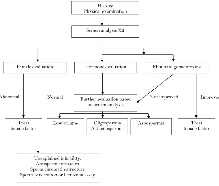
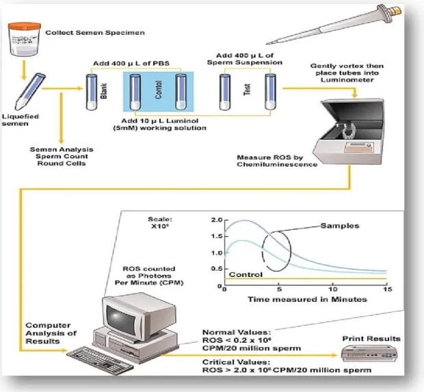
Figure 1.1: Male genital system ……… Figure 1.2: Hypothalamic-pituitary–gonadal (HPG) axis ...………..……… Figure 1.3: Seminiferous tubule ………. Figure 1.4: Spermatogenesis ………. Figure 1.5: Light and electron microscopic diagrams of human spermatozoon ... Figure 1.6: Diagram of a cross-section of the sperm axoneme ………... Figure 1.7: Diagrammatic representation of sperm developmental events………... Figure 1.8: World map containing percentages of infertility cases per region that are due to male factor………... Figure 1.9: General Algorithm for diagnostic evaluation of male infertility………. Figure 1.10: Immunobead test……….... Figure 1.11: Measurement of reactive oxygen species (ROS) in sperm
suspensions by chemiluminescence assay……… Figure 2.1: Factors contributing to oxidative stress-induced male infertility…………. Figure 2.2: Oxidants and antioxidants balance influencing overall sperm quality……..
Figure 2.3: General sources, mechanisms, and consequences of OS on male fertility………
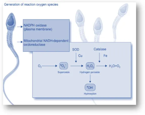
Figure 2.4: ROS Generation ……….. Figure 2.5: Antioxidant scavenging pathways of free radicals by superoxide dismutase (SOD) and catalase………... Figure 2.6 : Oxidative stress in male reproduction………. Figure 2.7: Biochemical pathway proposed to regulate sperm capacitation and
hyperactivation………. Figure 2.8: Biochemical pathway proposed to regulate the acrosome reaction (AR)……. Figure 2.9: The peroxidative process……… Figure 2.10: Selected natural and synthetic antioxidants……….. Figure 2.11: Reduction of nitroxide to hydroxylamine (1) and it’s oxidation to oxammonium cation (2) by single electron transfer ………. Figure 2.12: Nitroxide radical scavenging………..
5 6 8 9 12 12 15 17 26 35 37 40 41 42 43 44 45 50 51 56 58 58 59
Page | XVII Figure 2.13: Glutathione peroxidase (GPx) also scavenges hydrogen peroxide (H2O2),
along with glutathione (GSH), which becomes oxidized and is reduced/regenerated by glutathione reductase (GR) to allow further antioxidant function………... Figure 3.1: Number of people with diabetes worldwide and per region in 2017 and 2045 (20-79 years)………... Figure 3.2: Prevalence of people with diabetes by age and sex, 2017………. Figure 3.3: Schematic illustration of the metabolic cooperation between Sertoli and germ cells……….. Figure 3.4: Schematic representation of human sperm cell and localization of several glucose transporter isoforms in the distinct parts of the spermatozoon……….... Figure 5.1: Blood glucose levels of the different groups before and after diabetes induction ……….. Figure 5.2: Effect of diabetes mellitus and vitamins supplementation on the initial and final body weight of the adult male rats……… Figure 5.3: Effect of diabetes mellitus and vitamins supplementation on epididymis weight of adult male rats……….. Figure 5.4: Effect of diabetes mellitus and vitamins supplementation on testis weight of adult male rats………... Figure 5.5: Effect of diabetes mellitus and vitamins supplementation on testosterone blood levels of adult male rats………... Figure 5.6: Effect of diabetes mellitus and vitamins supplementation on FSH blood levels of adult male rats………... Figure 5.7: Effect of diabetes mellitus and vitamins supplementation on LH blood levels of adult male rats………... Figure 5.8: Effect of diabetes mellitus and vitamins supplementation on epididymal sperm number of adult male rats………... Figure 5.9: Paraffin section photograph……….. Figure 5.10: Effect of diabetes mellitus and vitamins supplementation on the Johnsen score………... 60 66 67 71 74 92 94 97 98 99 100 101 104 106 108
Page | XVIII
List of Tables
Table 1.1: Terminology in spermatogenesis………... Table 1.2: 2009 World Health Organization standard reference values for semen
characteristics……….. Table 3.1: Other specific types of diabetes……….. Table 4.1: Ingredients of standards commercial ruminant pellet………... Table 5.1: Blood glucose levels of wistar rats before diabetes induction, 5 days after
diabetes induction, and 4 weeks after treatment……..……….. Table 5.2: Body weight change results upon sacrifice wistar rats after treatment of
respective groups with vitamins C or/and E……… Table 5.3: Epididymal and testicular tissue weights results upon sacrifice wistar rats after treatment of respective groups with vitamins C or/and E…………... Table 5.4: Blood testosterone, LH, and FSH concentrations of male Wistar rats after
supplementation compared to control groups………... Table 5.5: Epididymal sperm count and testicular Johnsen score results of male
Wistar rats after treatment compared to control groups……….. 10 27 69 84 93 95 96 99 103
Page | XIX
List of Abbreviations and Acronyms
°C Degree Celsius
µl Microlitter
-1O2, Singlet Oxygen
4HNE 4-Hydroxynonenal
AGEs Advanced Glycated End Products
ALT Alanine Aminotransferase
AR Acrosome Reaction
ART Assisted Reproductive Technologies
ASA Antisperm Antibodies
ATP Adenosine triphosphate
BDE Bond Dissociation Enthalpy
BTB Blood–Testis Barrier
Ca2 + Calcium
cAMP Cyclic Adenosine 3',5'-Monophosphate
CASA Computer-Assisted Semen Analysis
CF Cystic Fibrosis
CFTR Cystic fibrosis transmembrane conductance regulator
cGy centigray
CPEMR Cell Phone-Generated Electromagnetic Radiation
CBAVD Congenital bilateral absence of the vas deferens CUAVD Congenital Unilateral Absence of the Vas Deferens
DAG Diacylglycerol
DHA Docosahexaenoic Acid
DM Diabetes mellitus
DNA Deoxyribonucleic acid
ELISA Enzyme-Linked Immunosorbent Assay
ERC Excess Residual Cytoplasm
FSH Follicle stimulating hormone
G6PD Glucose-6-Phosphate Dehydrogenase
GLUT Glucose Transporters
Page | XX
GLUT2 Glucose Transporter 2
GLUT3 Glucose Transporter 3
GLUT5 Glucose Transporter 5
GLUT8 Glucose Transporter 8
GLUT9a Glucose Transporter 9a
GLUT9b Glucose Transporter 9b
GnRH Gonadotrophine Releasing Hormone
GPx Glutathione Peroxidase GR Glutathione Reductase gr Gramme GSH Glutathione GU Genito-urinary Infections H2O2 Hydrogen Peroxide
HIV Human Immunodeficiency Virus
HOCL Hypochloric Acid
HPG hypothalamic-pituitary–gonadal
IBT Immunobead Test
ICSI Intracytoplasmic Sperm Injection
IP3 Inosital Triphosphate
IVF Fertilization In Vitro
KLK3 glycoprotein enzyme
LH luteinizing hormone
LHRH luteinizing hormone-releasing hormone
LOO Lipid Peroxyl
LOOH Lipid Peroxide
LPO Lipid Peroxidation
M Molar
MAR Mixed Antiglobulin Reaction
MCTs Membrane Transporters
MDA Malondialdehyde
min Minutes
mL Milliliter
Page | XXI ng/dL nanogramm/dicilitter nm Nanometers NO Nitric Oxide O2- Superoxide Ion O3 Ozone
OH- Hydroxyl Ion
OS Oxidative Stress
PFK Phosphofructokinase
pH potential Hydrogen
PIP2 Phosphatidylinosital-4,5-Biphosphate (
PKA Protein Kinase A
PLA2 Phospholipase A2
PSA Prostate-specific antigen
PTK Protein Tyrosine Kinase
PTPase Phosphotyrosine Phosphatase
PUFA Polyunsaturated Fatty Acids
RAGE Receptors for Advanced Glycated End Products
RO2 Peroxyl,
ROS Reactive Oxygen Species
SC Sertoli Cell
SGLT Sodium-Dependent Glucose Transporters
SMTP Sperm–cervical mucus penetration tests
SOD Superoxide Dismutase
SPA Sperm Penetration Assay
STZ Streptozotocin
T1D Type 1 Diabetes
T2D Type 2 Diabetes
TAC Total Antioxidant Capacity
TEMPO Tetramethylpiperidine-1-Oxyl3
TZI Teratozoospermia Index
WHO World Health Organization
Page | 1
Introduction
nfertility is an alarming health issue that concerns 48.5 million couples worldwide. In couple infertility, male infertility factors contribute themselves to 20-30% of cases and could then be responsible of 50% of overall cases (Agarwal et al., 2015). Previous research identified infertility causes from most to least common: sperm production issues, blockage of sperm transport, sperm antibodies, sexual issues (ejaculatory and erectile dysfunctions), endocrinology, idiopathic, and other causes (Parekattil and Agarwal, 2012). Over recent decades, with sophisticated analytical methods, several studies have shown a previously undetectable effect of diabetes on male fertility. In year 2017, the International Diabetes Federation (IDF) reported that 422 million adults were living with diabetes. The increasing diabetes incidence will result in an increased prevalence in men of reproductive age (IDF, 2017).
Diabetes mellitus (DM) constitutes a metabolic disorder of carbohydrates, lipids, and proteins, that induces pathology in various systems and tissues such as eyes, nerves, kidneys, and vessels as well (Basmatzou, 2016). Male reproductive functions could be affected, at multiple levels too, due to DM effects on the endocrine control of spermatogenesis, sperm production, erectile, and ejaculatory functions (Agbaje et al., 2007).
It is well known that glucose autoxidation, with increased oxidative stress, is the consequence of long term uncontrolled DM. This oxidative stress is extremely toxic for cells and may cause direct damages in proteins, lipids, and DNA, or affects normal cellular signaling, and gene regulating (Fernandes et al., 2011), as well as the alteration in antioxidant enzyme levels. In addition, spermatozoa are sensitive to reactive oxygen species (ROS) because of their elevated content of polyunsaturated fatty acids, and their incapacity to repair deoxyribonucleic acid (DNA) damage.
During twenty years, specialists have been foccusing in studying the treatment of infertility at low cost and high effectivness. They resorted to the use of antioxidants. These compounds, which have proven effective treatment of many cases of male infertility, are still
Page | 2 being studied and used to reduce oxidative stress, delaing infertility, caused by diabetes mellitus.
Because of the lack in data in this context, we carried out this study, aiming to assess the efficacy of supplementation with vitamin C and E, on the infertility of male diabetes patients, by conducting experiments on male Wistar rats, after diabetes induction by streptozotosin (STZ) injection, and assessing their sperm parameters, blood hormonal levels, and testis histological sections. We assessed the efficiency of these antioxidants either combined or separated.
Page | 4
Chapter 1
Male Infertility, Generalities
1.1 Sperm Physiology and Pathology………
1.2 Male Infertility………
5
Page | 5
Chapter 1
Male Infertility, Generalities
1.1 Sperm Physiology and Pathology
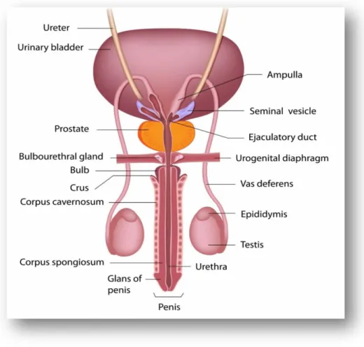
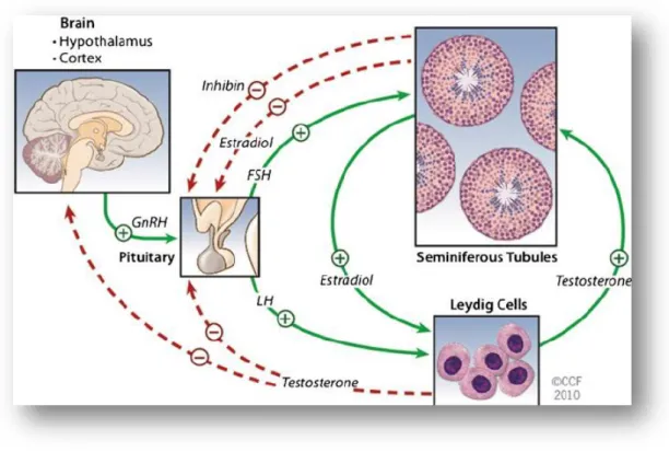
The male reproductive system is a complex and intricate system that produces spermatozoa or sex cells to carry the genetic material of the male. The components of the male reproductive system include both the external and internal sexual organs, as shown on Figure 1.1 and the hypothalamic-pituitary–gonadal (HPG) axis (Figure. 1.2).
Page | 6 The male reproductive system formed during the early embryonic development stages, becomes fertile during puberty and maintains the masculinity of adult male. The external genitalia include the scrotum, testes, and penis whereas the internal genitalia include the epididymis, seminal ducts, spermatic cords, seminal vesicles, ejaculatory ducts, bulbourethral or Cowper’s glands, and the prostate gland (Figure 1.1). The testes produce the male gametes (spermatozoa). The excurrent duct system matures, stores, and transports the gametes to the penis for expulsion, and the accessory glands produce and modify the contents of the semen (Durairajanayagam et al., 2015).
Figure 1.2: Hypothalamic-pituitary–gonadal (HPG) axis (Mohanty and Singh, 2017)
1.1.1
Spermatogenesis
Spermatogenesis is an extremely intricate process of cell differentiation, starting with germ cell (spermatogonia) development and culminating in the production of highly specialized spermatozoa. This process produces the genetic material required for species replication. Spermatogenesis occurs in the lumen of the seminiferous tubules. It was classically
Page | 7 believed that human spermatogenesis takes about 64 days in the testis (from spermatogonium to spermatid) with an additional 10–14 days in the epididymis for maturation of spermatozoa. Thus, the entire process takes about 70 ± 4 days to complete. However, a more recent report, published by Durairajanayagam et al., (2015), has suggested that the entire process from production to ejaculation of spermatozoa is completed within a shorter period: an average of 64 ± 8 days (with a range of 42–76 days). Spermatogenesis begins at puberty and occurs continually throughout the entire male adult life span in contrast to oogenesis, which is finite in women. The baseline number of precursor cells in the testes is regulated by Follicle Stimulating Hormone (FSH) (Durairajanayagam et al., 2015). Early in embryonic development, the gonocytes, which precede the spermatogonial germ cells formation, undergo active mitotic replication. Spermatogenesis involves a series of cellular events that start in the basal compartment and finishe in the apical compartment. The basal and the luminal compartments are kept separate by tight junctions. In the seminiferous tubules, the developing cells are arranged in a highly ordered sequence from the basement membrane toward the lumen (Figure 1.3) (Neto et al., 2016).
Page | 8 Figure 1.3: Seminiferous tubule. A cross section of the germinal epithelium in the seminiferous tubule. The germinal epithelium is divided by the Sertoli cell into two compartments, i.e., the basal and ad luminal compartments. Fully formed spermatozoa are
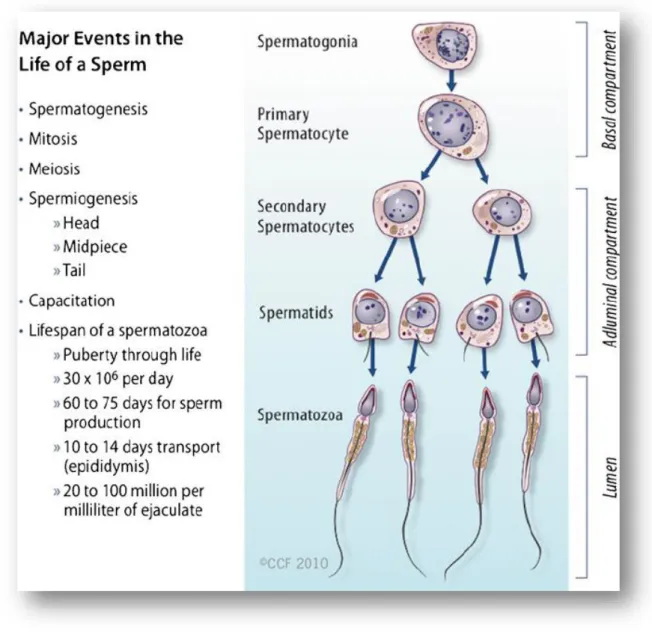
Page | 9 Spermatogenesis is a multistep process involving three major phases (mitosis, meiosis, and spermiogenesis), as well as other cellular events such as cells migration, differentiation, and apoptosis (Table 1.1). These events are highly regulated and the understanding of the molecular mechanisms that regulate the three spermatogenesis phases has been a major focus of study for decades (Figure 1.4) (Oliveira and Alves, 2015).
Figure 1.4: Spermatogenesis. Major events in the life of a sperm involving spermatogenesis, spermiogenesis, and spermiation (Durairajanayagam et al., 2015)
Page | 10 Table 1.1: Terminology in spermatogenesis (Oliveira and Alves, 2015)
Process Description
Spermatogoniogenesis Process of producing spermatogonia through multiple mitoses to amass a large population of stem cells, most of which undergo meiosis to produce spermatozoa
Spermatogenesis Process of differentiation of a spermatogonium into a spermatid Purpose: to produce (via mitosis and meiosis) the necessary genetic material for species replication
Spermatocytogenesis Process of producing spermatocytes that occurs in the basal compartment of the seminiferous tubules
Spermiogenesis A complex metamorphosis that transforms round spermatids (from the final division of meiosis) into a complex structure spermatozoon
Spermiation Process whereby a mature spermatid frees itself from the Sertoli cell and enters the tubular lumen
1.1.2 Sperm Structure
A normal human spermatozoon is 55 to 70 µm in length and has three main structural regions: the head, midpiece, and tail (flagellum). The principal function of the sperm head is the contribution of its haploid set of chromosomes to the oocyte at fertilization, whereas the midpiece and tail provide the motility essential for the spermatozoon to reach the fertilization site (Figure 1.5).
The sperm head contains the cell’s nuclear DNA, when the chromatin is heavily condensed, and its protamines are highly cross-linked so that the sperm nucleus is stabilized and effectively inactivated upon fertilization. This makes the head of the spermatozoon inflexible, which assists in penetration of the oocyte’s zona pellucida during fertilization. The anterior half to two-thirds of the sperm head is covered by the acrosome, the membrane-bound structure that originated from the Golgi complex during spermiogenesis (Patton and Battaglia, 2005).
Page | 11 The acrosome contains a number of hydrolytic enzymes, such as hyaluronidase and acrosin, which are required for fertilization. During fertilization, the acrosomal membrane fuses with the oocyte plasma membrane oocyte at numerous sites. This is followed by the acrosome reaction, an event characterized by acrosomal enzyme released from the head tip.
Among the common abnormalities of the sperm head are defective shape or size and the presence of numerous vacuoles (> 20 %) within the head surface. Shape defects include large, small, tapering, pyriform, amorphous, double heads, and various other combinations
The tail measures 40–50 μm in length (nearly ten times the length of the head) and provides motility for the cell. The sperm cell’s entire motility apparatus is contained in the tail, propelling the sperm body via waves generated in the neck region that pass along distally in a whiplash manner. The tail can be divided into: the midpiece (anterior portion); principal piece; and endpiece (posterior portion) (Durairajanayagam et al., 2015).
The sperm midpiece contains the mitochondria that generate energy via oxidative phosphorylation; the centriole, used by the fertilized oocyte in its first cell division; and the beginnings of axoneme, the motility apparatus. The mitochondria are arranged helically around the proximal part of the axoneme, and they supply the adenosine triphosphate (ATP) necessary for flagellar motility (Patton and Battaglia, 2005).
Ideally, the midpiece supports the head at exactly the center position. It should be slender as well (maximum width of 1 µm), yet thicker than the rest of the tail and between 7.0 and 8.0 µm in length. The tail diameter should be between 0.4 and 0.5 µm, measuring about 50 µm in length. The tail should have a well-defined endpiece, without any coiling or abnormal bending (over 90°). Throughout its length, the flagellum shows specific modifications. A central axoneme consists of nine microtubular doublets circularly arranged to form a cylinder around a central pair of single microtubules (Figure 1.6). In the middle piece, the axoneme is surrounded by an outer cylinder of nine outer dense fibers arranged as nine rod-like structures that run parallel to the axoneme; they play a role in protecting sperm against shearing forces, defining waveform and generating motility (Carrell, 2010). Motility plays a key role in sperm transport through the cervix; the sperm cells need to maintain motility despite being suspended in fluid secreted by the female reproductive organs. Moreover, motility is required
Page | 12 to avoid phagocytosis by polymorphonucleocytes found in female body fluid (Durairajanayagam et al., 2015).
Figure 1.5: Light and electron microscopic diagrams of human spermatozoon (Oehninger and Kruger, 2007)
Figure 1.6: Diagram of a cross-section of the sperm axoneme (Patton and Battaglia, 2005)
Page | 13
1.1.3 Sperm Function and Physiology
After production in the testes, immature spermatozoa are moved through the corpus and caput regions of the epididyma and are then stored in the proximal section of the cauda epididymis. The spermatozoa move a total of 6 m through the reproductive tract before leaving the urethra. The average transit time in the epididymis is estimated at 12 days. Sperm motion is driven by hydrostatic pressure that is created by the combination of fluids secreted by the seminiferous tubules and tubular peristalsis. Movement through the proximal epididymis is mediated by peripheral smooth muscle contractions, and movement throughout the epididymal head is mediated by contraction of the tunica albuginea. Fluidic rhythmic movements of the cilia, lining the walls of the ducts, and the cyclic contractions of contractile cells along the wall of the epididymal duct further propel seminal constituents. Epididymal duct contraction is believed to be regulated by cholinergic, adrenergic factors, and vasopressin (Mohanty and Singh, 2017).
The spermatozoa mature during epididymal transit and storage and acquire functional competence. The most obvious maturational change in spermatozoa is the motility acquisition when in contact with seminal plasma or physiological culture media - a process referred to as “activation.” Other changes occuring during epididymal maturation of spermatozoa are alterations of the plasma membrane, chromatin condensation and stabilization, and possibly some final modifications to the shape of the acrosome (Patton and Battaglia, 2005). These changes are induced by the fluid medium, secretions of the epithelium of the seminiferous tubules and epididymal lumen that the cells are exposed to. This epididymal fluid contains many substances such as sodium, glutamate, albumin, bicarbonate, transferrin, immobulin, inositol, potassium, L-carnitine, sialic acid, lactate, metalloproteins, proenkephalin, taurine, clusterin, glycerophosphorylcholine, and chloride. These substances are in part responsible for epididymal cell metabolism, activation of sperm motility, and regulation of fluid retention of sperm and epididymal cells (Mohanty and Singh, 2017).
At ejaculation, sperm are transported from their storage site and are mixed with prostatic fluid and seminal vesicle fluid before passage along the penile urethra. The first fraction of the ejaculate contains most of the spermatozoa, suspended in epididymal and prostatic fluid, whereas subsequent fractions contain both prostatic and vesicular fluid (Patton and Battaglia, 2005).
Page | 14 It is commonly stated that, the seminal plasma provides a nutritive and protective medium for the spermatozoa during their journey through the female reproductive tract. The main constituents of semen are listed below along with their functions:
- Water: fluid mechanism of transport for sperm; - Buffers: protect sperm in acidic vaginal environment,
- Nutrients: fructose, carnitine, vitamin C, and citric acid provide nourishment for spermatozoa;
- Mucus: acts as lubricant for sexual intercourse; - Spermatozoa: oocyte fertilization;
- Enzymes: semen clotting in vagina and further liquefaction of clot. Prostate-specific antigen (PSA) is a glycoprotein enzyme encoded by the KLK3 gene produced by the prostate for semen liquefaction and dissolving of the cervical mucus for sperm penetration;
- Prostaglandins: stimulate smooth muscle contraction and transport through both reproductive tracts and improve sperm motility;
- Immunity particles: lysozyme, immunoglobulin, and leukocytes act as antibacterial agents and wash out the urethra; zinc acts as antioxidant;
- Other cells: genitourinary tract epithelial cells and immature germ cells (Mohanty and Singh, 2017).
During intercourse, the spermatozoa are deposited into the vagina, near the cervical os, and must swim through the cervical mucus, accross the uterus, enter the oviduct, and reach the oocyte in its ampullary portion for fertilization to occur (Patton and Battaglia, 2005).
As with epididymal maturation, capacitation is required too before fertilization can occur. Capacitation takes place after ejaculation into the female reproductive tract. During capacitation, spermatozoa undergo a biochemical changes sequence that ultimately enable them to fertilize an ovum. The sperm plasmalemma is reorganized to support the subsequent acrosome reaction; seminal plasma factors are removed and modifications are made to the sperm membrane, sterols, lipids, glycoproteins, outer acrosomal membrane, and surface charge. The concentration of intracellular free Ca2+ increases as well. In particular, it is the
removal of cholesterol from the surface membrane that allows for the acrosome reaction to occur (Durairajanayagam et al., 2015). In addition, D-mannose binding lectins are involved
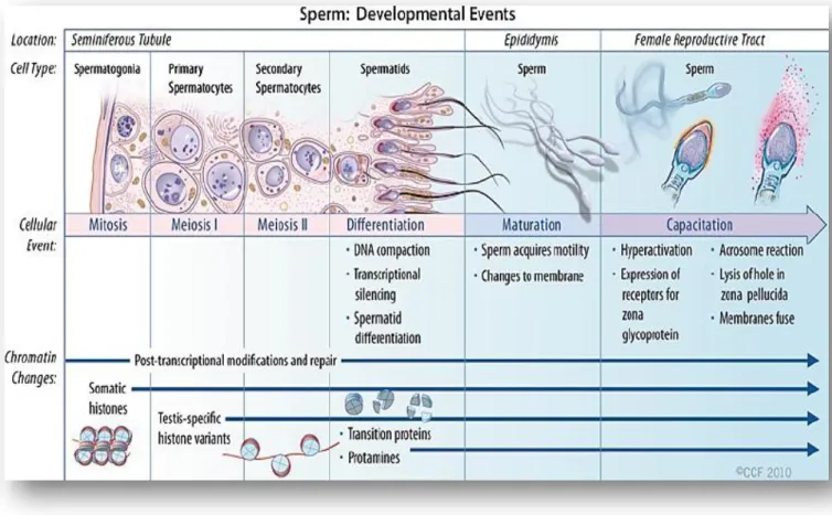
Page | 15 too in the binding of human sperm to the zona pellucida. Thus, all these series of changes are necessary to transform the stem cells into fully mature, functional spermatozoa equipped to fertilize an egg (Figure 1.7) (Zini and Agarwal, 2011).
Figure 1.7: Diagrammatic representation of sperm developmental events. Changes that occur during the development of a germ cell into a spermatozoon leading to its release and
subsequent maturation and storage in the epididymis, prior to its journey into the female reproductive tract (Zini and Agarwal, 2011)
1.1.4 Pathophysiology of Sperm and Infertility
The complexity of sperm structure and function means that it can be difficult to determine the pathophysiological reasons a man’s infertility. If the developmental or maturational processes are disturbed, this could result in issues, such:
Page | 16 - Low sperm concentration (from inefficient spermatogenesis);
- Poor sperm motility (caused by midpiece or axonemal abnormalities); - Abnormal sperm morphology (owing to errors in spermiogenesis).
This list is just illustrative, it could be extended to take in every step of each process, from the activation of spermatogonium to fertilization, and even further, because embryo development is affected by the quality of the nuclear DNA of fertilizing spermatozoon (Patton and Battaglia, 2005).
1.2 Male Infertility
1.2.1 Definition
Infertility is defined as failure to conceive after 12 or more months of regular unprotected intercourse. A comprehensive male infertility assessment should be pursued sooner than a year if there is a previous infertility history, a known risk factor, advanced female age or a specific request by the couple (Sabanegh, 2011).
It has been recorded that 48.5 million couples that have unprotected intercourse suffer from infertility worldwide. However, this statistic does not clearly define infertility by geographic region. In general, 50% of infertility cases are due to a solely female factor, pure male factor accounts for 20-30% of the problem, and the remaining 20-30% is due to a combination of both male and female factors.
At least 30 million men worldwide are infertile with the highest rates in Africa and Eastern Europe as illustrated on Figure 1.8 (Agarwal et al., 2015).
Page | 17 Figure 1.8: World map containing percentages of infertility cases per region that are due to
male factor (Agarwal et al., 2015)
The figure above demonstrates rates of infertility cases in each region studied (North America, Latin America, Africa, Europe, Central/Eastern Europe, Middle East, Asia, and Oceania) due to male factor involvement (Agarwal et al., 2015).
1.2.2 Etiology
1.2.2.1 Pre-testicular
a.
Hypo gonadotropic hypogonadismHypo gonadotropic hypogonadism affects fertility at multiple levels. Sperm production is deleteriously affected via a lack of testosterone and a lack of a stimulatory effect on the Sertoli/germ cell complex. Sexual function is negatively impacted with effects seen at the level of erectile function, ejaculatory function, and sexual desire too. There are several hypo gonadotropic hypogonadism etiologies. The most common are elevated prolactin,
Page | 18 medications, illicit drugs, and pituitary damage. Kallmann syndrome is another, albeit rare, cause of hypo gonadotropic hypogonadism (Parekattil and Agarwal, 2012).
b. Elevated prolactin
Elevated prolactin may cause hypogonadism by suppressing the release of GnRH. Symptoms of hypogonadism, especially erectile dysfunction and loss of libido, are the most common presenting symptoms in males with hyper-prolactinemia, though galactorrhea and gynecomastia may also be evident. Elevated prolactin may be secondary to various etiologies. The most common is a prolactinoma, (Carrell, 2010), renal failure, hypothyroidism and cirrhosis. Prolactin levels may be elevated too in certain systemic diseases such as systemic lupus erythematous, rheumatoid arthritis, celiac disease, and systemic sclerosis. Many drugs elevate prolactin levels, especially those which block the effects of dopamine, such as antipsychotics (Plessis, 2014).
c. Pharmacologic
Various medications may cause hypo gonadotropic hypogonadism. Estrogens and progestin may cause a decrease in testosterone levels via negative feedback to the hypothalamic-pituitary-gonadal axis. Marijuana is known to decrease testosterone levels by working on the endocannabinoid receptors present at multiple levels of the hypothalamic-pituitary axis. Both ethanol and cannabinoids suppress GnRH secretion at the level of the hypothalamus. Endocannabinoid receptors have also been found in the pituitary and may affect the hypothalamic-pituitary axis at that level as well. LHRH agonists and antagonists are used for the treatment of prostate cancer, precocious puberty, and gender reassignment surgeries (Carrell, 2010). In the male, both induce deep hypogonadism. LHRH antagonists directly and intuitively decrease LH and FSH levels. LHRH agonists produce a tonically stimulated state, which, unlike the physiologic circadian rhythmicity of normal LHRH stimulation, acts to decrease LH and FSH secretion. Narcotics may also produce profound hypogonadism. Nearly 40% of men using methadone were found to have total testosterone levels less than 230 ng/dL (Esteves and Agarwal, 2011).
Page | 19 d. Kallmann Syndrome
Kallmann Syndrome affects between one in 8,000–10,000 males. It is a spectrum of disease in which the primary manifestations are anosmia and hypo gonadotropic hypogonadism that leads to an absence of puberty (Plessis, 2014). Multiple genetic disorders can lead to Kallmann Syndrome. These most disorders commonly manifest through the same mechanism whereby GnRH secreting neurons fail to migrate to the hypothalamus. Lack of these neurons in the hypothalamus results in a lack of GnRH secretion and thus hypogonadism (Parekattil and Agarwal, 2012).
e. Hyper gonadotropic hypogonadism
One of the most common causes of hyper gonadotropic hypogonadism is Klinefelter Syndrome (Klinefelter’s). Klinefelter’s affects male fertility by altering spermatogenesis both directly and indirectly by altering the hormonal midium. Interestingly, sex hormone levels are normal until puberty. During puberty, they do rise to low-normal levels, however, by adulthood, serum testosterone levels are typically below normal. Histologic studies demonstrate gradual degeneration of the testes during development with hyperplasia of poorly functioning Leydig cells (Carrell, 2010).
1.2.2.2 Testicular etiologies
a.
VaricoceleA varicocele consists of a pampiniform plexus dilation likely caused by the absence or incompetence of the venous valves at the level of the internal spermatic vein. Varicoceles have long been associated with infertility. It affects multiple semen parameters; total sperm count, sperm motility, and sperm morphology that are all negatively affected. There are several theories about the underlying pathophysiology of a varicocele, with heat, renal metabolites, and hormonal abnormalities all playing a role. However, most agree that disruption of the countercurrent heat exchange mechanism in the testis, causing hyperthermia, is the most likely mechanism. The mechanism by which heat causes decreased sperm counts is poorly understood. However, one hypothesis is that high temperatures could increase the metabolic rate of testicular and epididymal sperm. Furthermore it could increase the oxidative damage
Page | 20 amount to both the structure and the DNA of the spermatocytes and spermatids ( Parekattil and Agarwal, 2012).
b. Cryptorchidism
Cryptorchidism is well known to affect fertility too, with bilateral cryptorchidism having more severe effects than unilateral, and with higher testes having worse function than lower testes. The pathophysiology is complex, with heat likely playing a partial but significant role. Other factors are implicated too, including the underlying genetics, hormonal status, and environmental exposures, that originally lead to the cryptorchidism (Ono and Sofikitis, 1997).
c. Testicular cancer
It has been proved that testicular cancer is strongly associated with infertility. There are multiple ways in which testicular cancer is related to and can contribute to reduce fertility. Both testicular cancer and impaired spermatogenesis may be related in their etiology of embryologic testicular dysgenesis. The testicular dysgenesis syndrome is a spectrum of disease that may involve cryptorchidism, hypospadias, decreased spermatogenesis, and testis cancer. In this syndrome, it is thought that all of these share an origin of abnormal fetal testis development. As a result of this abnormal development, any number of these symptoms may be present in a boy (Menon et al., 2008). Testicular tumors may directly contribute to infertility too by secreting hormones, which can down regulate sperm production in the contralateral testis. This is uncommon and has been seen with Leydig and Sertoli cell tumors as well as seminomas. Tumors may directly disrupt spermatogenesis by mass effect or by the effects of the inflammatory reaction to the tumor (Esteves and Agarwal, 2011). Hence, cancer treatments may decrease fertility (Kacem et al., 2014).
d. Ionizing radiation
Excellent data on the effects of ionizing radiation is available from two similar studies, which are unlikely to be repeated. Researchers in these studies prospectively irradiated the testes of prisoners with single or multiple doses of radiation up to 600 cGy. Sperm counts were followed, and serial testicular biopsies were performed. These studies showed that sperm counts declined when testes were irradiated and that decline was dose dependant. At low doses
Page | 21 of ~7.5 cGy, a mild decline of sperm counts was observed, and this decline increased to severe oligospermia by 30–40 cGy and azoospermia by 78 cGy. The time to recovery was seen to be dose-dependent too, with those receiving 20 cGy starting to develop a recovery of sperm counts by 6 months, those with 100 cGy at 7 months, 200 cGy at 11 months, and 600 cGy at 24 months. The percentage of men achieving a complete recovery even in time declined with increasing radiation doses (Rowley et al., 1974).
e. Chemotherapy
Chemotherapy typically targets rapidly dividing cells and thus has severe effects on the germinal epithelium. As such, the expected outcome of acute chemotherapy is a decline in spermatogenesis. BEP: Bleomycin, etoposide, and cisplatin or carboplatin, constitutes the most commonly used chemotherapy regimen for testicular germ cell tumors. The decrease in fertility seen post-BEP chemotherapy is likely the result of a direct reduction of spermatogenesis and not as a result of any change in the hormonal midium. Indeed, testosterone levels are not seen to be significantly reduced at 12 months’ post-chemotherapy, and FSH levels are appropriately elevated. FSH levels decline as spermatogenesis returns over the following 2–4 years. It should be noticed, however, that return of spermatogenesis is not guaranteed (Parekattil and Agarwal, 2012).
f. Genetic azoospermia/Oligospermia
It is estimated that 2–8% of infertile men have an underlying genetic abnormality, with this number rising to 15% in azoospermic men. Although the majority of male infertility does not have an identifiable genetic cause, two potential etiologies are Y chromosome micro deletions and karyotypic abnormalities. The two most common karyotypic abnormalities are Klinefelter’s (47, XXY) and chromosomal translocations. Robertsonian translocations represent a third significant genetic cause of infertility. They occur in 0.8% of infertile men, and this number rises to 1.6% in oligospermic individuals (Visser and Repping, 2009).
g. Environmental factors
Hyperthermia is considered to be a major contributor in the pathogenesis of infertility in men with varicocele and cryptorchidism. Many lifestyle factors have the potential to increase scrotal temperatures too, including underwear type, heated car seats, and occupational heat
Page | 22 exposure (Jung and Schuppe, 2007). Recently, cell phones have been implicated as possibly playing a role in decreasing male fertility. The mechanism by which cell phones affect semen parameters has not yet been elucidated, but one hypothesis is that cell phone-generated electromagnetic radiation (CPEMR) alters mitochondrial function and acts to increase reactive oxygen species (ROS) (Parekattil and Agarwal, 2012). Tobacco consumption has been implicated in the pathogenesis of numerous cancers and medical diseases. While this consumption significantly impacts female fertility comparing to male for whom the effect is less clear. Semen parameters, including sperm density, motility, and morphology, have all been shown to be worsened with tobacco use (Künzle et al., 2003; Collodel et al., 2009). However, a significant reduction in fertility has not yet been proven.
h. DNA damage
There are numerous etiologies of sperm DNA damage. Radiation, toxins, genital tract inflammation, varicocele, advanced paternal age, and testicular hyperthermia, all induce significant DNA damage (Belloc et al., 2009; Hammiche et al., 2010).
1.2.2.3 Post-testicular
a. Absence of the vas deferens
Congenital Bilateral Absence of the Vas Deferens (CBAVD) consitutes a condition strongly related to cystic fibrosis (CF) and has even been considered as a diagnostic criterion for CF.
Congenital unilateral absence of the vas deferens (CUAVD) is a different entity altogether (Donohue, 1989). While there is still a significant rate of CFTR mutations in men with CUAVD, especially when the obstructive azoospermia is present (Lissens et al., 1996). The bulk of CUAVD is the result of an embryologic Wolffian duct aberrancy (Shapiro et al., 2003). Since CUAVD not associated with a CFTR mutation is usually a unilateral and isolated phenomenon, fertility is often preserved (Parekattil and Agarwal, 2012).
Page | 23 b. Young’s Syndrome
Young’s Syndrome is a rare disorder, which presents clinically as obstructive azoospermia and chronic sinopulmonary infections (Handelsman et al., 1984). Thus, it could be difficult to differentiate clinically from cystic fibrosis variants and primary ciliary dyskinesia. Indeed, definitive diagnosis of Young’s Syndrome requires negative CFTR genetic testing as well as investigation of ciliary ultrastructure to rule out primary ciliary dyskinesia. Normal spermatogenesis is seen, and the obstructive azoospermia is due to intensified secretions in the vas deferens.
The etiology of Young’s Syndrome is unclear with childhood mercury exposure having been postulated to play a role in the past (Goeminne and Dupont, 2010).
c. EjDO/Seminal vesicle dysfunction
Ejaculatory duct obstruction is a common etiology of male infertility, occurring in 1–5% of men presenting with infertility (Smith et al., 2008). There are many causes of ejaculatory duct obstruction, including cystic fibrosis spectrum disease, Wolffian or Muellerian origin cysts, calcifications, tuberculosis and other genital urinary infections, calculi, and urinary tract instrumentation (Carson, 1984; Paick et al., 2001).
d. Vasectomy and vasectomy reversal
Vasectomy is a procedure that is intended to produce infertility, and it is successful in over 90% of cases (Labrecque et al., 2002). Some of the key determinants of success are related to aspects of surgical technique. The manner of ligating the ends, non- ligation versus clipping versus suture ligation, length of vas removed, as well as whether to fold vas ends are all controversial (Hallan and May, 1988; Adams and Wald, 2009). Two maneuvers which do seem to provide significant benefits are luminal cauterization and fascial interposition (Cook et al., 2007; Sokal and Labrecque, 2009). Reversal vasectomy may be performed in an attempt to return fertility to the sterilized man. The outcomes of reversal vasectomy are dependent on a number of factors. Surgical technique is a one, with the use of a microscope significantly improving pregnancy rates over loupe-assisted vasovasostomy (Jee and Hong, 2010). Time elapsed since fertility also plays a significant role with a 97% patency rate and
Page | 24 76% pregnancy rate being achieved if surgery is performed at less than 3 years since vasectomy (Belker et al., 1991).
e. Nerve injury
Nervous injury, affecting ejaculation, may occur at many levels and have a diverse etiology ranging from spinal cord injury to neural damage during retroperitoneal or pelvic surgery to neuropathy from systemic diseases. Ejaculatory dysfunction is present in 90% of spinal cord injury patients. The type and severity of ejaculatory dysfunction are dependent on the level and extent of the injury (Parekattil and Agarwal, 2012).
f. Medications
Medications could affect ejaculation process by altering adrenergic signaling. This is most clearly seen with alpha-1 antagonists. Recent studies have shown that the ejaculatory dysfunction induced by alpha-1 antagonists is actually a failure of emission (Hisasue et al., 2006; Kobayashi et al., 2008).
Antipsychotics have long been associated with sexual dysfunction, including ejaculatory dysfunction. Antipsychotics have effects on many different neurotransmitters including dopamine, norepinephrine, acetylcholine, and serotonin. Predictably, altered ejaculatory function with antipsychotics use correlates with anti-adrenergic actions of the antipsychotics (Smith et al., 2002).
g. Prostate resection
Surgery of the prostate is well known to cause retrograde ejaculation. Transurethral resection of the prostate as well as the laser photovaporization and enucleation all have a high likelihood of inducing retrograde ejaculation since removal of the proximal prostatic urethra severely diminishes the resistance to back flow of semen (Parekattil and Agarwal, 2012).
h. Coital
Abnormal coital practices may play a role in infertility when they interfere with semen deposition in the vagina or affect their timing with the female reproductive cycle. Similarly,