HAL Id: tel-03188139
https://tel.archives-ouvertes.fr/tel-03188139
Submitted on 1 Apr 2021
HAL is a multi-disciplinary open access
archive for the deposit and dissemination of sci-entific research documents, whether they are pub-lished or not. The documents may come from teaching and research institutions in France or abroad, or from public or private research centers.
L’archive ouverte pluridisciplinaire HAL, est destinée au dépôt et à la diffusion de documents scientifiques de niveau recherche, publiés ou non, émanant des établissements d’enseignement et de recherche français ou étrangers, des laboratoires publics ou privés.
Aptamer biosensor development for small molecule
detection
Lorena Zara
To cite this version:
Lorena Zara. Aptamer biosensor development for small molecule detection. Biomolecules [q-bio.BM]. Université Grenoble Alpes [2020-..], 2020. English. �NNT : 2020GRALV033�. �tel-03188139�
THÈSE
Pour obtenir le grade de
DOCTEUR DE L’UNIVERSITE GRENOBLE ALPES
Spécialité : Biologie Structurale et nanobiologie
Arrêté ministériel : 25 mai 2016
Présentée par
Lorena Zara
Thèse dirigée par Corinne Ravelet, Maître de Conférences,
Université Grenoble Alpes
Co encadrée par Jean-Jacques Toulmé, Directeur de recherche
émérite INSERM et Directeur Général Novaptech
préparée au sein du Département de Pharmacochimie
Moléculaire
dans l'École Doctorale Chimie et Sciences de Vivant
Développement de biocapteurs à
base d’aptamères pour la
détection de petites molécules
Thèse soutenue publiquement le 15 décembre 2020, devant le jury composé de :
Madame Claire DEMESMAY- GUILHIN
Professeur, Université Lyon I, Rapporteur
Monsieur Thierry NOGUER
Professeur, Université de Perpignan, Rapporteur
Monsieur Michael RYCKELYNCK
Professeur, Université de Strasbourg, Examinateur
Monsieur Eric DEFRANCQ
Professeur, Université Grenoble Alpes, Président
Madame Corinne RAVELET
Maître de Conférences, Université Grenoble Alpes, Directrice de Thèse
Monsieur Jean-Jacques TOULME
Directeur Général Novaptech, Bordeaux, Tuteur entreprise
Monsieur Eric DAUSSE
Ingénieur de Recherche INSERM, Université de Bordeaux, Membre invité
Monsieur Eric PEYRIN
1 Al mio amico geniale,
2
Acknowledgements
Je souhaiterais, tout d’abord, transmettre mes remerciements aux membres du jury de cette thèse, les professeurs Claire Demesmay-Guilhin et Thierry Noguer qui ont consacré une partie de leur temps à l’évaluation de ce travail et qui m’ont fait l’honneur d’avoir accepté́ la fonction de rapporteur. Je tiens également à remercier le professeur
Michael Ryckelynck pour avoir accepté d’examiner mon manuscrit, ainsi que le
professeur Eric Defrancq, qui a bien voulu présider le jury de ce doctorat.
Les travaux présentés dans ce manuscrit ont fait l'objet d'une convention CIFRE entre la
société́́ Novaptech de Bordeaux et le Département de Pharmacochimie Moléculaire
(DPM) de l’Université Grenoble Alpes. Je remercie cet organisme pour mon financement.
J’adresse toute ma gratitude à madame Marine Faussillon-Laville présidente de Novaptech et monsieur Jean-Jacques Toulmé directeur général ainsi que co-encadrant de ma thèse, sans qui cette convention CIFRE n'aurait jamais vu le jour. Je tiens également à les remercier pour la confiance qu’ils m’ont accordée en acceptant de co-financer ce projet.
Je remercie tout particulièrement ma directrice de thèse, madame Corinne Ravelet pour sa présence constante, ses conseils et pour toutes les heures qu'elle a consacré́ à diriger cette recherche. Je n’oublierai pas sa disponibilité dans la relecture des documents que je lui ai adressés. Enfin, j'aimerais également lui dire à quel point j’ai apprécié ses qualités, son soutien et sa compréhension, qui m’ont profondément aidé tout au long de ces dernières années.
Je tiens également à remercier monsieur le professeur Eric Peyrin pour son accueil à chaque fois que j'ai sollicité son aide, ainsi que pour ses critiques et conseils constructifs. Merci aussi pour sa bonne humeur quotidienne.
Je remercie toute l’équipe NOVA, en particulier madame Emmanuelle Fiore pour ses conseils et sa gentillesse. Ainsi, je souhaiterai également remercier tous les membres du DPM pour leur accueil chaleureux et leur soutien surtout pour la fin de la rédaction. Merci à Mathieu, Brayan, Pierre et à Silvia qui ont changé ma vie grenobloise.
Je remercie mes collègues de l’équipe Novaptech Laure, Raouia, Cynthia et Ellia pour leur aide, malgré la distance. Elles ont toujours été là, chaque fois que j’en ai eu besoin. Je les remercie aussi pour leurs échanges constructifs, leur soutien et pour avoir rendu agréable le travail au sein du laboratoire, à Bordeaux.
Un grand merci à Arghya Sett, pour son aide dans nos projets communs, je n’oublierai pas sa positivité, sa curiosité et l’amitié qui s’est créé entre nous pendant ces années. En
3 outre, je tiens à transmettre toute ma gratitude à monsieur Eric Dausse qui a été mon mentor et qui m’a apporté son soutien sans faille.
Enfin, je tiens à remercier tous mes amis de Bordeaux qui ont toujours cru dans mes projets et mes plus chers amis italiens qui m’ont donné la détermination, le support psychologique et l’enthousiasme pour persévérer dans mes recherches.
Pour conclure, je tiens à remercier ma famille qui a toujours accepté mes décisions, la distance et qui m’a fortement encouragée à atteindre mes objectifs.
4
Table of Contents
Abbreviations ... 5 List of Figures ... 7 List of Tables ... 8 Introduction ... 9 Bibliographic review ... 131. Small molecule detection ... 14
1.1. Aptamers (origins, structures, affinity and specificity) and their properties ... 15
1.2. Aptamers versus antibodies ... 17
1.3. SELEX for small molecules ... 19
1.4. Determination of binding affinity ... 24
2. Aptasensors and strategies for small molecules focused on pesticides ... 26
2.1 Strategies for the construction of aptamer-based biosensors ... 28
2.2 Aptasensors for pesticides ... 36
2.3 Light up aptasensors ... 40
3. Technologies used in the present work ... 43
3.1 Fluorescence anisotropy ... 44
3.2 Isothermal Titration Calorimetry (ITC) ... 46
Experimental Section... 51
1. Objectives of thesis ... 52
2. « Engineering light-up aptamers for the detection of RNA hairpins through kissing interaction. » ... 54
2.1 Strategy ... 54
2.2 Conclusions ... 70
3. « A malachite green light-up aptasensor for the detection of theophylline » ... 71
3.1 Strategy ... 71
3.2 Conclusions ... 92
4. « Anti-Pesticide DNA Aptamers Fail to Recognize their Targets with Reported Micromolar Dissociation Constants» ... 93
4.1 Strategy ... 93
4.2 Conclusions ... 119
Conclusions and perspectives ...120
5
Abbreviations
DNA Deoxyribonucleic acid GO-SELEX Graphene oxide-SELEX
HPLC High-performance liquid chromatography LC Liquid chromatography
MS Mass spectrometry Kd Dissociation constant PCR Polymerase Chain Reaction RNA Ribonucleic acid
SELEX Systematic Evolution of Ligands by Exponential enrichment ss single-stranded
UV Ultraviolet AFB1 Aflatoxin B1 AuNPs Gold nanoparticles BHQ1 Back hole quencher 1 CDs Carbon dots
D Dabcyl FAM/F Fluorescein
FRET Fluorescence resonance energy transfer
Au/MWCNT-rGONR multiwalled carbon nanotube-reduced graphene oxide nanoribbon HIV Human immunodeficiency virus
IFE Inner filter effect QDs Quantum dots
SPR Surface Plasmon Resonnance UCNPs Up-conversion nanoparticles
EIS Electrochemical impedance spectroscopy CV Cycle voltammetry
Hemin-rGO Hemin- functionalized reduced graphene oxide TAR Trans-activation response element
QCM Quartz crystal microbalance TO1 Thiazole orange 1
6 MGA Malachite Green Aptamer
TMR Tetramethylrosamine
NMR Nuclear magnetic resonance FP Fluorescence polarization Ka association constant
(FP-DA) Fluorescent polarization Displacement assay Msw Malaswitch
7
List of Figures
Fig. 1. Important small species targets. 14
Fig. 2. Aptamer structural motifs. 16
Fig. 3. Size comparison between an antibody (human IgG) and an aptamer (anti-thrombin
DNA aptamer). 18
Fig. 4. SELEX process for DNA. 20
Fig. 5. SELEX process for small molecules. 22
Fig. 6. Schematic illustration of a biosensor. 27
Fig. 7. General overview of aptasensing strategies. 29
Fig. 8. Schematic representation of the optimised signalling aptamers. 30
Fig. 9. Example of double-end labelled aptasensors. 31
Fig. 10. Example of a split aptasensor before and after the addition of dopamine. 32
Fig. 11. Example of a strand-displacement strategy. 34
Fig.12. Example of kissing complex. 35
Fig. 13. General overview of aptasensors for pesticides. 37
Fig. 14. Light up aptamer. 41
Fig. 15. Predicted structures of MGA and their targets. 42
Fig. 16. Allosteric light-up aptamer 43
Fig. 17. Schematic representation of fluorescence anisotropy (polarization) 44
Fig. 18. Representation of depolarized and polarized emission. 45
Fig. 19. Isothermal titration calorimeter. Example of results obtained by ITC. 48
Fig. 20. Illustration of the c-value on the shape of the binding isotherm. 49
Fig. 21. Schematic illustration of the phases of the project. 52
Fig. 22. Representation of the fluorescent kissing-complexes design. 54
Fig. 23. Representation of the fluorescent double-switch complex design. 71
Fig. 24. Secondary structure of theophylline-aptamer and chemical structure of
theophylline and caffeine. 72
Fig. 25. Secondary structure of theophylline-aptamer and chemical structure of
8
List of Tables
Table 1. Comparison of aptamers and antibodies. 19
Table 2. Some examples of aptamers selected with GO-SELEX and Capture SELEX. 24 Table 3. Summary of the aptasensor strategies for pesticide detection. 40
9
10 The need to detect small molecules (<1000 Daltons)1such as residues of pesticides or drugs, toxins, antibiotics or illegal drugs becomes even more important for protecting human health and the surrounding environment. It is also important to detect these molecules with sensitive and rapid assays, under environmental conditions, e.g. in small traces and complex matrices. Traditional analytical methods used to detect small molecules imply liquid or gas chromatography coupled to mass spectrometry detection (HPLC-MS, GC-MS).2,3 However, these methods are complicated, time-consuming and require qualified experimentors. Conventional binding ligands offer a valid alternative to these methods in biosensor platforms. Because of their capacity to recognize their target with high affinity and specificity, antibodies are among the most widely used as molecular recognition elements.4 However, they suffer from some limits concerning their production and characterization process. To overcome these issues, more and more researchers are turning towards other types of sensors, using alternative recognition elements such as aptamers. Aptamers are single-stranded DNA or RNA oligonucleotides that bind to their target with high affinity and specificity, by folding into a three-dimensional conformation. They can be selected in vitro7,8 against a wide variety of targets ranging from inorganic ions to cells, also including non-immunogenic and toxic compounds.9
Due to their synthetic nature, they can be easily labelled (with fluorophores, cholesteryl motif, spacer arm, etc.) or modified by chemical groups in order to improve their stability and use in complex media, such as biological fluids.
Compared to the traditional sensors such as antibody- or enzyme-based assays, aptamer-based biosensors have proved to be advantageous for small molecule detection and they have been largely adopted in a variety of applications in environmental monitoring. In the last years, many research groups were interested to develop new aptasensors for the detection of pesticide residues and, to this end, a large number of pesticide-specific aptasensors were developed. Most of them focuse on fluorescence, electrochemical, and colorimetric2technologies, 11-13 and they are principally engineered based on two approaches: the aptamer signalling and the strand displacement strategy.
In the assay format based on the aptamer signalling strategy, the aptamer interacts with the target and it undergoes significant structural changes which produce a detectable signal.10 The fabrication of those aptamer-based biosensors is greatly dependent on the
11 conformation of the aptamers (such hairpin conformation), or the specific target-aptamer complex structure. For these reasons, this mode belongs to structure-dependent assays.
The second, most used assay-format is based on the strand displacement strategy with a complementary oligonucleotide.14 The complementary sequence can be labelled with a fluorophore or employed as anchor to localize the aptamers. After incubation with the target, the duplex formed between aptamer and complementary sequences can be easily dissociated by competitive binding of the aptamer with the target. The aptamer-pesticide complex will be dissociated into the solution leading to a detectable signal. This mode is considered as structure-independent and it belongs to competitive assays.
Based on these two strategies, several pesticide-aptasensors have been developed. However, many of these aptasensors have demonstrated low selectivity and the inability to distinguish among analogous compounds. Thus, it is necessary to create new aptasensors more specific and applicable to complex matrix.
During my thesis project, the aim was to design an innovative and more specific aptasensor by coupling two strategies never used before for the pesticide detection: kissing-complex5,6 and light-up.The objectives were first to create aptasensors for small molecule model in order to show their potentiality and versatility for further application to the targeted pesticides.
This manuscript consists of 2 chapters. Chapter 1 is a literature review dealing with the aptamers for small molecules in general. A first part focuses on aptamer characteristics, their properties, their selection mode and the affinity characterization methods employed. A comparison of aptamers with antibodies is also described. A second part, deals with the strategies for the design of aptamer-based biosensors for small molecules, with a focus on pesticide aptasensors. Finally, the third part displays the technologies used in the experimental work (fluorescence anisotropy and ITC) and their application for the detection of small molecules.
The chapter 2 is dedicated to the experimental part of my thesis and it will be presented in the form of three research manuscripts:
- The first manuscript titled: « Engineering light-up aptamers for the detection of RNA hairpins through kissing interaction » has been published in the Analytical Chemistry
12 journal in July 2020. The Malachite green aptamer is an imperfect hairpin displaying a central loop and a bulge that constitute the fluorogenic dye (malachite green) binding site. In this study, this aptamer was modified in such a way that it can engage loop-loop (so-called kissing) interactions with RNA hairpins. Then, the malachite green kissing aptaswitch was used as a direct sensor of RNA hairpins.
- The second one titled « A malachite green light-up aptasensor for the detection of theophylline » is submitted to Biosensors and Bioelectronics, and is currently under review. We engineered a combination of hairpin aptamers containing two recognition elements: firstly, they bind a target; secondly, they display an apical loop that can generate loop-loop (kissing) complexes with another RNA hairpin. The first hairpin is specific for the malachite green. The second hairpin is another aptamer that binds to the molecule to be detected, the theophylline compound. Interestingly, the binding sites act in a concerted manner: the specific formation of the RNA-RNA complex requires the simultaneous presence of theophylline and malachite green and leads to the fluorescence emission from the dye. Thereby, the design and the characteristics of this new type of sensor could be used for analytical purposes in different areas of chemistry biochemistry and environment area.
- The third manuscript, which focuses on the characterization of two anti-pesticide aptamers with the aim at creating fluorescent aptasensors, will be submitted shortly to a peer-review journal. This publication compares the results obtained with two aptasensors for two organophosphorous pesticides (isocarbophos and phorate) with other aptasensors, widely used in the literature towards small molecules. Finally, it was shown that pesticide aptamers do not bind to their target.
Finally, a general conclusion and the perspectives of the project will conclude the manuscript.
13
14
1. Small molecule detection
Small molecules are chemical compounds having a molar mass lower than 1000g/mol.1,9 Their small size allows them to diffuse easily across the cell membrane and directly affect the intracellular site of action. Small molecules can possess large biological functions and applications. On one hand, they are beneficial, serving as research tools, drugs in medicine, pesticides and antibiotics in the agrochemical and food industry. On the other hand, they can be harmful such as toxins, narcotics causing pollution, diseases and others abnormalities.
As such, small molecule monitoring is crucial to determine the impact of these substances on human health and environment. Medical and pharmaceutical industries require fast and sensitive detection of small molecules such as molecular biomarkers or drugs. In agri-food industries, the detection of pesticides, toxins and heavy metals are fundamental to ensure food safety.15
Since the need to detect small molecules is increasing, reliable methods are required to identify these compounds with high sensitivity, low cost and simple to use.
Fig. 1. Important small species targets.
Although the detection of small species is a major challenge, due in part to their small size and limited availability of functional groups.9 In addition, detection of small species in
15 biological samples can require additional treatment steps, such as extraction, which can make difficult the outcome of the analysis.16,17
Small species can be detected in several manners.18 Traditional methods used are high-performance liquid chromatography (HPLC) in combination with UV and/or fluorescence detector19 or with mass spectrometry (LC-MS).2 In these methods, the complex sample is often pre-treated following steps which require time and laborious process, costly equipment and highly qualified personnel. For these reasons, it is necessary to develop rapid and cost-effective assays to quantify small species.
In the last years, conventional binding elements such as antibodies and enzymes used as biosensors, have been a helpful alternative to traditional techniques. Nevertheless, these approaches have some limitations. Compounds with low molecular weight are sometimes toxic and not immunogenic so that antibodies against these targets are difficult to be generated. While, enzyme-based assays often show low specificity caused by interference of similar species present in the sample matrix.20 In case of antibodies, haptens (small molecule conjugated to a carrier protein) are used to generate an immune response.4 The use of protein carrier has the advantage of inducing an immune response, in turn, collaborating to produce an anti-carrier response. Antibodies thus could recognize conjugates rather than the target molecules. In addition, antibodies as proteins are sensitive to enzymatic degradation or thermal denaturation with a limited shelf-life.21 Identify these issues allows to research the « ideal » molecular probe with high specificity, stable in structure, and preferably with a long shelf-life. Nearly 30 years ago, chemical antibodies also known as aptamers, have been suggested as a replacement affinity reagents.22–24
The technology of aptamers offers a great opportunity to develop non-traditional assays for a rapid detection and to engineer biosensors specific for small targets.16,20,25 In the last three decades, a considerable number of aptamer sequences that bind to small molecules with high affinity and selectivity have been isolated.9
1.1. Aptamers (origins, structures, affinity and specificity) and their properties
Nucleic acids are essential for any living organism. They are responsible for genetic information and protein synthesis and, in the recent decades, they have been largely used in various fields. Taking inspiration from their natural mechanisms, nucleic acids are
16 exploited for research, modulating gene expression and therapy. Moreover, nucleic acids can fold in a convoluted three-dimensional (3D) structure, which allows them to perform various functions including ligand binding and catalytic activity. Molecular biology now for 30 years, allows to synthetize and to select functional nucleic acids, having recognition properties towards a diverse range of analytes. These binding nucleic acids are known as aptamers.
Aptamers are single-stranded (ss) DNA (deoxyribonucleic acid) or RNA (ribonucleic acid) molecules able to specifically recognize their target molecule. In 1990, Gold and Ellington groups7,8 contributed to the introduction of aptamers and independently developed an in vitro selection process, called Systematic Evolution of Ligands by Exponential enrichment (SELEX), for isolating RNA sequences against targets. These oligonucleotides are then baptized as “aptamers” derived from Latin word « aptus » (meaning « to fit ») and Greek suffix « -meros » (meaning « part »).7,8
Aptamers can bind a wide range of targets with high affinity and specificity including, proteins,26 small molecules,27 ions,28 bacteria,29 virus30 and even whole cells.31
The molecular recognition of aptamers with a target compound is the result of intermolecular forces involving hydrogen bonds, stacking of aromatic rings, van der Waals forces, hydrophobic effect and electrostatic interactions.32 Further, the specific interaction between an aptamer and its cognate ligand is complemented through an induced fit mechanism, which requires the aptamer to adopt a unique folded structure to its target.33 Aptamers can fold into various structural motifs 34 including hairpin structures,35 pseudoknots or G-quadruplexes.36,37
Fig. 2. Aptamer structural motifs. a) hairpin structure; b) G-quadruplex; c) pseudoknots structure.38
Thanks to their unique three-dimensional fold, aptamers can recognize their cognate ligands with affinity, expressed in terms of equilibrium dissociation constant (Kd), in a
17 range from picomolar to millimolar. The ability of aptamers to recognize small molecules with high affinity has been widely demonstrated. McKeague and De Rosa have reported a list of all RNA and DNA aptamers selected against small molecules found in the literature from 1990 to 2012, including their binding affinity constants.9 For a long time now, aptamers are described as ideal molecular recognition probes for small molecules39 thanks to their high affinity, but also for the selectivity with which they recognize their target. The first example of a highly selective aptamer dates back to 1994, a functional oligonucleotide directed against the theophylline target.27 This RNA aptamer shows an affinity for theophylline 10000 times greater than for caffeine, although the two molecules vary their structures only in a single position. More importantly, a 10-fold improvement in discriminating power was reported as compared with antibodies.27 To date, such aptamer remains one of the most studied and used in different applications.40 Indeed, aptamers are capable of distinguishing between closely similar structures such as enantiomers. Several aptamers have demonstrated high affinity and enantioselectivity against various L and D amino acids,39,41 and some small molecule drugs such as ibuprofen or thalidomide.42,43
1.2. Aptamers versus antibodies
As antibodies, aptamers are known to be able to bind to their targets with high affinity and selectivity. Due to their similar recognition properties and because they are chemically synthesised, aptamers are also considered as artificial antibodies. For many years, antibodies are standard affinity reagents and they have been widely used to detect various targets, playing a dominant role in diagnostic, therapeutic and sensitive devices.44 The technology of aptamers has aroused much interest in the last years, demonstrating their inherent advantages over conventional antibodies.23 Besides of their superior characteristics such as high affinity, specificity, production method and low cost, aptamers are considered as a valid alternative to antibodies (Table 1). Firstly, due to their smaller size (Fig. 3.), aptamers are more suitable for improving the efficiency of affinity reagent grafted onto desired surfaces.
18 Fig. 3. Size comparison between an antibody (human IgG) and an aptamer (anti-thrombin DNA
aptamer).45
In contrast with antibodies that are produced in animals, all steps of aptamer generation occur in a test tube. The in vitro selection, in addition to being rapid process, allows to control and optimize the aptamer binding conditions. Additionally, they are considered to be chemically sturdy to resist at different conditions, including non physiological pH, temperature and ionic strength. In contrast with antibodies, aptamers can be selected in vitro against almost all types of target including, non-immunogenic and toxic molecules. Aptamers can be reversibly denatured after changing surrounding conditions such as temperature or salt conditions. Once selected, aptamer sequences can be quickly resynthesized at large scale using automated chemical solid-phase synthesis.9 Thereafter, for the characterization step, aptamers can be also modified with reporter molecules and shrewdly labelled at a specific position.
Despite the numerous advantages described above, aptamers also display some drawbacks. Firstly, they are susceptible to nuclease degradation which may affect their in vivo function. Furthermore, with their 4 nucleobases, aptamers have a lower chemical diversity than antibodies. However, to overcome these issues, some chemical modifications can be used in order to improve the nuclease resistance and increase the diversity of candidates.46–49
All these potential advances make the aptamers the prominent rivals of antibodies and powerful recognition elements for generating different types of biosensors (see below).
19
Aptamers Antibodies
Synthesis In vitro SELEX, take 4-6 weeks, cheap to synthesize
In vivo production in more than 6 months, laborious and expensive Stability Long self life (many years)
Temperature resistant Degradable by nucleases
Resistant by proteases
Limited shelf life Temperature sensible Resistant to nucleases Degradable by proteases Target potential Any targets from ion to whole cells Targets no-toxic and must cause an
immune response for antibodies production
Size Small molecules
(104 Da)
Relatively large by comparison (104-105 Da)
Reusability Good reusability through a
reversible conformation switch Low reusability for the irreversible conformation changes
Table 1. Comparison of aptamers and antibodies.
1.3. SELEX for small molecules
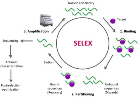
Aptamer oligonucleotides are selected from a large random library (typically 1013-1016 single-stranded unique sequences) for a wide variety of targets ranging from small molecules to whole cells. The diversity of the library depends on the length of the random region. Each oligonucleotide in the library is composed of 40-100 nucleotides, with a central random region (commonly in the 20-60 nucleotide range) and two fixed binding primer-sequences at each extremity.
Aptamers are generated by a method called SELEX (Systematic Evolution of Ligands by EXponential enrichment).7,8 This technique involves three interconnected steps: binding, partitioning and amplification (Fig. 4.). Firstly, DNA, RNA or modified nucleic acid library are incubated with the target of interest (step 1). After incubation, the DNA oligonucleotides specifically bound to the target are separated from the unbound ones (step 2). Bound sequences are collected, amplified by Polymerase Chain Reaction (PCR) (step 3) and then purified for subsequent re-starting the cycle. In the case of the RNA aptamer selection, the SELEX protocol further includes the transcription by T7 RNA polymerase and reverse transcription step before PCR.50
20 Fig. 4. SELEX process for DNA. Step1 Binding, Step 2 Partitioning, Step 3 Amplification.
For improving the binding step, negative or counter selection can be introduced. Negative selection, is the selection performed against the unmodified carriers (filter or beads) in the absence of the target; the counter selection could also be performed against some related molecule targets. This is an optional step used to discard the non-specific sequences, allowing aptamers to discriminate between very similar species.27
During the SELEX process, the evolution of the DNA pool can be monitored by quantification of target-bound oligonucleotides modified with a traceable label.51 This step is important to optimise the conditions and increase efficiency of selection in a controlled way. After several cycles of selection and amplification, the diversity of the pool decreases and the affinity of the oligonucleotides for target can increase.
The selection is stopped when the target-bound candidates are predominant in the pool of oligonucleotides or when no increase is observed during the next two or three SELEX rounds. The enriched pool obtained is then analysed and the candidate aptamers are identified by cloning and sequenced individually clones, or alternatively, with high-throughput sequencing and bioinformatic analysis. (Fig. 4.)
21 After sequencing, the most promising candidates are synthesized to be evaluated for their potential binding of the target, and characterized under different conditions. The affinity and specificity of individual candidates are evaluated by different methods. These two important characteristics of aptamers can be influenced by the conditions of the binding assay. Best aptamers are then optimized for incorporation in diagnostic, therapeutic or analytical applications.
To date, there is no standard selection method for any type of target. During the last three decades, the SELEX process has been continuously modified to make the selection more efficient, adapting it to new separation techniques and sometimes against difficult targets such as small molecules. SELEX method can be used to isolate aptamers against almost all types of target. Nevertheless, macromolecules are the most suitable targets because they contain more functional groups and provide a large surface for interaction with aptamers. Although the demand for the detection of small molecules (<1000g/mol) recently increased, the selection of aptamers against such class of targets is less prevalent.9
Aptamers screening for small molecules is accompanied by some specific difficulties as compared with the selection against proteins or other macromolecules. First of all, small molecules have limited functional groups that reduce the possibility to interact with aptamers. A major problem of performing SELEX against small molecules lies on the separation step between bound and unbound sequences. Due to the small size of the target, there is not an evident difference between unbound candidates and aptamer-ligand complex, complicating the partitioning step in homogenous phase. Consequently, immobilization of one of the two biomolecules is required.
The first conventional DNA SELEX against small molecules was performed by Ellington and Szostak for the selection of aptamers against organic dyes. In this case, the target is immobilized to a solid support matrix.8 Candidates that interact with the target are separated from sequences that do not bind after multiple washes. The sequences of interest are then eluted from the solid support and amplified by PCR for the next round. On one hand, with this method, the oligonucleotides pool is unmodified and free in solution, with no complication related to the conjugation of the pool. On the other hand, the immobilization of target generates some difficulties. As chemical modifications of the target are required to allow the immobilization, a novel epitope can be generated in such
22 a way that the selected aptamer may interact with the conjugate rather than the desired target. In addition, for certain small molecules, the immobilization on solid substrate is not easy to achieve.
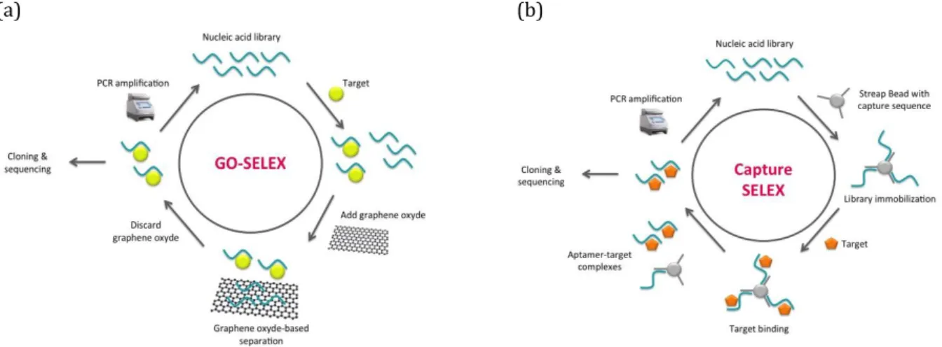
To overcome this problem of SELEX for small-molecules, target immobilization-free aptamer screening methods have emerged, such as GO-SELEX (Graphene oxide-SELEX) 52 (Fig. 5 a) and capture-SELEX (Fig. 5 b).53
(a) (b)
Fig. 5. SELEX process for small molecules. a) Scheme of graphene oxide-based SELEX (GO-SELEX). b) Scheme of Capture SELEX.
In GO-SELEX, the initial ssDNA library is adsorbed on graphene or graphene oxide sheets via - stacking and hydrogen bonds.54 The addition of the target can induce a release of sequences that interact with the target molecule and consequently, a separation from the unbound ssDNA pool still adsorbed on the graphene sheet. The separated sequences are recovered and amplified for the next selection cycles. GO-SELEX process is not based on the size difference, but on the competition among the oligonucleotides, the graphene sheets and the target molecules. It can therefore be applied to small molecule targets (Fig. 5a).
Another immobilization-free SELEX method is the Structure-Switching or Capture SELEX.53 In this process, the initial ssDNA library is designed to hybridize to a complementary capture sequence anchored to the solid substrate (magnetic beads or agarose). The addition of the target causes a conformational change or “structure switching” of ssDNA that can dissociate from the hybrid complex (Fig. 5b). The sequences
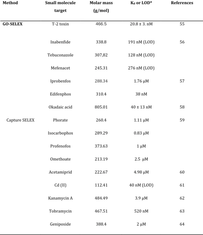
23 are then amplified and used for the subsequent SELEX round. Since they were first described GO-SELEX (2012) and Capture SELEX (2003) have been widely used in the selection of aptamers against different types of small molecules including, dyes or antibiotics and also poorly soluble targets such as toxins or pesticides. Table 2 lists some examples of aptamers successfully selected against small molecules using GO-SELEX or Capture SELEX.
24 *when is specified
Table 2. Some examples of aptamers selected with GO-SELEX and Capture SELEX.
1.4. Determination of binding affinity
The characterization of candidates is a fundamental step to validate the isolated sequences. An important parameter for the characterization of aptamers is the equilibrium dissociation constant Kd. The lower the Kd, the higher the affinity of the target for the aptamer. To determine the Kd, either aptamer or its cognate target is titrated
Method Small molecule target Molar mass (g/mol) Kd or LOD* References GO-SELEX T-2 toxin 466.5 20.8 ± 3. nM 55 Inabenfide 338.8 191 nM (LOD) 56 Tebuconazole 307,82 128 nM (LOD) Mefenacet 245.31 276 nM (LOD) Iprobenfos 288.34 1.76 µM 57 Edifenphos 310.4 38 nM Okadaic acid 805.01 40 ± 13 nM 58
Capture SELEX Phorate 260.4 1.11 µM 59
Isocarbophos 289.29 0.83 µM Profenofos 373.63 1 µM Omethoate 213.19 2.5 µM Acetamiprid 222.67 4.98 µM 60 Cd (II) 112.41 40 nM (LOD) 61 Kanamycin A 484.49 3.9 µM 62 Tobramycin 467.51 520 nM 63 Geniposide 388.4 2 µM 64
25 against the other molecule. There are several methods to determine the dissociation constant of aptamers, involving spectroscopy-based methods, mass-sensitive surface-based measurements or separation-surface-based methods.9 Currently, main methods used for the characterization of aptamers, are classified in two categories: immobilization of one binding partner (aptamer or its target) and immobilization-free technology. One immobilization-based method relies on the surface plasmon resonance (SPR) technique where one partner is immobilized on a solid surface. The immobilization-free technology can include fluorescence polarization/anisotropy (FA/FP)65, Isothermal Titration Calorimetry (ITC)66 and capillary electrophoresis (CE)67 approaches.
For the small molecule-aptamers the characterization is not easy. Aptamers are significantly larger than small molecules and the drastic difference size is one of the major limitations for the detection of these targets. To date, there is no universal method for determining the binding affinity and select the right assay for the accurate affinity measurements constitutes a real challenge. Each assay presents some limitations, including immobilization of target or aptamer, lack of sensitivity or inability to quantitatively measure the dissociation constant. Moreover, the same aptamer, characterised with different assays, can differently behave and display different binding-affinities.68 This inconsistency between Kd measuring methods could also be due to the method system (e.g. immobilization or free-solution). In order to overcome any limitation of each characterization approach, it is suitable to use multiple assays for the small molecule-aptamer characterization. The choice of surrounding conditions such as temperature, pH, or ionic strength is also fundamental for the characterization. The selection takes place in a given buffer and the functionality of the aptamers obtained could be specific for this buffer. Similarly, the aptamers are generated at a unique temperature. Because nucleic acids can fold into different conformations under different temperatures, aptamer misfolding can occur under particular conditions. For these reasons, it is recommended to maintain the same selection conditions for their characterization. Finally, two fundamental controls are required, i. e. the use of a non-binding aptamer and the use of at least one non-cognate target. Furthermore, to avoid changes that could compromise the final performance of the aptamer, it is important to choose the conditions of the SELEX according to the desired applications of biosensors.
26
2. Aptasensors and strategies for small molecules focused
on pesticides
The environmental contaminants are harmful substances present in soil, air and water. These compounds can be released from human sources such as industrial manufacturing, agricultural treatment and wastewater discharge, or they can be generated naturally in form of toxins by algae and bacteria blooms.69 During the last years, the increased production and use of these substances has caused serious problems for the food and environment contamination, putting the health of humanity at risk. Among these environmental contaminants, pesticides are one of the most widespread.70
Divided into three categories -fungicides, herbicides and insecticides-, pesticides include more than a thousand of active substances used to fight against organisms, which are considered as harmful (fungi, plants, insects). Their use in agriculture for the purpose of increasing crop yields and for improving the quality of agricultural products is very excessive. Consequently, the effects on the population exposed to pesticides have become devastating. The most detected pathologies are neurological diseases, damages to reproductive function, developmental alterations and cancers.71 This great threat to mankind is getting more and more attention from governments which impose stringent laws to minimize the usage of these substances in food and agricultural production.
To safeguard both human and ecosystem health, contaminant monitoring and, if possible, their removal is necessary.72 Many of these contaminants, such as pesticides, toxins, antibiotics and drugs, are small molecules (1000 g / mol) that are difficult to detect. Therefore, the development of methods that allow a rapid and specific detection of low-weight contaminants has become a real urgency. The limitations of traditional techniques have encouraged the development of new technologies, among them biosensors.16
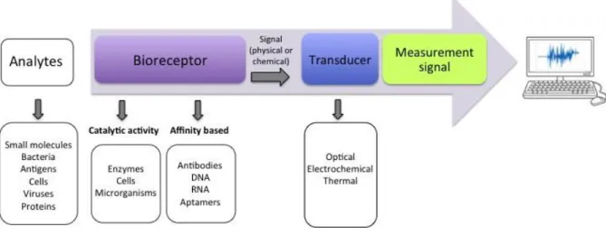
A biosensor (Fig. 6) is a small analytical device which combines a biological molecular recognition element with a physicochemical transducer73 that can signal the presence of the analyte of interest. The sensitive biological component interacts with the target and the transducer element transforms this interaction into a measurable signal. The biological component can be made up of antibody, enzyme, nucleic acid, etc. while a transducer may be optical, electrochemical, thermal, mass sensors, etc. A biosensor that
27 utilizes an aptamer as recognition molecular component is called aptasensor.22,74
Fig. 6. Schematic illustration of a biosensor. Upon binding of a bioreceptor towards an analyte, the transducer collects this interaction and converts it into a measurable signal.16
Biosensors for small molecule targets have not been extensively studied as much as those for protein targets because it is difficult to develop a recognition element for low-weight compounds. Although antibody- or enzyme-based assays are considered as standard sensing platform for the detection of proteins and small targets, they are not available for all compounds. A sandwich assay format, one of the most used methods in immunoassays, is not suitable for the detection of small molecules as these targets are generally embedded in the cleft of the first capturing probes, leaving a too small space for the interaction with the second detector probe.74
Another important issue for an assay development is related to the detector platform for signal production. To detect small molecules, mass-dependent detection methods such as quartz crystal microbalance (QCM) or surface plasmon resonance (SPR) are hardly advisable due to the small mass change produced upon molecular interaction.20
To address the challenges associated with the signal generation, aptamer-based biosensors have been developed and extensively adopted in a large variety of applications in environmental monitoring. Aptamers can selectively bind to analytes with affinity constants comparable to those of antibodies.75 However, aptasensors for pesticide
28 determination have recently gained considerable attention due to their advantages over antibodies. Hence, aptasensors are very promising and have made significant breakthroughs in the development of highly sensitive assays for small molecules detection.
2.1 Strategies for the construction of aptamer-based biosensors
In order to be used in aptasensors, an aptamer must have some fundamental requirement. Firstly, to be introduced to an aptasensor platform, aptamer must be modified in such a way it will be able to interact with the target and signal the target-binding event, simultaneously. Accordingly, it has to maintain the affinity and selectivity of the native aptamer and must quickly recognize the target molecule. Regarding the design process, a minimal knowledge of the secondary and tertiary structure aptamer-target complex is often necessary. It should be versatile and applicable to different readout methods. However, the choice of the design strategy to be adopted is a crucial point for the success of an aptasensor.
Since the first aptasensor created in 2000 by Ellington and colleagues, many aptasensing strategies have been designed and developed to make aptasensors simpler, applicable to various methods and versatile for all types of targets.76 The principal strategies developed are: a) signalling aptamer b) double end-labelled aptasensor, c) split aptasensor d) label-free aptasensor e) strand displacement strategy f) kissing complex.
29
Fig. 7. General overview of aptasensing strategies. a) signalling aptamer b) double end-labelled aptasensor, c) split aptasensor d) label-free aptasensor e) strand displacement strategy f)
kissing complex.
There are several examples of aptasensors based on these strategies designed for the detection of small molecules; most of these are proof-of-concept systems using ATP, theophylline or cocaine aptamers. In the next sub-chapters, more detailed information will be given on each aptasensing strategies.
Signalling aptamer
In aptasensors based on the signalling aptamer strategy, the signal transduction depends on the molecular recognition mechanism. The binding of the target causes a conformational change which will be used to generate a measurable signal (Fig. 7a). Although signalling aptamers have often shown a good affinity for the target, this approach has some limitations. Since many aptamers possess partially folded structure, the signal gain produced with this strategy is often low. In addition, in case of small molecules, the target can interact with aptamer without causing a large detectable conformational change. The application of this strategy is limited to some aptamers.76
30 Therefore, several research groups have investigated alternative methods to enhance the target-induced structural change and applied this approach to the small molecule detection. The first aptasensor design published in the literature was a signalling aptamer directed against ATP.77 In this aptasensor system, the aptamer is labelled with a fluorescent dye in proximity to the ligand-binding pocket. In the presence of the target, the structure of aptamer changes, increasing the fluorescent signal. Some years later, Ruta and colleagues described a small molecule aptamer assay based on this strategy (Fig. 8), using a directed fluorescence anisotropy (FA) technique.78 For this approach they engineered a biosensor based on the anti-tyrosinamide aptamer, labelled by a single fluorescent dye at one extremity. The presence of the target induces the conformational change of the aptamer, which modifiy the local motional freedom of the fluorescent label, resulting in an increase of fluorescence anisotropy signal. Based on this strategy Perrier and coworkers designed another signalling aptasensor with an anti-adenosine aptamer as a model. They rationally engineered the instability of the secondary structure of the aptamer which allows to demonstrate the conformational change of the aptamer in the presence of the target.79
Fig. 8. Schematic representation of the optimised signalling aptamer. The conformational change in the presence of the target molecule is translated into an increase of fluorescence anisotropy signal.78
Double-end labelled aptasensors
Afterwards, in order to engineer aptasensors with higher signal gain, aptasensor designs relying on a FRET pair rather than a single dye reporter have been developed. FRET aptasensors have been engineered based on a molecular beacon probe.80 A molecular beacon is a fluorescently bi-labelled DNA hairpin probe designed to bind complementary DNA or RNA sequences. It can report the presence of targets in a homogeneous solution. The extremities of the probes contain a FRET pair (or a fluorophore-quencher pair) whose
31 fluorescence signal changes (or is restored) when the beacon opens upon binding to the complementary target sequence. Other aptasensor strategies have been designed on the basis of this format.
Stojanovic and colleagues first implemented the molecular beacon-like format in a double end-labelled aptasensor (Fig. 9.).10 They re-engineered the anti-cocaine aptamer, introducing an instability in one stem of a three-way junction that forms the cocaine-binding pocket and labelling the resulting short terminal stem with a fluorophore and quencher pair. In the presence of cocaine, the formation of the three-way junction leads to the stem closure in such way that the fluorophore and quencher are brought in close proximity. This structural change causes the fluorescence decrease, which reports the presence of the cocaine. Moreover, this aptasensor has been tested in serum, demonstrating its selectivity over cocaine’s metabolites. By using this strategy to engineer an aptasensor, they have demonstrated that this design can provide a new set of analytical tool to detect small molecules in different media.
Recently, the same strategy has been used to engineer a new aptasensor against aflatoxin B1.81 Inspired by the Ellington model, they engineered the aptamer anti-aflatoxin 1 (AFB1) into a double-end labelled aptamer beacon (using a fluorophore and quencher pair). This aptamer molecular beacon assay showed high specificity for AFB1 (tested against other mycotoxins) and a potential detection (in the nanomolar range) in real samples. In the last years, the use of double end-labelled aptasensors has allowed the development of aptamer based fluorescent molecular beacon assays for the detection of small molecules such as cocaine or adenosine triphosphate (ATP), based on binding-induced structure change with the subsequent fluorescence increase or decrease.81,82
Fig. 9. Example of double-end labelled aptasensors.10 When the target molecule, in this case cocaine, is present in solution, a conformational change of the aptamer brings closer fluorescein and a quencher, leading to a fluorescent decrease.
32 Split aptasensor
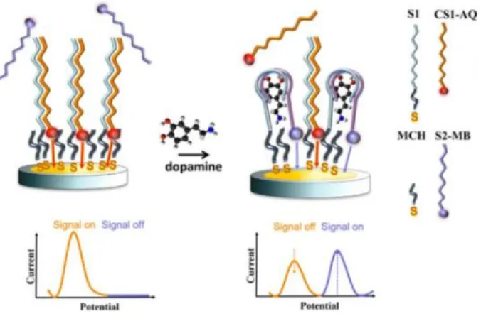
Biosensors can be in general classified into two groups, competitive and sandwich assays. In the sandwich assays, two probes such as antibodies or aptamers, are used simultaneously to capture the target through the recognition and binding of two distinct regions. The sandwich assay is mainly used for the macromolecule detection, especially proteins, with high sensitivity and specificity resulting from this dual recognition mechanism. However, it cannot be used for the detection of low molecular weight analytes for the reasons described above. To overcome this issue, Stojanovic and colleagues83 developed a split-aptamer-based strategy, in which the aptamer sequence was divided into two subunits that form a ternary complex in the presence of the target. To obtain a signal, the two subunits (labelled, for example, with a FRET or fluorescent-quencher pairs) have to be close enough to each other to allow signal transmission, which occurs only in the presence of a target. To transform an aptamer into a split aptasensor, it is fundamental to know the aptamer–target complex structure and understand the role of each nucleobases.84 This approach has been adopted for the detection of various ligands, especially small molecular targets, using different transduction methods including fluorescence, colorimetric, and electrochemical techniques,85–87 many based on anti-cocaine and ATP aptamers.84 Recently, a novel dual-signal ratiometric electrochemical biosensor based on a split aptamer for the detection of dopamine has been also engineered (Fig. 10).88 In this study, Guo and co-workers have demonstrated the efficiency of the split-aptasensor strategy in small molecule detection with good affinity (in micromolar range) while maintaining satisfactory reliability and sensitivity similar to those of the native aptamer.
33 Label-free aptasensors
The label-end strategy may suffer of some limitations. In some cases this target-induced fluorescence change results in a high detection limit, even under optimal conditions. This could originate from an inefficient proximity quenching,89 or the chemical modification of the aptamers.90 To overcome these concerns, label-free aptamer probes have been developed as an alternative strategy. This approach is dependent on the conformational change of the aptamers before and after binding to its target. This approach requires a ssDNA aptamer probe without any labelling and a fluorescent reporter such as double-stranded DNA (dsDNA) intercalators, single-double-stranded DNA (ssDNA) binding dye, or fluorescent conjugated polymer. The most common signal reporters for label-free fluorescence aptasensors is related to the use of DNA intercalation dyes. In aqueous solution, these dye molecules show a low fluorescence intensity while a strong fluorescence signal occurs when they intercalate into dsDNA.
Using the (Ru (phen)2(dppz)) intercalator dye, the Wang’s group has developed a label-free aptasensor for detecting ATP with high selectivity and high sensitivity, down to 1nM in homogeneous solution.91Some other fluorescent dyes such as SYBR Green I92 have been also used as a transducer for the aptamer-target recognition devoted to small molecules.
Label-free aptasensors can be similarly created using fluorogenic dyes (or fluorogens) such as Thiazole Orange,93 Hoescht,94 Malachite Green (MG)95 DFHBI,96 Patent blue97. Among them, MG shows extremely low fluorescence in homogeneous solution and strong fluorescence intensity (~2400-fold) after binding with its RNA aptamer.
Light-up aptamers are RNA sequences that specifically interact with a fluorogen to form a fluorescent complex.Based on the MG light-up aptamer, taking advantage of the unique fluorescence property of MG, Stojanovic and Kolpashchikov firstly proposed a modular aptameric sensor to detect various small molecules analytes (ATP, flavin mononucleotide and theophylline).98 These aptasensor designs consist of MG aptamer as signal domain, other aptamers (ATP, flavin mononucleotide or theophylline) as recognition domain and a connector module between the recognition and signal domains. The interaction of each aptamer with its cognate target leads to the exposure of the MG aptamer so that MG binds its aptamer and emits a high fluorescence intensity.
34 Recently other light-up aptamers were successfully selected. By taking advantage of their sensitive properties, they could be used to develop new label-free aptasensors to detect other small molecules.
Strand displacement strategy
The design of the above described aptasensors strategies cannot be applied to all aptamers. For instance, many aptamers do not form a defined stem structurewith a terminal hairpin. Likewise, most aptamers do not have a secondary structure that can be easily split into two fragments without affecting the aptamer functionality.
To overcome these drawbacks, a strategy based on the structure-switching concept was introduced in 2003 and is still today one of the most used in aptamer selection and aptasensor design for small molecules.14 This strategy is based on the target induced dissociation of a hybrid complex between an aptamer and a partially complementary sequence. It does not require any -or only minimal- aptamer modifications.53,99 This mechanism is based on the ability of aptamers to bind to both complementary sequence and their specific target. In the presence of the target, the aptamers can then switch from a nucleic acid helix to an aptamer-target complex. In « structure-switching » assays, the dissociation caused by the competitive binding of the aptamer to the target is converted into a measurable signal that detect the presence of the target.100 For instance, the aptamer can be functionalized with a fluorophore while the complementary sequences can be labelled with a quencher. In the absence of the target, the two nucleic acid strands form a duplex, leading to low fluorescence (Fig. 11). In the presence of target, the aptamer-target complex is formed and the complementary quenched sequence is displaced, leading to the fluorescence signal.
Fig. 11. Example of a strand-displacement strategy. In the absence of the target molecule, the aptamer, functionalized with a fluorophore, is hybridized with a complementary DNA strand, labelled with a quencher. After the addition of the target molecule, the displacement of the quencher strand leads to a fluorescence signal.101
35
Li and co-workers have shown the first proof-of-concept for strand displacement assay based on aptamers against ATP and thrombin.14 Since then, this strategy has been widely
used to detect various types of targets, being the most used for the small molecule signalling. Actually, it is considered as a highly generalizable method to engineer all aptamers into biosensor and it is introduced into a very different sensors.91,102
Kissing complex
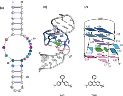
This strategy is based on the engineering an aptasensor for the detection of small molecules involved in the kissing complex system. The term « kissing complex » appeared for the first time in 1984, to describe the regulatory mechanism for the replication of the bacterial plasmid ColE1103 and it has been observed in the regulation of numerous
biological process such as virus assembly and plasmid replication.103–105Kissing or
loop-loop interaction is formed between two RNA via complementary base pairing and is
greatly dependent on loop parameters (base composition, sequence and size) in combination with the loop–loop helix - stem junctions (Fig. 12).More than 20 years ago,
this mechanism has been used to generate an RNA aptamer for the TAR-RNA element of HIV-1 and to select DNA aptamers specific for a DNA hairpin structure.106–108
Fig. 12 Example of kissing complex. Aptamer anti-TAR and HIV-1 TAR hairpin.108
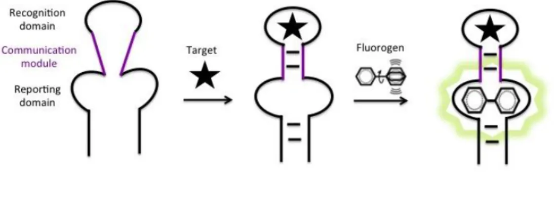
More recently, a novel aptamer biosensing concept based on the kissing complex strategy has been described by Toulmé and Peyrin groups.5,6 They introduced a new type of aptasensor to detect small molecules based on a dual recognition mechanism that includes a signalling partner that contributes to increase the stability of the complex with the target. This assembly is constituted by two partners: an aptamer called aptaswitch, able to shift its conformation from unfolded to folded (hairpin) structure in the presence of the target, and a second short RNA hairpin named aptakiss.
For the aptaswitch design, the apical loop of the native aptamer is substituted with a short RNA sequence able to recognize the aptakiss through loop-loop interactions forming a
36 kissing complex. Therefore, the resulting structure is formed when both partners are structured in hairpin structures, i.e. in the presence of the target. Aptakiss is then used as a transducer and it can be adapted to various kinds of aptaswiches. This strategy was conceived in order to enhance the specificity and the resulting signal gain of the biosensor. In recent years, this approach has been employed in the rational design of new small molecule biosensors. Toulmé and Peyrin groups designed some new aptasensors exploiting the formation of kissing complexes for sensing the presence of small molecules specifically recognized by their hairpin aptamers.5,6 The new aptaswitches rationally designed, baptized as Adenoswitch, GTPswitch, Theoswitch and Vasoswitch (specific for adenosine, GTP, theophyllline and vasopressine respectively) have been largely studied with different detection systems including SPR, fluorescence anisotropy,109colorimetry with AuNPs110and HPR with an ELAKCA system (Enzyme linked aptamer kissing complex assay).111 In addition, this last approach has been successfully applied to colorimetric tests, to detect the presence of theophylline in human plasma samples.
All these kissing-complex approaches have demonstrated a high sensitivity and specificity for the small molecules. The new kissing-aptasensors have proved to be a promising system also applied to multiple target sensing.
2.2 Aptasensors for pesticides
Due to their large application in agriculture, pesticides can be transported to the surface water and easily contaminate the agricultural product for the human consumption. Over the past years, various aptasensors have been successfully exploited for the detection of pesticide residues for the environmental monitoring.112,113 Most of these aptasensors are engineered based on two approaches: the aptamer signalling and the strand displacement strategy.
Based on these two strategies, the three most widely used types of the aptasensors are focused on fluorescence,114 electrochemical,115 and colorimetric116 technologies. (Fig. 13). For developing electrochemical and fluorescent aptasensors, a number of nanomaterials such as quantum dots (QDs), carbon dots (CDs), up-conversion nanoparticles (UCNPs), AuNPs, Graphene oxide (GO) and so on, have been employed for the detection of pesticide residues.
37
Fig. 13. General overview of aptasensors for pesticide. a) and d) fluorescent aptasensors, b) and e) electrochemical aptasensors, c) and f) colorimetric aptasensors.
Fluorescent aptasensors
Due to its high sensitivity and efficiency, the fluorescence technique is one of the most common approach for the analysis aptamer-target interactions.117 Generally, there are two sensing mechanisms in developing fluorescent aptasensors, i.e.:
-“signal-on” mechanism, where the presence of the target causes a recovery or great increase of fluorescence intensity, while in the absence it is quenched or minimal;
-“signal-off” mechanism where the presence of the target causes the quenching or a great decrease of the fluorescence intensity, while in the absence it is strong.
The change in the fluorescence intensity allows the quantitative detection of pesticides. Wang and his co-workers reported a non-competitive fluorescent aptasensor for the detection of acetamiprid pesticide based on the inner filter effect (IFE) of AuNPs toward
38 fluorescent carbon dots (CDs).118 This aptasensor system displayed high selectivity and excellent accuracy for acetamiprid detection with a linear range from 22.5 nM to 0.45 μM and a detection limit of 4.85 nM. In another study, Zhang and colleagues reported a FRET competitive aptasensor based on molecular beacon for the quantification of phorate, profenofos, isocarbophos and omethoate.13 The molecular beacon was labelled with FAM and DABCYL at the 5’ and 3’ end respectively, and contained a sequence partially complementary to the aptamer. Upon adding the pesticides, the aptamer preferably binds the targets, inducing the molecular beacon opening and a restore of fluorescence intensity. The limit of quantification (LOQ) detected for phorate, profenofos, isocarbophos, and omethoate reached 19.2, 13.4, 17.2, and 23.4 nM, respectively. Various other types of fluorescent aptasensors have been developed for the pesticide detection.119,120,121
Electrochemical aptasensors
In the past years, electrochemical aptasensors have shown great potential for pesticide detection due to their ability to multiplexed analysis, fast response, and low cost.122,123
Fei and co-workers developed an electrochemical aptasensor for the detection of acetamiprid residues using gold nanoparticles (AuNPs) decorated with multiwalled carbon nanotube-reduced graphene oxide nanoribbon (Au/MWCNT-rGONR) composites.124 This aptasensor, based on the variation of electron transfer resistance, was relevant to the formation of acetamiprid–aptamer complex on the modified electrode surface. This non-competitive electrochemical aptasensor showed excellent performances for the acetamiprid detection into a linear range of 50 fM to 10 μM and an extremely low detection limit of 17 fM. Rapini and colleagues developed a simple electrochemical DNA aptasensor for the detection of acetamiprid based on a competitive format and disposable screen-printed arrays.125 The dose–response curve was constructed between 0.25 and 2.0 mM acetamiprid concentration range with a limit of detection of 0.086 mM. Furthermore, the efficacy of this aptasensor has also been demonstrated in various fruit juice samples showing high recovery percentage. All these characteristics make this aptasensor particularly promising for the realization of a commercial kit.
39 Colorimetric aptasensors
Colorimetric methods have been extensively applied for the detection of pesticide contaminants. 112,126 Gold nanoparticles (AuNPs) are the most commonly used probes for the colorimetric sensing assays due to their facile preparation and surface modification. By using AuNPs, Wang and co-workers reported a simple non-competitive colorimetric aptasensor for the detection of omethoate.127 This system was based on the principle that single-stranded DNA (ssDNA)-wrapped gold nanoparticles (AuNPs) are resistant to salt-induced aggregation. In the presence of the target, the aptamer binds to omethoate and is released from the AuNP surface. The consequent aggregation of AuNPs changes the color of the sample from purple to red. This aptasensor showed good linearity between 0.1 and 10 μM, with a low detection limit of 0.1 μM. In addition, Abnous et al. recently developed a competitive colorimetric aptasensor to determine the presence of pesticide malathion.128 In the absence of the target, aptamer forms a double-strand DNA structure (dsDNA) with a complementary sequence and protect AuNPs against salt-induced aggregation. Upon malathion addition, the aptamer folds into a hairpin structure to form aptamer-malathion complex, releasing the complementary strands into the solution. As a result, the AuNPs underwent salt-induced aggregation, leading to a solution colour change from red to purple. The efficiency of this aptasensor has also been demonstrated in spiked human serum samples, showing its high sensibility to malathion with a rapid response (35 min) and a detection limit of 1 pM.
Several other pesticide-specific aptasensors have been developed, also demonstrating a lower limit of quantification (LOQ) and some of them are listed in the table 3.
40 Table 3. Summary of some the aptasensor strategy for pesticide detection.
2.3 Light up aptasensors
Light-up aptamers are generated for specific binding and activate their cognate fluorogenic ligand (small non-fluorescent dye that becomes fluorescent). In free unbound form, when the fluorogen molecule is excited, its energy is dissipated by non-radiative pathway, such as molecular vibration. However, when it binds to the aptamer, the planar
Pesticide residue Assay LOD References
Phorate, profenophos, isocarbophos, omethoate
Fluorimetric FRET: FAM and DABCYL
19 nM, 13 nM 17 nM, 23 nM 113 Phorate, profenophos, isocarbophos, omethoate Fluorimetric FRET: FAM and AuNPs
35 nM, 134 nM 384 nM 2.35 μM 129 Acetamiprid Fluorimetric FRET: QDs and Au NPs 7 nM 130 Acetamiprid Fluorimetric FRET: UCNPs and colorimetric AuNPs 3 nM 131 Isocarbophos, dursban phosalone, methamidophos, acephate trichlorfon Colorimetric
AuNPs aggregation Isocarbophos:100 ppb, others: 2000 ppb 132
Acetamiprid Colorimetric
hemin-rGO 40 nM 133
Acetamiprid Electrochemical AuNPs modified electrode;
EIS, CV