TRENDS
Development of bacteria-based bioassays for arsenic
detection in natural waters
Elizabeth Diesel&Madeline Schreiber&
Jan Roelof van der Meer
Received: 9 March 2009 / Revised: 30 March 2009 / Accepted: 31 March 2009 / Published online: 18 April 2009
# Springer-Verlag 2009
Abstract Arsenic contamination of natural waters is a worldwide concern, as the drinking water supplies for large populations can have high concentrations of arsenic. Tradi-tional techniques to detect arsenic in natural water samples can be costly and time-consuming; therefore, robust and inexpen-sive methods to detect arsenic in water are highly desirable. Additionally, methods for detecting arsenic in the field have been greatly sought after. This article focuses on the use of bacteria-based assays as an emerging method that is both robust and inexpensive for the detection of arsenic in groundwater both in the field and in the laboratory. The arsenic detection elements in bacteria-based bioassays are biosensor–reporter strains; genetically modified strains of, e.g., Escherichia coli, Bacillus subtilis, Staphylococcus aureus, and Rhodopseudomonas palustris. In response to the presence of arsenic, such bacteria produce a reporter protein, the amount or activity of which is measured in the bioassay. Some of these bacterial biosensor–reporters have been successfully utilized for comparative in-field analyses through the use of simple solution-based assays, but future methods may concentrate on miniaturization using fiber-optics or microfluidics platforms. Additionally, there are other potential emerging bioassays for the detection of arsenic in natural waters including nematodes and clams.
Keywords Bioassays . Biosensors . Escherichia coli . Reporter proteins
Arsenic in the environment
Arsenic is a ubiquitous metalloid in the environment: it can be found in natural systems all over the world from both natural and anthropogenic sources [1]. Natural sources of arsenic include rocks and minerals in the earth's crust, as
well as the sediments and soils derived from them [1,2].
Anthropogenic sources of arsenic include the application of organoarsenical pesticides, the burning of fossil fuels, the use of organoarsenical feed additives, mining activities, the disposal of industrial wastes, and the application of
arsenical desiccants and preservatives [1, 2]. Arsenic
contamination of natural waters is of particular concern as
arsenic is a known toxin and carcinogen [3, 4]. The most
recent crisis involving arsenic in water supplies is in Southeast Asia, including Bangladesh, Vietnam, India, and Cambodia, where naturally occurring arsenic contamination of groundwater has resulted in millions of cases of arsenic poisoning [4].
Several arsenic species are found in natural waters, with the dominant forms occurring as arsenite [As(III)] or arsenate [As(V)] as anions in solution. Arsenic can also complex with colloidal particles, and can also be found as organoarsenic compounds, depending on the source and the environmental conditions [2]. The toxicity of arsenic species in natural waters varies, with arsenite being the most toxic, and arsenate, monomethylarsonate, and dime-thylarsinate being less toxic [2]. Arsenic cycles through the hydrosphere via several key processes. Arsenic can be released from surface sources to soil water, and can be subsequently transported to groundwater and surface water DOI 10.1007/s00216-009-2785-x
E. Diesel
:
M. SchreiberDepartment of Geosciences, Virginia Tech, Blacksburg, VA 24061-0420, USA J. R. van der Meer (*)
Department of Fundamental Microbiology, University of Lausanne,
Bâtiment Biophore, Quartier UNIL-Sorge, 1015 Lausanne, Switzerland
supplies, and may eventually be discharged to the ocean. Arsenic can also be leached directly into groundwater supplies via dissolution of arsenic-bearing minerals in the aquifer matrix, and this contamination can then be trans-ported to surface and ocean waters. Uptake of or irrigation with arsenic-contaminated water results in contamination of crops or livestock, which can further aggravate the arsenic toxicity cycle in biota and people [5]. Several
biogeochem-ical processes control the release and transport of arsenic in
natural waters, including adsorption, oxidation–reduction,
and microbial activity. Arsenic adsorbs strongly to metal oxides and clays, which is a highly pH dependent process [6]. Oxidation and reduction of arsenic species impact speciation, which can thus impact mobility, as arsenite has been observed to be more mobile than arsenate [6]. Microbial activities play an important role in the dissolution of arsenic-containing minerals and in transforming arsenic speciation. For example, arsenate is reduced to arsenite by the common bacterial enzyme arsenite reductase [7]. Arsenate can also serve as an electron acceptor for anaerobic respiration with organic materials [8]. Arsenite can be oxidized to arsenate under aerobic conditions by certain types of bacteria that derive chemical energy from the reaction [8]. Furthermore, organoarsenicals are pre-sumed to be formed via microbial activities [9].
Detection of arsenic in environmental samples
Robust and inexpensive methods to detect arsenic in the field have become highly desirable, as standard laboratory-based techniques such as atomic absorption and atomic fluorescence spectrometry, inductively coupled plasma techniques, and high-pressure liquid chromatography, while
highly effective [10, 11], can be costly and
time-consuming. Regions that have the most extensive arsenic contamination are also the areas with the least access to these particular techniques, making the need for in-field arsenic detection even more urgent. In addition, certain countries such as Bangladesh have an extremely delocal-ized water supply system with millions of individual tube arsenic
resistance proteins Promoter
arsD arsA arsB arsC arsR RNA Polymerase As(III) As(III) As(III) As(III) As(III) As(III) As(III) As(III) As(III) As(III) As(III) As(V) As(V) ArsAB ArsAB ArsAB ArsAB ArsC ArsC ArsC Promoter
arsD arsA arsB arsC arsR As(III) As(III) As(III) As(III) As(III) As(III) As(III) As(III) As(III) As(III) As(III) As(V) As(V) ArsAB ArsAB ArsAB ArsAB ArsC ArsC Promoter repor ter arsR RNA Polymerase reporter protein
a
b
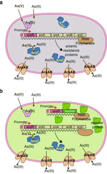
Fig. 1 Design principle of most bacteria sensor–reporters for arsenic. a When no arsenic enters the cell, the ArsR protein represses the transcription of the arsenic defense system genes (arsD, arsC, arsA, and arsB) from one particular DNA region upstream of the gene for itself (the operator–promoter site). In the presence of arsenite in the cell, ArsR loses affinity for the operator and RNA polymerase will transcribe the arsDCAB genes to produce the defense. ArsC is a reductase that reduces arsenate [(As(V)] to arsenite [As(III)], whereas ArsAB constitute an efflux pump for arsenite. b In the sensor–reporter strain, an extra copy of the operator–promoter DNA fused to the arsR gene and a gene for a reporter protein are added to the cell. In this case, when arsenite or arsenate is sensed by the cell, transcription for the reporter gene will also be unleashed and the reporter protein will be formed. The presence or activity of the reporter protein is subsequently measured
Light emission from luciferase
(rel. units) 0 200000 400000 600000 800000 1000000 0.0 0.2 0.4 0.6 0.8 1.0
Arsenite concentration (µM AsIII)
Fig. 2 Example of a calibration of a bioassay using an arsenic sensor– reporter strain. Cells of Escherichia coli (pJAMA-arsR) are incubated in an aqueous suspension with a series of known concentrations of sodium arsenite. After 2 h of incubation of the cells with arsenite, the substrate for the reporter protein (here, bacterial luciferase) is added and light emission is measured with a luminometer. Extracts from unknown samples are measured simultaneously and values are interpolated on the calibration curve, as indicated. (Graph and data redrawn with permission from Baumann and van der Meer [28])
T able 1 Overview of bioassays for measurement of arsenic using bacterial sensor –reporter strains Bacterial sensor –reporter strain Method of As detection Utilization Additional equipment As species detected Reported linear range of As(III) detection Field-tested References Escherichia coli MC1061 (pT OO21) Firefly luciferase + D -luciferin 96-well plate Y es As(III), As(V) 257 μ g/L –26 mg/L No [ 17 ] E. coli MC1061 (pT OO31) Firefly luciferase + D -luciferin 96-well plate, culture tube, fiberoptics Y es As(III), As(V) > 6 ug/L (fiberoptics) No [ 18 , 31 , 34 ] E. coli A W31 10 (pT OO31) Firefly luciferase + D -luciferin 96-well plate No As(III), As(V) NA No [ 18 , 34 ] E. coli (arsB::luxAB ) Bacterial luciferase + dodecanal substrate 4-mL cuvette Y es As(III), As(V), chromated copper arsenate NA No [ 7 ] E. coli MC1061 (parsRluxCDABE) Bacterial luciferase + D -luciferin 96-well plate, fiberoptics, 8-well strip Y es As(III), As(V) As(III): 8– 80 μ g/L, <1 8μ g/L (fiberoptics) As(V): < 8 0μ g/L, < 141 μ g/L (fiberoptics) No [ 31 , 32 , 35 ] E. coli DH5 α (pJAMA-arsR) Bacterial luciferase + decanal substrate 96-well plate Y es As(III), As(V), trimethylarsine oxide 8– 78 μ g/L Y es [ 19 , 25 , 28 , 36 ] E. coli DH5 α (pJAMA-arsR-ABS) Bacterial luciferase + decanal substrate 96-well plate Y es As(III), As(V) 8– 78 μ g/L No [ 19 ] E. coli DH5 α (pMV -arsR) β -Galactosidase + X-gal substrate Paper strip, 96-well plate No As(III), As(V) > 1 0μ g/L Y es [ 19 , 20 ] E. coli DH5 α (pMV -arsR-ABS) β -Galactosidase + X-gal substrate Paper strip, 96-well plate No As(III), As(V) ≥ 10 μ g/L Y es [ 19 , 20 ] E. coli DH5 α (pPR-arsR-ABS-LacZ) β -Galactosidase + X-gal substrate 96-well plate No As(III), As(V) > 5 0μ g/L No [ 20 ] E. coli DH5 α (pPR-arsR-ABS-RBS-LacZ) β -Galactosidase + X-gal substrate 96-well plate No As(III), As(V) 0.2 –6.0 μ g/L No [ 20 ] E. coli (pIRC140) Green fluorescent protein 96-well plate Y es As(III), As(V) 1– 100 μ g/L No [ 21 ] E. coli DH5 α (pPR-arsR) Green fluorescent protein Microscope slide Y es As(III), As(V) 8– 47 μ g/L No [ 19 ] E. coli DH5 α (pPR-arsR-ABS) Green fluorescent protein Microscope slide Y es As(III), As(V) 8– 234 μ g/L No [ 19 , 37 , 38 ] E. coli A W10 (pSD10) Green fluorescent protein Microfluidics device Y es As(III) 78 μ g/L –390 mg/L No [ 22 ] E. coli DH5 α (pArsR-ABS-CCP-R48Q) Cytochrome c peroxidase Agar plate, paper strip No As(III), As(V) 4– 30 μ g/L, > 1,000 μ g/L No [ 20 ] E. coli DH5 α (pArsR-ABS-CCP-K2.4) Cytochrome c peroxidase Agar plate, paper strip No As(III), As(V) 4– 30 μ g/L No [ 20 ] E. coli DH5 α (pArsR-ABS-CCP-PT1) Cytochrome c peroxidase Agar plate, paper strip No As(III), As(V) 4– 30 μ g/L, > 1,000 μ g/L No [ 20 ] E. coli DH5 α (pArsR-ABS-CCP-WT) Cytochrome c peroxidase Agar plate, paper strip No As(III), As(V) 4– 20 μ g/L No [ 20 ]
wells. This poses particular challenges for the logistics of measurement campaigns involving sampling, sample label-ing, transport, and administration. Direct, on-site rapid field tests could alleviate some of these logistics issues.
In response to the need for field-applicable arsenic detection techniques, there has been increased interest over the last decade in the development of sensors for the field detection of arsenic. Abiotic sensors, such as colorimetric tests, electrochemical sensors, and anodic stripping voltam-metric probes have been developed and tested in the field [11]. However, large-scale field testing of the most common mercury bromide colorimetric tests showed that they are not sufficiently accurate in the concentration range below 50μg/L, and, consequently, give rise to a large number of false-positive and false-negative results [12]. On the basis of this poor performance, the method has been optimized using an instrumental readout of the colorimetric signal, which resulted in significantly better arsenic concentration prediction [11]. Additionally, two new colorimetric kits (Quick Arsenic and Hach EZ) were recently developed and tested in the field [13]. Despite this improvement, there have been major concerns regarding the use of toxic chemicals (i.e., mercury bromide, zinc, release of arsine gas) in the colorimetric test [11], which merits the development of alternative protocols and methods.
Bacteria-based bioassays are one of the alternatives to the abiotic sensors created for arsenic detection [14]. Since most bacteria carry an extremely sensitive defense system against arsenite and arsenate, composed of an arsenate reductase and a highly effective arsenite efflux pump, they can be exploited for arsenic detection. Upon encountering arsenite, a dedicated sensory protein in the bacterial cell called ArsR will undergo a conformational change that unleashes expression of the defense system [15]. The way that this protein can invoke this response is by its action as a transcriptional repressor, which, in the absence of arsenic, binds to a specific DNA sequence (i.e., the operator) overlapping with the binding site for RNA polymerase (the
promoter; Fig.1). ArsR will, however, lose its affinity for
the DNA when binding arsenite and RNA polymerase can commence transcription [15].
To turn this natural defense system into a workable bioassay, the bacterial cell is equipped with a second copy of the operator–promoter sequence for ArsR, which is now transcriptionally fused to a gene for a so-called reporter
protein (Fig. 1). When arsenite enters the cell, both the
defense system and the reporter protein synthesis are derepressed. Interestingly for the development of a bioas-say, the reporter protein synthesis rate is dependent on the arsenite concentration sensed by the cell. One can therefore achieve a reporter protein response proportional to the arsenite concentration. Furthermore, because the ArsR reacts so sensitively to arsenite, the sensor–reporter cell
T able 1 (continued) Bacterial sensor –reporter strain Method of As detection Utilization Additional equipment As species detected Reported linear range of As(III) detection Field-tested References E. coli DH5 α (pArsR-ABS-CCP-R45I) Cytochrome c peroxidase Agar plate, paper strip No As(III), As(V) 20 –5,000 μ g/L No [ 20 ] E . coli DH5 α (pArsR-ABS-CCP-R48T) Cytochrome cperoxidase Agar plate, paper strip No As(III), As(V) 20 –5,000 μ g/L No [ 20 ] Bacillus subtilis BR151 (pT OO21) Firefly luciferase + D -luciferin 96-well plate Y es As(III), As(V) 257 –7,800 μ g/L No [ 17 , 34 ] B. subtilis (pMUT in-23) β -Galactosidase + accelerator -II Culture tube Y es As(III), As(V) 22 –7,800 μ g/L No [ 23 ] Staphylococcus aur eus RN4220 (pT OO21) Firefly luciferase + D -luciferin 96-well plate Y es As(III), As(V) 8– 257 μ g/L No [ 17 , 34 ] Rhodopseudomonas palustris (crtIBS ) Phytoene dehydrogenase (CrtI) 7.5-mL glass tubes Y es As(III), As(V) NA No [ 24 ] NA not analyzed
will invoke a reporter protein response at very low (extracellular) arsenite concentrations, typically significantly below 50μg/L. By calibrating the response of the sensor–reporter cells in the bioassay at various arsenite concentrations, one can thus infer the equivalent arsenite
concentration in an unknown sample (Fig. 2).
This principle has been demonstrated numerous times in
the literature, using different bacteria as a “host” for the
sensor–reporter construction such as Escherichia coli, Bacil-lus subtilis, Staphylococcus aureus, or more recently Rhodopseudomonas palustris. One of the interesting aspects of this technology is that there are a wide variety of possibilities for choosing the reporter protein [16]. In general, a reporter protein is selected that is easy to measure, has a high degree of selectivity (i.e., has no background in the host cell), and a high sensitivity (i.e., very few reporter protein molecules in the cell can be detected). Current examples are bacterial or eukaryotic luciferase,
autofluor-escent proteins,β-galactosidase, ice-nucleation protein, and
cytochrome c peroxidase. Luciferases, which will elicit bioluminescence, have been widely exploited because of their high selectivity and sensitivity. On the other hand, they impose a large energy cost on the host cell. Autofluorescent proteins have attracted wide interest because no cofactors are needed for the activity, as they permit single-cell detection and a variety of different fluorescence colors exist. This may
not be particularly important for a single target sensor–
reporter cell, but it becomes interesting when multiple chemical targets have to be measured simultaneously. β-Galactosidase is a very versatile enzyme for which a wide range of different substrates exist that enable
chemilumines-cence, fluoreschemilumines-cence, or colorimetric signal formation. Table1
presents an overview of the various bacterial arsenic sensor– reporters that have been developed and the ranges of detection sensitivity in the bioassays.
The majority of the microbial sensor-reporters for arsenic are based on E. coli with luciferase, green fluorescent protein
(GFP), β-galactosidase, or cytochrome c peroxidase as the
indicator for exposure to arsenic in solution. The assays conducted with bacterial sensor–reporters for arsenic in general consist of incubating an aquatic suspension of sensor cells with the sample and reading out the reporter signal after 2–4 h. The first E. coli sensor–reporters utilized bacterial and firefly luciferase as reporter proteins for arsenic
contamina-tion [7,17]. The reported linear detection range of bioassays
with E. coli expressing firefly luciferase was still relatively high, being 257μg/L to 26 mg/L of arsenite in solution [17]. Bioassays with subsequently reengineered E. coli sensor– reporters with bacterial and firefly luciferase were capable of detecting lower concentrations of arsenite that are at or below the most common drinking water standard of 10μg As/L [18, 19]. Bioassays with very low detection ranges for arsenic in solution (10–50µg/L) were also reported using E. coli
sensor–reporter cells expressing β-galactosidase. Assays with these cell lines display a linear detection range for arsenic concentrations in solution either less than 10µg/L or less than
50µg/L [19, 20]. Recently, we and others proposed the
concept of a traffic light system, in which strains with different reaction sensitivity to arsenic are applied that would render the analysis independent of external calibrations [20]. Assays with E. coli cells expressing GFP as a reporter protein
display a wider arsenic detection range than the
β-galactosidase reporters. The GFP reporter assays have a linear detection range for arsenic in solution ranging from 1 to 100µg/L [21], from 8 to 47µg/L and from 8 to 234µg/L [19], and from 78 to 390µg/L [22]. Cytochrome c peroxidase has also been utilized as a reporter to produce an E. coli arsenic biosensor. Bioassays with cytochrome c peroxidase reporter strains showed linear detection ranges for arsenic ranging from 4 to 20 or 30µg/L and from 20µg/L to 5 mg/L, as well as in ranges greater than 1 mg/L, dependent on genetic differences of the reporter strain [20].
The development of arsenic bioassays using B. subtilis, S. aureus, and R. palustris biosensor cells has occurred to a lesser extent. Only two B. subtilis sensor– reporter strains for arsenic have been created; one which
expresses firefly luciferase and the other β-galactosidase.
Bioassays with the firefly luciferase reporter achieved a linear detection range for arsenic of 257–7,800μg/L [17],
whereas with the β-galactosidase reporter a linear
detec-tion range for arsenic of 22–7,800μg/L was found [23]. The advantage of the B. subtilis system is that this bacterium forms spores that can be easily stored at room temperature for long periods of time. A reactivation period for the spores to form vegetative cells of around 12–14 h would then be necessary to obtain active cells that can be used for arsenic measurements. Only one each of the S. aureus and R. palustris bioreporters have been created.
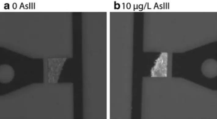
Fig. 3 Example of a microfluidics assay readout with sensor–reporter bacteria. Cells of E. coli (pPR-arsR-ABS) were incubated without (a) or with (b) 10 µg As(III)/L for 60 min. The intensity of the fluorescence produced from the reporter (here, green fluorescent protein) is imaged with a camera, from approximately 200 cells in a microcavity on the chip
The S. aureus bioreporter utilizes firefly luciferase and bioassays with this strain achieved a linear detection range for arsenic of 8–257μg/L [17]. Bioassays with the R. palustris sensor–reporter strain rely upon a spectrophoto-metic determination of color change that does not have a linear detection range [24].
There are many advantages and disadvantages to both biotic assays and abiotic sensors; however, sensor–reporter bacteria-based assays show a high promise for increased applicability in field deployment, not least because the
assays can be kept extraordinarily simple and the“sensor”
part (i.e., the bacterial cells) is self-reproductive at low cost. In a 2005 comparative study we provided compelling data on the performance of a bioassay using an E. coli sensor– luciferase reporter for arsenic in analyzing groundwater samples from Vietnam [25]. These data were obtained on simple aqueous suspension assays with the sensor–reporter cells and single-tube luminometers. Obviously, for direct field application one would like to have standalone assay systems with low or no maintenance requirements. This is still difficult to achieve, because of poor maintenance of the immediate reaction potential of the sensor–reporter cells upon long-term storage [26]. Most often, therefore, bio-assays apply freshly grown or deep-frozen cells to have the shortest reaction time. Future improvement may be achieved by embedding and drying techniques, or
applica-tion of cells to microfluidics devices (Fig.3) or fiberoptic
cables, and the development of in-field assays are all future capabilities for biological sensors that are currently under development.
Outlook on future bioassays
Bacterial sensor–reporter cells have proven to provide simple and quantitative assays, not only for arsenic determination, but in a variety of fields [27]. Bioassays with arsenic-specific bacterial sensor–reporter cells are usually carried out in an aqueous suspension with the target analyte, but this does not necessarily limited the assay to groundwater or drinking water analysis. Recently, we showed that a bioassay can be adapted to analyze arsenic in rice [28], and assays using bacterial sensor– reporters have also been applied to crab urine [29] and human blood [30]. Bioassays may also be developed for arsenic in soil using soil-water extracts, but similar as for groundwaters this may require optimization of procedures to avoid arsenic complexation with iron hydroxides [25].
Still, sensor–reporter-based bioassays are still hardly
ap-plied outside research laboratories. This may have a variety of reasons, such as them being genetically modified organisms, a lack of regulatory imperative to use biosensor tests as opposed to more regular chemical tests, and
absence of commercial enterprises marketing the sensor– reporter cells and the accompanying assays. There are many avenues for improvement and expansion of both the bacterial strains and the bioassays with the strains for use in the field, which would enable easier maintenance even for non-microbiology experts. For example, microbial biosensor cells are currently being applied to microfluidics devices for use as sensors in the field [22], as well as to
fiberoptic cables for in situ deployment [31,32].
Further developments may lie in combinations of bacterial sensor–reporters and other organisms. For example, bacterial sensor–reporters were used to measure toxicants in urine of sea crabs [29]. Most previous research has focused on the use of microbes as biological detectors of arsenic, but there are other organisms that could be used to detect arsenic in different and specialized ways. For example, it is possible that the nematode Caenorhabditis elegans extracted from arsenic-contaminated soils could be fed bacteria that fluoresce when exposed to arsenic, and a fluorescent response in the gut of the nematode would indicate the presence of arsenic. Additionally, the Asiatic clam Corbicula fluminea is a known bioaccumulator of arsenic [33], and it could be deployed in natural waters containing arsenic to serve as a proxy for the amount of arsenic bioaccumulation possibly seen in other aquatic species.
References
1. Mandal BK, Suzuki KT (2002) Talanta 58:201–235 2. Jain CK, Ali I (2000) Water Res 34:4304–4312
3. National Research Council (1999) Arsenic in drinking water. National Academy Press, Washington
4. Smith AH, Lingas EO, Rahman M (2000) Bull World Health Organ 78:1093–1103
5. Kim KW, Bang S, Zhu Y, Meharg AA, Bhattacharya P (2009) Environ Int (in press)
6. Smedley PL, Kinniburgh DG (2002) Appl Geochem 17:517–568 7. Cai J, DuBow MS (1997) Biodegradation 8:105–111
8. Oremland RS, Stolz JF (2003) Science 300:939–944 9. Cullen WR, Reimer KJ (1989) Chem Rev 89:713–764
10. Hung DQ, Nekrassova O, Compton RG (2004) Talanta 64:269– 277
11. Melamed D (2005) Anal Chim Acta 532:1–13
12. Rahman M, Mukherjee D, Sengupta MK, Chowdhury UK, Lodh D, Chanda CR, Roy S, Selim M, Quamrussaman Q, Milton AH, Shahidullah SM, Rahman MT, Chakraborti D (2002) Environ Sci Technol 36:5385–5394
13. Steinmaus CM, George CM, Kalman DA, Smith AH (2006) Environ Sci Technol 40:3362–3366
14. van der Meer JR, Tropel D, Jaspers MCM (2004) Environ Microbiol 6:1005–1020
15. Rosen BP (1995) J Basic Clin Physiol Pharmacol 6:251–263 16. Daunert S, Barrett G, Feliciano JS, Shetty RS, Shrestha S,
Smith-Spencer W (2000) Chem Rev 100:2705–2738
17. Tauriainen S, Karp M, Chang W, Virta M (1997) Appl Environ Microbiol 63:4456–4461
18. Tauriainen S, Virta M, Chang W, Karp M (1999) Anal Biochem 272:191–198
19. Stocker J, Balluch D, Gsell M, Harms H, Feliciano J, Daunert S, Malik KA, van der Meer JR (2003) Environ Sci Technol 37:4743– 4750
20. Wackwitz A, Harms H, Chatzinotas A, Breuer U, Vogne C, van der Meer JR (2008) Microb Biotechnol 1:149–157
21. Roberto FF, Barnes JM, Bruhn DF (2002) Talanta 58:181–188 22. Rothert A, Deo SK, Millner L, Puckett LG, Madou MJ, Daunert S
(2005) Anal Biochem 342:11–19
23. Date A, Pasini P, Daunert S (2007) Anal Chem 79:9391–9397 24. Yoshida K, Inoue K, Takahashi Y, Ueda S, Isoda K, Yagi K,
Maeda I (2008) Appl Environ Microbiol 74:6730–6738 25. Trang PTK, Berg M, Viet PH, Mui NV, van der Meer JR (2005)
Environ Sci Technol 39:7625–7630
26. Kuppardt A, Chatzinotas A, Breuer U, van der Meer JR, Harms H (2009) Appl Microbiol Biotechnol 82:785–792
27. Tecon R, van der Meer JR (2008) Sensors 8:4062–4080 28. Baumann B, van der Meer JR (2007) J Agric Food Chem
55:2115–2120
29. Lewis C, Beggah S, Pook C, Guitart C, Redshaw C, van der Meer JR, Readman JW, Galloway T (2009) Environ Sci Technol 43:423–428
30. Turner K, Xu S, Pasini P, Deo S, Bachas L, Daunert S (2007) Anal Chem 79:5740–5745
31. Hakkila K, Green T, Leskin P, Ivask A, Marks R, Virta M (2004) J Appl Toxicol 24:333–342
32. Ivask A, Green T, Polyak B, Mor A, Kahru A, Virta M, Marks R (2007) Biosens Bioelectron 22:1396–1402
33. Liao C, Jau S, Chen W, Lin C, Jou L, Liu C, Liao VH, Chang F (2008) Environ Toxicol 23:702–711
34. Tauriainen SM, Virta MPJ, Karp MT (2000) Water Res 34:2661– 2666
35. Tamminen MV, Virta MP (2007) Chemosphere 66:1329–1335 36. Harms H, Rime J, Leupin O, Hug SJ, van der Meer JR (2005)
Microchim Acta 151:217–222
37. Wells M, Gösch M, Harms H, van der Meer JR (2005) Microchim Acta 151:209–216
38. Wells M, Gösch M, Rigler R, Harms H, Lasser T, van der Meer JR (2005) Anal Chem 77:2683–2689