HAL Id: tel-02491320
https://tel.archives-ouvertes.fr/tel-02491320
Submitted on 26 Feb 2020
HAL is a multi-disciplinary open access archive for the deposit and dissemination of sci-entific research documents, whether they are pub-lished or not. The documents may come from teaching and research institutions in France or abroad, or from public or private research centers.
L’archive ouverte pluridisciplinaire HAL, est destinée au dépôt et à la diffusion de documents scientifiques de niveau recherche, publiés ou non, émanant des établissements d’enseignement et de recherche français ou étrangers, des laboratoires publics ou privés.
depending on prime-boost delay
Jean-Louis Palgen
To cite this version:
Jean-Louis Palgen. Characterization of the innate immunity elicited by vaccination and its interactions with adaptive immunity, depending on prime-boost delay. Innate immunity. Université Paris Saclay (COmUE), 2019. English. �NNT : 2019SACLS146�. �tel-02491320�
`ese
de
doctor
at
NNT
:2019SA
with adaptive immunity, depending on the
delay between prime and boost
Th`ese de doctorat de l’Universit´e Paris-Saclay pr´epar´ee `a l’Universit´e Paris-Sud Ecole doctorale n◦569 Innovation th´erapeutique, du fondamental `a l’appliqu´e (ITFA)
Sp´ecialit´e de doctorat : Immunologie Th`ese pr´esent´ee et soutenue `a Fontenay-aux-Roses, le 28 Juin 2019, par
J
EAN-L
OUISP
ALGENComposition du Jury : Isabelle Schwartz-Cornil Directrice de recherche,
INRA / Universit´e Paris-Saclay, D´epartement de virologie et
immunologie mol´eculaires Pr´esidente
Marc Dalod
Directeur de recherche,
CNRS / Universit´e d’Aix-Marseille / Inserm, CIML Rapporteur Helder Nakaya
Associate Professor,
University of S˜ao Paulo, Department of Clinical Analyses and Toxicology
Adjunct professor,
Emory University, Department of Pathology
Rapporteur
Nathalie Mantel Responsable d’unit´e,
Sanofi Pasteur, Virologie Recherche Examinatrice Jessica Quintin
Charg´ee de recherche,
Institut Pasteur, D´epartement de mycologie Examinatrice Anne-Sophie Beignon
Charg´ee de recherche,
Je profite de ces quelques lignes (ou plutôt quelques pages) pour remercier l’ensemble des personnes, qui de près ou de loin, directement ou indirectement, ont participé à l’aboutissement de ce projet. Je pense à tous mes collègues, amis et famille qui par leur présence, leurs conseils et leur soutien m’ont permis de mener à bien cette thèse. Je tiens à tous vous dire merci du fond du cœur.
En premier lieu, j’aimerais adresser de profonds remerciements à tous mes collègues de travail avec qui j’ai eu le plaisir de partager ces trois dernières années.
J’aimerais particulièrement remercier ma directrice de thèse Anne-Sophie Beignon pour son encadrement et son soutien pendant mes stages de master et les trois années de thèse qui ont suivi. Un grand merci pour sa constante positivité, sa réflexion scientifique et son écoute des autres, qui m’ont permis de développer plus avant ma curiosité scien-tifique et me lancer dans ce projet. Merci également pour tous les congrès internationaux auquel j’ai eu la chance de pouvoir participer et qui ont contribué à forger ce projet.
Je voudrais remercier le directeur d’IDMIT, Roger Le Grand, qui a accepté ma candi-dature en stage dans la laboratoire et m’a fait rencontrer Anne-Sophie. Merci également pour tous les conseils scientifiques.
J’aimerais aussi adresser un mot de remerciement aux membres de mon jury qui ont accepté d’évaluer mes travaux de thèses et pour toutes les discussions constructives lors de la soutenance. Merci à Marc Dalod (également pour son évaluation de ma mi-thèse), Helder Nakaya, Isabelle Schwartz-Cornil, Nathalie Mantel et Jessica Quintin.
J’adresse également un immense remerciement à Nicolas Tchitchek qui m’a fait re-découvrir la bio-informatique, pour ses suggestions constructives face à tous les problèmes, que ce soit dans les conceptions algorithmiques, ou la génération au pied levé d’un site web interactif, sans oublier son moral d’acier à toute épreuve (on ne lâche rien !).
Je remercie aussi tous les membres, présents et passés, du groupe d’Anne-Sophie, qui ont participé à la réalisation du projet ImMemory de recherche sur l’immunité vaccinale dans lequel s’est inscrite ma thèse. Un grand merci à David, André, Simon, Inana, Mal-vina, Hadjer, Gaëlle et Yanis.
J’aimerais remercier mes co-bureaux depuis trois ans, Julien L., Sixtine et Laura L., les doctorants avec qui j’ai eu la chance de vivre cette expérience de thèse, Candie, Cindy, Karunasinee alias Mo, Pierre, Lamine, Samuel, Adrien L.-P., André, Mélis, Néla, Mar-gaux, Matthieu et Thibaut, et tous ceux dont j’ai partagé le quotidien, Fanny, Marco, Laetitia B., Julie, Sophie B., Oscar, Ernesto (merci aussi pour le covoiturage !), Mario, Mylinda, Julien D., Rahima, Anna L., Quentin, Jamila, François, Brice, Salomé, Jean-Jacques, Nicolas G., Florian, Antoine, Diego, Mohammad, Sabine, Sophie L., Jérôme, Karine, Claude, Marie-Thérèse, Natalia, Stéphane, Louis, Yanis, Maxime E., Mathilde, Laetitia V. et Nastasia. Merci à tous pour votre présence, votre soutien, toutes les dis-cussions et tous les moments passés ensemble. Sans vous cette thèse n’aurait pas été la même, vous avez permis de rendre ces années inoubliables.
Je voudrais aussi adresser mes remerciements à tous ceux qui m’ont aidé que ce soit par des discussions scientifiques, par la gestion de l’expérimentation animale, des sys-tèmes informatiques (merci BatLab !) ou des formulations d’anglais. Merci à Oscar, Benoît F., Elisabeth, Frédéric M., Frédéric D., Brice, Jamila, Nadia, Antonio, Pauline, Romain, Vanessa, Delphine, Nathalie, Pascal, Christophe, Raphaël, Benoît D., Sébastien, Jean-Marie, Francis, Maxime P., Nina et Emma.
Enfin, je remercie tous les membres d’IDMIT, qu’ils soient doctorants, post-doctorants, directeurs d’étude, techniciens, ingénieurs, vétérinaires, commerciaux, stagiaires, maîtres de conférence, chargés de recherche, directeurs de recherche, ingénieurs-chercheurs..., qui font de ce laboratoire ce qu’il est aujourd’hui. Merci de créer ce cadre de travail et de vie unique qui a rendu cette thèse possible.
J’aimerais remercier aussi tous mes collègues enseignants de la faculté de Châtenay-Malabry, pour m’avoir fait découvrir le métier d’enseignant-chercheur. Merci en partic-ulier à Audrey, Dorine, Marion, Eva, Damien G. et Clémence pour leur accueil au 5ème étage et pour tous les moments passés ensemble à côté des TP et ED. Et merci également à tous les étudiants que j’ai eu en cours sans qui cette expérience d’enseignement n’aurait pas eu lieu.
Parce qu’à côté du travail de thèse, l’environnement et la vie hors laboratoire sont aussi importants, j’aimerais adresser quelques mots à mes amis pour leur soutien, leurs attentions et tous les moments vécus au cours non seulement de ces trois années mais aussi de toutes celles qui ont précédé.
Je voulais remercier Flora, Sandrine, Damien H., Yoann, Nadège et Guillaume pour toutes les après-midi et soirées jeux de société et cartes, accompagnées de thé, crêpes, gaufres. . . sans oublier une dose incommensurable de bonne humeur. Merci pour tous ces moments précieux au cours de ces dernières années !
J’aimerais remercier Aaron, Laura V., Hadrien et Anna P. pour toutes les excursions à Bordeaux (et les soirées Frog and Rosbif !), sans oublier les délicieuses tortillas y paellas españolas.
Je voudrais aussi remercier Léane pour toutes les sorties en Bourgogne et dans la cam-pagne lyonnaise.
J’aimerais également remercier Adrien S. pour son soutien pendant toutes ces années et tous les moments passés en Auvergne.
Enfin, je voudrais remercier toute ma famille, pour ces moments partagés, que ce soit au cours d’évènements particuliers (mariage, baptême, Noël, cousinades. . . ), ou simple-ment du quotidien, pour toutes les bonnes ondes que vous n’hésitez pas à partager. Merci pour tout ce que vous apportez.
Plus particulièrement, j’aimerais remercier mes frères, belle-sœurs et nièces, Marc, Sienna, Vincent, Clara, Méryl et Nora, pour tous les moments conviviaux partagés en-semble, pour le soutien que vous m’avez toujours apporté malgré les kilomètres qui ont pu nous éloigner.
Merci à Ghislain pour la découverte des Monts d’Or, du Beaujolais et des Dombes. Merci pour toutes les sorties randonnées, barbecues et méchouis. Merci pour tous ces moments passés ensemble.
Je voudrais aussi remercier mes parents. La liste de tout ce que vous m’avez apporté est trop longue pour tenir en quelques mots. Merci pour toutes ces années où vous m’avez accompagné et soutenu, merci d’avoir toujours cru en moi, merci pour toutes les valeurs humaines que vous m’avez transmises, merci pour tous ces moments infiniment précieux. Merci pour tout.
Enfin, un infini merci à Lise, mon amour, pour tous les moments que nous partageons, des excursions à travers le monde jusqu’aux instants du quotidiens que je chéris tout au-tant. Merci pour toutes tes attentions, pour ton soutien constant, merci de ta présence qui m’incite à toujours donner le meilleur de moi. Sans toi, rien n’aurait la même saveur. Notre aventure continue bientôt de l’autre côté de la Terre !
Table of contents
Acknowledgments vii
Contents xi
Table of contents . . . xi
List of figures . . . xiv
List of tables . . . xv
List of abbreviations xvii
Introduction
1
Vaccination: principle, successes and current limits . . . 3Innate immunity and vaccination . . . 10
Innate immune cells, an overview . . . 10
Innate cells as effector cells . . . 12
Recognition of pathogens . . . 12
Clearance of pathogens . . . 17
Innate cells as modulator cells . . . 22
Activation and shaping of adaptive immunity . . . 22
Modulation of innate cell effector functions by adaptive and innate immunity . . . 27
Resolution of inflammation . . . 29
Memory within innate immunity . . . 32
Trained innate myeloid cells . . . 32
“Memory“ NK cells . . . 35
Part I.
Methodology
41
Chapter 1. Experimental model and technologies used 43
Animal model: cynomolgus macaque . . . 43
Animal models in immunology, towards the choice of non-human primate . 43 Mouse models . . . 43
Non human primate models . . . 46
Non human primate models and vaccination, an overview . . . 48
Cynomolgus macaques as a model among non human primates . . . 49
Vaccine model: modified vaccinia virus Ankara . . . 50
From vaccinia virus to a new smallpox vaccine . . . 50
Modified vaccinia virus Ankara: development and immunity . . . 52
Modified vaccinia virus Ankara as a vaccine vector . . . 54
Modified vaccinia virus Ankara as vaccine model in the project . . . 55
Main experimental technology: mass cytometry . . . 56
The requirement of single-cell measurement . . . 56
Proteomics vs. transcriptomics: pros and cons . . . 58
Mass cytometry as the main analysis technics . . . 59
Toward a systems immunology approach . . . 62
Bioinformatic tools are required for mass cytometry data exploration . . . 62
viSNE vs. SPADE algorithm, pros and cons . . . 63
viSNE: visualization of t-distributed stochastic neighbor embedding results . . . 63
SPADE: spanning-tree progression analysis of density normalized events . . . 66
Multi-dimensional analysis tools . . . 69
Experimental approach, summary . . . 71
Chapter 2. Validation of the experimental model: comparison of blood innate immune cells in human and cynomolgus macaques 75 Overview . . . 75
"In Depth Comparative Phenotyping of Blood Innate Myeloid Leukocytes from Healthy Humans and Macaques Using Mass Cytometry" . . . 77
Part II.
Innate responses induced by a prime-boost vaccination
follow-ing a classical vaccination schedule
93
Chapter 3. Innate myeloid responses after a prime-boost vaccination fol-lowing a classical schedule 95 Overview . . . 95"Prime and Boost Vaccination Elicit a Distinct Innate Myeloid Cell Immune Response" . . . 99
Chapter 4. NK cell responses after a prime-boost vaccination following
a classical schedule 119
Overview . . . 119
"NK cell immune responses differ after prime and boost vaccination" . . . 122
Part III.
Impact of a shortened delay between prime and boost on
vaccine-induced immune responses
143
Chapter 5. Innate myeloid cell responses during an early boost vaccina-tion schedule and correlavaccina-tion with adaptive immune memory145 Overview . . . 145"The timing of vaccine boosting regulates the induction of memory innate myeloid cells and the quality of the secondary antibody response" . . . 149
Chapter 6. NK cell responses during an early boost vaccination schedule and correlation with adaptive immune memory 223 Overview . . . 223
"Boost vaccination timing impacts the quality of NK cell response, which corre-lates with a differential humoral response" . . . 225
Conclusions and perspectives
271
Results summary . . . 273Opened questions . . . 277
Innate and cellular adaptive immunity interactions . . . 277
NK cell phenotyping . . . 278
Innate immune memory duration . . . 279
Enhanced functions of “likely trained” cells and adaptive immunity activation281 “Likely trained” innate immune cell ontogeny . . . 282
Innate immune training related pathways . . . 285
Overtraining and innate immune exhaustion . . . 286
Impact of the immunization route . . . 287
Vaccine efficacy assessment . . . 288
Harnessing innate immune training to improve vaccine design . . . 289
Concluding remarks . . . 291
References
293
Appendix
349
List of figures
1 Principle of vaccination. . . 4
2 Overview of vaccination impact on public health nowadays. . . 7
3 Rationale for prime-boost strategy. . . 9
4 Classical hematopoiesis overview. . . 11
5 A wide range of receptors directly recognizing pathogen/vaccine. 13 6 Indirect recognition of pathogen through antibody or comple-ment binding. . . 15
7 Recognition of infected cell signature via MHC molecules. . . 16
8 Signaling pathways activated upon PRR interaction with its lig-ands. . . 18
9 Signaling pathways activated upon indirect pathogen recognition. 19 10 Signaling pathways activated upon NK receptor engagement. . . . 20
11 Consequences of innate immune cells activation by pathogens or vaccines. . . 21
12 Activation of T cells by innate immune cells. . . 25
13 Overview of direct anti-pathogen activity of adaptive response. . 26
14 Scheme of crosstalk between innate and adaptive immunity re-sulting in pathogen clearance. . . 28
15 Integrated scheme of inflammation resolution. . . 31
16 Current view of trained immunity in vaccination. . . 34
17 Diverse populations within “memory” NK cells. . . 37
18 Graphical abstract of the project aims. . . 39
19 Phylogenetic tree of mammals. . . 45
20 Non-human primates mirror human diseases. . . 47
21 Poxvirus viral cycle. . . 51
22 MVA sensing by the immune system. . . 53
23 Principle of mass cytometry. . . 59
24 Representation of the high dimensionality generated by mass cy-tometry. . . 62
25 Examples of viSNE representation. . . 64
26 The SPADE algorithm. . . 68
27 Main data representations from the packages developed and used in this project. . . 70
28 Experimental strategy. . . 72
29 Characterization of innate responses after prime and boost with respect to vaccine schedule. . . 275
30 Associations between innate responses and adaptive responses. . . 276
31 Potential impact of a late boost on the resulting immune response.280 32 Generation of trained innate immune cells. . . 283
List of tables
1 Current vaccines. . . 6 2 Main technologies available to decipher vaccine-induced immune
response. . . 57 3 High-dimensional technologies to study NK cell compartment. . 278
ADCC: antibody dependent cell cytotoxicity ADCP: antibody dependent cell phagocytosis AIDS: acquired immune deficiency syndrome
ANRS: French agency for research on AIDS and viral hepatitis APC: antigen-presenting cell
APRIL: A proliferation-inducing ligand BCG: bacillus Calmette-Guérin
BCR: B cell receptor
CCL: chemokine (C-C motif) ligand CCR: chemokine (C-C motif) receptor CD: cluster of differentiation
cDC: classical DC
CEV: cell-associated enveloped virus
cGAS: cyclic guanosine monophosphate adenosine monophosphate synthase CHIKV: chikungunya virus
CITE-seq: cellular indexing of transcriptomes and epitopes by sequencing CLP: common lymphoid progenitor
CMP: common myeloid progenitor CR: complement receptor
CXCL: chemokine (C-X-C motif) ligand CXCR: chemokine (C-X-C motif) receptor CyTOF: cytometry by time of flight
DAMP: damage/danger associated molecular pattern DC: dendritic cell
DCIR: dendritic cell immunoreceptor
DC-SIGN: dendritic cell-specific intracellular adhesion molecule-3-grabbing non-integrin DDA: dimethyldioctadecylammonium
DENV: dengue virus
DNA: desoxyribonucleic acid ds: double-stranded
EBV: Epstein-Barr virus
EEV: extracellular enveloped virus Fc: constant fraction
FoxP3: forkhead box 3 GAG: glycosaminoglycan
GBV-B: hepatitis G virus B /GB-virus B GMP: granulocyte-monocyte progenitor HCMV: human cytomegalovirus
HBV: hepatitis B virus HCV: hepatitis C virus HEV: hepatitis E virus
HIV: human immunodeficiency virus HLA: human leukocyte antigen HSC: hematopoietic stem cell
iC3b: inactive complement molecule C3b iE-DAP: D-glutamyl-meso-diaminopimelic acid IEV: intracellular enveloped virus
IFN: interferon Ig: immunoglobulin IL: interleukin
ILC: innate lymphoid cell IMV: intracellular mature virus
IP-10: interferon gamma-induced protein 10 (alternative name: CXCL10) ITAM: immunoreceptor tyrosine-based activating motif
ITIM: immunoreceptor tyrosine-based inhibitory motif KLRG1: killer-cell lectin like receptor G1
KSHV: Kaposi’s sarcoma associated herpes virus LabEx: French laboratory of excellence
LAPV: live-attenuated pertussis vaccine
LASSO: least absolute shrinkage and selection operator LDA: linear discriminant analysis
LGP2: laboratory of genetics and physiology 2 LPS: lipopolysaccharide
LRR: leucine rich repeat
MCMV: murine cytomegalovirus
MCP-1: monocyte chemoattractant protein 1 (alternative name: CCL2) MDA5: melanoma differentiation-associated protein 5
MDSC: myeloid-drived suppressor cell MDP: muramyldipeptide
MEP: megakariocyte-erythrocyte progenitor MHC: major histocompatibility complex
Mincle: macrophage inducible Ca2+-dependent lectin receptor MIP: monocyte inflammatory protein
MPLA: monophosphoryl lipid A
mTOR: mechanistic target of rapamycin MSI: mean signal intensity
MVA: modified vaccinia virus Ankara
NACHT: neuronal apoptosis inhibitor protein MHC class 2 transcription activator in-compatibility locus protein from Podospora anserina telomerase-associated protein NALP: NACHT, LRR and PYD containing domain
NKG2: Natural killer cell protein group
NLRP: NOD-like receptor family pyrin domain containing
NOD: nucleotide-binding oligomerization domain-containing protein oxLDL: oxidized low-density lipoprotein
PAMP: pathogen associated molecular pattern PBMC: peripheral blood mononuclear cell pDC: plasmacytoid DC
PFU: plaque-forming unit
poly(I:C): polyinosinic-polycytidilic acid PRR: pattern recognition receptor PYD: pyrin domain
REAP-seq: RNA expression and protein sequencing assay RhCMV: rhesus cytomegalovirus
RhLCV: rhesus lymphocryptovirus RIG-I: retinoic acid-inducible gene I RNA: ribonucleic acid
RNAseq: RNA sequencing ROS: reactive oxygen species RRV: rhesus macaque rhadinovirus sCD40L: soluble CD40 ligand
SHIV: simian/human immunodeficiency virus SIV: simian immunodeficiency virus
SPADE: spanning-tree progression analysis of density normalized events ss: single-stranded
stHIV: simian tropic HIV SVV: simian varicella virus Tcm: central memory T cell TCR: T cell receptor
Tem: effector T cell
Tfh: follicular helper T cell TGF: transforming growth factor Th: helper T cell
TLR: Toll-like receptor TNF: tumor necrosis factor Treg: regulatory T cell TRIM: tripartite motif VACV: vaccinia virus
VEGF: vascular endothelial growth factor
VIGIV: vaccinia immune globulin intravenous (human)
viSNE: visualization of t-distributed stochastic neighbor embedding algorithm VZV: varicella-zoster virus
WHO: world health organization YFV: yellow fever virus
Vaccination: principle, successes and current limits
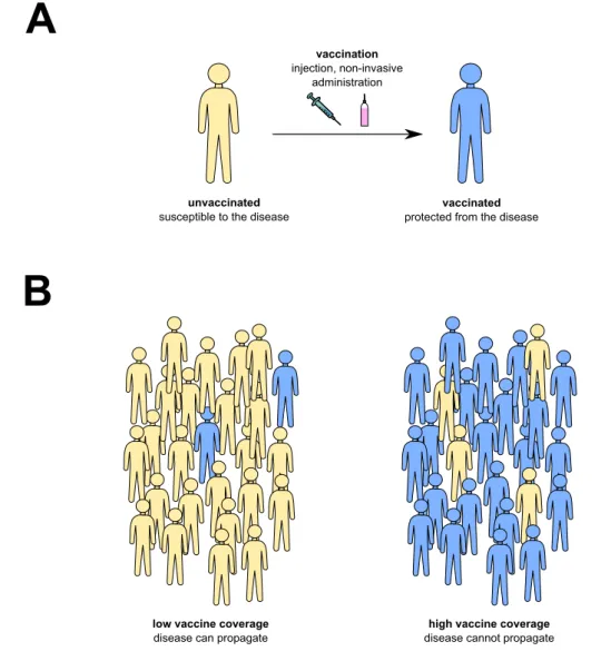
Vaccination, as defined by the World Health Organization, consists of the injection of any biological that enhances immunity toward a given disease. It relies on the ability of the immune system to remember previous encounters with a given pathogen, the so-called immune memory. As a consequence, the immune system can react more rapidly and ef-ficiently at the next pathogen encounter, which usually prevents the disease to occur. This also impedes the transmission of the disease from person to person and thus also ensures group protection at the public health level (Figure 1). That is why vaccination was described as one of the main advances ever made along with access to clean water, sanitation and antibiotics discovery (Greenwood, 2014;Rappuoli et al.,2014).
Examples of vaccination can be found several centuries ago, with reports of inoculation of extract of smallpox sore into healthy people, a relatively unsafe process (potentially lethal) called variolation, which could reduce risks of infection but also propagate the disease. Such processes were still widely in use in the middle 18th century (Greenwood, 2014; Rappuoli et al., 2014). The field of vaccination knew a massive expansion with the work of Jenner in 1796 who immunized people with animal poxviruses that cause very mild symptoms in humans, but prevented them to develop the much more severe small-pox (Tognotti, 2010;Plotkin, 2014). A century after, in 1880, Louis Pasteur successfully inoculated dead/attenuated pathogens and prevented infection in the animals and hu-mans inoculated. He notably developed the first vaccine against rabies. Afterwards, with the progresses made in molecular biology, recombinant vaccines were designed, consisting of purified proteins associated with adjuvants molecules designed to elicit a sufficiently intense inflammation. This technics notably led to the development of the first hepatitis B vaccine (Plotkin,2014).
A
unvaccinated
susceptible to the disease
vaccinated
protected from the disease
vaccination
injection, non-invasive administration
B
low vaccine coverage
disease can propagate
high vaccine coverage
disease cannot propagate
Figure 1. Principle of vaccination. Vaccination induces protection at (A) the indi-vidual level and (B) the population level, called herd immunity.
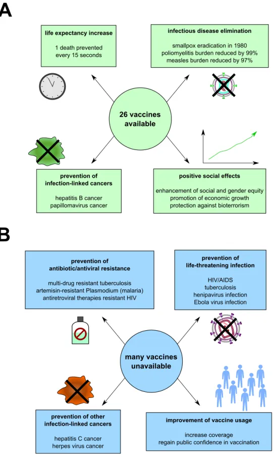
Nowadays, 26 vaccines are referenced by the WHO (including phase III vaccine trials) to prevent a wide variety of pathogens and diseases: cholera, dengue, diphteria, hepatitis A, B and E, Haemophilus influenzae B, human papillomavirus, influenza viruses, japanese encephalitis, malaria, measles, menincogoccal meningitis, mumps, pertussis, pneumococ-cal disease, poliomyelitis, rabies, rotavirus, rubella, tetanus, tick-borne encephalitis, tu-berculosis, typhoid fever, chickenpox/shingles and yellow fever (Table 1). They consist either of live-attenuated micro-organisms (e.g. yellow fever virus vaccine), inactivated micro-organisms (e.g. inactivated poliovirus vaccine), recombinant microbe proteins with adjuvants (e.g.. hepatitis B virus vaccine), or toxoids –non toxic modified toxins– (e.g. diphteria vaccine).
Great successes were met in the past decades, with the eradication of smallpox in 1980 (WHO), the near-eradication of poliomyelitis (99% reduction of cases, with 29 cases reported to WHO in 2018), or the drastic reduction of cases of measles in the past decades (from around 4,000,000 deaths in 1980 vs. less than 100,000 deaths in the last few years) (Figure 2A). However, more than two centuries after the invention of the term vaccine, infectious diseases still remain one major threat to population with more than 10 mil-lion death every year (WHO). Vaccines are still lacking for many complex pathogens, including but not limited to, human immunodeficiency virus (HIV, causing the acquired immunodeficiency syndrome -AIDS), respiratory syncytial virus (RSV, causing neona-tal bronchiolitis) or henipaviruses (causing lethal encephalitis). In addition, for some pathogens, existing vaccines are insufficiently efficient to allow for a good protection, such as influenza virus -seasonal flu (Wong & Webby,2013;Sautto et al.,2018), mycobacterium tuberculosis –tuberculosis (Andersen & Doherty, 2005; Voss et al., 2018), dengue virus –dengue (McArthur et al., 2013; Bos et al., 2018), or Plasmodium –malaria (Snounou et al., 2005; Frimpong et al.,2018) (Figure 2B).
Difficulties to develop new vaccines can rely on the complexity of the pathogen it-self, due to its ability to evade immune response (e.g. rapid genetic variation for HIV (Barouch,2008), polymorphism of immunogenic antigen for Plasmodium (Hisaeda et al., 2005), genetic shift and drift for influenza viruses (van de Sandt et al., 2012)). But the main reason for this impediment in vaccine design is that despite decades of studies of the immune system and immune responses to pathogens, we are still nowadays far to completely understand all the aspects of the immune response.
For example, only few live vaccines (17D yellow fever virus vaccine and vaccinia virus smallpox vaccine) elicit a life-lasting immune protection (Amanna et al.,2007;Wrammert et al., 2009), while most vaccines, including live-attenuated (e. g. measles and mumps vaccine), inactivated (e.g. rabies virus vaccine) and recombinant (e. g. hepatitis B) vaccines require several injections to develop a long-lasting protection in most people –a first immunization called prime and latter ones called boost(s) (Ramshaw & Ramsay, 2000; Woodland, 2004). The sole prime can induce a short-lasting and/or only partial protection from the disease (Figure 3A). In addition, since responses to vaccines dif-fer between individuals, prime may only induce a protective immunity in a fraction of the population (Figure 3B). Overall prime-boost strategies aim to enhance individual response by recalling a primary immune memory, and enhance the frequency of vaccine responders among the population, to ensure protection from the disease at both level.
Determination of the best vaccination schedule is still empirically defined, based on clinical trials that may miss the optimal settings. For example the schedule reported by WHO for diphteria, tetanus, poliomyelitis and pertussis combination vaccine is 2, 4, and 11 months old in France, 2, 3, 4 and 15 months in Belgium, and 2, 4, 6, 15-24 months old in Switzerland, but we miss an objective argumentation to rationally choose any of these schedules.
Disease and main pathogen
targeted Type of vaccine Limitations
Cholera
Vibrio cholerae inactivated bacterium
-Dengue Dengue virus
live-attenuated viral vector YFV-based
recommended only for individuals with pre-existing
immunity against DENV Diphteria
Corynebacterium diphtheriae toxoid
-Viral hepatitis Hepatitis A virus
inactivated or
live-attenuated virus
-Viral hepatitis
Hepatitis B virus recombinant protein
-Viral hepatitis
Hepatitis E virus recombinant protein
-Bacterial pneumonia/meningitis
Haemophilus influenzae B recombinant saccharide
-Viral genital cancers
Human papillomavirus recombinant protein
-Flu
Influenza virus inactivated virus
low efficiency (around 50% each year) Japanese encephalitis
Japanese encephalitis virus
inactivated or live-attenuated
virus
-Malaria
Plasmodium falciparum recombinant protein
short-term and partial protection Measles
Measles virus live-attenuated virus
-Bacterial meningitis
Neisseria meningitidis recombinant saccharide
-Mumps
Mumps virus live-attenuated virus
-Whooping cough Bordetella pertussis
inactivated bacterium or recombinant protein
prevention of the symptoms but not infection (recombinant
vaccine) Pneumococcal diseases
Streptococcus pneumoniae recombinant protein
-Poliomyelitis Poliomyelitis virus
inactivated or live-attenuated virus
rare paralysis with the live-attenuated vaccine Rabies
Rabies virus inactivated virus
-Viral diarrhea
Rotavirus live-attenuated virus
-Rubella
Rubella virus live-attenuated virus
-Tetanus
Clostridium tetani toxoid
-Tick-borne encephalitis
Tick-borne encephalitis virus inactivated virus
-Tuberculosis
Mycobacterium tuberculosis live-attenuated bacterium
poor protection against adult pulmonary disease Typhoid fever Salmonella typhi live-attenuated bacterium or recombinant saccharides -Yellow fever
-A
B
26 vaccines available
life expectancy increase
1 death prevented every 15 seconds
infectious disease elimination
smallpox eradication in 1980 poliomyelitis burden reduced by 99%
measles burden reduced by 97%
prevention of infection-linked cancers
hepatitis B cancer papillomavirus cancer
positive social effects
enhancement of social and gender equity promotion of economic growth protection against bioterrorism
prevention of antibiotic/antiviral resistance
multi-drug resistant tuberculosis artemisin-resistant Plasmodium (malaria)
antiretroviral therapies resistant HIV
prevention of life-threatening infection
HIV/AIDS tuberculosis henipavirus infection Ebola virus infection
prevention of other infection-linked cancers
hepatitis C cancer herpes virus cancer
improvement of vaccine usage
increase coverage
regain public confidence in vaccination
many vaccines unavailable
Figure 2. Overview of vaccination impact on public health nowadays. Current successes (A) and unmet challenges (B) of vaccination, based on WHO estimations, are represented.
This sheds light on the fact that despite decades of studies on vaccine-induced response, we still miss the complete picture and are unable to capitalize our current knowledge in simple parameters that vaccinologists could use to modulate immunity (e.g. route of immunization, type of antigens, number of injections, delay between each injection. . . ). Going further into the rational design of vaccine requires a better characterization of the initial trigger of vaccine-induced immune response, the innate immunity, and how this impacts the adaptive immune response that is known to mediate memory.
A
unvaccinated
susceptible to the disease
fully vaccinated
protected from the disease
prime vaccination
B
no vaccination
disease can easily propagate
prime vaccination
(high vaccine coverage) disease can partially propagate
prime vaccinated
partially protected from the disease (e.g. attenuated symptoms)
boost vaccination
time adaptive
immune response
boost vaccination
(high vaccine coverage) disease cannot propagate
initial state primary response secondary response
effector phase
memory phase
effector phase
memory phase
Figure 3. Rationale for prime-boost strategy. (A) Impact of prime-boost at indi-vidual level. Prime vaccination induces a primary memory, which can be only partially protective, whereas boost vaccination recalls the primary memory, giving rise to a sec-ondary immune memory that is likely more protective. (B) Impact of prime-boost strat-egy at the population level. Prime vaccination results in only a fraction of the population that is fully protected from the disease, whereas after boosting, individuals whose
pri-Innate immunity and vaccination
Innate immune cells, an overview
Vaccines are designed to mimic at most the infection of pathogen, including the trig-gering of a strong and robust immune response, but obviously without the pathogenicity associated with the pathogen. As a consequence, the detection of a vaccine follows similar pathways as a pathogen, including the recognition by innate immune cells.
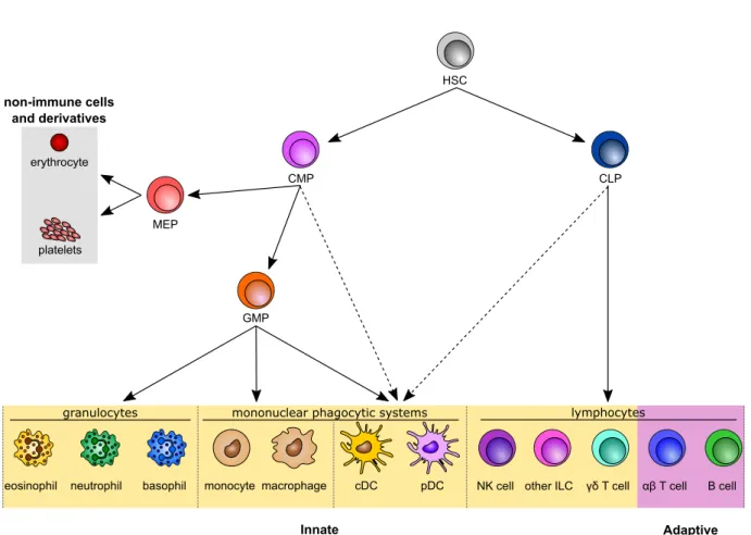
Innate immunity is composed of a wide range of cells, including both myeloid and lymphoid ones. They essentially arise from bone marrow hematopoiesis (Orkin & Zon, 2008; Laurenti & Göttgens, 2018), although some new cell generation can also occur in other organs, including yolk sac during development and adult liver (Taniguchi et al., 1996; Palis & Yoder, 2001; Yamamoto et al., 2016) (Figure 4). Note that beyond the simplified overview represented here, several intermediate cells are involved in the gen-eration of a wide range of fully differentiated immune cells, including a high plasticity potential of cell progenitors (Manz et al., 2001). In addition, the development of new recent technologies allowed to unveil an unprecedented heterogeneity among immune cell precursors, suggesting that immune cell development is likely more complex than what was initially thought (Perié & Duffy, 2016).
Innate immune cells share the ability to react rapidly upon pathogen infection or vac-cine injection. They are often described as the first line of defense of the immune system, although non-immune cells (e.g. epithelial cells, fibroblasts) are actually the first to en-counter pathogen in most cases. Overall, innate immune cells accomplished numerous functions.
HSC MEP GMP CMP CLP erythrocyte platelets
cDC pDC NK cell other ILC γδ T cell αβ T cell B cell monocyte macrophage
eosinophil neutrophil basophil granulocytes Innate immune cells Adaptive immune cells non-immune cells and derivatives
mononuclear phagocytic systems lymphocytes
Figure 4. Classical hematopoiesis overview. A simplified overview of the generation of the major immune cell populations is displayed. Intermediate populations between the different progenitors and precursors are not represented, neither the heterogeneity within each progenitor population. Dotted arrows indicate suggested alternative differentiation pathways of DCs. Dotted lines separate each compartment (granulocytes, monocytes, DCs and lymphocytes). Innate and adaptive immune cells are highlighted in yellow and purple frames respectively. Non-immune cells and derivatives arising from hematopoiesis are highlighted in a gray frame. HSC: hematopoietic stem cell. CMP common myeloid progenitor. CLP: common lymphoid progenitor. MEP: megakaryocyte-erythrocyte pro-genitor. GMP: granulocyte-monocyte propro-genitor. DC: dendritic cell. cDC: classical DC. pDC: plasmacytoid DC. NK: natural killer. ILC: innate lymphoid cell.
Innate cells as effector cells
Recognition of pathogens
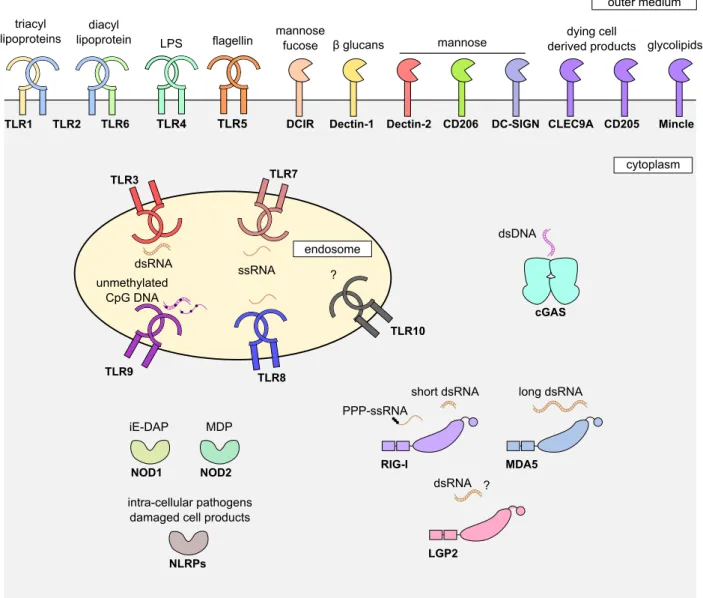
Once a pathogen or a vaccine enters the body, it can be directly recognized by receptors expressed by innate immune cells, but also non-immune cells, called pattern recognition receptors (PRR) (Takeuchi & Akira, 2010). These receptors can specifically recognize patterns associated with danger, damage or pathogen (Damage/Danger/Pathogen associ-ated molecular pattern DAMP or PAMP). The list of PRR and associassoci-ated DAMP/PAMP is wide and still growing (Akira et al.,2006;Takeuchi & Akira,2010;Cai et al.,2014). We will here just give an overview of the main PRR triggering direct pathogen recognition (Figure 5).
Toll-like receptors (TLR) are transmembrane proteins recognizing several patterns as-sociated with micro-organisms, including proteins (e.g. TLR2), saccharides (e.g. TLR4), or nucleic acids (e.g. single-stranded RNA and derived products -TLR7 and TLR8- or extra-nuclear DNA -TLR9) (Medzhitov, 2001; O’Neill et al., 2013; Tanji et al., 2015; Zhang et al., 2016; Ohto et al., 2018). RIG-like receptors (RLR) especially recognize double-stranded RNA (Reikine et al.,2014;Yoneyama et al.,2015;Hur,2019). NOD-like receptors (NLR) are cytosolic receptors that can recognize peptidoglycans (e.g. NOD1), proteins (e.g. NLRP1), or nucleic acids (e.g. NLRP3) (Kanneganti et al., 2007; Franchi et al., 2009); mutations in NLR genes were recently linked with cancer progression ( Sax-ena & Yeretssian,2014). C-type lectin like receptors (CLR) mainly recognized sugars (e.g. mannose, β glucan) present in fungi, viruses, but also in auto-immune diseases, allergy or cancer (Dambuza & Brown, 2015; Saijo & Iwakura, 2011; Shrimpton et al., 2009; Lu et al., 2018). Finally the recently discovered cGAS protein can recognize cytosolic DNA dimers through a complex pathway (Sun et al., 2013; Wu et al., 2013; Li et al., 2013; Cai et al., 2014). It was also recently shown that it could detect HIV nuclear DNA, in cooperation with the NONO protein that targets a conserved region of the HIV capsid (Lahaye et al., 2018).
Since recognition by PRR is one the most initial events in immune response, it is a cru-cial event in vaccine-induced immune response. Actually, PRR can be targeted by vaccine adjuvants used with recombinant vaccines (which are not immunogenic enough by them-selves, in contrast to live-attenuated vaccines), to elicit a strong immune response. For example, among adjuvants widely used in clinical development, poly(I:C) (polyinosinic-polycytidilic acid) and its derivatives activate TLR3 and RLRs, MPLA (monophosphoryl lipid A) activates TLR4, flagellin activates TLR5, imiquimol activates TLR7 and CpG containing oligonucleotides activate TLR9 (Coffman et al., 2010; Vasou et al., 2017). Note that the activation of distinct PRR trigger qualitatively different innate responses, as shown for TLR stimulation (Kwissa et al., 2012). Surprisingly, for alum, the most widely used adjuvant, it is not fully clear which receptor is at play, though NLRP3 in-flammasome seems important (Coffman et al., 2010).
TLR1 TLR2 TLR6 triacyl lipoproteins diacyl lipoprotein TLR4 LPS TLR5 flagellin dsDNA cGAS NOD1 NOD2 iE-DAP MDP RIG-I MDA5 PPP-ssRNA
short dsRNA long dsRNA
LGP2
dsRNA ?
Dectin-1 Dectin-2 DC-SIGN
DCIR
mannose
fucose β glucans mannose
CLEC9A dying cell derived products CD205 NLRPs intra-cellular pathogens damaged cell products
cytoplasm outer medium TLR7 TLR8 TLR9 TLR3 dsRNA unmethylated CpG DNA ssRNA ? TLR10 endosome CD206 Mincle glycolipids
Figure 5. A wide range of receptors directly recognizing pathogen/vaccine. The main human pattern recognition receptors are represented with their cognate lig-ands, when known. TLR: Toll-like receptor. CD: cluster of differentiation. DCIR: dendritic cell immunoreceptor. DC-SIGN: dendritic cell-specific intracellular adhesion molecule-3-grabbing non-integrin. Mincle: macrophage inducible Ca2+-dependent lectin receptor. cGAS: cyclic guanosine monophosphate adenosine monophosphate synthase. NOD: nucleotide-binding oligomerization domain-containing protein. NLRP: NOD-like receptor family pyrin domain containing. RIG-I: retinoic acid-inducible gene I. MDA5: melanoma differentiation-associated protein 5. LGP2: laboratory of genetics and phys-iology 2. LPS: lipopolysaccharide. ss: single-stranded. ds: double-stranded. iE-DAP: D-glutamyl-meso-diaminopimelic acid. MDP: muramyldipeptide.
In parallel, live-attenuated or dead pathogens used for vaccines are self-adjuvanted since they are strongly recognized by multiple PRRs. For example, yellow fever vaccine is recognized by TLR2, 7, 8 and 9 to trigger a robust immune response (Querec et al., 2006). Measles vaccine activate TLR2, 4, 5, 7 and 8, as well as RIG-I (Kennedy et al., 2012). Influenza vaccine is recognized by several PRRs including TLR3,7 and 8, RIG-I, NLRP3 inflammasome, as well as NLRs (Iwasaki & Pillai, 2014).
In addition to this direct PRR-mediated recognition, pathogens/vaccines can be de-tected through antibody-binding. Antibodies are immunoglobulin molecules synthesized by plasmablasts and plasma cells, which are specific to antigens. Following a previous encounter with the same pathogen/vaccine by the immune system, antibodies against the same pathogen/vaccine can be constitutively secreted in the body. They will thus bind to their target and stain the pathogen/vaccine for the innate immune cells. The recogni-tion of these antibody-coated pathogen/vaccine is driven by so called Fc receptors (FcRs) that can recognize the constant fraction of antibodies, each receptors displaying its own specificity (in term of antibody classes) and affinity (Bruhns & Jönsson,2015; Cho et al., 2006) (Figure 6A). An intra-cellular FcR, named TRIM21, which can recognize IgM and IgG was also identified (McEwan et al., 2013).
Besides, complement molecules, consist of proteins (constitutively present in the body, but inducible upon infection/vaccination) that activate themselves when bound to a pathogen or a vaccine, or an antibody-bound pathogen/vaccine, eventually leading to membrane disruption of the pathogen/vaccine (Merle et al.,2015a;b). Complement-bound pathogens can be recognized by so-called complement receptors (CR) expressed on im-mune and non-imim-mune cells –e.g. erythrocytes expressing CR1 (Merle et al., 2015a; Dustin, 2016) (Figure 6B).
Note that infected cells can also be labelled by both antibodies and complement molecules, and produce DAMPs.
Eventually, the abnormal immune signature of infected cell can be detected (Figure 7). Indeed, all cells express molecules called major histocompatibility complex (MHC) of class I that carries peptides deriving from the degradation of the proteins expressed within the cells (Neefjes et al., 2011; Rock et al., 2016). Should the cell be infected then pathogen/vaccine protein derived peptides will be presented by MHC I molecules on cell surface. In addition non-classical MHC molecules induced by cellular stress (e.g. MICA, MICB) will be expressed on cell surface. The MHC signature can be detected by the CD8 T cells in the adaptive part of the immune system and the natural killer (NK) cells on the innate side of the immune system (Vivier et al., 2008; Neefjes et al., 2011; Campbell & Hasegawa, 2013; Rock et al., 2016). CD8 T cells are specific of a given MHC I bound antigen, which they recognize through their T cell receptor (TCR). By contrast, NK cells have a broader repertoire of ligands, MHC class I molecules as well as non-classical MHC molecules, that are recognized by several NK receptors, whose expression is highly stochastic among NK cells (Wilk & Blish, 2018). NK cells can also be activated by a downregulation of MHC molecules, which can be induced by infection.
CR1 C3b CR2 CR3 CR4 iC3b C3d iC3b CD11b CD18 CD11c CRIg C3b iC3b CD64 CD32 CD16 FcεRI CD23 CD89 IgG IgE IgA IgM FcαµR Pathogen antibodies cytoplasm outer medium Pathogen cytoplasm outer medium
A
B
Figure 6. Indirect recognition of pathogen through antibody or complement binding. Mechanisms of indirect detection of pathogen via bound antibodies (A) or bound complement (B) is displayed. The main receptors are displayed in each panel. Fc: constant fraction. CD: cluster of differentiation. CR: complement receptor. iC3b: inactive C3b.
non-infected cell
NK cell
CD8 T cell steady state associated
MHC signature infection/stress associated MHC signature inhibitory signal activating signal NK receptors TCR infected cell
Figure 7. Recognition of infected cell signature via MHC molecules. Cells infected by a pathogen present peptides on MHC I that differ from non-infected cells. The infected cells also present stress-induced non-classical MHC molecules. Eventually MHC class I molecule expression can be downregulated upon infection. NK cells detect this overall abnormal MHC signature through several NK receptors. CD8 T cells detect a sole MHC I carrying a given antigen, through their T cell receptor (TCR).
Clearance of pathogens
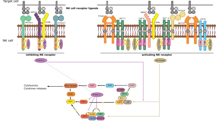
The recognition of a pathogen will trigger a cascade of activation involving several pathways (e.g. MyD88/TRAF, TRIF, ZAP kinases), depending on the receptor involved, such as PRR (Takeuchi & Akira,2010; Shaw et al., 2010; Loo & Gale, 2011; Wen et al., 2013; Reikine et al., 2014; Cai et al., 2014; Hoving et al., 2014; Sellge & Kufer, 2015; Balka & De Nardo,2018) (Figure 8), FcR (Sánchez-Mejorada & Rosales,1998; Getahun & Cambier,2015) (Figure9A) and complement receptors (Bohana-Kashtan et al.,2004; Dustin, 2016) (Figure 9B). Regarding the particular case of NK cells, the balance be-tween inhibitory signals (steady-state associated MHC signature) and activating signals (for example infection associated MHC signature) provided by a target cell determines whether an NK cell will be activated or not (Vivier et al.,2008;Chan et al.,2014) (Figure 10).
Note that despite the precise molecular characterization of the pathways involved, this knowledge at cell level does not allow to draw a comprehensive picture of the impact of each receptor activation at the immune system level.
Overall, these cascades will activate innate immune cells triggering several functions (Figure 11). Upon activation, some cells such as neutrophils, eosinophils, monocytes, macrophages and DCs can phagocyte the whole pathogens/vaccines they recognized ( Gor-don, 2016; Rosales & Uribe-Querol, 2017). This can be done following PRR-mediated recognition, complement-based recognition or antibody-mediated recognition (in this case, it is called antibody dependent cell phagocytosis ADCP). The process to facilitate pathogen uptake by phagocytic cells is called opsonization. Besides, some cells, mainly NK cells, NKT cells and γδ T cells in the innate immunity can secrete cytotoxic molecules that can disrupt pathogen/vaccine or infected cell membranes (e.g. perforin) and proteases that can trigger an apoptosis cascade in infected cells (e.g. granzyme) (Trapani, 2001; Trapani & Smyth, 2002; Caligiuri, 2008; Osińska et al., 2014). NK cell cytotoxicity can be triggered by an aberrant MHC I signature as well as by antibody-bound pathogen, the latter process being called antibody dependent cell cytotoxicity (ADCC). Besides, a specificity of neutrophils is their ability to expulse DNA out of their nucleus to capture microbes into DNA fibers, a process called neutrophil extra-cellular trap (NET) (Kaplan & Radic, 2012; Yang et al., 2016; Delgado-Rizo et al., 2017; Boeltz et al.,2019).
Eventually, upon activation every innate immune cells, as well as non-immune cells, can produce a wide range of cytokines (small proteins modulating cell behavior). In particular, they produce so-called inflammatory cytokines (such as IL-1α, 1α, MIP-1β, CCL5, IL-12. . . ). In addition to cytokines, some complement proteins, the so-called anaphylotoxins (C3a, C4a and C5a), also share these pro-inflammatory properties (Klos et al., 2009). These molecules will be recognized by immune and non-immune cells and initiate a process called inflammation.
Inflammation notably includes the recruitment of effector cells through the binding of chemokines (cytokines driving effector cell chemotaxis) on their cognate receptor on innate cells, the enhanced phagocytosis on phagocytes (e.g. macrophages, DCs, neutrophils), the increased permeability of vascular vessel to allow for recruitment of effector cells, and the generation and release of effector cells from bone marrow (Akdis et al., 2016; Pietras, 2017). Also, some cytokines produced have direct anti-pathogen effects and stimulate in-trinsic immunity -a constitutive immunity of individual cells, mediated by endogenously expressed proteins inhibiting pathogen replication. For example type I interferon (e.g. IFNα and IFNβ) induces the production of anti-viral genes in all cells, the so-called inter-feron stimulated genes (ISG), in addition to its immunomodulatory effect on immune cells (Yan & Chen,2012;Schneider et al.,2014; McNab et al., 2015). All cells cannot produce all cytokines (e.g. pDCs are specialized in IFN I production, NK cells are main producers of IFN II (IFNγ) (Akdis et al.,2016)), but all will contribute to the overall inflammatory state. Inflammation is a positively self-regulated process, since a consequence of cell ac-tivation by pro-inflammatory molecules is the release of more pro-inflammatory molecules.
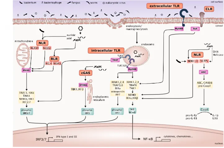
MyD88 ASC MyD88 STING MVAS TRIF cell surface cytosol CLR extracellular TLR intracellular TLR RLR cGAS NLR NLR Syk
IRF3/7 IFN type I and III
nucleus
NF-κB cytokines, chemokines...
Figure 8. Signaling pathways activated upon PRR interaction with its ligands. The signaling cascade is indicated for each of the aforementioned PRR. Red/orange: PRRs. Magenta: adapter protein. Yellow: downstream components. Green: transcrip-tion factor. Modified from Metzger et al. (2018).
Syk
Syk Syk Syk
Pl3K Pl3K membrane complement receptors cytoskeleton (phagocytosis) Calmodulin Gene Activation
Figure 9. Signaling pathways activated upon indirect pathogen recognition. Exemplified signaling pathways induced by indirect pathogen recognition via IgG re-ceptors (FcγR) and complement rere-ceptors. Only activating receptor signaling pathway is displayed here, and not inhibiting receptor signaling pathway (e.g. CD32b signaling pathway). Modified from Rosales (2017).
Src Ki nase P P P Src Kinase PTNP6/11 PTNP6/11 Target cell NK cell
NK cell receptor ligands
inhibiting NK receptor activating NK receptor
Cytotoxicity Cytokines releases
Figure 10. Signaling pathways activated upon NK receptor engagement. Exem-plified signaling pathways induced by the engagement of some of the main NK receptors. ITIM: immunoreceptor based inhibitory motif. ITAM: immunoreceptor tyrosine-based activating motif. Modified from Kelley et al. (2005).
Innate cells infected cell pro-inflammatory cytokines chemokines cytotoxic molecules NET ...
induction of apoptosis and anti-microorganism activity activation upo n detection activation recruitment killing phagocytosi s
Figure 11. Consequences of innate immune cells activation by pathogens or vaccines. Upon activation, innate cells will produce inflammatory cytokines (activating themselves and promoting apoptosis of infected cells), produce chemokines (attracting more effector cells) and kill pathogen and infected cells (e.g. through phagocytosis, cyto-toxic activity, NET (neutrophil extra-cellular trap)).
In term of spatio-temporal dynamics, the resident cells at the site of infection or im-munization (such as macrophages, dendritic cells, ILCs, γδ T cells on the immune part, but also non-immune cells, such as epithelial cells) will trigger the initial inflammatory response recruiting more effector cells, essentially neutrophils, the most abundant and short-lived cell population in blood (Pillay et al.,2010), but also monocytes, which could notably differentiate in macrophages and potentially cDCs (Gonzalez-Mejia & Doseff, 2009). This initial response can clear the pathogen/vaccine from the body, especially if few infectious agents or vaccine particles were present. But in addition to this effector function, a second function of the innate immune system is to activate, modulate and shape not only itself but also the second arm of the immune system, the adaptive one, constituted by T and B cells.
Innate cells as modulator cells
Activation and shaping of adaptive immunity T cell activation and polarization
Each T cell is able to recognize one sole antigen, encoded by non-self cells or micro-organisms, that is presented on an MHC molecule after processing. This recognition is mediated through its TCR arising from somatic rearrangements, during T cell matura-tion. (Zúñiga-Pflücker,2004;Koch & Radtke, 2011).
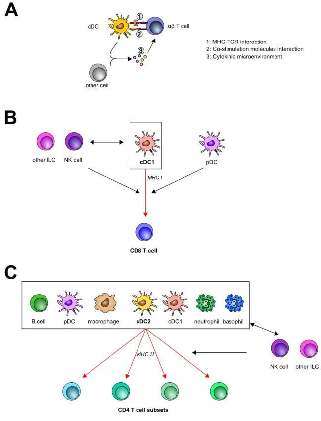
Cells such as dendritic cells, monocytes/macrophages and B cells, carry MHC class II molecules (in addition to class I), which can present antigen, not expressed within the cell (conversely to class I) but from pathogens/vaccines or infected cells they internal-ized (Neefjes et al., 2011; Rock et al., 2016). They are thus called professional antigen-presenting cells. Among them, cDCs are almost the only ones that can activate previously naive T cells (priming), while the other APCs can restimulate primed T cells. cDCs can migrate from the site of infection/immunization to secondary lymphoid organs (lymph nodes, spleen, gut associated lymphoid tissue, nasal associated lymphoid tissue) through lymphatic vessels. Note that some cDCs also reside within lymphoid organs. In these tissues, cDCs can present MHC II carrying antigen to so-called CD4 T lymphocytes, and MHC I carrying antigen to so-called CD8 T lymphocytes, to activate them (Figure 12A). T cell priming was also shown to occur in bone marrow in several contexts, including can-cer and infections (Schirrmacher et al., 2003). Strikingly, following vaccination with a smallpox vaccine, transport of antigen to the bone marrow was made by neutrophils and not cDCs in mice (Duffy et al., 2012). Still, in mice also, blood circulating antigens are eventually presented to T cells by cDCs (Feuerer et al., 2003).
All cDCs can do both presentations (to CD4 and CD8 T cells) but not with the same efficacy; actually two main subclasses of cDCs are defined: cDC1s and cDC2s (Schlitzer et al., 2015; Vu Manh et al., 2015; Collin & Bigley, 2018). cDC1s essentially present antigen captured during phagocytosis on MHC class I molecules (a process called cross-presentation) and are more efficient in antigen presentation to CD8 T cells (Figure12B), though they also activate CD4 T cells via MHC II (Bedoui et al., 2009; Eickhoff et al., 2015). cDC2s essentially present antigen on MHC II molecules and are major activator of CD4 T cells (Figure 12C), though they can also perform cross-presentation (Segura et al.,2013;Sheng et al.,2017). Note also that other cDC population exist, such as tissue resident Langherans cells or monocyte-derived DCs (Vu Manh et al., 2015).
Three signals are required to activate T cells, the first is the interaction between MHC (MCH I for CD8 T cells and MHC II for CD4 T cells) and the TCR, the second is the in-teraction between co-stimulation molecules expressed on both cells (e.g. CD80/86-CD28, CD40-CD40L), the last and third signal is the cytokine micro-environment that is essen-tial to polarize T cell differentiation.
Several subpopulations of CD4 T cells were identified based on their cytokine produc-tion, transcription factors expression, immune functions, and protective capacity (O’Garra, 2000;Zhu & Paul,2008;Zhu et al.,2010;Geginat et al.,2014;Crotty,2015;Raphael et al., 2015;DuPage & Bluestone,2016). For example, among CD4 helper T cells (Th), Th1 cells are described as potentiators of macrophage activation and NK cell activation through IFNγ production, whereas Th2 cells as activator of eosinophils, basophils and mast cells via IL-4, IL-5 and IL-13 production (Raphael et al., 2015; DuPage & Bluestone, 2016). Follicular helper T cells (Tfh) were shown to be crucial in B cell activation and antibody production and maintenance, notably via IL-21 production (Tangye et al., 2013). By contrast, CD8 T cells are usually more homogeneously described as producer of cytotoxic molecules and proteases, allowing them to kill infected cells they recognized via MHC I mediated antigen presentation (Zhang & Bevan, 2011; Halle et al., 2017).
The differentiation in one or the other T cell subset is dependent of multiple factors, including the tissue of T cell activation and the quality of the initial innate immune response (relying notably on the site of injection, the PRR targeted, the cDC subpop-ulation activated and the inflammation induced). Accordingly, vaccine response can be shifted towards one of the other T cell response, based on the adjuvant used. For ex-ample, MPLA formulated with cationic DDA (dimethyldioctadecylammonium) liposome (DDA/MPL) was described as inducing a Th1 response, including a potent IFNγ produc-tion (Rosenkrands et al.,2005), whereas alum was described as an inducer of a Th2 with poor CD8 T cell mediated immunity (HogenEsch,2002). As adjuvant in mice vaccination, lipopolysaccharide (LPS) stimulates TLR4 and lead to generation of T helper 1 (Th1) cells in the lymphoid tissue, but Th17 cells in the gut (McAleer & Vella, 2010). In com-bination with a tuberculosis candidate vaccine in mice also, the liposome system CAF01 induced Th1/Th17 cells, while squalene-based oil-in-water emulsion triggered Th1/Th2 responses (Ciabattini et al.,2016). Also, cyclic dinucleotides targeting the cGAS pathways induced preferentially Th1/Th17 cells during tuberculosis vaccination of mice (Van Dis et al., 2018). Note that adjuvants inducing distinct T cell response, also induced distinct early innate response, both qualitatively and quantitatively, further supporting the deep interconnection between innate and adaptive immunity (Korsholm et al., 2010).
Overall, given the key role of dendritic cells at the interface between innate and adap-tive immunity, better targeting them to trigger a more efficient antigen presentation is a promising filed of research in vaccinology (Dubsky et al., 2005; Palucka & Banchereau, 2013). For instance, targeting some DC-expressed PRRs (such as DCIR and CD205), was shown to potentiate and orientate the subsequent T cell response (Dudziak et al., 2007; Soares et al., 2007).
Interestingly, other innate immune cells, not classified as professional APCs, were shown to participate directly or indirectly to T cell activation and shaping. Indeed, neu-trophils and in a lesser extent eosinophils and basophils, which are usually not classified as APCs were shown to express MHC class II molecules and acquire a functional ability to present antigen in vitro and ex vivo in both mice and humans (Abi Abdallah et al., 2011; Vono et al., 2017; Lin & Loré, 2017; Costa et al., 2019). NK cells and cDCs interaction
was proved crucial in CD8 T cells activation, allowing for CD4 T helper cells independent activation (Mocikat et al., 2003; Adam et al., 2005; Ge et al., 2012). NK cells were also shown to regulate the differentiation of T helper cells induced by cDCs (Martín-Fontecha et al., 2004), to initiate IFN I production required for CD8 T cell response induction (Cocita et al., 2015), to kill infected cDCs -dampening T cell activation (Andrews et al., 2010)-, and kill CD4 T cells -dampening CD8 T cells activation (Welsh & Waggoner,2013).
B cell activation and polarization
B lymphocytes can recognize one sole native antigen (unprocessed), on the variable part of their B cell receptor (BCR), deriving from genetic recombinations (Batista & Har-wood, 2009; Pieper et al., 2013). The antigens recognized by B cells can be captured as they circulate through lymphoid organs and/or be presented unprocessed by macrophages and DCs in these organs (Heesters et al., 2016).
The mature B cells are activated only when co-activated by both the circulating anti-gen (either released from the pathoanti-gen/infected cell directly in the lymph or carried by immune cells toward the lymph nodes) and by CD4 T cells. Few exceptions exist, with some extremely potent antigens able to induce a B cell response in the absence of CD4 T cell co-stimulating signal (Levinson et al., 1995; Goodyear & Silverman, 2005). Upon activation, B cell will undergo a complex maturation process, including several cycle of mutations of the BCR sequence, the so-called somatic hyperrmutation (Dörner & Rad-bruch, 2005; Pieper et al., 2013; Suan et al., 2017). This maturation process, tightly regulated by Tfh, will allow for the generation of new B cell clone carrying high affinity BCR. From these B cells, antibody secreting cells will be induced, that can secrete the soluble form of the BCR, the antibody (Figure 13). NK cells were shown to be involved in B cell differentiation and maturation (Gao et al., 2001; 2008), as well as modulation of antibody generation, especially via Tfh regulation (Cook et al.,2015;Rydyznski et al., 2018). Inflammatory monocytes also take part in this modulation (Sammicheli et al., 2016), as well as neutrophils (Costa et al., 2019). Antibody secreting cells will produce antibody that will bind pathogen and/or the infected cell. This can impair pathogen ability to move and/or enter target cell (neutralizing effect), but also activate FcR ex-pressed by innate cells as previously described. Note that some FcR are also exex-pressed by adaptive cells.
Adaptive immune response: effector and memory phase
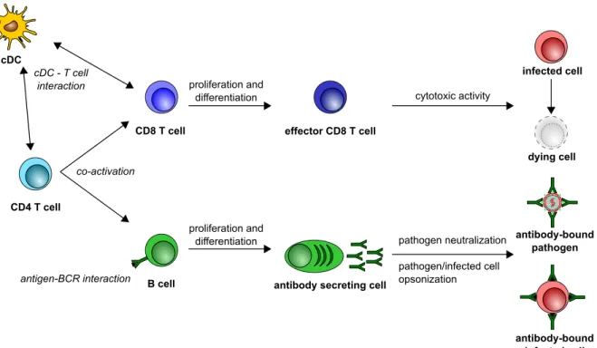
T and B cells, activated in the lymph node, will be recruited into the inflamed tissue via interaction between chemokines receptor and chemokines produced at the site of in-fection/injection. They will also participate in the elimination of the pathogens/vaccines, via the previously mentioned functions (e.g. cytotoxicity, pathogen neutralization). This corresponds to the effector phase of the adaptive immunity (Figure 13).
CD4 T cell subsets
MHC II
cDC2 neutrophil basophil macrophage
B cell
NK cell other ILC pDC CD8 T cell cDC1 NK cell MHC I pDC other ILC cDC αβ T cell 1: MHC-TCR interaction
2: Co-stimulation molecules interaction 3: Cytokinic microenvironment other cell 2 3
A
B
C
1 cDC1Figure 12. Activation of T cells by innate immune cells. (A) General mechanism of T cell priming by a dendritic cell in the lymph node. The three signals received by T cells are indicated. For cytokine-mediated signal 3, cDC can be assisted by another cell. (B-C) Overview of cells involved in MHC I-dependent CD8 T cell (re-)activation (B), and MHC II-dependent CD4 T cell (re-)activation (C). Antigen-presenting cells (APCs) are indicated in the frame, the main APC able to prime the T cell is indicated in bold. Cells that do not directly present antigen but modulate antigen presentation or orientate
cDC cDC - T cell interaction CD8 T cell CD4 T cell B cell antibody-bound pathogen antigen-BCR interaction co-activation effector CD8 T cell cytotoxic activity proliferation and differentiation pathogen neutralization pathogen/infected cell opsonization
antibody secreting cell
infected cell dying cell antibody-bound infected cell proliferation and differentiation
Figure 13. Overview of direct anti-pathogen activity of adaptive response. The different adaptive cells are represented, with their main direct impacts on pathogen clearance after activation. Note that the different subsets within each population are not represented, neither are the different steps of differentiation process.
While the pathogen/vaccine is being cleared, some long-lived (years) subsets will arise among these adaptive cells. These subsets can react more rapidly at any subsequent in-fection with the same pathogen, the previously mentioned immune memory (Dörner & Radbruch, 2005; Farber et al.,2014; Omilusik & Goldrath, 2017; Phan & Tangye, 2017). Some of these subsets will be maintained in the periphery (e.g. Tcm cells), whereas other will patrol in the tissues (e.g. Trm cells). Antibodies will be produced in the long term by long-lived antibody producing cells and will result in the constitutive presence of pathogen-specific antibodies in the serum (Yoshida et al.,2010;Brynjolfsson et al.,2018).
The maintenance of these long-lived cells requires notably cytokine signals (e.g. IL-7 and IL-15 for T cells, IL-6 and APRIL for plasma cells) (Sallusto et al., 2010). Besides, restimulation of primary memory cells at recalls will give rise to qualitatively distinct sec-ondary memory responses (e.g increased cytokine production, increased antibody affinity) (Masopust et al.,2006;Peixoto et al.,2007;Blanchard-Rohner et al.,2009;MacLeod et al., 2010; Wirth et al., 2010; Zabel et al., 2014).
These memory populations as well as resulting antibodies are the ones vaccines aim to induce. Indeed, these T and B cell populations (and antibodies) will mediate a more efficient response towards subsequent pathogen encounter, thus potentially protecting the body from the corresponding infectious agents. Since T cells and B cells recognize specific antigens, this highlights the crucial role the choice of the antigen(s) vaccines should contain to be efficient (Flower et al.,2010; Rueckert & Guzmán, 2012).
Modulation of innate cell effector functions by adaptive and innate immunity Adaptive cells produce cytokines that modulate innate cells functions. For example, CD8 T cells produce IFNγ, notably enhancing phagocytosis by macrophages/monocytes and DCs (Zhang & Bevan,2011). CD4 T cells, including memory cells, produce cytokines that will alter innate cell behavior, notably cytokine production, and allow for a more efficient innate response at recall (Strutt et al.,2010;2011). Memory CD8 T cells increase innate effector functions (Narni-Mancinelli et al.,2007;Soudja et al.,2014;Schenkel et al., 2014;Ariotti et al.,2014). T cells modulate NK cells mediated IFNγ production and cyto-toxic activity in infectious contexts (He et al.,2004) and following vaccination (Horowitz et al., 2010). B cells are known to produce cytokines, including IFNγ and IL-12, that will regulate innate cells in addition to T cells (Lund,2008). They can also participate to inflammation through production of TNFα, lymphotoxin and IL-6 (Vazquez et al.,2015). Eventually, antibodies will form immune complexes labelling pathogens and infected cells for phagocytosis or cell killing by innate cells.
Besides, innate cells mutually interact with one another, modulating their functions. For example, NK cells are potent producers of IFNγ and TNFα, which will activate DC and macrophages for phagocytosis. Upon activation, DCs and macrophages produce type I IFN, as well IL-12, IL-15 and IL-18 that will activate NK cells (Vivier et al., 2008). Modulation of DC activation and maturation was proved crucial for the efficient removal of infectious agents (Alter & Altfeld, 2011). In addition, direct cell-cell contact between monocytes and NK cells was shown to regulate NK cell activity (Michel et al., 2012).
Overall, all innate lymphoid cells (ILCs) are potent modulators of adaptive immunity activation (Withers, 2016; Vivier et al., 2018). Since modalities of vaccination strongly impact ILC response (e.g. route of immunization (Li et al., 2018)), ILCs stand as a promising target for vaccine optimization. Few adjuvants/self-adjuvanted vaccines are known or designed to directly target NK cells. Though, since NK cells can express PRRs (notably TLRs and NLRs), many adjuvants likely activate NK cells as a bona fide mech-anism (Martinez et al.,2010;Souza-Fonseca-Guimaraes et al., 2012;Adib-Conquy et al., 2014). In addition, NK cell activity can be enhanced via the induction of Th1 response (Jost et al., 2014; Martins et al., 2014; Van den Bergh et al., 2014). Also, for therapeu-tic vaccines, injection of NK cell–susceptible targets (e.g. MHC deficient cells) enhanced NK-cell mediated potentiation of adaptive responses (Kelly et al.,2002;Krebs et al.,2009).
Besides, neutrophils and monocytes/macrophages can mutually potentiate their ac-tivity during inflammation (Prame Kumar et al., 2018). Moreover, neutrophils can take part in cDCs activation and function (van Gisbergen et al.,2005). Eventually, neutrophils and NK cells deeply modulate their mutual activity via cytokine production (Costantini & Cassatella, 2011), including for example potentiation of NK cells functions (Amano et al., 2015). Overall, several studies highlighted the important role of neutrophils in vaccination (Di Pilato et al., 2015; Trentini et al., 2016; Musich et al., 2018).
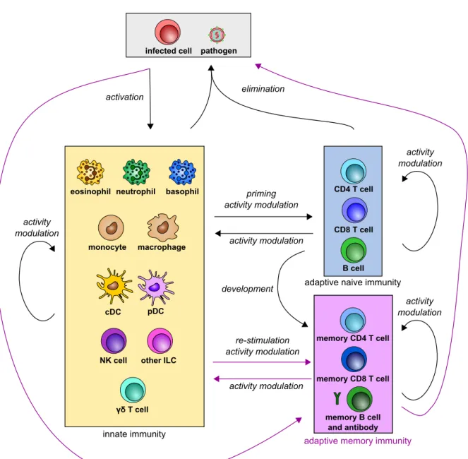
These cross-interactions of both innate and adaptive cells usually end up with the clearance of the pathogen/vaccine (Figure 14).
priming activity modulation activity modulation cDC pDC macrophage monocyte
eosinophil neutrophil basophil
NK cell other ILC
γδ T cell activity modulation pathogen infected cell activation elimination innate immunity CD8 T cell CD4 T cell B cell
adaptive naive immunity
re-stimulation activity modulation activity modulation activity modulation memory CD8 T cell memory CD4 T cell memory B cell and antibody
adaptive memory immunity
activity modulation development
Figure 14. Scheme of crosstalk between innate and adaptive immunity re-sulting in pathogen clearance. Black arrows indicate interactions occurring at ev-ery pathogen encounters. Purple arrows indicate interactions at recall responses only. Pathogen/infected cells activate innate immunity, triggering a crosstalk between innate and adaptive immunity, resulting in pathogen clearance. At a second encounter, pathogens and infected cells activate innate cells and restimulate memory responses, resulting in a faster pathogen clearance.