HAL Id: tel-02377630
https://tel.archives-ouvertes.fr/tel-02377630
Submitted on 24 Nov 2019
HAL is a multi-disciplinary open access
archive for the deposit and dissemination of sci-entific research documents, whether they are pub-lished or not. The documents may come from teaching and research institutions in France or abroad, or from public or private research centers.
L’archive ouverte pluridisciplinaire HAL, est destinée au dépôt et à la diffusion de documents scientifiques de niveau recherche, publiés ou non, émanant des établissements d’enseignement et de recherche français ou étrangers, des laboratoires publics ou privés.
Identification and Characterization of Two Novel
Syncytin-Like Retroviral Envelope Genes, Captured for
a Possible role in the Atypical Structure of the Hyena
Placenta and in the Emergence of the Non-Mammalian
Mabuya Lizard Placenta a
Mathis Funk
To cite this version:
Mathis Funk. Identification and Characterization of Two Novel Syncytin-Like Retroviral Envelope Genes, Captured for a Possible role in the Atypical Structure of the Hyena Placenta and in the Emergence of the Non-Mammalian Mabuya Lizard Placenta a. Virology. Université Paris Saclay (COmUE), 2018. English. �NNT : 2018SACLS106�. �tel-02377630�
Identification and characterization of
two novel syncytin-like retroviral
envelope genes, captured for a
possible role in the atypical structure
of the hyena placenta and in the
emergence of the non-mammalian
Mabuya lizard placenta
Thèse de doctorat de l'Université Paris-Saclay préparée à l'UMR 9196, Gustave Roussy
École doctorale n°582 cancérologie: biologie, médecine, santé (CBMS)
Spécialité de doctorat: aspects moléculaires et cellulaires de la biologie
Thèse présentée et soutenue à Villejuif, le 23 mai 2018, par
Mathis Funk
Composition du Jury : Uriel Hazan
Professeur des université, ENS Paris-Saclay (– UMR 8113) Président Jean-Luc Battini
Directeur de recherche, IRIM (– UMR 9004) Rapporteur Olivier Schwartz
Directeur de recherche, Institut Pasteur (– UMR 3569) Rapporteur Pascale Chavatte-Palmer
Directrice de recherche, INRA (– UMR 1198) Examinatrice François Mallet
Directeur de recherche, bioMérieux (– EA 7426) Examinateur Thierry Heidmann
Directeur de recherche, CNRS (– UMR 9196) Directeur de thèse
N N T : 2 0 18 S A C LS 1 06
Acknowledgments
I would first like to thank the members of the jury for taking the time to read the present manuscript, which turned out a bit longer than I had planned. I would like to thank Uriel Hazan for accepting to be the president of this jury, book-ending his involvement in my studies. What had started at the ENS Cachan and continued during my Master’s degree at the Institut Pasteur, finally reaches its culmination with the present work, on a topic that Uriel suggested I look into. I would like to sincerely thank Jean-Luc Battini and Olivier Schwartz for their critical reading and evaluation of the present manuscript and their positive feedback. Finally, I want to thank Pascale Chavatte-Palmer and François Mallet, for taking the time to read my work and for accepting to be a part of the jury.
I would, of course, also like to thank Thierry Heidmann for giving me the opportunity to work in his laboratory for the past four years, for being always available when I was in need of a guiding hand and present when I needed some prodding. Thank you for not only helping me to get these results, but also for the countless hours we spent to turn them into actual scientific articles.
I am grateful to Anne Dupressoir and Gérard Pierron for spending time reading through my manuscript and their words of encouragement and advice.
I also want to thank Christa Kuhn for her enthusiasm and encouragement, which played an important part in getting me to where I am today.
Je souhaite remercier l’intégralité de l’unité 9196, en particulier Marie et Anne pour avoir partagé leurs savoirs, leurs conseils ainsi que leurs bureaux, Cécile Vernochet pour son aide inestimable, Seila pour son soutien administratif et émotionnel, Grégoire pour son optimisme inébranlable et infectieux, Caroline, Cécile Lemaître et Anthony pour les bon moments passés ensemble, et ce-dernier tout particulièrement pour nos nombreuses discussions (profondes et/ou inutiles) et son énergie aussi inépuisable qu’insupportable. Un grand merci également à Guil-laume, ex-membre de l’unité, pour m’avoir encadré pendant mes débuts et avoir toujours été disponible pour mes questions.
J’aimerais remercier le gang des pasteuriens, premiers parmi eux mes ex-colocataires, Matthieu et Thomas, merci de m’avoir supporté pendant ces trois ans et quelques de vie com-mune au 20 avenue d’Ivry, qui s’est transformé en haut-lieu de la science, accueillant maints et maints congrès réunissant la fine fleur de la virologie et de l’immunologie parisienne. I would
like to thank Oksana, one of the kindest and most generous persons I have had the pleasure to know and someone you can always count on. Merci à Simon l’invisible, ton rétro nous a apporté de nombreuses heures de bonheur. Merci à Vincent le sale cylon pour les côtes de bœuf et à Essia pour ses couscous, toujours aussi bons que son humeur. Merci aux deux Juju pour la bonne ambiance, et je vous souhaite bien du courage, à Juju pour supporter Matthieu et à Juju pour finir ta thèse. Merci aussi à Anaïs, Laura, Cécile, Alix, Marion et tous les autres qui ont rendu la vie parisienne tellement plus agréable.
J’aimerais également remercier mes nouveaux ex-colocataires, Raphaël, Ferretti et Viking, pour m’avoir permis de garder l’appartement quelques mois de plus, puis pour m’avoir accueilli sur notre canapé qui, entretemps, était devenu le leur.
On a somewhat stranger note I would like to thank all of the friends I have never met, but who have been there to play, talk and just hang out in the virtual parts of this world. Thank you to all sigmas, especially 48, Mori Kaal and koreamax, but also willemd, :goatdrügs:, Cal, and even Thuneral, Slap Chop and Koahi.
A great thank you to the Aqueduct Acrobats who have been able to observe my slow and steady descent into placenta and retrovirus induced madness over the past few months, especially Topaz with his awful K/D, expensive haircuts and shared disgust of terrible book adaptations, mrcompson for our academia themed diversions and unmasking the terrible truth behind omics-conferences, and Malloot, everyone’s favorite tactical espionage action ground team leader and platforming liability. I also want to thank Norns and Tarth, our resident Americans, as well as Charisma and Waka.
Ich möchte natürlich auch meiner Familie danken, meiner Mutter, meiner Schwester und meinen beiden Brüdern, von deren Hilfe ich immer ausgehen konnte und deren Gesellschaft und Vertrauen mein Leben bereichert haben. Ich danke meiner Oma und meinem Opa für ihre Ermutigungen und Liebe. J’aimerais remercier Patrice, qui a été pour moi pendant ces 25 dernières années le meilleur père que j’aurais pu souhaiter, et qui m’a poussé à être curieux et intéressé en espérant de peut-être, un jour, être aussi instruit et intéressant que lui. Ich möchte hier auch ganz besonders meiner Tante Jutta danken, die innerhalb von ein paar Tagen die ganze Einführung durchgelesen und korrigiert hat.
Last but not least, I want to thank all of the people that I have shamefully forgotten to mention here, please forgive me for this oversight.
I am still unsure of how I made it this far, but one thing I know: I would not have made it without you. The words above pale in comparison to what I owe you all, I have written and rewritten them many times and I know I’ll never be able to express the feeling appropriately, so all I can do is apologize and say it once more, thank you all so very much.
Contents
Acknowledgments
iiiAbbreviations
xiI
Introduction
1
1 Placenta
5 1.1 Challenges of viviparity . . . . 5 1.1.1 Maternofetal exchanges . . . 5 a. Nutrients 6 b. Oxygen 8 c. Waste 91.1.2 Placentation and the maternal immune system . . . 9
a. Major histocompatibility complex expression 9
b. Uterine natural killer cells 10
c. Adaptive immunity 11
1.1.3 Maternofetal communication . . . 11
a. Placental hormones 11
b. Extracellular vesicles 12
1.2 Mammalian eutherian placentas . . . . 13 1.2.1 Origin and evolution of the mammalian placenta . . . 13
a. Monophyletic origin 13
b. Placental diversity 13
1.2.2 Placental types by maternofetal interface . . . 16
a. Epitheliochorial placentation 18
b. Synepitheliochorial placentation 20
c. Endotheliochorial placentation 22
d. Hemochorial placentation 22
1.3 Viviparity in non-mammalian species . . . . 26 1.3.1 Prevalence of viviparity outside of mammals . . . 26
a. Invertebrates 26
b. Non-mammalian vertebrates 27
1.3.2 Examples of non-mammalian placentas . . . 30
a. Non-vertebrates: the placenta of salps 30
b. Fish: placentation in sharks 31
c. Lizards: placentation in Mabuya and other Scincidae 32
2 Retroviruses
382.1 Exogenous Retroviruses . . . . 39 2.1.1 Genomic orgarnisation . . . 39
a. Non-coding features 39
b. Gag 41
c. Pro and Pol 42
d. Env 44
e. Regulatory and accessory proteins 44
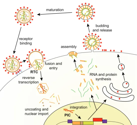
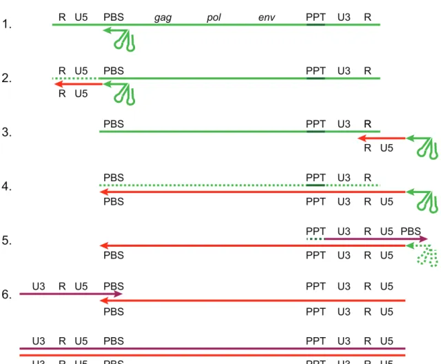
2.1.2 Retroviral phylogeny . . . 48 a. Subfamily Orthoretrovirinae 48 b. Subfamily Spumaretrovirinae 51 2.1.3 Viral cycle . . . 53 a. Viral entry 53 b. Reverse transcription 53 c. Genome integration 57 d. Protein expression 60
e. Assembly and budding 61
f. Maturation 64
2.2 Endogenous Retroviruses . . . . 66 2.2.1 Endogenization of retroviral sequences . . . 67
a. Infection of germinal cells 67
b. Intra-genome spreading of ERVs 69
c. Groups of ERVs 71
2.2.2 Impact of retroviral integration on the host . . . 72
a. LTR integration can drive ectopic gene expression 72
b. Impact of retroviral gene acquisition on host physiology 76 2.2.3 Fate of ERVs and control by the host . . . 78
a. Epigenetic regulation of ERV expression 78
b. Accumulation of mutations and solo LTR formation 79
c. Exaptation and purifying selection 80
3 Syncytins
813.1.1 Functional domains . . . 81
a. Domains of the SU 81
b. Domains of the TM 83
3.1.2 Env induced membrane fusion . . . 86
a. Implication of the cognate receptor in fusion 86
b. Model of viral fusion protein class I induced membrane fusion 89
3.2 Syncytins in mammals . . . . 90 3.2.1 Human syncytins: Syncytin-1 and Syncytin-2 . . . 90
a. Syncytin-1 92
b. Syncytin-2 94
3.2.2 Mouse syncytins: Syncytin-A and Syncytin-B . . . 95
a. Discovery 95
b. Expression in 2 segregated layers 95
c. Impact of Syncytin knockout on placental physiology 96
3.2.3 Other mammalian syncytins . . . 98
a. Rabbit: Syncytin-Ory1 98
b. Cat and dog: Syncytin-Car1 99
c. Old world monkey: EnvV2 99
d. Cattle and sheep: Syncytin-Rum1 and Fematrin-1 99
e. Squirrel and marmot: Syncytin-Mar1 100
f. Tenrec: Syncytin-Ten1 100
g. Short-tailed opossum: Syncytin-Opo1 100
3.2.4 Syncytin-like Env . . . 101
a. Conserved ERV Env with placental expression in human 101
b. Non-human syncytin-like genes 102
3.3 Syncytins and evolution . . . 103
3.3.1 Ancestral syncytin and syncytin replacement . . . 103
a. Example of EnvV2, a decaying syncytin in humans 104
b. Baton pass hypothesis 105
3.3.2 Syncytin diversity and placental diversity . . . 106 3.3.3 Other roles of syncytins . . . 108
a. Cell-cell fusion in muscles 109
b. Osteoclast fusion 109
c. Syncytins and pathologies 110
4 Objectives
1111 Implication of ERV env genes in non-mammalian Mabuya
placen-tation
1141.1 Summary . . . 114
1.2 A syncytin in a placental lizard . . . 117
2 Implication of ERV env genes in placental structural transitions in
Hyaenidae
135 2.1 Summary . . . 1352.2 Hyena-specific retroviral envelope gene capture and placenta evolution . . . 139
III
Discussion
167
1 Two novel Env with a potential placental role and a new SLC1A5 Env
168 1.1 Hyena-Env2 is a conserved syncytin-like envelope gene expressed during hyena placentation . . . 1681.2 Hyena-Env3 is a member of the SLC1A5 interference group with a peculiar tropism . . . 169
1.3 Syncytin-Mab1: characterization of a non-mammalian syncytin and its re-ceptor . . . 170
2 Implication of syncytins and syncytin-likes in placental evolution
172 2.1 Retroviral insertions have been a constant driving force of vertebrate evolution172 2.2 Syncytin-Mab1 and convergent evolution of animal placentas . . . 1742.2.1 Convergent use of ERV env genes in placentation . . . 174
2.2.2 Syncytin-Mab1 as the first example of a founding syncytin . . . 175
2.3 Syncytins and syncytin-likes and divergent evolution of the mammalian placenta . . . 175
3 Syncytin-likes and alternative envelope functions in the placenta
178 3.1 Growing evidence of conservation of non-fusogenic Env . . . 1783.2 Alternative placental roles for Env . . . 178
3.2.1 Immunosuppression . . . 178
3.2.2 Differentiation and proliferation . . . 180
3.2.3 Invasion . . . 180
3.3 Syncytin-likes in epitheliochorial placentation . . . 183
IV
Bibliography
187
V
Résumé Français
223
List of Figures
1 Dynamics of placental glucose and amino-acid transport . . . 7
2 Timed tree of major mammalian clades. . . 14
3 Diversity of eutherian placentas . . . 17
4 Types of maternofetal interface in eutherians . . . 19
5 Heterologous syncytium formation in ruminant placentas . . . 21
6 Remodeling of maternal blood vessels by extravillous trophoblast cells in humans 24 7 Matrotrophy in Animalia clades . . . 28
8 Mabuya placental structure . . . . 35
9 Organization of retroviral virions and genomes . . . 40
10 Organization of the complex HIV-1 genome . . . 45
11 Phylogeny of retroviral genera based on RT or TM analysis . . . 49
12 Retroviral replication cycle . . . 54
13 Reverse transcription . . . 56
14 Retroviral genome integration . . . 59
15 Model of ESCRT mediated viral budding . . . 64
16 Retroviral maturation . . . 65
17 Retroviral endogenization . . . 68
18 ERV amplification and fate . . . 70
19 ERV family representation . . . 72
20 LTR insertional mutagenesis . . . 74
21 Detailed structure of Env . . . 82
22 Sequence of the ISD and CXnC1-2motif and rationale of the mice immunosup-pression assay . . . 85
23 Retroviral Env mediated cell fusion . . . 86
24 Nature of retroviral receptors and SLC1A5 RBD . . . 88
25 Phylogeny of mammals with known syncytins positioned . . . 91
27 Compared evolutionary fates of HERV-W Env and EnvV2 in simians . . . 104
28 Phylogeny of retroviral syncytins among retroviral genera based on RT or TM analysis . . . 107
29 Differences in syncytin transcript initiation . . . 108
1 Phylogeny of mammals with emphasis on carnivorans . . . 143
2 Characterization of the identified Crocuta crocuta envelope candidates . . . 145
3 Entry date and conservation of env genes in Carnivora . . . 147
4 Alignment of Hyena-Env2 genes from all four extant hyena species . . . 148
5 Hyena-Env2 associated provirus and its genomic location . . . 150
6 The Hyena-Env2 associated virus is a gammaretrovirus . . . 151
7 Comparison of carnivoran and hyena placentas . . . 153
8 Expression pattern of syncytin-Car1 and Hyena-Env2 . . . 154
9 Fusogenicity assay for the three hyena envelope proteins . . . 155
10 Viral titers obtained in the pseudotyping assay using all three hyena envelopes . 156 11 Hyena-Env3 belongs to the SLC1A5 interference group of envelopes . . . 158
Abbreviations
APC: antigen presenting cell
ALV: avian leukosis virus
BaEV: baboon endogenous virus
BLV: bovine leukemia virus
BNC: binucleate cell
CA: capsid protein
CCD: catalytic core domain
CT: cytotrophoblast
CTD: C-terminal domain
EIAV: equine infection anemia virus
enJSRV: endogenous Jaagsiekte sheep retrovirus
Env: envelope
ER: endoplasmic reticulum
ERV: endogenous retrovirus
ESCRT: endosomal sorting complex required for transport
ETn: early transposon
EVT: extravillous trophoblast
FLV: feline leukemia virus
Gag: group-specific antigen
GaLV: gibbon ape leukemia virus
HA: hemagglutinin
HBZ: HTLV-1 basic zipper factor
HEMO: human endogenous MER34 (medium re-iteration family 34) ORF
HERV: human endogenous retrovirus
HIV: human immunodeficiency virus
HLA: human leukocyte antigen
hPL: human placental lactogen
HR: heptad repeat
HTLV: human T-lymphotropic virus
IAP: intra-cisternal A-type particle
IAPE: IAP-related retroviral element containing an env gene
ICTV: International Committee on Taxonomy of Viruses
IN: integrase
ITIM: immunoreceptor tyrosin-based inhibitory motif
JSRV: Jaagsiekte sheep retrovirus
KoRV: koala retrovirus
KRAB-ZFP: Krüppel associated box zinc finger proteins
LTR: long terminal repeat
Ly6E: lymphocyte antigen 6E
MA: matrix protein
MFSD2A: major facilitator superfamily domain containing 2 A
MHC: major histocompatibility complex
MHR: major homology region
MLV: murine leukemia virus
MPMV: Mason-Pfizer monkey virus
MPZL1: myelin protein zero-like 1
My: million years
Mya: million years ago
NAHR: non-allelic homologous recombination
NC: nucleocapsid protein
Nef: negative factor
NK: natural killer
NTD: N-terminal domain
ORF: open reading frame
PBS: primer binding site
PIC: pre-integration complex
Pol: polymerase
PPT: polypurine tract
Pro: protease
PRR: proline-rich region
PtdIns(4,5)P2: phosphatidylinositol-4,5-biphosphate
PTLV: primate T-lymphotropic virus
RDR: RD114 and D-type receptor
Rev: regulator of expression of viral proteins
Rex: Rev homolog
RSV: Rous sarcoma virus
RT: reverse transcriptase
RTC: reverse transcription complex
SIV: simian immunodeficiency virus
SLC: solute carrier
ST: syncytiotrophoblast
SU : surface subunit
Tat : trans-activator
Tax : trans-activating regulatory protein
TM : transmembrane subunit
TSD: target site duplication
uNK: uterine natural killer
UTR: untranslated region
Vif: viral infectivity factor
Vpr: viral protein r
Vpu: viral protein u
Part I
The field of virology is a relatively new one: less than two centuries ago the notion of a pathogen smaller than even bacteria was proposed by Pasteur, but he lacked the means to prove its existence. Years after these speculations, Chamberland invented his famous filter, able to remove even bacteria from a solution passed through it. It was this discovery that enabled Beijerinck in 1898 to show that filtered sap from plants infected with what is now known as tobacco mosaic virus was able to infect other plants. He concluded from this that the causative pathogen was smaller than a bacteria and called it a virus. From there virology advanced in leaps and bounds and in the following decades a great number of viruses were identified and linked to diverse diseases.
In 1911 Rous published his discovery of what would later become known as Rous sarcoma virus (RSV, Rous, 1911). While this was not the first retrovirus to be discovered, and while Rous himself did not know about the true nature of the infectious agent he described, this discovery would lead to the creation of both the fields of tumor viruses and retrovirology. In the 1960s, cancers caused by RSV in egg-laying hens were becoming an ever more important problem, leading to many new investigations into the characteristics and behavior of the virus. In 1961 Crawford & Crawford (1961) isolated the virus on a density gradient and determined that it had an RNA genome. Further research in the following years, making use of Mendelian genetics, both in live chicken and in infected cells, suggested that the viral genes could be stably inherited, which would imply that they had become part of the genome. This posed a major problem since at the time the "central dogma of biology" postulated that DNA could be transcribed into RNA, but that the opposite was impossible. How then could an RNA virus integrate its genes into a DNA genome? Nevertheless, in 1964 Temin introduced the provirus hypothesis, coining this term in the context of eukaryote viruses, and postulating that RSV could convert its RNA genome into a DNA genome which could then be inserted into the host genome (Temin, 1964). This hypothesis proved unpopular until the discovery 6 years later of reverse transciptase (RT, Baltimore, 1970; Temin & Mizutani, 1970), which finally delivered the key element: an enzyme able to convert RNA information into DNA information, and which led to the naming and creation of the Retroviridae family by the International Committee on Taxonomy of Viruses in 1975 (ICTV, 1975).
genome, endogenous retroviruses (ERV) were discovered in parallel with the family itself, first in chicken and mice and later in humans and other vertebrates. The identification of retroviral sequences that were an integral part of host genomes (sequencing of the human genome later revealed that ERVs represent 8% of the genome Lander et al., 2001) led to the search for the function of these elements, and a number of them have been identified as protecting their host from infection by exogenous retroviruses. A role not linked to viral infection, and probably the most remarkable example of ERV function, is the exaptation of syncytins in mammals. The first such gene was described in 2000 as participating in the fusion of trophoblastic cells in the human placenta (Blond et al., 2000; Mi et al., 2000). Since then numerous other syncytins have been identified in other mammalian placentas and their role in trophoblast fusion has been confirmed using KO mice.
To delve further into these topics, I will first discuss the characteristics of mammalian and non-mammalian placentas, a pre-requisite for discussion of syncytin role and acquisition. I will then present retroviruses in greater detail and finally I will talk specifically about captured envelope genes, especially syncytins.
1. Placenta
The transition from oviparity (egg-laying) to viviparity (live-bearing) occured on numerous occa-sions during vertebrate evolution (Blackburn, 2015), most famously in Eutherians. In viviparous species, by definition, the embryo is not exposed to the external environment and the exchanges necessary for embryonic development must take place between the embryo and the mother. These exchanges are rendered possible by the placenta. The placenta is a highly variable organ, both in structure and functionality. Its simplest definition was given by Mossman in his seminal 1937 work on mammalian placentas: "an apposition or fusion of the fetal membranes to the uterine mucosa for physiological exchange" (Mossman, 1937). This definition encompasses two related notions: the placenta joins maternal and fetal tissues, and it forms an exchange interface between mother and fetus.
1.1
Challenges of viviparity
1.1.1
Maternofetal exchanges
The first major challenge facing viviparous species is for the mother to act as the go-between of the embryo and the exterior. This means that the mother must not only provide the substances required for embryo development but also that all embryonic waste products must be taken up by the mother and eliminated. The importance of these exchanges during development is very variable depending on the species (Blackburn, 2015). In some cases, the embryo obtains most nutrients from its own yolk and is reliant on the mother mostly for gas and water exchanges, in others the embryo also needs to acquire almost all nutrients necessary for development. These two extremes are called lecithotrophy and matrotrophy, respectively, and form the two ends of a continuum of reproductive strategies that differ by the degree of implication of the mother and the functional capacities of the placenta (Blackburn, 2015). Of note, while almost all oviparous species are by necessity lecithotrophic, the mammalian monotremes (platypus and echidnas) have a unique matrotrophic oviparous mode of reproduction in which females ovulate small eggs with a 3 mm diameter yolk which takes up maternal nutrients before oviposition, to reach a final diameter of 17 mm (Hughes, 1993). While this is mentioned here for completeness, "oviparity" will refer to the much more common lecithotrophic oviparous mode of reproduction from here on. Combining the two distinctions of oviparity/viviparity and lecithotrophy/matrotrophy allows classification of vertebrate reproductive strategies into four major categories, see Table 1.
1. Placenta
1.1
Table 1 – Classification of vertebrate reproductive modes. The examples provided for each reproductive
type are not exhaustive. Adapted from Blackburn (2015).
oviparity lecithotrophy
matrotrophy
viviparity lecitotrophic oviparity:
all birds and crocodilians, most squamates, teleosts, some chondrichtyans
lecitotrophic viviparity:
many squamates,
some teleosts, chondrichtyans
matrotrophic viviparity:
all eutherians and marsulpials, several teleosts and chondrichtyans, a few clades of squamates
matrotrophic oviparity:
only monotremes
Since most of our species of interest will be matrotrophic viviparous, the description of placental characteristics will focus on this subgroup, however even within this group placental structures show a great amount of variability (see 1.2.1.b.).
a. Nutrients
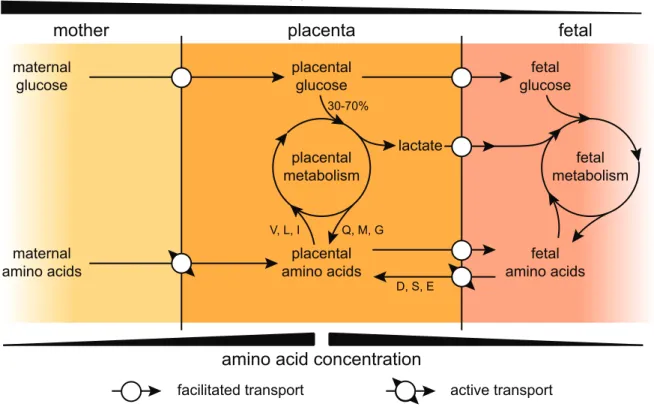
In matrotrophic placentation, nutrients that must be supplied to the embryo include carbohy-drates, most amino-acids, fatty acids and metal ions. In mammals, glucose is one of the most important fetal nutrients, providing much of the energy required during fetal growth (Wooding & Burton, 2008, chap. 2). In eutherians, maternal glycemia is higher than fetal glycemia, es-tablishing a concentration gradient that induces glucose transport from the mother to the fetus. In order to transfer the necessary amount of glucose from maternal to fetal blood, diffusion is facilitated using glucose transporters (glucose transporter type 1 in all mammalian placentas and additionally type 3 in some, like rat, horse and ewe, see Wooding & Burton, 2008, chap. 2). Depending on the species, 30-70% of the glucose provided by the mother is used in placental tissues to permit growth and functionality of the placenta itself and does not reach the embryo (Fig. 1, Wooding & Burton, 2008, chap. 2), though some of the products of placental glucose metabolism, like lactate, will enter the fetal circulation to serve as metabolites, lessening the impact of placental glucose consumption (Fowden, 2010). Indeed, lactate is another major actor of fetal metabolism (up to 25% of total fetal energy requirements, see Wooding & Burton, 2008, chap. 2). Similarly to glucose, lactate transport makes use of transporters for facilitated diffusion, but in this case most of the lactate is produced by the placenta itself and maternal transplacental provision of this metabolite seems absent (Fowden, 2010), leading to higher concentrations of lactate in fetal than in maternal blood.
Contrarily to carbohydrates, amino acid concentrations are higher in fetal than in maternal blood in most species, indicating that there is a net uptake of amino acids by the fetus and that active transport mechanisms must be involved (Fig. 1, Vaughan & Fowden, 2016). Interestingly, placental amino acid concentrations are even more elevated than fetal concentrations and the placenta exchanges amino acids both with the maternal and fetal circulations. Most amino acids
1.1
1.1. Challenges of viviparity maternal glucose placental glucose fetal glucose maternal amino acids placental amino acids fetal amino acids placental metabolism fetal metabolism lactate 30-70% glycemia placenta mother fetalamino acid concentration
D, S, E V, L, I Q, M, G
facilitated transport active transport
Figure 1 – Dynamics of glucose and amino-acid transport. Schematic representation of placental
transport of glucose (top) and amino acids (bottom) and implication of the placental metabolism. Examples of amino-acids involved in placental metabolism are given as one letter codes and based on the example of sheep. Concentration gradients are represented as black wedges.
are simply imported by active transport into the placental tissues and then cross into the the fetal circulation according to their concentration gradient, this is notably the case for essential amino acids that, by definition, cannot be synthesized by the placenta or fetus and must be provided by the mother (Vaughan & Fowden, 2016). For some amino acids, transit is much more complicated, this is the case for aspartate, serine and glutamate in sheep for example. The net placental uptake from maternal blood for these three amino acids is null, they are instead taken from the fetal circulation to satisfy placental metabolic needs, including ATP generation and protein synthesis (Vaughan & Fowden, 2016). In addition, the sheep placenta is a net consumer of valine, leucine and isoleucine, but it provides more glutamine, methionine and glycine to the fetus than it takes up from the mother (Vaughan & Fowden, 2016). The placental role in amino acid supply to the fetus reinforces the notion, that had been introduced with lactate in the previous paragraph, that the placenta is an active middleman between mother and fetus, and that its role is not simply passive (Fig. 1).
Being hydrophobic by nature, lipids such as free fatty acids, glycerol and cholesterol can be supplied by diffusion across the membranes separating mother and fetus, especially since maternal concentrations are higher than fetal concentrations for most. For others like the essential long chain fatty acids, active transporters participate in establishing a higher concentration in fetal than in maternal blood (Wooding & Burton, 2008, chap. 2).
1. Placenta
1.1
The final nutrient category discussed here concerns ions. For some like calcium, the transfer from maternal blood to the placenta can simply occur by facilitated diffusion through calcium channels. Once in the placenta, it is bound to calcium binding proteins, so as to isolate it from sensitive cellular signaling mechanisms that rely on cytoplasmic Ca2+ concentration, and then transferred to the fetus using calcium ATPase pumps (Wooding & Burton, 2008, chap. 2). Iron transport is a bit more complex since iron ions are not present in their free form in the organism. Instead they are bound to transport proteins (uteroferrin in horse and pig or transferrin) or are acquired by breakdown of red blood cells. Uteroferrin and transferrin are secreted by maternal uterine glands and enter the placenta by endocytosis. Iron ion acquisition from red blood cells concerns mainly carnivorans and ruminants in which specialized regions of the placenta (called hemophagous zones) break down these cells after phagocytosis (Wooding & Burton, 2008, chap. 2). Of note, in some placentas (human for example) a similar endocytic method is used to transfer a few specific proteins from the mother to the fetus, such as immunoglobulin G (Wooding & Burton, 2008, chap. 2).
The substances acquired directly from maternal blood are grouped under the term "hemotroph" while the substances secreted by the mother and taken up through endocytosis are termed "his-totroph".
b. Oxygen
As with many other organs and organisms, both the placenta and the fetus need a constant supply of O2. Since biological membranes are highly permeable to gases like O2, there is no need for facilitated diffusion or active transport, instead diffusion occurs according to Fick’s law along the concentration gradient, leading to a lower oxygenation of fetal blood than maternal blood. The two main factors influencing the efficiency of O2 transfer from the mother to the fetus are blood flow velocity and relative disposition of the blood vessels (see Fig. 3C Wooding & Burton, 2008, chap. 2). Interestingly both of these factors are very variable across mammalian placentation, indicating that there is not necessarily a single efficient strategy for O2 transport since all mammals are able to produce viable offspring.
The placental O2 consumption is high, higher than that of the fetus when normalized by weight and on par with that of the fetal brain (Fowden, 2010). In sheep, about 75% of the O2 consumed by the placenta is used to generate ATP using a variety of substrates, including glucose and amino acids, as discussed in 1.1.1a.. This ATP is then used primarily for synthesis of placental proteins and to fuel active transport against electrochemical gradients. 15-20% of the O2 uptake of sheep placentas is used to produce intermediaries necessary for synthesis of progesterone and other steroid hormones (Vaughan & Fowden, 2016). The proportion of O2 that actually reaches the fetal circulation is very dependent on the species and sometimes the gestational stage. In sheep it varies from 80% at the beginning to about 45% towards the end of gestation, while in horse and pig it remains at a constant 55% or 65% respectively (Vaughan & Fowden, 2016).
1.1
1.1. Challenges of viviparity
c. Waste
It is important to remember that both the placenta and the fetus are metabolically active and that it is not only necessary to provide them with the necessary substrates, but it is also necessary to eliminate some of the metabolic byproducts. This is for example the case with CO2and urea, both being able to return to the maternal circulation by simple diffusion, as one is a gas and the other a small uncharged molecule able to cross biological membranes. In sheep during late gestation, it is estimated that each day 7.8 g of carbon per kg of fetal weight enter the fetal circulation and 4.6 g of carbon per kg need to exit the fetal circulation in the form of CO2 and urea (Battaglia & Meschia, 1978), illustrating the need for the placenta to permit transport in both directions.
1.1.2
Placentation and the maternal immune system
Since during sexual reproduction the paternal and maternal genes mix and give rise to a new hybrid genome, the fetus is a foreign tissue in the eyes of the maternal immune system. In oviparity this does not pose a major problem since mother and fetus are separated by a physical barrier, the egg shell (reviewed in Wooding & Burton, 2008, chap. 9). In viviparity however, close contact between fetal and maternal tissues is absolutely necessary for transplacental exchanges (see 1.1.1). This intimate contact necessitates the establishment of a particular immunological context to avoid attack of fetal tissues by the maternal immune system, as is seen in other equivalent allograft situations (reviewed in Moffett & Colucci, 2014). Here again, the placenta plays a crucial role as it is the point of contact between maternal and fetal tissues. The cells of the fetus itself generally do not interact with the maternal immune system, as they are shielded by placental tissues (reviewed in Moffett & Colucci, 2014). Interactions between the placenta and maternal immune system are complex and highly dependent on the considered species, as such the following discussion will only focus on a few major aspects. An important aspect that will not be detailed here seems to be the presence of anti-inflammatory molecules, in the absence of which even syngeneic pregnancies (in which the fetus does not present foreign genetic material) result in pregnancy failure in rodent models (reviewed in Moffett & Loke, 2006).
a. Major histocompatibility complex expression
In mammals the adaptive immune system relies on the expression of major histocompatibility complex proteins (MHC). These proteins are recognized by and display antigens to B and T lymphocytes, leading to immune responses if a foreign element is detected. In humans the MHC molecules are called human leukocyte antigens (HLA) and cells usually express HLA-A, -B and -C at their surface, however in the placenta most cells do not express any HLA (Apps et al., 2009). This absence of MHC expression effectively renders the placental cells invisible to the adaptive immune system, protecting them from maternal attack and especially cytotoxic T cells.
1. Placenta
1.1
The presence of mostly MHC null cells is also observed in rodent, pig, horse and ruminant placentas for example (reviewed in Wooding & Burton, 2008, chap. 9), and as such seems to be a common mechanism of fetal tolerance.
While this is true for the bulk of placental cells, some subsets still express MHC molecules, this is mostly the case for invasive cells. In horses for example, some cells that migrate into the maternal tissue do express MHC proteins, only becoming MHC null once they have reached their destination. In cows the so-called binucleate cells (BNC) fuse with maternal cells, giving rise to heterologous trinucleate syncytia that express MHC molecules but do not seem to present allogenic antigens (reviewed in Wooding & Burton, 2008, chap. 9). The human placenta also presents invasive cells that express MHC proteins: the extravillous trophoblasts (EVT). These cells separate from the rest of the placenta and invade maternal tissues where they induce a major remodeling of blood vessels in order to increase placental blood supply by the mother (see Fig. 6, reviewed in Wooding & Burton, 2008, chap. 8). At their surface the EVT cells express the unique combination of HLA-C, -E and -G (Apps et al., 2009). HLA-E and -G are non classical HLA molecules and display a very restricted expression profile, HLA-G being exclusively expressed in EVT (Apps et al., 2009). The presence of these HLA molecules is crucial for the regulation of maternal vascular remodeling, as they provide both activatory and inhibitory signals to uterine natural killer (uNK) cells (see below, Parham & Moffett 2013).
b. Uterine natural killer cells
During pregnancy in humans up to 70% of uterine lymphocytes are uNK lymphocytes and an equivalent population of NK has been described in rodents (reviewed in Wooding & Burton, 2008, chap. 9), suggesting that they play an important role in all highly invasive (hemochorial, see 1.2.2.d.) placentas. Despite their name and unlike systemic NK cells, uNK cells are only weakly cytotoxic in vitro and do not kill MHC-null placental cells (Ritson & Bulmer, 1989; King et al., 1989). Using NK specific receptors at their surface, uNK interact with the HLA expressed at the surface of EVTs, receiving both activating and inhibiting signals (reviewed in Parham & Moffett, 2013). When activated, uNK cells release cytokines and proteases that regulate EVT migration and assist in vascular remodeling (reviewed in Burton & Fowden, 2015). The dialog between EVT and uNK plays a critical role in pregnancy as it has been shown that a spectrum of pregnancy complications, referred to as the "great obstetrical syndromes" (preeclampsia, intrauterine growth restriction, pre-term labor and abruption placentae among others), are linked to defects in remodeling of the maternal circulation by EVT (reviewed in Brosens et al., 2011). While the non-classical HLA-E and -G show very little genetic variation, HLA-C presents several different epitopes that interact with distinct receptors on the uNK cells. Some combinations of placental HLA-C epitopes and maternal uNK receptors have been shown to increase the risk of pregnancy complications as they bias the EVT/uNK interaction, favoring uNK inhibition over activation (Hiby et al., 2010).
1.1
1.1. Challenges of viviparity
c. Adaptive immunity
Adaptive immunity in mammals is based on the recognition of antigens by B or T lymphocytes, said antigens being presented either by generic cells using class I MHC or by specialized antigen presenting cells (APC) using class II MHC. As discussed above, most placental cells are MHC-null, preventing direct recognition as foreign tissues by the adaptive immune system (Apps et al., 2009). Regarding MHC expressing placental cells, it has been shown that HLA-G, one of the MHC types expressed on the surface of human EVT cells, binds to inhibitory receptors on APC with very high affinity (Shiroishi et al., 2003) and that this binding interferes with APC maturation, resulting in lowered presentation of allogenic peptides through class II MHC as well as induction of immunosuppressive T cells in vitro (Ristich et al., 2005). Since this mechanism necessitates a direct contact between the HLA-G expressing EVT and the maternal APC, it is restricted to the placental context and does not impact APC functionality in the rest of the maternal immune system.
Apart from effector T cells, regulatory and helper T cells also play an important role in adaptive immunity. Tilburgs et al. (2008) show that during pregnancy fetus-specific protective regulatory T cells migrate to the placental interface and suppress effector T cells. The role and impact of T helper cells on pregnancy is less clear. It seems that during pregnancy Th1 responses (which favor cytotoxicity and are effective against viral infections) are decreased in favor of Th2 responses (antibody generation) (reviewed in Moffett et al., 2017). This shifted balance is the reason why pregnant women are more susceptible to some viral infections (like influenza, reviewed in Mertz et al., 2017) and why during pregnancy the severity of Th1 linked autoimmune disorders decreases while that of Th2 linked disorders increases (reviewed in Ostensen, 1999; Marder et al., 2016). Moffett et al. (2017) stress the fact that this observed shift does not seem to have any impact on reproductive success and consider it to be a side effect of the particular hormonal context established during pregnancy.
Of note, maternal B cells are rarely observed near the placental tissues, indicating that there is no substantial local generation of EVT-specific antibodies (Moffett et al., 2017).
1.1.3
Maternofetal communication
Establishing the link between the mother and fetus, the placenta is responsible for acting as an intermediary and communicating with the maternal side so as to establish correct conditions for fetal growth.
a. Placental hormones
Part of this communication uses classical hormonal pathways, the placenta being a major en-docrine organ whose hormones have important effects on maternal physiology during pregnancy (reviewed in Burton & Fowden, 2015). As discussed in 1.1.1.a., all nutrients necessary for
pla-1. Placenta
1.1
cental and fetal growth have to be supplied by the mother, who also must fulfill the needs of her own metabolism. This is particularly true for glucose, the main substrate of placental and fetal metabolism (reviewed in Wooding & Burton, 2008, chap. 2) and an important metabolite in adults. In humans the main hormones of placental origin involved in these mechanisms are progesterone, human placental lactogen (hPL) and human placental growth hormone (hGH) (re-viewed in Freemark, 2006; Burton & Fowden, 2015). Expression and impact of these hormones on the maternal metabolism can be separated into two phases: early to mid-gestation and late gestation.
During early to mid-gestation, secretion of progesterone and hPL by the placenta increase progressively (reviewed in Freemark, 2006; Burton & Fowden, 2015). Both of these hormones are appetite stimulants and have been associated with weight gain and fat deposition in mice and humans (Grueso et al., 2001; Freemark, 2006), leading to a disruption of energetic homeostasis and a buildup of reserves. In addition hPL participates in establishing leptin resistance during pregnancy, attenuating the negative feedback loop of fatty tissues on food intake normally mediated by this hormone (reviewed in Ladyman et al., 2010). Finally, hPL also stimulates beta-cell proliferation in the pancreas of the mother, leading to increased insulinaemia and thus fat deposition and energy storage (reviewed in Burton & Fowden, 2015). Taken together, these effects lead to a 10-15% increase in food intake and a 60% increase in insulin secretion during this first phase, which starts even before a significant fetal energy demand arises and corresponds to a pre-preemptive increase in energy storage (reviewed in Freemark, 2006).
In late pregnancy there is a significant secretion of hGH by the syncytiotrophoblast of the placenta. This growth hormone is capable of inducing insulin resistance (Barbour et al., 2002) and the resulting 45-70 % decrease in maternal insulin sensitivity leads to breakdown of fat reserves and mobilization of glucose into the maternal bloodstream, from where it can then be absorbed by the placenta (see 1.1.1.a.).
In addition to the roles in nutrient provision discussed above, fetal hormones also play an important role in the establishment and maintenance of pregnancy (and interrupting the menstrual cycle) and can be involved in the process leading to parturition at the end of gestation (reviewed in Wooding & Burton, 2008).
b. Extracellular vesicles
In addition to the soluble secreted factors described above, the placenta releases a great amount of extracellular vesicles (Salomon et al., 2014). These particles are lipid-bilayer particles that can contain proteins, lipids, RNAs and DNA, be classified as exosomes (40-120 nm) or microvesicles (0.2-1 µm), and participate in inter-cellular communication processes (reviewed in Adam et al., 2017). Salomon et al. (2014) show that the concentration of exosomes in plasma of pregnant woman is increased 50-100 fold when compared to non-pregnant women, depending on the stage of pregnancy. The role of these particles in maternofetal communication is still poorly understood, though it has been shown that placenta generated extracellular vesicles can interact
1.2
1.2. Mammalian eutherian placentas
with the maternal immune system and have an immunosuppressive effect (Sabapatha et al., 2006).
1.2
Mammalian eutherian placentas
1.2.1
Origin and evolution of the mammalian placenta
a. Monophyletic origin
Since the placenta is composed of soft tissue, there is no substantial fossil record of placentation in extinct species, complicating studies of placental origin and evolution. Nevertheless, in mammals the placenta is very probably of monophyletic origin and the time of emergence can be estimed using mammalian phylogeny (Lillegraven et al., 1987). The class Mammalia can be split into three subgroups with regard to placentation: i) the monotremes that are oviparous and do not present a placenta, ii) the marsurpials which present a short-lived placenta and iii) the eutherians or placentalia (see Fig. 2). Eutherians present long gestation periods and complex placentas and regroup most of the extant mammalian species. Since both eutherians and marsupials are placental, the parsimony principle suggests that the emergence of the mammalian placenta occurred in the last common ancestor of the two clades, after their separation from monotremes. This common origin is supported by the comparison of eutherian and marsupial placentation traits, though it cannot be entirely excluded that two indendent emergences of placentation occurred (Lillegraven et al., 1987; Blackburn, 2015). In any case the similarity of involved processes and tissues in eutherian placentation clearly suggest that at least in this branch the origin of the organ is monophyletic (Lillegraven et al., 1987).
The fossil calibrated timed tree established by dos Reis et al. (2012) indicates that a placental origin common to marsupials and eutherians would have occurred about 175 Mya. In accordance with the "long fuse" model of eutherian evolution (relatively recent diversification), the radiation of this clade occurred about 90 Mya, long after emergence of a common mammalian placenta (dos Reis et al., 2012), indicating that the different adaptations and specializations observed in modern day eutherian placentas have evolved from this common primitive placenta during the last 90 My.
b. Placental diversity
While eutherian placentas have a single shared origin, they have since evolved into the organ exhibiting the greatest amount of inter-species diversity (reviewed in Enders & Carter, 2004; Wooding & Burton, 2008, chap. 1). This becomes immediately apparent when one tries to establish a system of classifying the placentas of different species, as no single system allows for easy and clear classification (Fig. 3, reviewed in Chavatte-Palmer & Tarrade, 2016).
1. Placenta
1.2
MAMMALIAN PLACENTA Euarchontoglires Laurasiatheria Xenartha Afrotheria monotremes eutherians marsupials 0 Mya 50 100 150 200 Jura ssi c Cre tace ou s Te rtiaryFigure 2 – Timed tree of major mammalian clades. Placental clades are indicated in red, branch length
is proportional to time, see scale at the bottom (dos Reis et al., 2012). A red arrow marks the emergence of the mammalian placenta.
Classification by shape. The most straightforward way of classifying different eutherian placentas is to simply go by external appearance, but even here there are two possible approaches. The first is based on description of the distribution of areas of high maternofetal exchange at the surface of the placenta (Fig. 3A). These areas present folds or extruding placental villi that increase the exchange area and the distribution of such regions can follow four major patterns. In diffuse placentas (e.g. pig, horse, Friess et al., 1980; Samuel et al., 1974) there are no particular areas of the placenta concentrating villi or folds. Cotyledonary or placentomal placentas (e.g. ruminants, Björkman, 1969) present several spots of increased maternofetal exchange surface. The number of these so-called cotyledons ranges from 5 in deer to 150 in giraffe (reviewed in Wooding & Burton, 2008, chap. 1), illustrating the high amount of potential variability even within a given category. In zonary placentas (e.g. carnivorans, Leiser & Koob, 1993) the folds are grouped into a single band that circumvents the placenta. Finally, in discoid placentas the exchange zone takes the form of one (e.g. human, rodents) or two (e.g. rhesus monkey) discs at the surface of the placenta (reviewed in Chavatte-Palmer & Tarrade, 2016; Hafez, 2017).
The second shape-based classification relies on the way the above described folds and villi interdigitate fetal and maternal tissues (Fig. 3B). The simplest structure is found in folded placentas (e.g. pig, Friess et al., 1980) where placental folds simply interlock with corresponding maternal endometrial folds. Carnivores present the slightly more complex lamellar form of placentation in which the folds are more extensive and branched (Leiser & Koob, 1993). Humans as well as ruminants and horses present villous placentas where placental tissues form tree-like structures with numerous primary villi that branch out into secondary and tertiary villi that fill corresponding maternal recesses (Enders, 1965; Björkman, 1969; Samuel et al., 1974). Finally the most complex form is found in labyrinthine placentas (e.g. rodents, Enders, 1965) in which the placental tissues are crossed by an elaborate assembly of maternal channels, forming a large interconnected mesh-like structure. We can already see that both types of classification establish
1.2
1.2. Mammalian eutherian placentas
different groups, horse being grouped with pigs in the first but with humans and ruminants in the second system. Another important and recurrent factor to these classifications is that they take into account the definitive placental structures and not the placental development.
Classification by invasiveness. Schlafke & Enders (1975) distinguish between four major degrees of invasiveness of eutherian placentas during early development. The lowest degree of invasion, interdigitation, is observed in pigs where maternal and fetal epithelia are simply apposed and both maintain their integrity (Dantzer, 1985). Displacement describes an interme-diate phenotype in which individual cells or groups of cells of the uterine epithelium slough off after apposition with the fetal epithelium, leading to penetration of placental cells beyond the maternal epithelial barrier. This is observed in rats and mice for example (reviewed in Wooding & Burton, 2008, chap. 8). Ruminant placentas are an example of the third type of early invasion of maternal tissues: fusion. Fetal BNC fuse with cells of the maternal epithelium, resulting in heterologuous syncytia (Wooding et al., 1981, see also 1.2.2.b.). This process allows penetration of the maternal epithelium by cells of (partial) fetal origin without loss of integrity of the former. The final type of placental invasion during implantation is intrusion and can be found in horse and human, albeit to different degrees. In the horse placenta, individual binucleated cells sepa-rate from the placenta and pass through the body of uterine epithelial cells (Allen et al., 1973). In human placentas the entire conceptus crosses the uterine epithelium (reviewed in Wooding & Burton, 2008, chap. 8).
In addition to this classification based on invasiveness during the very early implantation step, definitive placental structures can also be classified in terms of invasiveness (Fig. 4A). As stated above, the pig placenta results mostly from the apposition of maternal and placental epithelia, without loss of integrity of the former (Friess et al., 1980; Dantzer, 1985). This represents the least invasive type of placentation. From there on there exists a spectrum of invasiveness, the horse placenta for example displays a bit more invasion: while epithelia maintain their integrity, a subset of binucleated placental cells cross the maternal epithelium and migrate into the endometrium (Allen et al., 1973, see also 1.2.2.a.). This spectrum culminates in the so-called hemochorial placentas where maternal tissues have been largely degraded and the placenta bathes directly in maternal blood (see 1.2.2.d.).
Classification by other factors. Another possible classification of eutherian placentas is based on the amount of maternal tissue lost during birth, however this is closely linked to the previous one: in low-invasion placentation the maternal and placetal tissues are simply apposed so that during parturition they separate cleanly, while in high-invasion placentation the deeper relationship between maternal and placental tissues necessitates tearing of the former (reviewed in Hafez, 2017).
It is also possible to distinguish placentas with regards to the relative disposition of maternal and placental blood vessels (Fig. 3C). The disposition of blood vessels has a major impact on
1. Placenta
1.2
the efficiency of exchanges between them. In a concurrent system (parallel blood flows) for example, the theoretical maximal efficiency is 50% while a countercurrent system allows for a theoretical maximum of 100%. Perhaps unsurprisingly, no eutherian placenta employing a fully concurrent system has been described to this day. An example of a placenta presenting an almost perfect countercurrent system can be found in guinea pigs, while other species present intermediate arrangements like cross-current (e.g. cats) and villous (e.g. humans)(Fig. 3C, reviewed in Wooding & Burton, 2008; Hafez, 2017).
The final major classification is based on the histological nature of the maternofetal interface and the amount of layers that separate the maternal and fetal circulations (see Fig 4A). Wooding & Burton (2008, chap. 1) suggest that this way of categorizing placental structures is more relevant to the physiological functions of eutherian placentas and as such it will be presented in more detail than the other classifications.
1.2.2
Placental types by maternofetal interface
This classification was first proposed by Grosser (1909) and is based on his observation that while all fetal tissue layers (endothelium, connective, epithelium called trophoblast) were al-ways present in every eutherian placenta, the amount of maternal tissue layers (epithelium, connective, endothelium) was variable. Grosser proposed four categories of placenta: Placenta
epitheliochorialis, where all maternal tissues are still present and the trophoblast is in contact
with the maternal epithelium, Placenta syndesmochorialis, in which the maternal epithelium is absent and the trophoblast is in direct contact with connective tissue, Placenta
endotheliochori-alis, in which both the maternal epithelium and connective tissue have been destroyed and the
trophoblast is in direct contact with the maternal endothelium, and Placenta haemochorialis, in which all maternal tissues have disappeared and the trophoblast is in direct contact with maternal blood.
Most of this classification proved remarkably accurate for being based only on light mi-croscopy, but the category of syndesmochorial placentation was based on a mistake. Grosser (1909) had concluded that in ruminant placentas the maternal epithelium was destroyed and replaced by the fetal trophoblast, but closer investigation with more elaborate methods revealed that both epithelia remain present until term. This led to ruminant placentas being described as epitheliochorial for a time, however as reviewed in Wooding (1992), ruminant placentas present fusion events between fetal and maternal cells, separating them from all other epitheliochorial placentas and justifying their belonging to a separate category: synepitheliochorial placentation. Grosser (1909) had also suggested that this classification might represent an evolutionary progression, that epitheliochorial placentas are the most basic and least efficient, having six cell layers separating maternal and fetal circulations, while hemochorial placentas are the most evolved and efficient, having only three layers in-between both circulations. This supposed
1.2
1.2. Mammalian eutherian placentas
diffuse cotyledonary zonary discoidal
e.g. pig e.g. cattle e.g. cat e.g. human
e.g. pig e.g. cat e.g. human e.g. guinea pig
concurrent countercurrent
villous crosscurrent
lamellar villous labyrinthine
folded F M 0 100 50 50 F M 99 100 0 1 F M M 0 100 100 x+y y x M100 100-x F 0 F M F M F M F M
A
B
C
classification by gross morphology
classification by interdigitation type
classification by vascular arrangement
e.g. none e.g. guinea pig
e.g. cat e.g. human
x
Figure 3 – Diversity of eutherian placentas as illustrated by several classification systems (A)
Classification by gross morphology. In many eutherian placentas (except diffuse) there are distinct regions specialized for maternofetal exchanges . The shape and distribution of these regions can help separate placentas into distinct categories. Areas specialized for maternofetal exchange are indicated in dark red. (B) Classification by interdigitation. While all placentas interdigitate with maternal tissues, the complexity of the interdigitation is variable and four major categories can be established. Orange and F indicate fetal tissues while yellow and M indicate maternal tissues. (C) Classification by vascular arrangement. The relative disposition of maternal and fetal blood vessels has a major impact on the efficiency of maternofetal exchanges. Fetal blood vessels are represented in red, maternal vessels in pink. The qualitative concentration of an imaginary solute is indicated on the schemes when possible and arrows denote transfer of the solute, thickness indicates amount transferred.
1. Placenta
1.2
significant difference in efficiency based on the number of layers separating maternal and fetal blood has since been disproved as Grosser did not take into account the finer details of placental structure that can have major effects on the permeability of placental layers and the efficiency of exchanges (reviewed in Wooding & Burton, 2008; Chavatte-Palmer & Tarrade, 2016; Hafez, 2017). In the following parts the different types of placentation shall be described using representative species and with particular focus on the structure of the maternal interface, cell-cell fusion events and the invasiveness of fetal tissues.
a. Epitheliochorial placentation
In epitheliochorial placentation, all three layers of maternal tissue remain present and conserve their integrity during gestation (Fig. 4A). The maternal epithelium and trophoblast are apposed and their microvilli interdigitate. The trophoblast does not present cell-cell fusion events, re-maining in the form of individualized trophoblast cells called cytotrophoblasts (CT). Pig and horse placentas are the most studied representatives of this type of placentation.
Pig. At first glance, the pig placenta seems like the simplest eutherian placenta, it is lowly invasive, diffuse, folded and epitheliochorial, in other words an apposition of membranes that then fold to increase the available exchange surface (Friess et al., 1980; Dantzer, 1985). This results in the presence of the full complement of six layers separating maternal and fetal bloodstreams, hinting at low exchange efficiency. In reality this is not the case, while all layers remain present until term, their thickness is decreased considerably at the tip and sides of the folds, and the blood vessels indent the epithelial layers reducing the interhemal distance to as little as 2 µm, instead of 20-100 µm at the base of the fold (reviewed in Wooding & Burton, 2008, chap. 5). Comparing the weight of fetal mass supplied per gram of placental mass suggests that, according to this particular metric, even with six layers to cross the pig placenta is more efficient (9 g of fetus/g of placenta) than human placentas that present only three interhemal layers (6 g of fetus/g of placenta) (reviewed in Dantzer et al., 1988).
Horse. The horse placenta is structurally very similar to that of pigs, also resulting from a simple apposition of fetal trophoblast and maternal epithelium. Its general structure however, is villous rather than folded (Samuel et al., 1974) and its efficiency might be even higher (14 g of fetus supplied by 1 g of placenta, reviewed in Dantzer et al., 1988). A particularity of horse placentas is the presence of a subpopulation of binucleate invasive cells. These cells start as a so-called "chorionic girdle", encircling the fetus (Allen et al., 1973). The girdle cells are binuclear, express MHC I molecules (Donaldson et al., 1990) and detach from the conceptus to invade maternal tissues. They cross the uterine epithelium by traversing the body of epithelial cells, rather than by crossing at the tight inter-cellular junctions (Allen et al., 1973). Once on the other side they aggregate to constitute what is called endometrical cups, stop expressing MHC molecules and start secreting hormones that assist placental growth, before finally being
1.2
1.2. Mammalian eutherian placentas
haemo-chorial epithelio-chorial blood vessel uterine epithelium cytotrophoblast (CT) synepithelio-chorial endothelio-chorial syncytiotrophoblast (ST) blood vessel heterologous syncytium
Invasion of maternal tissues
m o th e r fe tu s MAMMALIAN PLACENTA Eutheria + + + + + + + + Monotrema Marsupiala Afrotheria Xenarthra Haplorrhini Strepsirrhini Rodentia Lagomorpha Insectivora Carnivora Chiroptera Perissodactyla Suidae Ruminantia Cetacea + fusion -0 Mya 50 100 150 200
Jurassic Cretaceous Tertiary
A
B
Figure 4 – Schematic representation of the four types of maternofetal interface in eutherian mam-mals and their repartition on the species tree. (A) Schematic representations of the four types of
maternofetal interfaces found in eutherians, arranged in order of invasiveness of fetal tissues. A line delimits the maternal and fetal side. (B) Placental interface types are distributed paraphyletically. For the latter the placental type is indicated using the same colors as the frames in (A) and the presence or absence of fused cells is indicated by a plus or minus respectively. Branch length is proportional to time as indicated in the scale below the tree (dos Reis et al., 2012).
1. Placenta
1.2
destroyed by cells of the maternal immune system (Allen & Moor, 1972; Allen et al., 1973; Donaldson et al., 1990). The presence of this invasive subpopulation is especially striking since apart from it epitheliochorial placentas are non-invasive.
b. Synepitheliochorial placentation
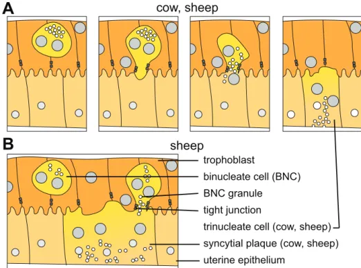
Ruminant placentas are the only synepitheliochorial placentas described to this day. At first glance they are very similar to epitheliochorial ones, presenting all six layers at term. They were in fact classified as such until Wooding (1992) established the synepitheliochorial category based on the presence of cell-cell fusion events at the maternofetal interface. In contrast to the epitheliochorial placentas described above, here binucleated trophoblast cells fuse with cells of the maternal epithelium, forming heterologous syncytia that directly connect the fetal and maternal sides (see Fig 4A, reviewed in Wooding (1992)). The degree of syncytialization of the interface depends on the species, it is for example low in cattle, but high in sheep, the two most studied synepitheliochorial placentas and thus the two common examples of each category.
Binucleate cells in synepitheliochorial placentation The appearance of BNC is a character-istic of synepitheliochorial placentation (reviewed in Wooding, 1992; Wooding & Burton, 2008, chap. 6). These cells are generated by division of trophoblastic nuclei without cytokinesis and represent about 15-20% of trophoblast cells (Wooding, 1983). Mature BNC are very large and present a well developed translational apparatus as well as numerous microvesicles containing dense granules (reviewed in Wooding, 1992; Wooding & Burton, 2008, chap. 6). As in horse placentas (see 1.2.2.a. above), the BNC are migratory and invade the uterine epithelium, however the employed mechanism is very different.
BNC mature between two trophoblastic cells before extending a pseudopod towards the maternofetal interface. This pseudopod squeezes between the two neighboring trophoblast cells and separates their tight junctions, replacing them with its own (Morgan & Wooding, 1983). This maintains the integrity of the placental epithelium while allowing the BNC to contact the maternal epithelium. Once the pseudopod has made contact with the maternal epithelium, a number of specialized vesicles flock to its tip to extend the membrane surface area (Wooding & Burton, 2008). This process induces a break in the microvillous surface of the maternofetal interface since it generates a flat membranous area. The flat area of the BNC membrane bulges into the flattened membrane of the maternal epithelium and both membranes fuse, allowing the BNC cell to inject its contents into what was the maternal epithelial cell (Wooding et al., 1981). Following the fusion event the membrane of the BNC that remains between trophoblast cells gets resorbed by the trophoblasts, re-establishing a tight junction contact between the trophoblast cells (Morgan & Wooding, 1983). For a summary of these processes, see Fig. 5A. The fusion of the BNC with a maternal epithelial cell creates a heterologous syncytium in which nuclei and cell contents are both of placental and maternal origin. This also gives the contents of the BNC cell, and especially its dense vesicles, access to the basal lamina of the maternal epithelium and
1.2
1.2. Mammalian eutherian placentas
B
A
cow, sheep sheep trophoblast binucleate cell (BNC) BNC granuletrinucleate cell (cow, sheep) syncytial plaque (cow, sheep) uterine epithelium
tight junction
Figure 5 – Schematic representation of the fusion processes observed between fetal BNC and maternal epithelium in ruminant placentas. (A) Fetal BNC mature between two trophoblast cells
before extending cytoplasmic projections towards the maternofetal interface, replacing adherent junctions with their own in the process. Following fusion, the BNC cytoplasm mixes with the maternal epithelial cell cytoplasm and the BNC granules are transferred to the basal membrane where they secrete their contents. The trophoblast absorbs the rest of the BNC membrane and re-establishes adherent junctions. (B) In sheep type epitheliochorial placentation, BNC can fused into an already existing heterologous syncytium to form syncytial plaques. The mechanism is the same as for the formation of trinuclear cells. Adapted from Cornelis et al. (2013)
exocytosis of these vesicles is observed specifically at the basal membrane of the fused cells, towards the maternal circulation (Wango et al., 1990). The granules contain placental hormones (placental lactogen and pregnancy associated glycoproteins for example, reviewed in Wooding & Burton, 2008, chap. 6) and this very particular migration and fusion process allows their release close to the maternal circulation, bypassing the maternal epithelial barrier.
Syncytia in synepitheliochorial placentation While in all ruminant placentas fusion events implying BNC occur, the fate of the fused cells depends on the species. In some like sheep or goats, large sycnytial plaques develop at the maternofetal interface of the definitive placenta through repeated fusion of new placental cells into existing syncytia (Fig. 5B, Wooding et al., 1981). By counting the amount of nuclei present in the syncytial plaques, Wooding (1984) show that the total number is always odd, indicating that BNC are the only cells that fuse into the syncytium. Every syncytial plaque starts as a trinucleate cell, as described above, and can reach up to 25 nuclei, corresponding to about a dozen more BNC fusing into the syncytium (Wooding, 1984). In addition to providing a more direct access to the maternal circulation for secretions of the BNC, the syncytial plaque layer also has an amplified microvillous border to increase