1
PBLS,PALS;
Pediatric
Basic
Life
Support, Pediatric Advanced
life Support
■PBLS, PALS Task Force Co-Chairmen Kunio Ohta, Naoki Shimizu
■PBLS, PALS Task Force Members
Mitsuji Iwasa, Tatsuo Iwasaki, Hiroya Ushinohama, Takamori Kanazawa, Junji Kamizono, Sasa Kurosawa, Hiroshi Kurosawa, Osamu Saito, Takekatsu Saito, Seiichi Sato,
Nobuaki Shime, Takehiro Niitsu, Masahiko Nitta, Keiichiro Mizuno, Takashi Muguruma, ■Editorial Board
Kunio Ohta, Tetsuya Sakamoto, Naoki Shimizu, Hiroshi Nonogi, Tetsuo Hatanaka ■Co-Chairmen
Kazuo Okada, Seishiro Marukawa
■1 Introduction
1. Definition of Infant and Child
Since it is reasonable for puberty to be considered the boundary between children and adults from physiological perspectives internationally, “Infant” refers to a person younger than one year of age, and “child” is defined as a person older than one year of age but prior to puberty (including junior high-schoolers). A term “child”, in a broad sense, may sometimes refer to any person prior to puberty and may include an infant.
Neonate refers to a baby in the first 28 days after birth, for whom neonatal resuscitation is applied. In pre-hospital emergency care or the pediatric intensive care unit, however, the neonate within the first 28 days after birth may receive treatment in the same way as an infant.
2. The Chain of Survival and Bow-Tie Concept
The Chain of Survival, which is now a common concept for children and adults, consists of the linking of four actions: 1) prevention of cardiac arrest; 2) immediate recognition of cardiac arrest
and activation of the EMS; 3) basic life support (including AED); and 4) advanced life support (including post-cardiac arrest care).
This is the result of the fact that the bow-tie concept that was emphasized in pediatric
resuscitation in the 2005 guidelines, has become increasingly valued including among cases with adults.
1) Prevention of Cardiac Arrest
This is the concept that include prevention of injuries resulting from unexpected accidents, prevention of disease, and prevention of cardiac arrest by recognizing warning signs of disease. For infants and children, importance of prevention of injuries resulting from unexpected accidents has especially been emphasized. Prevention here also includes improvement of the emergency medical system.
2) Immediate recognition and Activation of EMS
This is the concept that covers immediate recognition of cardiac arrest, activation of the EMS or Medical Emergency Team (MET)/ Critical Care Response Team (CCRT).
As for the cause of cardiac arrest in infants and children, cardiac etiology is rare. The common etiology is respiratory failure with subsequent cardiac arrest (respiratory etiology). Infants and children who have experienced cardiac arrest will have poor outcomes. If found with only respiratory arrest and given treatment before reaching cardiac arrest, however, their survival rates are reportedly 70% or more. Hence it is crucial for the improvement in survival rates to recognize respiratory disorders and shock which directly lead to cardiac arrest in infants and children at an early stage and to deal with the situation promptly.
■2 Causes of Death in Children and Prevention of Cardiac Arrest
The primary cause of death in children over 1 year of age in Japan is injuries.
Many of the injuries are preventable, and forestalling these injuries, which might cause cardiac arrest, is important. They should be regarded as preventable injuries rather than inevitable accidents. Education for the general public on the prevention of injuries is necessary.
1. Motor Vehicle Accidents
Deaths of children under the age of 6 years in motor vehicles have been increasing at three times the rate of those of all ages even after the use of child safety seats was mandated in 2000.The low usage rate of 50% and the poor installation technique have been pointed out as the cause of the increasing death toll.
2. Bicycle Accidents
3
30,000 per year (Tokyo Metropolitan Police Department, 2009). This figure is decreasing but accounts for an increasing portion of total traffic fatalities. Although bicycle helmets are known to significantly reduce the severity of head injury that is closely related to deaths from bicycle accidents, public awareness of helmet safety is still low in Japan. There are also many cases where children under the age of 2 years fall off child bicycle seat, which is particularly seen in Japan.
3. Foreign Body Ingestion and Aspiration
Approximately 60% of deaths from foreign body ingestions and aspirations in children occurred in infants under the age of one year, and 90% occurred in children under the age of 5 years. Using an empty toilet paper roll to approximate the size, anything that comes through the roll can be an object that might be ingested or aspirated by children and infants. It is necessary to provide guidance for the prevention according to children’s stage of development at the infant medical check-up.
4. Submersion injuries
In Japan, there are many submersion injuries in bathtubs. In households with preschool age children, precautions for various possible cases are necessary, such as draining water from the bathtub after bathing, or installing a lock on the upper part of the bathroom door.
5. Fire-related injuries
Eighty percent of deaths from fire in children and infants occurred at one’s own home.
Installing smoke detectors and fire sprinklers in one’s home helps reduce deaths from fire, yet they cannot prevent fire breakout from playing with fire by children who stay at home unattended. While specifying flame-retardant materials and developing child-resistant lighters is being discussed, the major premise is to recognize parental guidance as an indispensable basis for preventing deaths from fire-related injuries.
■3 Early Recognition of Respiratory Disturbances and Shock
There is a tendency among many cases physicians to begin with seeking for diagnosis, incorrectly believing that treatments should not precede diagnosis. Even in the absence of a diagnosis in the initial treatment for emergency patients, however, physiological understanding of circulatory and respiratory function and prompt assessment based on the child’s vital signs will contribute to the immediate start of initial treatment. Trying to identify the disease while stabilizing the patient’s condition will eventually lead to further advanced treatment.
1. Respiratory Disturbances
Respiratory disturbances are classified according to severity into the two levels; respiratory distress and respiratory failure.
1) Respiratory Distress
Respiratory distress refers to a condition where normal or near-normal level of oxygenation and/or ventilation is maintained despite the presence of respiratory efforts such as grunting, tachypnea, retraction, or nasal flaring.
2) Respiratory Failure
Respiratory failure refers to a deteriorating condition of respiratory distress with the presence of abnormal level of oxygenation and/or ventilation.
3) Initial Treatment for Respiratory Disturbances
When respiratory distress is recognized, start oxygen administration. If accompanied with hypoxemia, supply highly concentrated oxygen. If accompanied with hypoventilation, assist breathing using a bag valve mask, while trying to determine whether a brief assistance is all that is needed or if tracheal intubation is required.
2. Shock
Shock is an acute life-threatening systemic pathological condition. Poor tissue perfusion makes oxygen and nutrient supply balance inadequate to meet metabolic demands. It results in oxygen deficiency in each cell level and leads to fatal progression of metabolic acidosis.
Typical signs of shock are deteriorated mental status, tachycardia, bradycardia, diminished pulse, drop in blood pressure, prolonged capillary refill time (>2 seconds), cold extremities and decreased urine output.
1) Compensated Shock
Compensated shock is defined as a state where systolic blood pressure is maintained within each age group’s normal range despite the reduction of stroke volume. This compensatory mechanism owes it to increased heart rate and systemic vascular resistance, which together operate to keep blood pressure within normal range.
2) Decompensated Shock (Hypotensive Shock)
When compensated shock becomes exacerbated with the compensatory mechanisms failing to maintain adequate vital organ perfusion, and blood pressure fall down below the normal limit for each age group, decompensated shock (hypotensive shock) with hypotension will develop.
3) Initial Treatment for Shock
Shock results from a number of causes. As for initial treatment, 10-20ml/kg of isotonic fluids (normal saline or Ringer’s solution) should be promptly administered regardless of the cause.
5
Hypotonic fluid should not be used. Re-evaluation of the patient should be conducted following the prompt initial evaluation. Repeated administrations of isotonic fluids will be performed if necessary, while searching for the cause of shock.
As oxygen demand in the tissues of the body exceeds supply in the state of shock, supplemental oxygen should be supplied.
■4 Systems: MET/CCRT and PICU
1. Medical Emergency or Rapid Response Team
It was shown that METs or RRTs are effective to prevent in-hospital respiratory arrests or cardiac arrests. The introduction of a MET or RRT was associated with a decrease in pediatric hospital mortality in 1 LOE 3 meta-analysis1 and 3 pediatric LOE 3 studies with historical
controls2-4. The introduction of a MET or RRT was associated with
• a decrease in respiratory but not cardiac arrest in 1 LOE 35 study with historical controls
• a decrease in total number of preventable cardiac arrests in 1 LOE 3 retrospective chart review6
• a decrease in preventable cardiac arrests in 1 LOE 34 study
• a decrease in total number of cardiac arrest and a decrease of out-of–pediatric intensive care unit (PICU) mortality in 1 LOE 33 pediatric cohort study using historical controls
For the prevention of respiratory and cardiac arrest in general wards (out-of-PICU ), introduction of METs or RRTs can be considered on the premise that PICU can be established.
2. Pediatric Intensive Care Unit (PICU)
PICUs have already well developed in other countries, while not yet fully established in Japan. Evidence suggests that centralization of critically ill or traumatized children to PICUs improves outcomes (LOE 47). One study in Japan shows that centralization of pediatric critically ill or
traumatized children to PICUs improved patients’ outcomes (J-LOE 48).
Ideally patients after cardiac arrest should be managed by a trained PICU team, and when post-cardiac resuscitation care is necessary, interfacility transfer should be coordinated as soon as possible. Preferably the interfacility transport team should consist of personnel with broad experience in the management of critically ill patients such as pediatric intensivists or pediatric emergency physicians. Japan has been lagging behind in development and expansion of PICUs. Centralization of critically ill and traumatized children to PICU and establishment of systems are strongly called for in Japan.
■5 Pediatric Basic Life Support: PBLS
1. Introduction
When a lay rescuer gives CPR to a child, they should follow the same Basic Life Support (BLS) guidelines as for the adult. However, those who contact children on a routine basis, such as
parents or family members of children, nursery staff, school teachers, lifeguards and sports coaches are encouraged to learn Pediatric Basic Life Support (PBLS). When healthcare providers respond to a child with cardiac arrest, they should follow PBLS.
Although in the guidelines, a series of skills are sequentially presented so as for the procedures to go step by step, if more than one rescuer is present, it is recommended to perform certain steps at the same time, e.g., initiation of CPR and EMS activation. These procedures are shown in the algorithm. Each heading number corresponds to the number in each box. (This section does not deal with the neonate, which is described in the section of Neonatal Resuscitation.)
2. Changes in Guidelines
Changes in PBLS 2010 guidelines from 2005 are as follows:
- For the purpose of increasing the likelihood of bystander CPR during pediatric out-of-hospital cardiac arrest, CPR should be started with chest compression as in the case with adults. Since the effectiveness of rescue breathing is evident in the case of cardiac arrest in children, it is emphasized to give rescue breath as soon as possible.
- The pulse check to determine cardiac arrest has been proved to be unreliable. Cardiac arrest is determined by no responses and the abnormal breathing.
- AEDs with energy dose attenuator can be used for preschool age children (approximately until age 6).
7
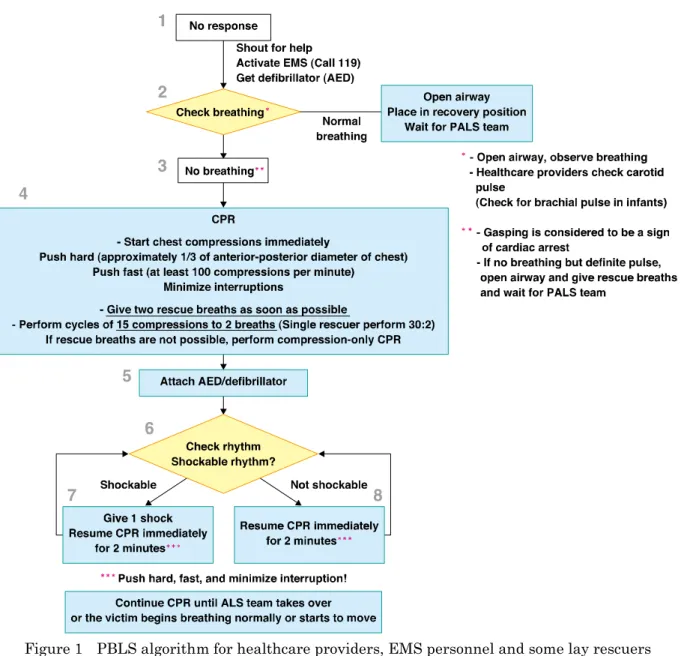
Figure 1 PBLS algorithm for healthcare providers, EMS personnel and some lay rescuers (parents, preschool teachers, etc)
3. PBLS Algorithm
1) Check for Victim’s Response and EMS Activation [Box 1]
Ensure the safety of the victim and rescuer.
Speak loudly to the victim, patting them lightly on the shoulder. If there is no response or no purposeful movement, the person should be determined "unresponsive". For infants, stimulate the sole of the infant’s foot to try to elicit a response.
If the victim is unresponsive, shout for help and ask people around to activate the EMS (call 119). And ask to bring a defibrillator (AED, if available nearby). The EMS dispatcher is supposed to dispatch an ambulance immediately upon suspicion of cardiac arrest based on the report from the caller. The rescuers follow telephone instructions to assess the victim and to perform CPR.
Rescuers stay at the scene and start chest compressions. If in-hospital emergency call is available, activate it and ask for help and for get crash cart.
2) Recognition of Cardiac Arrest [Box 2, 3]
If the victim is unresponsive and not breathing or breathing abnormally (gasping), rescuers assume cardiac arrest and begin CPR immediately. The rescuer should take no more than 10 seconds to check for breathing. Gasping is considered to be absence of normal breathing, and is a sign of cardiac arrest.
Healthcare providers and EMS personnel should first open the airway and check respiration. However, opening the airway should not preclude a proper respiration check or delay the start of CPR. Trained healthcare providers can check for a pulse while observing respiration. Delays of starting CPR due to pulse check should be avoided. Untrained rescuers do not have to check for a pulse.
If the victim is breathing normally, keep the airway open and wait for help and the EMS personnel. In the meantime, continue observing the victim's breathing. If the victim stops breathing, begin CPR immediately. If the rescuer needs to leave the victim to ask for help, the victim should be placed in the recovery position.
There may be rare occasions where the victim has no breathing but has a pulse. The rescuer should open the airway and provide rescue breathing. When the victim’s pulse rate is below 60 beats per minute, the rescuer should follow the bradycardia algorithm. If there is a palpable pulse of more than or equal to 60 beats per minute but there is no spontaneous breathing or there is inadequate breathing, the rescuer should provide rescue breaths at a rate of about 12 to 20 breaths per minute (1 breath every 3 to 5 seconds). Subsequently while waiting for the arrival of the PALS team, the rescuer should check for the pulse frequently so as not to delay beginning chest compressions in case of cardiac arrest.
3) CPR [Box 4]
(1) Chest compressions
All rescuers should provide chest compressions to victims of cardiac arrest. Location of compressions is the lower half of the sternum. It is reasonable to refer to it as “the center of the chest.”
A strong emphasis on delivering high quality chest compressions remains essential:
- Push hard approximately one-third of the anterior-posterior diameter of the chest for infants and children
- Compress at a rate of at least 100 compressions per minute - Minimize interruptions of chest compressions
(2) Opening the Airway and Ventilation
As soon as the rescue breather is ready, the rescuer should open the victim’s airway and give 2 breaths. Duration of each breath is about one second to make chest rise. If prompt rescue breath is impossible or not affordable, the rescuer should immediately begin chest compressions.
9
Open the airway using the head tilt-chin lift maneuver. The trained rescuer can use the jaw thrust maneuver if necessary. For the victims with suspected spinal injury, the jaw thrust maneuver should be the first choice. Use the head tilt-chin lift maneuver if the jaw thrust does not open the airway.
As pediatric cardiac arrest is likely to be respiratory origin, it is important to open the airway and start rescue breath as quickly as possible. Therefore, in the case of in-hospital patients considered at risk for cardiac arrest, preparedness of rescue breath is recommended.
(3) Compression-Ventilation Ratio
For 2-rescuer CPR, a compression-ventilation ratio of 15:2 is reasonable. For single-rescuer CPR, a ratio of 30:2 as for adult is reasonable.
When an advanced airway such as a tracheal tube is in place, compressions should not be interrupted for ventilations. Rescue breath should be performed around 10 times per minute.
When rescue breath is difficult to give, the rescuer should perform compression-only CPR.
4) ECG analysis [Box 5, 6]
Rescuers, including healthcare providers, should continue CPR without checking for a pulse until a defibrillator/AED arrives.
Whether or not using an AED or manual defibrillator, chest compressions should be continued until just before ECG analysis. Defibrillator which can be converted to AED mode, where the heart rhythm is automatically analyzed, are helpful to healthcare providers who usually have few chances to perform CPR.
For preschool-age children and infants, the rescuer should use a defibrillator with energy dose attenuator. If dose attenuator is not available, adult systems can be substituted. In such cases, the user should make sure that the pads do not overlap or even touch each other.
Pads should be placed on the exposed chest in an anterior-lateral position. The anterior-posterior position is alternatively acceptable.
5) Shockable [Box 6 to 7]
When using an AED, the rescuer should follow the audio instructions from the AED and deliver the electric shock.
When using a manual defibrillator, the shock should be delivered if ventricular fibrillation or pulseless ventricular tachycardia is recognized. Immediately after delivering a shock, two-minute CPR should be resumed starting with chest compressions. Subsequently every two minutes, the rescuer is required to check the heart rhythm on the monitor, deliver the shock if necessary, and continue CPR.
6) Not shockable [Box 6 to 8]
When using an AED, the rescuer should follow the audio instructions from the AED and resume CPR.
When using a manual defibrillator, if the QRS complex that shows the possibility of ROSC is recognized, the rescuer should check for a pulse. If a pulse is detected, the post-ROSC monitoring and management should be started.
In the case of pulseless electrical activity or asystole, two-minute CPR should be immediately resumed starting with chest compressions. Subsequently every two minutes, the rescuer is required to check the heart rhythm on the monitor and continue CPR.
7) Continuation of BLS
Rescuers should continue CPR until sufficient circulation is restored in the victim, or EMS providers or other responders take over the care of victim to provide advanced life support. CPR should not be discontinued until the victim regains obvious ROSC (such as regular respiration or purposeful movement).
4. Foreign Body Airway Obstruction; FBAO
In children 1 year of age or older with FBAO, rescuers should activate EMS. Back blows, abdominal thrusts, and chest thrusts for obstruction relief should be tried. These techniques must be repeated rapidly until the relief of the obstruction.
If the choking infants are still responsive but cannot make an effectively strong cough, rescuers are recommended to move the victim’s head downward and try back blows and chest thrusts.
If the victim with FBAO becomes unresponsive, the rescuer should immediately begin CPR. Lay rescuers can begin CPR starting with chest compressions as in usual cases of cardiac arrest. It is reasonable for healthcare providers to start CPR with rescue breath. For unresponsive victims of FBAO, direct removal may be considered only when solid material is visible in the airway.
5. CPR
1) Recognition of Cardiac Arrest
Rescuers should observe movements of the victim’s chest and abdomen. When no breathing is recognized, CPR should be started (Class I). Lay rescuers do not need to open the airway when assessing breathings. They should focus on the movement of the chest and abdomen instead. No more than 10 seconds should be taken to check for breathing.
Gasping is considered to be absence of normal breathing, and is a sign of cardiac arrest. Gasping, also known as agonal breathing, is an abnormal breathing-like movement occasionally seen immediately after cardiac arrest. Gasping is sometimes seen in adults, but rarely seen in children and infants.
Healthcare providers and EMS personnel should first open the airway and check the respiration of the unresponsive victim. The process of opening airway should not cause negligence of checking breathings or delay in starting CPR.
Lay rescuers should not check for a pulse to determine cardiac arrest (Class III). Healthcare providers will check for a pulse while observing respiration. However, healthcare providers not
11
well trained or unfamiliar with CPR could skip checking a pulse as same as lay rescuers. Delays in starting CPR due to pulse check should be avoided (Class III). If the rescuer is uncertain of the presence or absence of a pulse, they should focus on checking for respiration. Once recognizing a lack of breathing, CPR should be immediately started.
2) Pulse Check Versus Check for Signs of Life
Palpation of a pulse (or its absence) is not reliable as thesole determinant of cardiac arrest and need for chest compressions.If the victim is unresponsive, not breathing normally, and thereare no signs of life, lay rescuers should begin CPR. In infantsand children with no signs of life, healthcare providers should begin CPR unless they can definitely palpate a pulse within10 seconds (Class I).
Thirteen LOE 5 studies9-21 observed that neither laypersonsnor healthcare providers are able to
perform an accurate pulsecheck in healthy adults or infants within 10 seconds. In 2 LOE5 studies in adults22, 23 and 2 LOE 3 studies in children withnonpulsatile circulation24, 25, blinded healthcare
providers commonly assessed pulse status inaccurately and their assessment often took >10 seconds. In the pediatric studies, healthcareprofessionals were able to accurately detect a pulse by palpationonly 80% of the time. They mistakenly perceived a pulse whenit was nonexistent 14% to 24% of the time and failed to detecta pulse when present in 21% to 36% of the assessments. The averagetime to detect an actual pulse was approximately 15 seconds,whereas the average time to confirm the absence of a pulse was30 seconds. Because the pulseless patients in these studies were receiving extracorporealmembrane oxygenation (ECMO) support, one must be cautious in extrapolating these data to the arrest situation; all pulselesspatients did have perfusion and therefore had signs of circulationas evidenced by warm skin temperature with brisk capillary refill.All patients evaluated were in an intensive care unit (ICU)setting without ongoing CPR.
3) Chest Compressions
The rescuer should place the victim in a supine position26 with the rescuer kneeling beside the
victim27.
To maximize the effectiveness of chest compressions, it is reasonable to place the victim on a firm surface if possible (Class IIa)(LOE 528-30). Air-filled mattresses should be deflated when
performing CPR (Class I)(LOE 531). There is insufficient evidence for or against the use of
backboards during CPR. When backboard is used, attention should be paid to avoid delays in initiation of CPR, to minimize interruptions in CPR, and to prevent line/tube displacement. Chest compressions for victims lying on a bed reportedly turn out to be shallow since some of the force intended to compress the chest results in mattress displacement rather than chest compression (LOE 432, LOE 528, 33-35). No studies have examined the risks or benefits of moving the patient from
a bed to the floor to perform CPR.
4) Chest Compressions for Children: Hand Position
in 2005 CoSTR (“the rescuer should compress the lower half of the victim’s sternum”) when performing external chest compressions for children or adults in cardiac arrest. Therefore it is reasonable to adopt the use of “the lower half of the sternum” as the hand position for chest compressions (Class IIa).
5) One- Versus 2-Hand Chest Compression in Children
There are no outcome studies comparing 1- versus 2-hand chestcompressions for children in cardiac arrest. Evidence from 1LOE 5 randomized crossover child manikin study36 showed that
higher chest-compression pressures are generated by healthcareprofessionals using the 2-hand technique. Two LOE 5 studies37, 38 report no increase in rescuer fatigue comparing 1-hand with
2-hand chest compressions delivered by healthcare providersto a child-sized manikin.
Either a 1- or 2-hand technique can be used for performing chestcompressions in children (Class IIb).
6) Chest Compressions for Infants: Two-Finger Technique /Two Thumb–
Encircling Hands Technique
In infant victims, lay rescuer or single healthcare provider should compress the sternum with two fingers placed in the center of the chest (Class I). The two thumb–encircling hands technique is recommended for healthcare providers when two or more rescuers are present (Class I). The rescuer is required to encircle the infant’s chest with both hands, spread their fingers around the thorax, and place their thumbs together over the center of the chest. If the rescuer is alone or they cannot physically encircle the victim’s chest, they should compress the chest with two fingers.
The two thumb–encircling hands technique is preferred because it produces higher coronary artery perfusion pressure, more consistently results in appropriate depth or force of compression39-42, and may generate higher systolic and diastolic pressures43-46. However, there are
insufficient data for or against the need for a circumferentialsqueeze of the chest when performing the 2-thumb technique ofexternal chest compression for infants.
7) Chest Compression Depth
Evidence from anthropometric measurements in 3 good-qualityLOE 5 case series47-49 showed
that in children the chestcan be compressed to one third of the anterior-posterior chestdiameter without causing damage to intrathoracic organs. One LOE 5 mathematical model based on neonatal chest computed tomography scans50 suggests that one third anterior-posterior chest
compressiondepth is more effective than one fourth compression depth andsafer than one half anterior-posterior compression depth.
A good-quality LOE 551 adult study found that chest compressionsare often inadequate, and a
good-quality LOE 4 pediatric study48 showed that during resuscitation of patients >8 yearsof age,
compressions are often too shallow, especially following rescuer changeover. Evidence from 1 pediatric LOE 4 systematic review of the literature52 showed that rib fractures are rarely
13
Given these facts, 2010 CoSTR states “In infants, rescuers should be taught to compress the chestby at least one third the anterior-posterior dimension or approximately1 inches (4 cm). In children, rescuers should be taught to compress the chest by at least one third the anterior-posterior dimensionor approximately 2 inches (5 cm).” A study from Japan (J-LOE 453) ,
however, showed that the average chest diameter of Japanese children aged between 1 and 7 is from 109.2 to 141.4mm. One-third of this makes 36.4 to 47.1mm, and compression at a depth of 5cm is too deep. Therefore, Japanese guidelines recommend compressing the chest by one-third the anterior-posterior diameter (Class I).
8) Chest Decompressions
While allowing complete recoil of the chest after each compression may improve circulation (Class IIa), care should be taken not to let compressions become too shallow.
9) Chest Compression Rate
It is reasonable for lay rescuers and healthcare providers to perform chest compressions at a rate of at least 100 compressions per minute. There is insufficient evidence to indicate a recommended upper limit in chest compression rate. Duration of interruptions in compressions should be minimized so as to maximize the number of compressions delivered per minute (Class I).
10) Feedback for Chest Compression Quality
When more than one rescuer is present, it is reasonable for rescuers and EMS personnel to monitor and try to improve the CPR quality, ensuring adherence to recommended compression and ventilation rates and depths (Class IIa). Real-time chest compression-sensing and feedback/prompt technology may be useful adjuncts during resuscitation efforts.
11) Pulse Check during CPR
Interrupting chest compressions for a pulse check is not recommended unless there is an obvious reaction (normal breathing or purposeful movement) that clearly shows ROSC (Class III). Healthcare providers should also continue CPR without checking for a pulse if there is no monitor available (Class I). It is reasonable to check for a pulse if an organized rhythm is visible on the monitor (Class IIa).
12) Switching Rescuers
It may be reasonable for another rescuer to take over after a period of no longer than 1 to 2 minutes, to prevent deterioration in the quality of compressions (Class IIb). Switching should be done with the minimum of interruptions in the compressions (Class I).
13) Opening the Airway
Opening and maintaining the airway is essential to ensure effective ventilation (Class I). For unresponsive children, open the airway using the head tilt-chin lift maneuver (Class IIa). The trained rescuer can use the jaw thrust maneuver if necessary as for victims with suspected spinal injury (Class IIb). Use the head tilt– chin lift maneuver if the jaw thrust does not open the airway. As the jaw lift maneuver can be harmful, it requires careful attention to adaptive decision making and practice.
14) Tidal Volume and Ventilation Rate
It is reasonable to achieve chest rise with each breath given (Class IIa). The rescuer should avoid hyperventilation during CPR, regardless of etiologies of cardiac arrest (ie. cardiac or respiratory) (Class III). In infants and children, a reduction in minute ventilation to less than baseline for age is reasonable to avoid the harmful effects of hyperventilation (Class IIa).
15) Barrier Devices
The risk of disease transmission in out-of-hospital is very low and initiating rescue breath without a barrier device is reasonable. If available, rescuers may consider using a barrier device (Class IIb). However, safety precautions should be taken both in the in-hospital and out-of-hospital situations if the victim is known to have a serious infection (e.g., human immunodeficiency virus (HIV), tuberculosis, hepatitis B virus, or severe acute respiratory syndrome (SARS) ) (Class I). Healthcare providers on duty must always follow standard precautions when performing CPR (Class I).
16) Barrier Devices (Healthcare Providers)
When two or more experienced rescuers are present, ventilation using a bag-valve-mask is reasonable (Class IIa). It may be effective that one rescuer use both hands to open the airway and maintain a tight mask-to-face seal while another compresses the ventilation bag (Class IIa). Holding the mask to the victim's face with both hands can ensure a better mask seal (LOE 554, 55).
In the case of in-hospital pediatric patients considered at risk for respiratory or cardiac arrest, oxygen and BVM should be readily available (Class I).
17) CPR Initiation Procedures
As most cardiac arrests in children are of respiratory origin, it is important to open the airway and start rescue breathing as promptly as possible. It is desirable that devices for rescue breathing and oxygen is readily available in the setting where PBLS may be performed. In PBLS, once rescue breath is ready, the rescuer should open the victim’s airway and perform 2 rescue breaths. If immediate rescue breath is unavailable, the rescuer should immediately begin chest compressions. As soon as it is ready, open the airway and perform 2 rescue breaths. Subsequently, chest compressions and rescue breath should be performed at a ratio of 30:2 for single-rescuer
15
CPR, or 15:2 for 2-rescuer CPR.There is no direct evidence in human or animal studies that starting adult and pediatric CPR with 30 chest compressions produce a better outcome than starting with 2 rescue breaths.
18) Optimal Compression-Ventilation Ratio for Infants and Children
There are insufficient data to identify an optimal compression-ventilationratio for CPR in infants and children. In 4 LOE 5 manikin studies56-59 examining the feasibility of
compression-ventilationratios of 15:2 and 5:1, lone rescuers could not deliver thedesired number of chest compressions per minute at a ratio of5:1. In 5 LOE 5 studies60-64 using a variety of
manikinsizes comparing compression-ventilation ratios of 15:2 with30:2, a ratio of 30:2 yielded more chest compressions with no,or minimal, increase in rescuer fatigue. One LOE 5 study65 of
volunteers recruited in an airport to perform 1-rescuer laypersonCPR on an adult-sized manikin observed less "no flow time" withthe use of a 30:2 ratio compared with a 15:2 ratio.
One LOE 5 observational human study66 comparing resuscitationsby firefighters before and
after the change from a recommended15:2 to 30:2 compression-ventilation ratio reported more chestcompressions per minute with the 30:2 ratio, but the rate ofROSC was unchanged. Three LOE 5 animal studies67-69 showedthat coronary perfusion pressure, a major determinant of
success in resuscitation, rapidly declines when chest compressions are interrupted; once compressions are resumed, several chest compressionsare needed to restore coronary perfusion pressure to preinterruptionlevels. Thus, frequent interruptions of chest compressions prolongthe duration of low coronary perfusion pressure and flow andreduce the mean coronary perfusion pressure.
Three LOE 5 manikinstudies65, 70, 71 and 3 LOE 551, 72, 7361,72,73 in- and out-of-hospitaladult
human studies documented long interruptions in chest compressionsduring simulated or actual resuscitations. Three LOE 5 adult studies74-76 demonstrated that these interruptions reduced
ROSC.
In 5 LOE 5 animal studies67-69, 77, 78 chest compressionswithout ventilations were sufficient to
resuscitate animalswith VF-induced cardiac arrest. Conversely in 2 LOE 5 animalstudies79, 80
decreasing the frequency of ventilation was detrimental in the first 5 to 10 minutes of resuscitation of VF-inducedcardiac arrest.
One LOE 5 mathematical model81 suggested that the compression-ventilationratio in children
should be lower (more ventilations to compressions) than in adults and should decrease with decreasing weight. TwoLOE 5 studies of asphyxial arrest in pigs82, 83 showed thatventilations
added to chest compressions improved outcome compared with compressions alone. Thus, ventilations are more importantduring resuscitation from asphyxia-induced arrest than during resuscitation from VF. But even in asphyxial arrest, fewer ventilationsare needed to maintain an adequate ventilation-perfusion ratioin the presence of the low cardiac output (and consequently low pulmonary blood flow) produced by chest compressions.
In order to simplify instruction for teaching and improve skill retention, it is reasonable for the single-rescuer to perform CPR in infants and children at a 30:2 compression to ventilation ratio
(Class IIa). After 30 chest compressions, 2 effective ventilations should be promptly given so as to minimize the interruption of chest compressions. When healthcare providers are performing 2-rescuer CPR, a compression-to-ventilation ratio of 15:2 where one is performing chest compression and the other is keeping the airway open is reasonable (Class IIa). When an advanced airway management device is in place, compressions should not be interrupted for ventilations. The rescuer in charge of ventilation should perform rescue breath at a rate of approximately 10 times per minute and avoid hyperventilation (Class I).
(1) Newborns (Out of the Delivery Area) Without an Endotracheal Airway
There are insufficient data to identify an optimal compression-ventilationratio for all infants in the first month of life.
One LOE 5animal study67 showed that coronary perfusion pressure declinedwith interruptions
in chest compressions; after each interruption,several chest compressions were required to restore coronaryperfusion pressure to preinterruption levels. One LOE 5 adulthuman study75 and 2 LOE
5 animal studies68, 76 showed thatinterruptions in chest compression reduced the likelihood of
ROSC in VF cardiac arrest.
One LOE 5 1-rescuer manikin study59 showed that more effectiveventilation was achieved with
a 3:1 ratio than with a 5:1, 10:2,or 15:2 ratio. One LOE 5 mathematical study of cardiovascular physiology84 suggested that blood flow rates in neonates arebest at compression rates of >120/min.
Although based on limited data, in the case of 2-rescuer CPR in cardiac arrest with cardiac etiology, a 15:2 ratio might be more effective than 3:1 (Class IIb). To simplify instruction for teaching, for term infants in the first month of life and neonates, ratios and CPR methods should be adjusted to those most commonly used in their respective environments.
(2) Newborns (Out of Delivery Area) With a Tracheal Tube
There is insufficient evidence to determine if an intubatedneonate has a better outcome from cardiac arrest using a 3:1compression-ventilation ratio and interposed ventilations comparedwith continuous chest compressions without pause for ventilations(asynchronous compressions and ventilations).
Two LOE 5 adult74, 76 and 2 LOE 5 animal67, 77 studies demonstratedthat interruptions in chest
compressions reduced coronary perfusionpressure, a key determinant of successful resuscitation in adults, and decreased ROSC. There are no equivalent studies evaluating the impact of interrupted chest compressions in asphyxiatedneonates or neonatal animal models.
In 1 LOE 5 piglet study85 of VF arrest, myocardial blood flowincreased using simultaneous chest
compressions and high–airwaypressure ventilations in a 1:1 ratio as compared with conventional CPR at a 5:1 ratio. Another LOE 5 VF piglet study86 demonstratedequivalent cardiac output but
worsened gas exchange using a1:1 compression-ventilation ratio (ie, simultaneous compressions and ventilations) with high airway pressures compared with conventionalCPR at a 5:1 ratio.
One LOE 582 study in nonintubated asphyxiated piglets resuscitated with a 5:1
compression-ventilation ratio showed that ventilationsare important for successful resuscitation. One LOE 5 study in intubated asphyxiated piglets87 showed that the additionof ventilations
17
resulted in lower arterial CO2 tension (PaCO2) without compromising hemodynamics when
compared with compressionsalone. One LOE 5 manikin study88 found that healthcare providers
were unable to achieve the recommended rate of ventilations during infant CPR at a 3:1 compression-ventilation ratio, with20% delivering a net rate of 40 breaths per minute after 5 minutesof resuscitation. There are no studies that evaluate the impactof continuous compressions on minute ventilation, gas exchange,or the outcome of resuscitation during CPR for intubated neonates.
Given these study results and the necessity of simplifying education, for intubated term infants in the first month of life and neonates, ratios and CPR methods should be adjusted to those most commonly used in their respective environments (Class I). For intubated neonates in need of CPR in the settings outside of the delivery room, newborn nursery, or NICU (e.g., pre-hospital setting, emergency department or PICU), or for those in cardiac arrest with cardiac etiology regardless of place, infant CPR should be performed in accordance with the guidelines (i.e., chest compressions should not be interrupted for ventilation)(Class I).
19) Compression-Only CPR
Evidence from 1 LOE 2 large out-of-hospital pediatric prospectiveobservational investigation89
showed that children with cardiacarrest of noncardiac etiology (asphyxial arrest) had a higher 30-day survival with more favorable neurologic outcome if theyreceived standard bystander CPR (chest compressions with rescuebreathing) compared with chest compression-only CPR. Standard CPR and chest compression-only CPR were similarly effectiveand better than no bystander CPR for pediatric cardiac arrestfrom cardiac causes. Of note, the same study showed that morethan 50% of children with out-of-hospital cardiac arrest did not receive any bystander CPR. Compression-only CPR was as ineffectiveas no CPR in the small number of infants and children with asphyxialarrest who did not receive ventilations.
Two LOE 5 animal studies82, 83 demonstrated improved survivalrates and favorable neurologic
outcome with standard CPR comparedwith no CPR. One LOE 5 animal study87 showed that
blood gasesdeteriorated with compression-only CPR compared with standardCPR in asphyxial arrests.
Data from 1 LOE 5 animal study83 indicated that compression-onlyCPR is better than no CPR
for asphyxial arrest but not as effectiveas standard CPR, and 6 LOE 5 clinical observational studiesin adults90-95 showed that compression-only CPR canresult in successful resuscitation from
an asphyxial arrest.Moreover, in 10 LOE 5 animal studies67, 77, 78, 96-102and 7 LOE5 adult clinical
observational studies90-95, 103 compression-only bystander CPR was generally as effective as
standard 1-rescuerbystander CPR for arrests from presumed cardiac causes.
For adults in cardiac arrest, if minimizing of interruption to chest compressions is impossible, rescuers, even healthcare providers, are recommended to focus on chest compressions rather than rescue breath in CPR. However, as most cardiac arrest in children and infants is of respiratory origin, the best resuscitation for such victims in cardiac arrest due to hypoxia should be prompt
initiation of ventilation and chest compressions. Thus, rescuers should provide conventional CPR (rescue breathing and chest compressions) for in-hospital and out-of-hospital pediatriccardiac arrests (Class I). However, rescuers who cannot provide rescue breathshould at least perform chest compressions for infants and childrenin cardiac (Class I).
20) AED Use in Children
The use of AED pads with pediatric attenuation capabilities or AEDs in the pediatric mode was limited to children between 1 and 8 years of age. The recent CoSTR 2010, however, has lowered the lower border of the age range and now an AED is applicable to infants as well.
In Japan, due to the age range in the Japanese schooling systems, there was confusion at the scene in the use of pads; children aged 6-7 (usually first or second grade) need pediatric pads, while children aged 8 or older (usually second or older grade) require adult pads. Risk of miss-applying pediatric pads to children aged 8 years or older was also reported. Given this situation, the Japanese guidelines define the age range of pediatric pad use as preschool age (until about the age of 6 years) for convenience.
This means that the adult pads will be used for children aged 6-7. A use of adult pads for this age group has previously been safely practiced when pediatric pads are not available, and a large body of evidence suggests that estimated energy dose per bodyweight given via adult pad to these children is safe., the adult pads have been applied to those in that age group in the case of the absence of pediatric pads. In addition, many studies have ensured the safety of the number of joules delivered, estimated from the average weight of Japanese children in that age group.
21) Pad Position
Pad position in children does not change theROSC rate104, and there is no clear evidence that it
alters transthoracic impedance either105-108. Transthoracic impedance was increased in 1 adult
LOE 5109 studyby placing the pads too close together and in 1 LOE 5110 studywhen the pads were
placed over the female breast. Additionally,1 adult LOE 5111 study showed that placing the apical
pad ina horizontal position lowers transthoracic impedance.
For preschool-age children, pediatric pads with attenuation capabilities or an AED in the pediatric mode should be used (Class I). If pediatric pads are unavailable and no other choice is left, adult pads can be substituted (Class I).
There is insufficient evidence to alter the current recommendations to use the largest size paddles/pads that fit on the infantor child's chest without touching each other or to recommend one paddle/pad position or type over another.
22) AED Use in Infants
Three112-114 studies showed that infantsin cardiac arrest (in- and out-of-hospital) may have
shockablerhythms. Evidence from 3 LOE 5115-117 studies showed thatmany AED devices can safely
19
The optimal energy dose for defibrillation in infants has notbeen established, but indirect data from 5 LOE 5 animal studies118-122 showed that the young myocardium may beable to tolerate
high-energy doses. In 3 LOE 5 animal studiesa pediatric attenuator used with an adult-dose biphasic AEDshock was as effective and less harmful than monophasic weight-baseddoses123 or
biphasic adult doses124, 125.
Two LOE 4 case reports126, 127 described survival of infantswith out-of-hospital cardiac arrest
when AED use was coupledwith bystander CPR and defibrillation using an AED. Two pediatric LOE 5 case reports128, 129 noted successful defibrillation withminimal myocardial damage and good
neurologic outcome usingan AED with adult energy doses.
The AED can be used on infants under 1 year of age in out-of-hospital VF/ pulseless VT (Class I). If AED with dose attenuator are not available, adult systems can be substituted.
For treatment of out-of-hospital VF/pulseless VT in infants,the recommended method of shock delivery by device is listedin order of preference below. If there is any delay in the availabilityof the preferred device, the device that is available shouldbe used. The AED algorithm should have demonstrated high specificityand sensitivity for detecting shockable rhythms in infants.The order of preference is as follows:
1. Manual defibrillator 2. AEDwith dose attenuator 3. AED without dose attenuator
23) Defibrillator Pads and Paddles for Infants
In Japan, so-called “pediatric pads” and “pediatric paddles” for manual defibrillators are actually intended for use in infants aged up to 1 year who weigh less than approximately 10 kg. However, the term “pediatric” has generated confusion among practitioners. In the current guidelines, such pads and paddles are referred to as “infant” pads and “infant” paddles.
One LOE 5 study in adults130 demonstrated that shock successincreased from 31% to 82% when
pad size was increased from 8x8 cm to 12x12 cm. Three pediatric LOE 4105, 131, 132, 3 adultLOE 5110, 130, 133, and 3 LOE 5 animal108, 134, 135 studies demonstratedthat transthoracic impedance decreases
with increasing pad size.Decreased transthoracic impedance increases transthoracic currentand, thus, presumably, transmyocardial current.
24) Removal of Foreign Body from Infant’s Airway
In responsive children 1 year of age or older with foreign-body airway obstruction (FBAO), rescuers should activate emergency medical systems (Class IIa) and try back blows, abdominal thrusts, and chest thrusts (Class IIa). More than one technique may be required to relieve the obstruction. These techniques must be repeated rapidly until the relief of the obstruction.
If the choking infants are still responsive but cannot make a strong cough effectively, rescuers are recommended to try back blows and chest thrusts (Class IIa). In these rescue maneuvers, it is
reasonable to move the victim’s head downward as the most common cause of FBAO is liquid (Class IIa). If the choking infants are still coughing strongly, rescuers move them onto their sides encouraging their coughing so that they can spit out the obstructing liquids.
If the victim with FBAO becomes unresponsive, the rescuer should immediately begin CPR (Class I). Lay rescuers can begin CPR starting with chest compressions as in usual cases of cardiac arrest. It is reasonable for healthcare providers to start CPR with rescue breath (Class IIa). For unresponsive victims of FBAO, direct removal may be considered only when solid material is visible in the airway (Class IIb).
As with CPR, relief of FBAO is an urgent procedure that should be taught to lay-persons. Evidence for the safest, most effective, and simplest methods was sought. More than one technique may be needed for relief of FBAO; there is insufficient evidence to determine which should be used first. Case series studies and case reports have documented successful relief of FBAO in conscious victims using back blows (LOE 4136, 137), abdominal thrusts (LOE 4138-140), and chest thrusts (LOE
4136、LOE 5141).
Thirty-two case reports142, 143 have documented life-threatening complications associated with
the use of abdominal thrusts. A randomized trial of maneuvers to clear the airway in cadavers (LOE 5144) and 2 prospective studies in anesthetized volunteers (LOE 5141, 145) showed that higher
airway pressures could be generated by using the chest thrust rather than the abdominal thrust. In a few case reports a finger sweeping was effective in relieving FBAO in unconscious adults and children aged 1 year or older (LOE 4136, 137, 146136, 137, 146). Some case reports documented harm to the
victims or biting of the rescuer’s finger with finger sweeping (LOE 4147and LOE 5148-150). According
to a retrospective study of fifty FBAO, only the time spent between the emergency call and the hospital arrival was a significant factor of survival for discharge151.
It is distinctive that liquids are the most common cause of FBAO in children under 1 year of age137. At this time, there is insufficient evidence for a treatment recommendation specific for
obese or pregnant patients with FBAO.
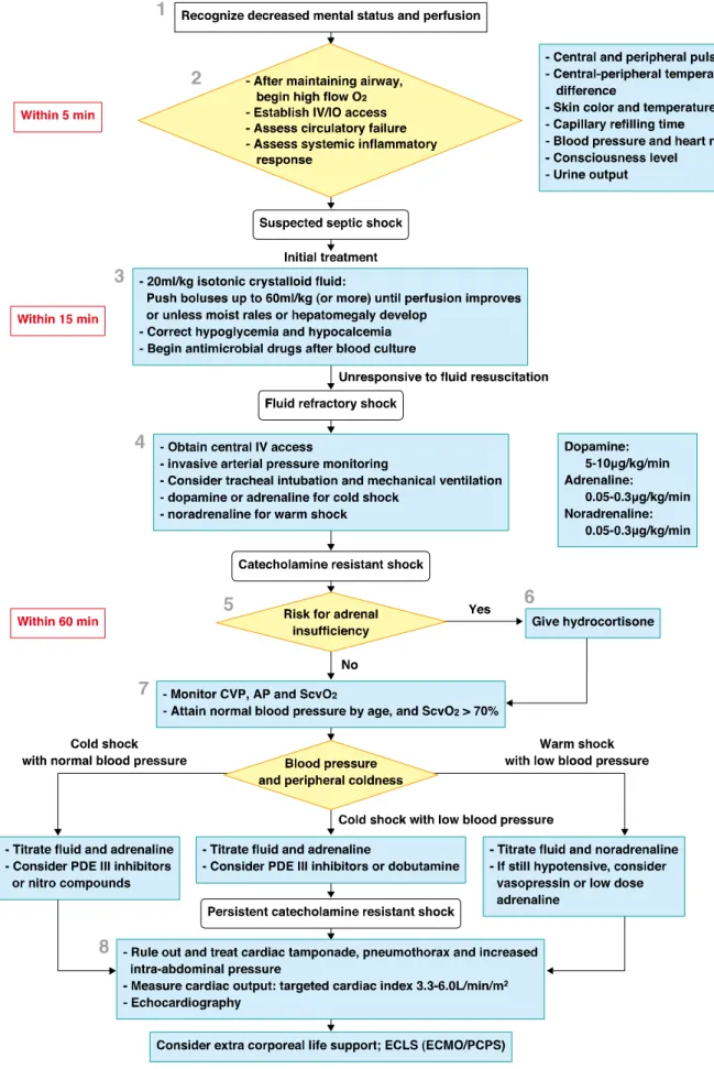
■6 Pediatric Advanced Life Support: PALS
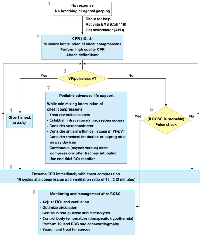
1. Cardiac Arrest Algorithm
The PALS Cardiac Arrest Algorithm is a set of actions for those who perform CPR on a routine basis to treat infants and children in cardiac arrest.
1) PBLS
[ Box 1 ]
Observe the chest and abdominal movements in unresponsive infants and children. Once recognizing that there is no breathing, follow the PALS Algorithm.
[ Box 2 ]
Begin CPR immediately. Administrate oxygen, attach an ECG monitor and a pulse oximeter, and get a defibrillator ready.
21
[ Box 3 to 4 to 5 ]Determine the victim’s cardiac rhythm on the ECG. VF and pulseless VT are shockable rhythms. The initial energy dose should be 4J/kg. After giving a shock, resume CPR beginning with chest compressions immediately.
After 2 minutes of CPR, check the rhythms. If there is still VF or pulseless VT, give another shock. The energy dose should be 4J/kg also in the second and subsequent shocks. Drug dosing should be promptly done after rhythm checking.
[ Box 6 ]
Asystole and PEA are nonshockable rhythms.
The most common ECG patterns in infants and children with cardiac arrest are asystole and PEA.
For infants and children in asystole or PEA, resume CPR and give chest compression as continuously as possible. While one rescuer is performing CPR, another rescuer should prepare adrenaline administration. Standard doses (IV dose of 0.01mg/kg, endotracheal dose of 0.1mg/kg) should be given for the initial and subsequent administrations.
Figure 2 Pediatric cardiac arrest algorithm
2) PALS
[ Box 7 ]
When ROSC is not achieved in PBLS, PALS is necessary. Continuous, effective chest compressions are the key to success not only in PBLS but also in PALS. Interruption of chest
23
compressions must be avoided in PALS as well as at any other time excepting for rescue breathing, ECG analysis, pulse check, or shock delivery.
(1) Identification and Treatment of Reversible Causes
Identifying and treating reversible causes for the cardiac arrest is required at every step in resuscitation while performing high quality CPR. Although searching for the causes is usually conducted by investigating the circumstances at the time of cardiac arrest, review of the victim’s medical history, physical examination and arterial blood gas or electrolytes tests that yield results rapidly can occasionally be useful. Echocardiography can be useful for diagnosing pericardial effusion and pulmonary thromboembolism. However, there is insufficient evidence to support or refute the routine use of echocardiography.
(2) Establishment of Intravenous or Intraosseous Access
While continuing to perform CPR, peripheral intravenous access or intraosseous access should be established in order for fluids and medications to be successfully delivered. When peripheral intravenous access is not readily attainable, intraosseous access is recommended.
(3) Vasopressors
There is insufficent evidence to suggest that adrenaline improves survival to hospital discharge or neurological outcome. However, the use of adrenaline may be considered in cardiac arrest since there is evidence that adrenaline may improve the rate of ROSC and short-term survival. Adrenaline should be given at 0.01mg/kg per dose (maximum dose of 1mg) with additional dosage every 3-5 minutes. There is insufficient evidence to support or refute the routine use of vasopressin.
(4) Antiarrhythmics
Administration of antiarrhythmics may be considered in refractory VF or pulseless VT(pVT). There is, however, insufficient evidence that antiarrhythmics administration improves the rate of ROSC or survival. In Japan, amiodarone, nifekalant and lidocaine are commonly used as antiarrhythmics for VF/ pVT. Amiodarone may be used for the treatment of shock-refractory or recurrent VF/pVT (Class IIb). Nifekalant may be considered in victims in cardiac arrest and with shock refractory VF/pVT (Class IIb). If amiodarone or nifekalant is not available, lidocaine may be considered although it is less effective (Class IIb).
(5) Tracheal Intubation and Supraglottic Airway Devices
The tracheal tube is considered the optimal method of managing the airway in CPR. Since tracheal intubation is a high risk procedure, it requires adequate training and ongoing skills maintenance for secure and prompt intubation. Prolonged attempts at tracheal intubation are harmful if they lead to interruption of chest compressions. Interruption of chest compressions should be minimized when performing tracheal intubation (Class I).
professionals trained in their use may be considered as a method of airway management during CPR. These devices can be used as a backup in a difficult or failed tracheal intubation. There is inadequate evidence to define the optimal timing of advanced airway placement during cardiac arrest. In the case of shortage of rescuers, early placement of these devices might help the rescuers focus on other effective treatment without having to deal with manual airway management. (6) Continuous Chest Compressions
When a tracheal tube is in place, continious chest compressions should be performed without pause for ventilations (asynchronous compressions and ventilations). Chest compressions should be given at least 100 times per minute, and about 10 rescue breaths per minute should be given. When a supraglottic airway device is in place, continuous, uninterrupted chest compressions can be performed if adequate ventilation can be provided.
(7) Capnometry
Waveform capnography is recommended to confirm and continuously monitor the position of a tracheal tube in victims of cardiac arrest, and it should be used in addition to clinical assessment of auscultation and direct visualization. If waveform capnography is not available, a nonwaveform CO2 detector, a colorimetric exhaled CO2 detector or oesophageal detector device (for children who
weigh 20kg or more) in addition to physical examination is an alternative.
3) Postresuscitation Care
[ Box 8 ]
Comprehensive treatment protocols for patients with ROSC include management of ventilation and circulation, controlling blood glucose and electrolytes, and therapeutic hypothermia.
(1) Adjustment of Concentration of Inspired Oxygen and Ventilation
Although it is necessary to avoid hypoxemia after ROSC, administration of highly concentrated oxygen may adversely affect brain damage. There is insufficient evidence that the routine ventilation with 100% oxygen is harmful. However, it is reasonable to adjust the fraction of inspiratory oxygen using PaO2 and SpO2 as indicators in the early treatment for patients with
ROSC (Class IIa). Hyperventilation after ROSC may reduce cerebral blood flow. After restoration of circulation, routine hyperventilation leading to hypocapnia should be avoided in order to prevent additional cerebral ischemia.
(2) Circulation Management
There is insufficient evidence that early hemodynamic optimization following ROSC improves outcomes. Although there is insufficient evidence regarding administration of IV fluids after ROSC, it is reasonable to use IV fluids as a part of comprehensive treatment based on the pathophysiology after ROSC (Class IIa). There is also insufficient evidence to support the efficacy of the use of vasopressors and/or inotropes, the continuous administration of amiodarone,
25
nifekalant or lidocaine, or the use of mechanical circulatory support such as intra-aortic balloon pumping. Although there is limited clinical data concerning the efficacy of circulation management, hemodynamic optimization for improved organ perfusion was performed based on the pathophysiology after ROSC.
(3) Blood Glucose and Electrolytes Control
It is appropriate to monitor blood glucose levels and avoid hypoglycemia as well as hyperglycemia following cardiac arrest (Class I). It is necessary to stay alert for hypoglycemia especially during control of blood glucose using insulin . There is insufficient evidence at present to identify the specific target glucose concentration range for the control of hyperglycemia in infants and children after ROSC. It is better not to use glucose containing fluids during CPR.
Hyponatremia causes plasma osmolality to fall, which may result in cerebral edema. The use of hypotonic fluids may cause iatrogenic cerebral edema. Whereas the negative effects of hyponatremia have already been pointed out in other countries, little attention is paid to that in medical setting in Japan. In the management after ROSC, hyponatremia should be avoided especially when abnormalities are seen in the central nervous system (Class III).
(4) Temperature Control (Therapeutic Hypothermia)
Victims with high body temperature after ROSC have poor outcomes. Hyperthermia after ROSC needs aggressive treatment (Class I). Therapeutic hypothermia (to 32–34 degrees Celsius for 12-24 hours) may be beneficial for adolescents who remain comatose (not responding in a meaningful way to verbal commands) following resuscitation from out-of-hospital VF cardiac arrest (Class IIa). The use of therapeutic hypothermia in infants and children is not refuted. Therapeutic hypothermia may also be beneficial for victims who remain comatose following resuscitation from in-/out-of-hospital PEA or asystole.
(5) 12-Lead ECG and Echocardiography
Lethal arrhythmia and myocardial disorder are important reversible causes of sudden cardiac arrest. After ROSC, 12-lead ECGs should be recorded and differential diagnoses on lethal arrhythmia should be carried out.
Echocardiography is useful not only for searching for causes, but also for assessing cardiac function. In addition, it can be used non-invasively and without having to move the patient. Given this, it is reasonable to use echocardiography after ROSC (Class IIa).
(6) Search for Causes and Treatment
It is necessary to search for causes of cardiac arrest and subsequently treat the patient after ROSC. Treating the cause is essential for preventing a recurrence of cardiac arrest and hemodynamic optimization.
2. Assessment
1) Focused Echocardiogram to Detect Reversible Causes of Cardiac Arrest
In 1 small LOE 4 pediatric case series152 cardiac activity was rapidly visualized by
echocardiography without prolonged interruptionof chest compressions, and this cardiac activity correlatedwith the presence or absence of a central pulse. In 1 pediatricLOE 4 case report153,
echocardiography was useful for diagnosingpericardial tamponade as the cause of cardiac arrest and wasuseful in guiding treatment.
In 8 LOE 5 adult case series154-161, echocardiographic findingscorrelated well with the presence
or absence of cardiac activityin cardiac arrest. These reports also suggested that echocardiography may be useful in identifying patients with potentially reversiblecauses for the arrest.
There is insufficient evidence to recommend for or against theroutine use of echocardiography during pediatric cardiac arrest. Echocardiography may be considered to identify potentially treatablecauses of an arrest when appropriately skilled personnel areavailable, but the benefits must be carefully weighed against the known deleterious consequences of interrupting chest compressions.
2) End-tidal CO
2(PETCO
2) and Quality of CPR
Three LOE 5 animal studies162-164, 4 LOE 5 adult165-168, and 1 LOE 5 pediatric series169 showed a
strong correlation between PETCO2 and interventions that increase cardiac output during
resuscitation from shock or cardiac arrest. Similarly3 LOE 5 animal models170-172 showed that
measures that markedlyreduce cardiac output result in a fall in PETCO2.
Two LOE 5 adult out-of-hospital studies173, 174 supported continuousPETCO2 monitoring during
CPR as a way of determining return of spontaneous circulation (ROSC), particularly if the readingsduring CPR are >15 mm Hg (2.0kPa). In 1 LOE 4175 and 2 LOE5 adult case series176, 177,
an abrupt and sustained rise in PETCO2 often preceded identification of ROSC.Two LOE 4
pediatric cases series169, 178, 8 LOE 5 adult174, 179-185, and 1 LOE 5 animal study163 showed that a low
PETCO2 (<10mm Hg [1.33 kPa] to <15 mm Hg [2.0 kPa]) despite 15 to 20minutes of advanced life
support (ALS) is strongly associatedwith failure to achieve ROSC. On the basis of 2 LOE 5 animal studies175, 186 and 2 adult LOE 5 case series174, 182, PETCO2 afterat least 1 minute of CPR may be
more predictive of outcome thanthe initial value because the initial PETCO2 is often increasedin
patients with asphyxial cardiac arrest.
The wide variation for initial PETCO2 during resuscitation limitsits reliability in predicting
outcome of resuscitation and itsvalue as a guide to limiting resuscitation efforts. Two LOE5 animal studies175, 186 and 2 large LOE 5 adult trials174, 182 suggestedthat the initial PETCO2 is
higher if the etiology of the cardiacarrest is asphyxial rather than if it is a primary cardiac arrest.Interpretation of the end-tidal CO2 during resuscitation isaffected by the quality of the
measurement, the minute ventilationdelivered during resuscitation, the presence of lung disease that increases anatomic dead space, and the presence of right-to-leftshunting.
27
LOE 5 adult188-190 and 2 LOE 5 animal191, 192 studies, adrenaline (and other systemic
vasoconstrictiveagents) transiently decreased PETCO2.
Continuous capnography or capnometry monitoring, if available,may be beneficial by providing feedback on the effectivenessof chest compressions. Whereas a specific target number cannotbe identified, if the PETCO2 is consistently <15 mm Hg, itis reasonable to focus efforts on improving
the quality of chest compressions and avoiding excessive ventilation (Class IIa). Although a threshold PETCO2 may predict a poor outcome fromresuscitation and might be useful as a guide
to terminationof CPR, there are insufficient data to establish the thresholdand the appropriate duration of ALS needed before such evaluationin children. The PETCO2 must be interpreted with
caution for1 to 2 minutes after administration of adrenaline or othervasoconstrictive medications because these medications may decreasethe PETCO2.
3. Airway and Ventilation
Opening and maintaining a patent airway and providing ventilationsare fundamental elements of pediatric CPR, especially because cardiac arrest often results from, or is complicated by, asphyxia.There are no new data to change the ILCOR 2005 recommendationto use manual airway maneuvers (with or without an oropharyngealairway) and bag-mask ventilation (BMV) for children requiringairway control or positive-pressure ventilation for short periods in the out-of-hospital setting. When airway control or BMV isnot effective, supraglottic airways may be helpful when usedby properly trained personnel.
When performing tracheal intubation, data suggest that the routineuse of cricoid pressure may not protect against aspiration andmay make intubation more difficult.
Routine confirmation of tracheal tube position with capnography/capnometryis recommended with the caveat that the PETCO2 in infants andchildren in cardiac arrest may be below detection
limits forcolorimetric devices.
Following ROSC, toxic oxygen byproducts (reactive oxygen species,free radicals) are produced that may damage cell membranes,proteins, and DNA (reperfusion injury). Although there are no clinical studies in children (outside the newborn period) comparing different concentrations of inspired oxygen during and immediatelyafter resuscitation, animal data and data from newborn resuscitation studies suggest that it is prudent to titrate inspired oxygen after return of a perfusing rhythm to prevent hyperoxemia.
1) Concentration of Supplementary Oxygen
There are no studies comparing ventilation of infants and children in cardiac arrest with different inspired oxygen concentrations. Two LOE 5 meta-analyses of several randomized controlled trialscomparing neonatal resuscitation initiated with room air versus100% oxygen193, 194 showed increased survival when resuscitationwas initiated with room air. Seven LOE 5 animal
studies195-201 suggested that ventilationwith room air or an FIO2 of <1.0 during cardiac arrest may