REVIEW
Secreted venom allergen-like proteins of
helminths: Conserved modulators of host
responses in animals and plants
Ruud H. P. Wilbers
ID1*
, Roger Schneiter
2, Martijn H. M. Holterman
ID1, Claire Drurey
ID3,
Geert Smant
ID1, Oluwatoyin A. Asojo
ID4, Rick M. Maizels
ID3, Jose L. Lozano-Torres
ID1 1 Laboratory of Nematology, Plant Sciences Group, Wageningen University and Research, Wageningen,The Netherlands, 2 Division of Biochemistry, Department of Biology, University of Fribourg, Fribourg, Switzerland, 3 Wellcome Centre for Molecular Parasitology, Institute of Infection, Immunity and Inflammation, University of Glasgow, United Kingdom, 4 Department of Chemistry and Biochemistry, Hampton University, Hampton, Virginia, United States of America
Abstract
Despite causing considerable damage to host tissue at the onset of parasitism, invasive
hel-minths establish remarkably persistent infections in both animals and plants. Secretions
released by these obligate parasites during host invasion are thought to be crucial for their
persistence in infection. Helminth secretions are complex mixtures of molecules, most of
which have unknown molecular targets and functions in host cells or tissues. Although the
habitats of animal- and plant-parasitic helminths are very distinct, their secretions share the
presence of a structurally conserved group of proteins called venom allergen-like proteins
(VALs). Helminths abundantly secrete VALs during several stages of parasitism while
inflict-ing extensive damage to host tissue. The tight association between the secretion of VALs
and the onset of parasitism has triggered a particular interest in this group of proteins, as
improved knowledge on their biological functions may assist in designing novel protection
strategies against parasites in humans, livestock, and important food crops.
Introduction
Upon infection, helminth parasites establish an intricate relationship with their host.
Hel-minths cause considerable damage during host invasion, migration through host tissues, and
feeding on host cells [
1
,
2
], but infections by these parasites can nonetheless be very persistent
and last for several decades. Helminths are masters in manipulating host defense responses [
1
,
3
], thereby creating a suitable environment for their survival and simultaneously limiting
excessive damage due to host immune responses.
Excretory/secretory (ES) products are regarded as the tools employed by helminth parasites
to control host defense responses. Recently, it was shown that ES products of helminth
para-sites reflect their diversity in lifestyles and hosts and therefore have little in common between
plant and animal parasites [
4
]. However, members of the alternatively named Sperm-coating
protein/Tpx/antigen 5/pathogenesis-related-1/Sc7 (SCP/TAPS) or cysteine-rich secretory
a1111111111
a1111111111
a1111111111
a1111111111
a1111111111
OPEN ACCESSCitation: Wilbers RHP, Schneiter R, Holterman
MHM, Drurey C, Smant G, Asojo OA, et al. (2018) Secreted venom allergen-like proteins of helminths: Conserved modulators of host responses in animals and plants. PLoS Pathog 14(10): e1007300.https://doi.org/10.1371/journal. ppat.1007300
Editor: James B. Lok, University of Pennsylvania,
UNITED STATES
Published: October 18, 2018
Copyright:© 2018 Wilbers et al. This is an open access article distributed under the terms of the
Creative Commons Attribution License, which permits unrestricted use, distribution, and reproduction in any medium, provided the original author and source are credited.
Funding: This work was supported by ALW Grant
84713008 from the Netherlands Organisation for Scientific Research. The funders had no role in study design, data collection and analysis, decision to publish, or preparation of the manuscript.
Competing interests: The authors have declared
proteins/antigen 5/pathogenesis-related 1 (CAP) protein superfamily are ubiquitously present
in ES products of helminth species that parasitize plants and animals. Although a uniform
nomenclature was proposed previously [
5
], helminth CAP proteins still go by different names,
including activation-associated secreted proteins (ASPs) or most commonly used venom
aller-gen-like proteins (VALs or VAPs).
The expression of VALs is specifically up-regulated during parasitic phases of the life cycle
of helminths, which could point to a role in host–parasite interactions [
6
–
9
]. The presence of
VALs in secretions of both plant- and animal-parasitic helminths suggests that these proteins
are important for the establishment of persistent infections in both plants and animals. It is
possible that conserved structural properties in VALs provide a diverse group of parasites a
robust platform for modulating host responses in both plant and animal kingdoms. However,
a question that remains unanswered is whether VALs from plant and animal parasites could
have conserved functions based on common biochemical properties of these secreted proteins.
Recent reports have shed new light on structural properties, biochemical modes of action,
and functions of secreted VALs of parasitic helminths. However, most of these reports have
been published in specialized journals dedicated to either medical and veterinary biology or
plant pathology. Here, we present an interdisciplinary review of the latest findings from
phylo-genetic analyses, x-ray crystallography, and functional studies on VALs from parasitic
hel-minths. The aim of this review is to explore conserved mechanisms underlying the role of
VALs as modulators of host responses during parasitism with an emphasis on their potential
impact on common concepts in plant pathology and human and/or animal parasitology.
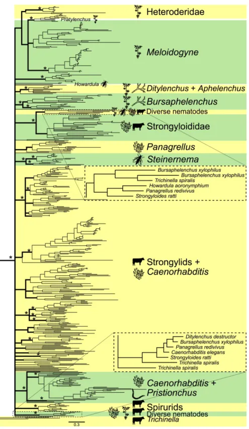
Phylogenetic analysis of nematode VALs reveals no clear links with
parasitism
Multiple lineages within the nematode phylum have independently evolved the ability to
para-sitize plants or animals. Modelling the evolutionary history of nematode VALs, based on
over-all sequence diversity, may reveal links between patterns of diversification within this protein
family and particular lifestyles or host organisms. To this end, we generated a Bayesian tree of
available VAL sequences from plant- and animal-parasitic nematodes as well as their
free-liv-ing close relatives from the distal end of the nematode tree (
Fig 1
and
S1 Fig
,
S1 Table
) [
10
].
First of all, we observed no separate clade of VALs uniquely associated with parasitism among
nematodes. So, based on overall sequence diversity in VALs, we found no clear distinction
between VALs from parasitic nematodes and free-living nematodes. Similarly, we also found
no evidence for a clear separation between VALs from different lineages of plant parasites and
VALs from different lineages of animal parasites, which could have pointed at specific
adapta-tions to living on either host plants or animals. Instead, several VALs from plant-parasitic
nematodes (
Ditylenchus destructor and Bursaphelenchus xylophilus), animal-parasitic
nema-todes (
Howardula aoronymphium, Strongyloides ratti, and Trichinella spiralis), and free-living
nematodes (
Panagrellus redivivus) clustered together (Fig 1
insets). Moreover, VALs from
related parasitic and free-living nematode species clustered into separate clades, which points
at functional diversification in a common ancestor of these parasitic and free-living lineages.
By contrast, some clusters contain large numbers of homologous VALs from the same
nema-tode genus, which indicates extensive recent functional diversification. The large difference in
numbers of VALs present in nematode genomes is striking, ranging from a few (
H.
aoronym-phium, Brugia malayi) to over a hundred (Ancylostoma caninum). Gene family expansion also
does not seem to be linked to a particular lifestyle or host organism. Altogether, our
phyloge-netic analysis revealed no clear link between overall sequence diversity in VAL genes in
nema-todes and parasitism in plant and animals.
Fig 1. Bayesian tree of VALs in nematodes. Asterisks indicate main branches (bold) well-supported by the Bayesian
or maximum likelihood analysis. Icons represent feeding types (plant parasitism, fungal feeding, bacterial feeding, nematode predation, insect parasitism, and vertebrate parasitism). Figure insets reveal two clusters with nematode species of plant- and animal-parasitic and free-living species. Taxon names, support values, and methods for construction can be found inS1 Fig. VAL, venom allergen-like protein.
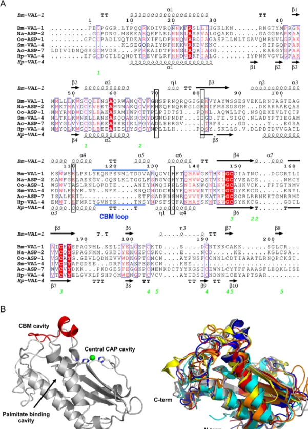
Helminth VALs bind lipids and other hydrophobic structures
Helminth VALs are classified as SCP/TAPS proteins, which include a range of structurally
related proteins found in a wide range of eukaryotes [
5
]. Ample structural information exists
on eukaryotic SCP/TAPS proteins, but so far only a handful of structures for helminth VALs
have been resolved. Similar to eukaryotic SCP/TAPS proteins, the core of helminth VALs is a
structurally conserved approximately 15 kDa cysteine-rich CAP domain (Pfam00188), which
typically has limited sequence identity [
11
–
26
]. SCP/TAPS proteins are generally made up of
single CAP domains, which are sometimes covalently linked to other functional domains [
20
,
27
]. A higher degree of structural complexity is seen in helminth VALs as they can comprise a
single CAP domain with the ability to form homodimers (like
Oo-ASP-1 from Ostertagia
oster-tagi [15
]), two covalently linked CAP domains (like
Na-ASP-1 from Necator americanus [28
]),
or even four covalently linked CAP domains (
P. redivivus gene Pan_g9869.t1, genome
PRJNA186477 WormBase ParaSite). In addition, helminth VALs can be found fused to other
protein family sequences such as the ShK toxin domains (Pfam01549) in
Toxocara canis [29
].
The CAP domain itself adopts a characteristic alpha-beta-alpha sandwich fold with flexible
loop regions and is further stabilized by disulfide bonds [
11
,
13
,
14
]. The lengths of the flexible
loops,
β-strands, and α-helices vary between different proteins, making it difficult to accurately
model the structures of these proteins. An alignment of the amino acid sequences from single
CAP domain VALs of helminths, for which the structures have been resolved, illustrates that
conservation among these sequences is mainly found in structural properties, like disulfide
bonds,
α-helices, and β-strands (
Fig 2A
). Superimposing VAL structures on top of each other
nicely illustrates that loop regions in between these secondary structures as well as N- and
C-termini are very distinct (
Fig 2B
). Importantly, these loop regions constitute up to 50% of the
overall VAL structure.
All reported SCP/TAPS protein structures have a large central CAP cavity, which are either
cysteine-rich secretory protein (CRISP)-like or non-CRISP-like. The CRISP-like CAP cavity is
characterized by a tetrad of residues, consisting of two histidine and two glutamic acid residues
that bind divalent cations including Zn
2+and Mg
2+[
14
,
20
,
30
–
38
]. This tetrad was shown to
be important for Zn
2+binding and the heparan-sulfate dependent inflammatory modulation
mechanisms of the cobra CRISP protein natrin [
38
]. However, the majority of available
sequences of plant and animal parasite VALs are suggestive of non-CRISP-like protein
struc-tures because they lack the histidine residues of the CAP tetrad [
12
]. Furthermore, our recent
analysis of the
Bm-VAL-1 structure reveals that the central CAP cavity is connected to other
cavities by channels that can serve as pathways for water molecules, cations, and small
mole-cules [
16
].
Given the shared structural topology of the CAP domain, it seems likely that CAP proteins
exert a fundamentally similar biochemical function within their respective environments [
20
,
39
,
40
]. However, the exact nature of this shared biochemical function among CAP proteins
remains poorly defined. Recent evidence shows that distinct pockets in the CAP domain bind
lipids, such as leukotrienes, sterols, and negatively charged phospholipids [
25
,
41
,
42
]. CAP
proteins from yeast (pathogen-related yeast protein [Pry1] and Pry2) bind sterols and fatty
acids at two distinct lipid-binding sites [
42
,
43
]. These two well-defined lipid-binding pockets
are a flexible loop that binds sterols (called the caveolin-binding motif [CBM] loop) and a
hydrophobic channel, which is formed by two parallel
α-helices, the so-called
palmitate-bind-ing cavity. Furthermore, it was shown that the palmitate-bindpalmitate-bind-ing cavity of tablysin-15 from the
saliva of the horsefly
Tabanus yao binds leukotrienes and thereby inhibits their
pro-inflamma-tory effects [
25
].
Fig 2. Comparison of helminth VALs. (A) The sequences of helminth VALs with known structure (see below) are aligned with
clustalW2, and the secondary structural features are illustrated with the coordinates ofBm-VAL-1 and Hp-VAL-4 using ESPript.
The different secondary structural elements shown are alpha helices as large squiggles labeled (α), 310-helices as small squiggles
labeled (η), beta strands as arrows (β), and beta turns (TT). Identical residues are highlighted in solid red, and conserved residues are shown in red. The locations of the cysteine residues involved in disulfide bonds are numbered in green. The location of the CBM loop is shown in blue. The position of amino acid residues constituting the CAP tetrad are marked with a black box. (B)
Lipid-binding properties have recently also been demonstrated for helminth VALs using
mutant yeast cells that lack their endogenous CAP proteins Pry1 and Pry2 and are deficient in
sterol export. Expression of
Na-ASP-2, Sm-VAL-4, Hp-VAL-4, or Bm-VAL-1 in these mutant
cells restores their ability to export sterol [
12
,
16
,
44
]. These results also indicate that the CAP
tetrad residues are not required for sterol export as both
Sm-VAL-4 and Hp-VAL-4 lack the
tetrad histidine residues. Sterol binding by
Bm-VAL-1, on the other hand, can be inhibited
with EDTA and therefore seems to be dependent on the binding of divalent cations. Besides
sterol binding, BmVAL-1 is also able to bind palmitate in vitro with comparable affinity as
tablysin-15 [
16
]. The two parallel
α-helices that constitute the palmitate-binding cavity are
structurally well conserved among other helminth VALs (
Fig 2B
), which suggests that these
VALs likely bind palmitate or similar hydrophobic ligands as well. These studies introduce
VALs as a new protein family of parasitic helminths with lipid-binding properties, just like
nematode polyprotein antigens and/or allergens (NPAs), nematode fatty acid-binding proteins
(nemFABPs), and fatty acid- and retinol-binding proteins (FARs) [
45
].
Altogether, helminth VALs share a conserved alpha-beta-alpha sandwich structure that is
typical for CAP proteins. However, the length of different
α-helices and β-strands and the
composition of loop regions determine approximately 50% of their overall structure. This
sup-ports the idea that the CAP domain may act as a stable but versatile molecular scaffold for
diversification of the entire VAL gene family. Also, evidence indicates that this stable CAP
domain allows helminth VALs to sequester small hydrophobic ligands [
12
,
16
]. This could be a
potential mechanism used by VALs to modulate immune responses in their host.
Biological functions of VALs from plant parasites
Plant-parasitic nematodes use an oral stylet to perforate plant cell walls, deliver secretions into
cells, and extract low molecular weight compounds from living plant cells. VALs have attracted
attention because they are among the very few molecules in stylet secretions that seem to be
important for persistent nematode infections in plants and animals [
46
]. Following the
discov-ery of
Mi-VAP-1 from the root-knot nematode Meloidogyne incognita in 2000 [17
,
47
],
research has focussed on the role of VALs as activators and suppressors of host immune
responses. For instance, while studying the function of
Gr-VAP-1 from the potato cyst
nema-tode
Globodera rostochiensis, it was found that binding of this protein to the extracellular
cyste-ine protease Rcr3
pimin tomato triggers a defense-related hypersensitive response mediated by
the surface-localized immune receptor Cf-2 [
8
]. Interestingly, tomato genotypes carrying the
Rcr3
pimprotein but lacking the matching immune receptor Cf-2 were more susceptible to
nematode infections than plants lacking both Rc3
pimand Cf-2, suggesting that
Gr-VAP-1
interacts with Rcr3
pimto suppress host immunity.
The expression of VAL genes in plant-parasitic nematodes is regulated in association with
host invasion and subsequent migration through host tissues [
8
,
19
,
48
–
54
]. Proteomic
analy-ses of chemically induced secretions from infective juveniles showed the presence of VALs
along with different classes of plant cell wall-degrading enzymes [
8
]. Plant-parasitic nematodes
utilize a large repertoire of plant cell wall-degrading enzymes to break down the protective
layer of cellulose and other carbohydrate polymers around cells during migration. A
and coordination of the divalent cation Zn2+is shown in green. Superimposed structures of helminths VALs with a single CAPdomain are given to illustrate structural differences among the different family members. The represented structures areNa-ASP-2
in blue [13],Oo-ASP-1 in orange [15],Ac-ASP-7 in cyan [90],Sm-VAL-4 in yellow [44],Hp-VAL-4 in red [12], andBm-VAL-1 in
gray [16]. ASP, activation-associated secreted protein; CAP, cysteine-rich secretory proteins/antigen 5/pathogenesis-related 1; CBM, caveolin-binding motif; VAL, venom allergen-like proteins.
knockdown of stylet-secreted cell wall-degrading enzymes by RNA interference in infective
juvenile nematodes can halt host invasion [
55
]. Similarly, a knockdown of VAL expression in
migratory plant-parasitic nematodes can also reduce their ability to migrate inside host plants
and successfully establish a permanent feeding site [
50
,
56
,
57
]. These findings suggest that
sty-let-secreted VALs are important for modulating host responses, particularly during the
migra-tion of the nematodes through host tissues and the early stages of an infecmigra-tion.
Ectopic expression of nematode VALs (
Gr-VAP-1, and Hs-VAP-1 and Hs-VAP-2 from
Heterodera schachtii) in the extracellular matrix of transgenic plants significantly increases
their susceptibility to plant-parasitic nematodes [
57
]. Interestingly, these plants also proved to
be more susceptible to infections by fungi, bacteria, and oomycetes, all of which have entirely
different infection strategies [
57
]. Furthermore, ectopic expression of nematode VALs
sup-presses the growth inhibition response that normally occurs when young plants are constantly
exposed to the flagellin peptide flg22. Flg22 is recognized as a pathogen-associated molecular
pattern by surface-localized immune receptors in plants [
58
,
59
]. Whole transcriptome
analy-sis of plants ectopically expressing nematode VALs suggests that enhanced susceptibility
involves plant cell wall modifications, lipid signalling, and extracellular protein processing
[
57
]. In a different experimental setup, ectopic nematode VALs suppress a defense-related
hypersensitive response in plant cells mediated by surface-localized, but not by cytoplasmic,
immune receptors. Taken together, stylet-secreted VALs of plant-parasitic nematodes most
likely enhance overall susceptibility of host plants to nematode infections by suppressing plant
innate immunity mediated by surface-localized receptors.
Biological functions of VALs from animal parasites
Initially, the major focus of study for animal parasite VALs was their immunogenic properties.
Following the discovery in the 1990s that dominant secreted proteins of hookworm larvae are
the VAL family members ASP-1 and ASP-2 [
22
,
28
], they were considered ideal candidates for
vaccine development. Subsequently, VALs from a wide range of animal and human parasites
have shown substantial degrees of protective immunity as vaccines [
60
–
65
].
Na-ASP-2 was
explored as a potential human hookworm vaccine candidate, but despite successes in animal
experiments [
66
–
68
], vaccine development was halted due to allergic reactions in previously
exposed individuals in a clinical trial [
69
]. Although adverse, this outcome emphasized the
immunogenicity of the VALs in natural helminth infection.
The ubiquitious presence and frequent dominance of VALs in helminth ES products—and
their up-regulation during parasitic phases of the life cycle—point to a major role in
host–par-asite interactions. VALs are among the most abundant secreted proteins in the intestinal cattle
parasites
Cooperia oncophora and O. ostertagi, where they are prevalently upregulated in
para-sitic stages [
70
], and in the rodent model nematodes
Heligmosomoides polygyrus [6
] and
Nip-postrongylus brasiliensis [9
]. A fascinating example is presented by
Strongyloides nematodes,
which can follow either a free-living life cycle or a parasitic cycle through mammals; multiple
VAL family members show preferential expression in the parasitic adult worm compared to
the free-living form of the same species [
7
]. Furthermore, predicted VALs from the trematode
Schistosoma mansoni are up-regulated during parasite infective stages, indicating a role in
invasion of the human host [
71
].
Sm-VAL-4 has been detected in human skin following
inva-sion of
S. mansoni cercariae [72
]. In the filarial nematode
B. malayi, Bm-VAL-1 is highly
expressed in the mosquito-borne L3 stage prior to entry into the mammalian host, and is
re-expressed subsequently by later stages [
73
]. ASP/VAL genes of
A. caninum and T. canis, the
dog hookworm and roundworm respectively, are abundantly expressed in invasive and
migra-tory larval stages [
74
,
75
], as are VALs in the adult stages of
A. caninum, Haemonchus
contortus, and N. americanus [
76
–
78
]. Environmental niche is also linked to VAL expression,
which is significantly higher in mucosal-dwelling larvae of
Teladorsagia circumcincta
com-pared to larvae from the lumen [
79
]. VALs therefore seem to be expressed at stages of the
para-site life cycle where maximal contact occurs between parapara-site and host, whether this is
transmission, tissue migration, or feeding.
Location of expression in the parasite also hints at function, with many helminth VALs
being expressed in secretory glands. Staining for the glycan found on
H. polygyrus VAL-1 and
VAL-2 identified a series of structures on the exterior cuticle in contact with host tissues [
6
].
Several
A. caninum ASP proteins localize to the pharyngeal glands and glandular esophagus,
which produce hookworm ES secretions [
26
,
80
].
S. mansoni VALs also localize to the
esoph-ageal gland [
81
].
Ov-ASP-1, an ASP/VAL transcript from the human parasite Onchocerca
vol-vulus—causal agent of river blindness—is localized in the glandular esophagus of L2 and L3
larvae and secreted via degranulation following the invasion of the host [
63
].
Though both timing and location of expression of VALs point to roles in parasite–host
interactions, relatively few of these proteins have a well-defined physiological function.
Na-ASP-2 binds to human B cells via CD79A, triggering downregulation of receptor signaling
pathways [
82
].
Na-ASP-2 also induces neutrophil migration and accumulation within tissues,
a potentially pro-inflammatory activity that may aid tissue migrating larvae through increased
tissue permeability [
83
]. This contrasts with other VALs that suppress immune responses once
a parasite is tissue-dwelling. For example, neutrophil inhibitor factor (NIF) from
A. caninum
binds to neutrophils via the integrin CD11b/CD18 and blocks their adhesion to vascular
endo-thelial cells and oxidative bursts [
84
–
86
]. Hookworm platelet inhibitor (HPI) is secreted from
adult
A. caninum at the site of intestinal attachment, where it inhibits platelet aggregation and
adhesion, allowing continuous feeding without blood clotting [
87
,
88
].
Sm-VAL-9 from S.
mansoni induces differential expression of matrix metalloproteinases in macrophages and
modulates host extracellular matrix remodeling gene expression in both its vertebrate and
snail hosts [
89
].
Importantly, care should be taken when results obtained from in vitro studies with
recom-binant helminth proteins are extrapolated to the in vivo situation. First of all, these proteins
are often studied at seemingly supraphysiological concentrations. Furthermore, differences in
protein folding and post-translational modifications (e.g., N-glycosylation) between native
and recombinant helminth proteins could significantly influence binding to target cells or
bio-logical activity.
Taken together, the pattern of animal-parasitic VAL evolution and expression suggests that
most family members fulfill core functions at the host–parasite interface, with rapid
diversifi-cation and adaptation to each host species.
Concluding paragraph
The expression pattern of VALs during invasion and migration of host tissues by both
plant-and animal-parasitic helminths suggests that these proteins might have conserved mechanisms
of immune modulation in their respective host. However, sequence analysis suggests an
inde-pendent evolution of these functions. In recent years, several VAL family members have been
functionally characterized (summarized in
S2 Table
) and revealed modulation of similar
bio-logical processes in plant and animal hosts, like suppression of innate immune responses and
remodeling of the extracellular matrix. Furthermore, VALs reveal a conserved CAP domain
that allows them to sequester small hydrophobic ligands (e.g., cholesterol and palmitate), but
little is known about endogenous ligands that are bound during parasitism. Therefore, future
research should focus on the specificity for different lipid ligands and how these ligands can be
involved in the immunomodulatory properties of helminth VALs.
Supporting information
S1 Fig. A Bayesian phylogenetic tree of the functional domains of VAL genes in
nema-todes. For multidomain proteins, each domain was included separately.
Trichinella spiralis
served as an outgroup. The alignment was created in BioEdit version 7.2.5 using ClustalW 1,4
and was further manually refined. The Bayesian tree was created using MrBayes version 3.2.6
and run for 10 million generations with 4 chains in 4 parallel runs using a mixed amino acid
substitution model. Runs converged after a burnin of 2 million generations and used the
WAG substitution model. Posterior probabilities are given above the branches. Displayed
below the branches are the bootstrap percentages of a fast maximum likelihood tree run on the
same dataset with RAxML version 8.2.10 using the WAG substitution model with 1000
boot-straps. All sequences that were used to constrict this tree are listed in
S1 Table
. VAL, venom
allergen-like protein; WAG, Whelan and Goldman.
(PDF)
S1 Table. Sequences used in the creation of the phylogenetic trees. Sequences were gathered
by blasting published genomes, EST databases and GenBank with VAL sequences or taken
from literature references. Genomes were blasted on Wormbase Parasite (
http://parasite.
wormbase.org
) with the exception of
B. xylophilus (GeneDB:
http://www.genedb.org
),
S. ratti (https://www.sanger.ac.uk/cgi-bin/blast/submitblast/strongyloides
),
P. coffeae
(
https://www.ncbi.nlm.nih.gov
), and
H. aoronymphium (http://nematodes.org/downloads/
959nematodegenomes/blast/db/Howardula_aoronymphium_clc_1.fna
). EST databases were
blasted on Nematode.net (
http://nematode.net
). The
G. rostochienis, P. coffeae, and H.
aoro-nymphium sequences were extracted from unannotated genome data. Identical sequences
were merged in a consensus sequence. EST, expressed sequence tag.
(XLSX)
S2 Table. Overview of helminth VAL functional characterization. Collection of helminth
VALs as discussed in this review with current nomenclature, structural information (including
RCSB PDB reference [
https://www.rcsb.org
]) and information on lipid binding, interactions
and function are given when available. PDB, Protein Data Bank; RCSB, Research
Collabora-tory for Structural Bioinformatics; VAL, venom allergen-like protein.
(XLSX)
References
1. Goverse A, Smant G. The activation and suppression of plant innate immunity by parasitic nematodes. Annu Rev Phytopathol. 2014; 52:243–65.https://doi.org/10.1146/annurev-phyto-102313-050118 PMID:24906126
2. Allen J, Wynn T. Evolution of Th2 immunity: a rapid repair response to tissue destructive pathogens. PLoS Pathog. 2011; 7(5):e1002003.https://doi.org/10.1371/journal.ppat.1002003PMID:21589896
3. Maizels R, McSorley H. Regulation of the host immune system by helminth parasites. J Allergy Clin Immunol. 2016; 138(3):666–75.https://doi.org/10.1016/j.jaci.2016.07.007PMID:27476889
4. Cuesta-Astroz Y, Oliveira F, Nahum L, Oliveira G. Helminth secretomes reflect different lifestyles and parasitized hosts. Int J Parasitol. 2017; 47(9):529–44.https://doi.org/10.1016/j.ijpara.2017.01.007 PMID:28336271
5. Cantacessi C, Campbell B, Visser A, Geldhof P, Nolan M, Nisbet A, et al. A portrait of the "SCP/TAPS" proteins of eukaryotes—Developing a framework for fundamental research and biotechnological out-comes. Biotechnol Adv. 2009; 27(4):376–88.https://doi.org/10.1016/j.biotechadv.2009.02.005PMID: 19239923
6. Hewitson J, Harcus Y, Murray J, van Agtmaal M, Filbey K, Grainger J, et al. Proteomic analysis of secre-tory products from the model gastrointestinal nematode Heligmosomoides polygyrus reveals domi-nance of venom allergen-like (VAL) proteins. J Proteomics. 2011; 74(9):1573–94.https://doi.org/10. 1016/j.jprot.2011.06.002PMID:21722761
7. Hunt V, Tsai I, Coghlan A, Reid A, Holroyd N, Foth B, et al. The genomic basis of parasitism in the Stron-gyloides clade of nematodes. Nat Genet. 2016; 48(3):299–307.https://doi.org/10.1038/ng.3495PMID: 26829753
8. Lozano-Torres J, Wilbers R, Gawronski P, Boshoven J, Finkers-Tomczak A, Cordewener J, et al. Dual disease resistance mediated by the immune receptor Cf-2 in tomato requires a common virulence target of a fungus and a nematode. Proc Natl Acad Sci U S A. 2012; 109(25):10119–24.https://doi.org/10. 1073/pnas.1202867109PMID:22675118
9. Sotillo J, Sanchez-Flores A, Cantacessi C, Harcus Y, Pickering D, Bouchery T, et al. Secreted prote-omes of different developmental stages of the gastrointestinal nematode Nippostrongylus brasiliensis. Mol Cell Proteomics. 2014; 13(10):2736–51.https://doi.org/10.1074/mcp.M114.038950PMID: 24994561
10. van Megen H, van den Elsen S, Holterman M, Karssen G, Mooyman P, Bongers T, et al. A phylogenetic tree of nematodes based on about 1200 full-length small subunit ribosomal DNA sequences. Nematol-ogy. 2009; 11:927–S27.
11. Asojo O. Structure of a two-CAP-domain protein from the human hookworm parasite Necator ameri-canus. Acta Crystallogr D Biol Crystallogr. 2011; 67(Pt 5):455–62.https://doi.org/10.1107/
S0907444911008560PMID:21543848
12. Asojo O, Darwiche R, Gebremedhin S, Smant G, Lozano-Torres J, Drurey C, et al. Heligmosomoides polygyrus Venom Allergen-like Protein-4 (HpVAL-4) is a sterol binding protein. Int J Parasitol.
13. Asojo O, Goud G, Dhar K, Loukas A, Zhan B, Deumic V, et al. X-ray structure of Na-ASP-2, a pathogen-esis-related-1 protein from the nematode parasite, Necator americanus, and a vaccine antigen for human hookworm infection. J Mol Biol. 2005; 346(3):801–14.https://doi.org/10.1016/j.jmb.2004.12.023 PMID:15713464
14. Asojo O, Koski R, Bonafe N. Structural studies of human glioma pathogenesis-related protein 1. Acta Crystallogr D Biol Crystallogr. 2011; 67(Pt 10):847–55.https://doi.org/10.1107/S0907444911028198 PMID:21931216
15. Borloo J, Geldhof P, Peelaers I, Van Meulder F, Ameloot P, Callewaert N, et al. Structure of Ostertagia ostertagi ASP-1: insights into disulfide-mediated cyclization and dimerization. Acta Crystallogr D Biol Crystallogr. 2013; 69(Pt 4):493–503.https://doi.org/10.1107/S0907444912050019PMID:23519657
16. Darwiche R, Lugo F, Drurey C, Varossieau K, Smant G, Wilbers R, et al. Crystal structure of Brugia malayi venom allergen-like protein-1 (BmVAL-1), a vaccine candidate for lymphatic filariasis. Int J Para-sitol. 2018.
17. Ding X, Shields J, Allen R, Hussey R. Molecular cloning and characterisation of a venom allergen AG5-like cDNA from Meloidogyne incognita. Int J Parasitol. 2000; 30(1):77–81. PMID:10675748
18. Fernandez C, Szyperski T, Bruyere T, Ramage P, Mosinger E, Wuthrich K. NMR solution structure of the pathogenesis-related protein P14a. J Mol Biol. 1997; 266(3):576–93.https://doi.org/10.1006/jmbi. 1996.0772PMID:9067611
19. Gao B, Allen R, Maier T, Davis E, Baum T, Hussey R. Molecular characterisation and expression of two venom allergen-like protein genes in Heterodera glycines. Int J Parasitol. 2001; 31(14):1617–25. PMID: 11730789
20. Gibbs G, Roelants K, O’Bryan M. The CAP superfamily: cysteine-rich secretory proteins, antigen 5, and pathogenesis-related 1 proteins—roles in reproduction, cancer, and immune defense. Endocr Rev. 2008; 29(7):865–97.https://doi.org/10.1210/er.2008-0032PMID:18824526
21. Guo M, Teng M, Niu L, Liu Q, Huang Q, Hao Q. Crystal structure of the cysteine-rich secretory protein stecrisp reveals that the cysteine-rich domain has a K+ channel inhibitor-like fold. J Biol Chem. 2005; 280(13):12405–12.https://doi.org/10.1074/jbc.M413566200PMID:15596436
22. Hawdon J, Narasimhan S, Hotez P. Ancylostoma secreted protein 2. cloning and characterization of a second member of a family of nematode secreted proteins from Ancylostoma caninum. Mol Biochem Parasit. 1999; 99(2):149–65.
23. Shikamoto Y, Suto K, Yamazaki Y, Morita T, Mizuno H. Crystal structure of a CRISP family Ca2+-chan-nel blocker derived from snake venom. J Mol Biol. 2005; 350(4):735–43.https://doi.org/10.1016/j.jmb. 2005.05.020PMID:15953617
24. Wang J, Shen B, Guo M, Lou X, Duan Y, Cheng X, et al. Blocking effect and crystal structure of natrin toxin, a cysteine-rich secretory protein from Naja atra venom that targets the BKCa channel. Biochemis-try. 2005; 44(30):10145–52.https://doi.org/10.1021/bi050614mPMID:16042391
25. Xu X, Francischetti I, Lai R, Ribeiro J, Andersen J. Structure of protein having inhibitory disintegrin and leukotriene scavenging functions contained in single domain. J Biol Chem. 2012; 287(14):10967–76. https://doi.org/10.1074/jbc.M112.340471PMID:22311975
26. Zhan B, Liu Y, Badamchian M, Williamson A, Feng J, Loukas A, et al. Molecular characterisation of the Ancylostoma-secreted protein family from the adult stage of Ancylostoma caninum. Int J Parasitol. 2003; 33(9):897–907. PMID:12906874
27. Gibbs G, O’Bryan M. Cysteine rich secretory proteins in reproduction and venom. Soc Reprod Fertil Suppl. 2007; 65:261–7. PMID:17644967
28. Hawdon J, Jones B, Hoffman D, Hotez P. Cloning and characterization of Ancylostoma-secreted pro-tein—A novel protein associated with the transition to parasitism by infective hookworm larvae. J Biol Chem. 1996; 271(12):6672–8. PMID:8636085
29. Tetteh K, Loukas A, Tripp C, Maizels R. Identification of abundantly expressed novel and conserved genes from the infective larval stage of Toxocara canis by an expressed sequence tag strategy. Infect Immun. 1999; 67(9):4771–9. PMID:10456930
30. Kratzschmar J, Haendler B, Eberspaecher U, Roosterman D, Donner P, Schleuning W. The human cysteine-rich secretory protein (CRISP) family. Primary structure and tissue distribution of CRISP-1, CRISP-2 and CRISP-3. Eur J Biochem. 1996; 236(3):827–36. PMID:8665901
31. Magdaleno L, Gasset M, Varea J, Schambony A, Urbanke C, Raida M, et al. Biochemical and confor-mational characterisation of HSP-3, a stallion seminal plasma protein of the cysteine-rich secretory pro-tein (CRISP) family. FEBS Lett. 1997; 420(2–3):179–85. PMID:9459306
32. Mason L, Tribolet L, Simon A, von Gnielinski N, Nienaber L, Taylor P, et al. Probing the equatorial groove of the hookworm protein and vaccine candidate antigen, Na-ASP-2. Int J Biochem Cell B. 2014; 50:146–55.
33. Milne T, Abbenante G, Tyndall J, Halliday J, Lewis R. Isolation and characterization of a cone snail pro-tease with homology to CRISP proteins of the pathogenesis-related protein superfamily. J Biol Chem. 2003; 278(33):31105–10.https://doi.org/10.1074/jbc.M304843200PMID:12759345
34. Udby L, Johnsen A, Borregaard N. Human CRISP-3 binds serum alpha B-1-glycoprotein across spe-cies. Bba-Gen Subjects. 2010; 1800(4):481–5.
35. Udby L, Lundwall A, Johnsen A, Fernlund P, Valtonen-Andre C, Blom A, et al. beta-Microseminoprotein binds CRISP-3 in human seminal plasma. Biochem Bioph Res Co. 2005; 333(2):555–61.
36. Urayama S, Harada Y, Nakagawa Y, Ban S, Akasaka M, Kawasaki N, et al. Ascidian sperm glycosyl-phosphatidylinositol-anchored CRISP-like protein as a binding partner for an allorecognizable sperm receptor on the vitelline coat. J Biol Chem. 2008; 283(31):21725–33.https://doi.org/10.1074/jbc. M802631200PMID:18524765
37. Volpert M, Mangum J, Jamsai D, D’Sylva R, O’Bryan M, McIntyre P. Eukaryotic expression, purification and structure/function analysis of native, recombinant CRISP3 from human and mouse. Sci Rep-Uk. 2014; 4.
38. Wang Y, Kuo J, Lee S, Liu J, Hsieh Y, Shih Y, et al. Cobra CRISP functions as an inflammatory modula-tor via a novel Zn2+- and heparan sulfate-dependent transcriptional regulation of endothelial cell adhe-sion molecules. J Biol Chem. 2010; 285(48):37872–83.https://doi.org/10.1074/jbc.M110.146290 PMID:20889969
39. Cantacessi C, Gasser R. SCP/TAPS proteins in helminths—where to from now? Mol Cell Probes. 2012; 26(1):54–9.https://doi.org/10.1016/j.mcp.2011.10.001PMID:22005034
40. Schneiter R, Di Pietro A. The CAP protein superfamily: function in sterol export and fungal virulence. Biomol Concepts. 2013; 4(5):519–25.https://doi.org/10.1515/bmc-2013-0021PMID:25436594
41. Van Galen J, Van Balkom B, Serrano R, Kaloyanova D, Eerland R, Stuven E, et al. Binding of GAPR-1 to negatively charged phospholipid membranes: unusual binding characteristics to phosphatidylinositol. Mol Membr Biol. 2010; 27(2–3):81–91.https://doi.org/10.3109/09687680903507080PMID:20095951
42. Choudhary V, Schneiter R. Pathogen-Related Yeast (PRY) proteins and members of the CAP super-family are secreted sterol-binding proteins. P Natl Acad Sci USA. 2012; 109(42):16882–7.
43. Darwiche R, Mene-Saffrane L, Gfeller D, Asojo O, Schneiter R. The pathogen-related yeast protein Pry1, a member of the CAP protein superfamily, is a fatty acid-binding protein. J Biol Chem. 2017; 292 (20):8304–14.https://doi.org/10.1074/jbc.M117.781880PMID:28365570
44. Kelleher A, Darwiche R, Rezende W, Farias L, Leite L, Schneiter R, et al. Schistosoma mansoni venom allergen-like protein 4 (SmVAL4) is a novel lipid-binding SCP/TAPS protein that lacks the prototypical CAP motifs. Acta Crystallogr D Biol Crystallogr. 2014; 70(Pt 8):2186–96.https://doi.org/10.1107/ S1399004714013315PMID:25084337
45. Franchini G, Porfido J, Shimabukuro M, Burusco M, Belgamo J, Smith B, et al. The unusual lipid binding proteins of parasitic helminths and their potential roles in parasitism and as therapeutic targets. Prostag Leukotr Ess. 2015; 93:31–6.
46. Jasmer D, Goverse A, Smant G. Parasitic nematode interactions with mammals and plants. Annu Rev Phytopathol. 2003; 41:245–70.https://doi.org/10.1146/annurev.phyto.41.052102.104023PMID: 14527330
47. Ding X, Shields J, Allen R, Hussey R. Molecular cloning and characterisation of a venom allergen AG5-like cDNA from Meloidogyne incognita. Int J Parasitol. 2000; 30(1):77–81. PMID:10675748
48. Duarte A, Curtis R, Maleita C, Tiago I, Abrantes I. Characterization of the venom allergen-like protein (vap-1) and the fatty acid and retinol binding protein (far-1) genes in Meloidogyne hispanica. Eur J Plant Pathol. 2014; 139(4):825–36.
49. Duarte A, Maleita C, Abrantes I, Curtis R. Tomato root exudates induce transcriptional changes of Meloidogyne hispanica genes. Phytopathol Mediterr. 2015; 54(1):104–8.
50. Kang J, Koh Y, Moon Y, Lee S. Molecular properties of a venom allergen-like protein suggest a parasitic function in the pinewood nematode Bursaphelenchus xylophilus. Int J Parasitol. 2012; 42(1):63–70. https://doi.org/10.1016/j.ijpara.2011.10.006PMID:22142561
51. Peng H, Gao BL, Kong L, Yu Q, Huang W, He X, et al. Exploring the host parasitism of the migratory plant-parasitic nematode Ditylenchus destuctor by expressed sequence tags analysis. PLoS ONE. 2013; 8(7):e69579.https://doi.org/10.1371/journal.pone.0069579PMID:23922743
52. Petitot A, Dereeper A, Agbessi M, Da Silva C, Guy J, Ardisson M, et al. Dual RNA-seq reveals Meloido-gyne graminicola transcriptome and candidate effectors during the interaction with rice plants. Mol Plant Pathol. 2016; 17(6):860–74.https://doi.org/10.1111/mpp.12334PMID:26610268
53. Wang X, Li H, Hu Y, Fu P, Xu J. Molecular cloning and analysis of a new venom allergen-like protein gene from the root-knot nematode Meloidogyne incognita. Exp Parasitol. 2007; 117(2):133–40.https:// doi.org/10.1016/j.exppara.2007.03.017PMID:17481609
54. Yan X, Cheng X, Wang Y, Luo J, Mao Z, Ferris V, et al. Comparative transcriptomics of two pathogenic pinewood nematodes yields insights into parasitic adaptation to life on pine hosts. Gene. 2012; 505 (1):81–90.https://doi.org/10.1016/j.gene.2012.05.041PMID:22705985
55. Rehman S, Butterbach P, Popeijus H, Overmars H, Davis E, Jones J, et al. Identification and Character-ization of the Most Abundant Cellulases in Stylet Secretions from Globodera rostochiensis. Phytopa-thology. 2009; 99(2):194–202.https://doi.org/10.1094/PHYTO-99-2-0194PMID:19245333
56. Duarte A, Maleita C, Egas C, Abrantes I, Curtis R. Significant effects of RNAi silencing of the venom allergen-like protein (Mhi-vap-1) of the root-knot nematode Meloidogyne hispanica in the early events of infection. Plant Pathol. 2017; 66(8):1329–37.
57. Lozano-Torres J, Wilbers R, Warmerdam S, Finkers-Tomczak A, Diaz-Granados A, van Schaik C, et al. Apoplastic Venom Allergen-like Proteins of Cyst Nematodes Modulate the Activation of Basal Plant Innate Immunity by Cell Surface Receptors. PLoS Pathog. 2014; 10(12).
58. Chinchilla D, Bauer Z, Regenass M, Boller T, Felix G. The Arabidopsis receptor kinase FLS2 binds flg22 and determines the specificity of flagellin perception. Plant Cell. 2006; 18(2):465–76.https://doi. org/10.1105/tpc.105.036574PMID:16377758
59. Gomez-Gomez L, Boller T. FLS2: an LRR receptor-like kinase involved in the perception of the bacterial elicitor flagellin in Arabidopsis. Mol Cell. 2000; 5(6):1003–11. PMID:10911994
60. Gonzalez-Hernandez A, Van Coppernolle S, Borloo J, Van Meulder F, Paerewijck O, Peelaers I, et al. Host protective ASP-based vaccine against the parasitic nematode Ostertagia ostertagi triggers NK cell activation and mixed IgG1-IgG2 response. Sci Rep-Uk. 2016; 6.
61. Hewitson J, Filbey K, Esser-von Bieren J, Camberis M, Schwartz C, Murray J, et al. Concerted activity of IgG1 antibodies and IL-4/IL-25-dependent effector cells trap helminth larvae in the tissues following vaccination with defined secreted antigens, providing sterile immunity to challenge infection. PLoS Pathog. 2015; 11(3):e1004676.https://doi.org/10.1371/journal.ppat.1004676PMID:25816012
62. Kalyanasundaram R, Balumuri P. Multivalent vaccine formulation with BmVAL-1 and BmALT-2 confer significant protection against challenge infections with Brugia malayi in mice and jirds. Res Rep Trop Med. 2011; 2011(2):45–56.https://doi.org/10.2147/RRTM.S13679PMID:21760754
63. MacDonald A, Tawe W, Leon O, Cao L, Liu J, Oksov Y, et al. Ov-ASP-1, the Onchocerca volvulus homologue of the activation associated secreted protein family is immunostimulatory and can induce protective anti-larval immunity. Parasite Immunol. 2004; 26(1):53–62. https://doi.org/10.1111/j.0141-9838.2004.00685.xPMID:15198646
64. Meyvis Y, Geldhof P, Gevaert K, Timmerman E, Vercruysse J, Claerebout E. Vaccination against Ostertagia ostertagi with subfractions of the protective ES-thiol fraction. Vet Parasitol. 2007; 149(3– 4):239–45.https://doi.org/10.1016/j.vetpar.2007.08.014PMID:17881131
65. Schallig H, vanLeeuwen M, Cornelissen A. Protective immunity induced by vaccination with two Hae-monchus contortus excretory secretory proteins in sheep. Parasite Immunol. 1997; 19(10):447–53. PMID:9372512
66. Loukas A, Bethony J, Brooker S, Hotez P. Hookworm vaccines: past, present, and future. Lancet Infect Dis. 2006; 6(11):733–41.https://doi.org/10.1016/S1473-3099(06)70630-2PMID:17067922
67. Mendez S, D’Samuel A, Antoine A, Ahn S, Hotez P. Use of the air pouch model to investigate immune responses to a hookworm vaccine containing the Na-ASP-2 protein in rats. Parasite Immunol. 2008; 30(1):53–6.https://doi.org/10.1111/j.1365-3024.2007.00994.xPMID:18086017
68. Xiao S, Zhan B, Xue J, Goud G, Loukas A, Liu Y, et al. The evaluation of recombinant hookworm anti-gens as vaccines in hamsters (Mesocricetus auratus) challenged with human hookworm, Necator americanus. Exp Parasitol. 2008; 118(1):32–40.https://doi.org/10.1016/j.exppara.2007.05.010PMID: 17645877
69. Diemert D, Pinto A, Freire J, Jariwala A, Santiago H, Hamilton R, et al. Generalized urticaria induced by the Na-ASP-2 hookworm vaccine: implications for the development of vaccines against helminths. J Allergy Clin Immunol. 2012; 130(1):169–76 e6.https://doi.org/10.1016/j.jaci.2012.04.027PMID: 22633322
70. Heizer E, Zarlenga D, Rosa B, Gao X, Gasser R, De Graef J, et al. Transcriptome analyses reveal pro-tein and domain families that delineate stage-related development in the economically important para-sitic nematodes, Ostertagia ostertagi and Cooperia oncophora. BMC Genomics. 2013; 14:118.https:// doi.org/10.1186/1471-2164-14-118PMID:23432754
71. Chalmers I, McArdle A, Coulson R, Wagner M, Schmid R, Hirai H, et al. Developmentally regulated expression, alternative splicing and distinct sub-groupings in members of the Schistosoma mansoni venom allergen-like (SmVAL) gene family. BMC Genomics. 2008; 9:89. https://doi.org/10.1186/1471-2164-9-89PMID:18294395
72. Hansell E, Braschi S, Medzihradszky K, Sajid M, Debnath M, Ingram J, et al. Proteomic analysis of skin invasion by blood fluke larvae. PLoS Negl Trop Dis. 2008; 2(7):e262.https://doi.org/10.1371/journal. pntd.0000262PMID:18629379
73. Murray J, Gregory W, Gomez-Escobar N, Atmadja A, Maizels R. Expression and immune recognition of Brugia malayi VAL-1, a homologue of vespid venom allergens and Ancylostoma secreted proteins. Mol Biochem Parasitol. 2001; 118(1):89–96. PMID:11704277
74. Datu B, Gasser R, Nagaraj S, Ong E, O’Donoghue P, McInnes R, et al. Transcriptional changes in the hookworm, Ancylostoma caninum, during the transition from a free-living to a parasitic larva. PLoS Negl Trop Dis. 2008; 2(1):e130.https://doi.org/10.1371/journal.pntd.0000130PMID:18235850
75. Stroehlein A, Young N, Hall R, Korhonen P, Hofmann A, Sternberg P, et al. CAP protein superfamily members in Toxocara canis. Parasite Vector. 2016; 9.
76. Mohandas N, Young N, Jabbar A, Korhonen P, Koehler A, Amani P, et al. The barber’s pole worm CAP protein superfamily—A basis for fundamental discovery and biotechnology advances. Biotechnol Adv. 2015; 33(8):1744–54. PMID:26239368
77. Morante T, Shepherd C, Constantinoiu C, Loukas A, Sotillo J. Revisiting the Ancylostoma Caninum Secretome Provides New Information on Hookworm–Host Interactions. PROTEOMICS. 2017; 17(23– 24):1700186.
78. Tang Y, Gao X, Rosa B, Abubucker S, Hallsworth-Pepin K, Martin J, et al. Genome of the human hook-worm Necator americanus. Nat Genet. 2014; 46:261.https://doi.org/10.1038/ng.2875PMID:24441737
79. McNeilly T, Frew D, Burgess S, Wright H, Bartley D, Bartley Y, et al. Niche-specific gene expression in a parasitic nematode; increased expression of immunomodulators in Teladorsagia circumcincta larvae derived from host mucosa. Sci Rep. 2017; 7(1):7214.https://doi.org/10.1038/s41598-017-07092-0 PMID:28775251
80. Bethony J, Loukas A, Smout M, Brooker S, Mendez S, Plieskatt J, et al. Antibodies against a secreted protein from hookworm larvae reduce the intensity of hookworm infection in humans and vaccinated laboratory animals. Faseb J. 2005; 19(9):1743–+.
81. Rofatto H, Parker-Manuel S, Barbosa T, Tararam C, Wilson R, Leite L, et al. Tissue expression patterns of Schistosoma mansoni Venom Allergen-Like proteins 6 and 7. Int J Parasitol. 2012; 42(7):613–20. https://doi.org/10.1016/j.ijpara.2012.04.008PMID:22575701
82. Tribolet L, Cantacessi C, Pickering D, Navarro S, Doolan D, Trieu A, et al. Probing of a Human Prote-ome Microarray With a Recombinant Pathogen Protein Reveals a Novel Mechanism by Which Hook-worms Suppress B-Cell Receptor Signaling. J Infect Dis. 2015; 211(3):416–25.https://doi.org/10.1093/ infdis/jiu451PMID:25139017
83. Bower M, Constant S, Mendez S. Necator americanus: The Na-ASP-2 protein secreted by the infective larvae induces neutrophil recruitment in vivo and in vitro. Experimental Parasitology. 2008; 118(4):569– 75.https://doi.org/10.1016/j.exppara.2007.11.014PMID:18199436
84. Moyle M, Foster D, Mcgrath D, Brown S, Laroche Y, Demeutter J, et al. A Hookworm Glycoprotein That Inhibits Neutrophil Function Is a Ligand of the Integrin Cd11b Cd18. J Biol Chem. 1994; 269(13):10008– 15. PMID:7908286
85. Rieu P, Sugimori T, Griffith D, Arnaout M. Solvent-accessible residues on the metal ion-dependent adhesion site face of integrin CR3 mediate its binding to the neutrophil inhibitory factor. J Biol Chem. 1996; 271(27):15858–61. PMID:8663417
86. Schnyder-Candrian S, Maillet I, Le Bert M, Brault L, Jacobs M, Ryffel B, et al. Neutrophil Inhibitory Fac-tor Selectively Inhibits the Endothelium-Driven Transmigration of Eosinophils In Vitro and Airway Eosin-ophilia in OVA-Induced Allergic Lung Inflammation. J Allergy (Cairo). 2012; 2012:245909.
87. Chadderdon R, Cappello M. The hookworm platelet inhibitor: functional blockade of integrins GPIIb/IIIa (alphaIIbbeta3) and GPIa/IIa (alpha2beta1) inhibits platelet aggregation and adhesion in vitro. J Infect Dis. 1999; 179(5):1235–41.https://doi.org/10.1086/314724PMID:10191228
88. Del Valle A, Jones B, Harrison L, Chadderdon R, Cappello M. Isolation and molecular cloning of a secreted hookworm platelet inhibitor from adult Ancylostoma caninum. Mol Biochem Parasit. 2003; 129(2):167–77.
89. Yoshino T, Brown M, Wu X, Jackson CJ Ocadiz-Ruiz R, Chalmers I, et al. Excreted/secreted Schisto-soma mansoni venom allergen-like 9 (SmVAL9) modulates host extracellular matrix remodelling gene expression. Int J Parasitol. 2014; 44(8):551–63. PMID:24859313
90. Osman A, Wang C, Winter A, Loukas A, Tribolet L, Gasser R, et al. Hookworm SCP/TAPS protein struc-ture—A key to understanding host-parasite interactions and developing new interventions. Biotechnol Adv. 2012; 30(3):652–7. PMID:22120067