Focused Ultrasound to Disrupt the Blood-Brain Barrier
by MASSACHUSETT
by OFTECHNO
Lisa Hsu Treat
APR 15
B.S. Physics, French
LIBRAR
Dickinson College, 2000
Submitted to the Division of Health Sciences and Technology in Partial Fulfillment of the Requirements for the Degree of Doctor of Philosophy in Nuclear Science and Medical Engineering
at the
MASSACHUSETTS INSTITUTE OF TECHNOLOGY
ARCHIVES
February 2009
© 2009 Massachusetts Institute of Technology. All rights reserved.
Signature of Author: ...
Harvard-MIT Division of Health Sciences and Technology October 28, 2008
Certified by: ... ...
ervo Hynynen, Ph.D. Professor of Medical Biophysics, University of Toronto Director of Imaging Research, Sunnybrook Health Sciences Centre Thesis Supervisor
A ccepted by: ... ... ... Ram Sasisekharan, Ph.D. Edward Hood Taplin Professor of Health Sciences and Technology Director, Harvard-MIT Division of Health Sciences and Technology
INSTITUTE LOGY
Focused Ultrasound to Disrupt the Blood-Brain Barrier
byLisa Hsu Treat
Submitted to the Division of Health Sciences and Technology on October 28, 2008 in Partial Fulfillment of the Requirements for the Degree of Doctor of Philosophy in
Nuclear Science and Medical Engineering
ABSTRACT
The clinical application of chemotherapy to brain malignancies has been severely limited because many potential therapeutic agents are typically unable to penetrate the blood-brain barrier (BBB). A novel approach to overcome this barrier uses focused ultrasound to induce localized BBB disruption in a targeted region of the brain and magnetic resonance imaging (MRI) to guide and monitor the procedure. The purpose of this thesis was to develop a technique using MRI-guided focused ultrasound for trans-BBB drug delivery applications.
This thesis demonstrates that MRI-guided focused ultrasound can be used to achieve consistent and reproducible BBB disruption without invasive craniotomy in rats, to enable doxorubicin to accumulate in normal brain at clinically therapeutic levels, and to increase the antitumoral efficacy of doxorubicin in a rodent model of aggressive glioma. Using a microbubble-based ultrasonographic contrast agent, focal BBB opening was consistently achieved using transcranial focal pressures of 1.2 MPa or greater; locations in the posterior brain exhibited consistent BBB disruption with applied focal pressures of 0.8 MPa or greater. When combined with systemic administration of liposomal doxorubicin, we achieved local drug concentrations of 900 ± 300 ng/g tissue in the brain with minimal tissue effects, and up to 5400 ± 700 ng/g tissue with more significant tissue damage, while accumulation in non-targeted contralateral brain tissue remained significantly lower (p < 0.001). In addition, MRI signal enhancement in the sonicated region correlated strongly with doxorubicin concentration in tissue (r = 0.87), suggesting that contrast-enhanced MRI may provide useful feedback on drug penetration. Finally, glioma-bearing rats treated with ultrasound-enhanced chemotherapy exhibited significantly longer median survival times (31 versus 25 days; p = 0.0007) and slower tumor growth (average tumor volume doubling time, 3.7 ± 0.5 days, versus 2.3 ± 0.3 days) than nontreated rats; rats which received standard intravenous chemotherapy showed no significant difference in survival or tumor growth rate. In sum, this thesis research provides pre-clinical data toward the development of MRI-guided focused ultrasound as a noninvasive method for the delivery of agents such as doxorubicin across the BBB to treat patients with diseases of the central nervous system.
Thesis Supervisor: Kullervo Hynynen
I would like to thank my thesis advisor, Kullervo Hynynen, for his extraordinary support and patience throughout my graduate career. The past six years have been filled with ups and downs, and he has been there for me every step of the way. His wisdom, compassion, and renowned uncanny insight have contributed immeasurably to my growth as a scientist and as a person. I am also indebted to Nathan McDannold, for his patience, guidance, and hands-on mentorship, and to Fred Bowman and Jacquelyn Yanch, for their invaluable input and constant encouragement.
It has been a great pleasure to conduct my thesis research as a member of the Focused Ultrasound Laboratory at the Brigham and Women's Hospital. I am grateful to all its current and former members and affiliates, who define its spirit and sense of community. I am especially grateful to Yongzhi Zhang, Natalia Vykhodtseva, Karen Tam, Scott Raymond, Randy King, and Sue Agabian, for their contributions to this thesis research; to Greg Clement, Jason White, and Caleb Farny (aka the Peanut Gallery), for their entertaining antics; to Subha Maruvada, Krisztina Fisher, Shipra Sharma, and Keiko Fujiwara, for their moral support; and to Sham Sokka, for introducing me to the lab.
My graduate experience has been defined by the love and support of the close-knit community built by the Harvard-MIT Division of Health Sciences and Technology. Too many to list, I credit all my friends and mentors in HST for helping me to find my place when I first arrived at MIT and for giving me the strength to carry through to the end. I have found ardent supporters in Cathy Modica, Randy Gollub, Valerie Pronio-Stelluto, and Caroline Boudoux, who have collectively taught me both to accept who I am and to aspire to be more. It has truly been my honor and privilege to be a part of the HST family.
I thank my friends in the MIT Nuclear Science and Engineering Department, Rachel Batista, Yoonsun Chung, Jeongik Lee, Heongpil Ham, Xiaofeng Qian, John Bernard, and Jeff Coderre, for their support, especially during my early graduate years.
The journey that led me to this point began long before I set foot in Cambridge. I would like to thank my undergraduate mentors, Charles Bloch, John Luetzelschwab, Hans Pfister, Priscilla Laws, and Ken Laws, for their guidance and faith in me.
I am grateful to all the individuals and organizations whose financial and material support made this work possible, including the National Institutes of Health, through grants #R01EB003268 and #U41RR019703; the National Institute of Biomedical Imaging and Bioengineering, through the Medical Engineering and Medical Physics Neuroimaging Training Program funded by grant #5T32EB001680; and MIT, through graduate research fellowships from the Whitaker Health Sciences Fund and from HST. Additional funding was provided by a gift from the Brudnick family, and cell cultures were provided by the University of California-San Francisco/Neurosurgery Tissue Bank.
Finally, I thank all my family and extended families - the Hsus, Wangs, Treats, Grants, Martis, and Benezeches - for their continued love and support. I dedicate this thesis to my mother, Helen Hsu, who has made me everything I am and will be, and to my husband, Adam Treat, whose unconditional love gives me hope for tomorrow.
List of Figures ... 8
List of Tables ... ... 9
Abbreviations ... 10
1 Introduction ... 11
1.1 M otivation ... ... . . . .... 11
1.2 The Blood-Brain Barrier ... ... 15
1.2.1 Role ... ... 15
1.2 .2 H istory ... ... 15
1.2.3 Blood supply to the brain... ... 16
1.2.4 Physiology ... .. . ... ... ... 16
1.2.4.1 Physical barrier... 17
1.2.4.2 Enzymatic barrier ... 18
1.2.4.3 Efflux barrier ... ... ... 18
1.2.5 Endogenous transport across the BBB ... ... 19
1.2.5.1 Free diffusion of small molecules ... ... 19
1.2.5.2 Catalyzed transport of small or large molecules ... 19
1.2.6 Strategies for drug delivery to the brain ... 20
1.2.6.1 Catheter-based BBB disruption... 20
1.2.6.2 Pharmacological modification... 22
1.2.6.3 Other nonvascular routes to CNS drug delivery ... 24
1.3 Focused Ultrasound (FUS) ... 26
1.3.1 B ackground... .. ... ... .. .. . ... 26
1.3.2 History of therapeutic ultrasound in the brain ... 28
1.3.2.1 Development of ultrasound applications through the skull ... 29
1.3.2.2 Development of targeting and monitoring methods... . 30
1.3.3 Ultrasound-induced BBB disruption ... ... ... 31
1.3.3.1 BBB disruption using ultrasound alone... ... 31
1.3.3.2 Microbubble-enhanced ultrasound for BBB disruption ... 32
1.3.3.3 Physical mechanisms... 34 1.3.3.4 Biological mechanisms ... 35 1.4 Doxorubicin (DOX) ... . ... 37 1.4.1 Clinical use ... ... ... ... 37 1.4.2 Mechanism of action... 38 1.4.3 Spectroscopy... ... ... 39
1.5 Scope of this thesis ... ... 41
2 M aterials and M ethods ... 43
2.1 A nim als and Equipm ent ... ... 43
2.1.1 A nim als... 43
2.1.2 U ltrasound ... ... 44
2.1.2.1 Transducer ... ... 44
2.1.2.2 Transducer characterization ... ... 44
2.1.2.4 Derated measurements accounting for energy losses in bone and tissue.. 46
2.1.3 Experimental set-up ... 49
2.1.4 Magnetic resonance imaging ... 50
2.1.5 Fluorometry ... 51
2.2 Blood-brain barrier disruption using transcranial MRI-guided focused ultrasound... 53
2.2.1 Sonications... ... 53
2.2.2 Image analysis ... ... 54
2.2.3 Trypan blue staining ... 54
2.2.4 H istologic analysis... ... ... ... 54
2.3 Targeted delivery of doxorubicin to normal brain by ultrasound-mediated blood-brain barrier disruption ... ... 55
2.3.1 C hem otherapy ... 55
2.3.2 Ultrasound ... ... ... 55
2.3.3 Fluorometric assay... 57
2.3.3.1 Extraction and quantification of doxorubicin... ... 57
2.3.4 Statistical analysis ... ... 58
2.3.5 Effect of microbubble concentration on DOX delivery to the brain ... 58
2.3.6 Histologic analysis... ... ... 59
2.3.7 Fluorescence imaging ... 60
2.4 Therapeutic efficacy of ultrasound-enhanced chemotherapy in a rodent model of aggressive glioma ... ... ... 61 2.4.1 Cell culture ... ... 61 2.4.2 Tumor implantation ... 61 2.4.3 Study design ... 62 2 .4 .4 Sonication s... 63 2.4.5 C hem otherapy ... 64
2.4.6 Magnetic resonance imaging ... 64
2.4.7 Image analysis ... ... 66
2.4.8 Survival analysis ... ... 66
2.4.9 Histologic analysis... ... ... 67
3 Blood-brain barrier disruption using transcranial MRI-guided focused ultrasound ... 68
3.1 Results ... ... ... 68
3.1.1 Visual confirmation of ultrasound-induced BBB disruption ... 68
3.1.2 Threshold response of ultrasound-induced BBB disruption... 68
3.1.3 Variations in sensitivity to ultrasound-induced BBB disruption by anatomical location in the brain ... 70
3.1.4 MRI guidance of ultrasound-induced BBB disruption ... 71
3.1.5 Histologic results ... 72
3.2 Discussion ... ... ... 74
4 Targeted delivery of doxorubicin to normal brain by ultrasound-mediated blood-brain barrier disruption ... 77
4.1 Results ... ... . . ... ... ... 77
4.1.1 Therapeutic Levels of Doxorubicin Delivered to the Rat Brain ... 77
4.1.2 Correlation of MRI signal enhancement and DOX delivery ... 79
4.1.3 Histologic findings... 80
4.1.4 Distribution of DOX revealed by fluorescence images ... 83
5 Therapeutic efficacy of ultrasound-enhanced chemotherapy in a rodent
model of aggressive glioma ... 87
5 .1 R esults ... ... . . ... 87
5.1.1 Improved survival in rats with implanted glioma after treatment with ultrasound-enhanced chem otherapy... ... 87
5.1.2 Delayed tumor growth in rats with implanted glioma after treatment with ultrasound-enhanced chemotherapy ... .. ... 89
5.1.3 Histologic findings... .. ... 91
5.2 D iscussion ... ... ... .... ... ... 94
6 Conclusions and recommendations for future work ... . 97
6.1 Conclusions ... ... ... 97
6.2 Recommendations for future work... 104
Figure 1-1: Correlation of intratumoral DOX concentration and patient response rate ... 37
Figure 1-2: Fluorescence spectra for doxorubicin in aqueous solution ... 40
Figure 2-1: Characteristic beam plots for the 1.7-MHz transducer ... 45
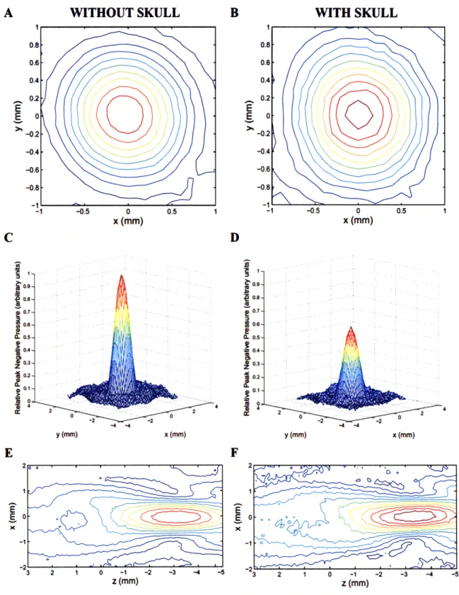
Figure 2-2: Comparison of beam plots with and without rat skull in beam path. ... 48
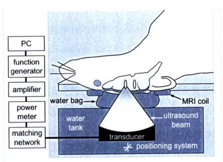
Figure 2-3: Diagram of the experimental set-up ... ... 50
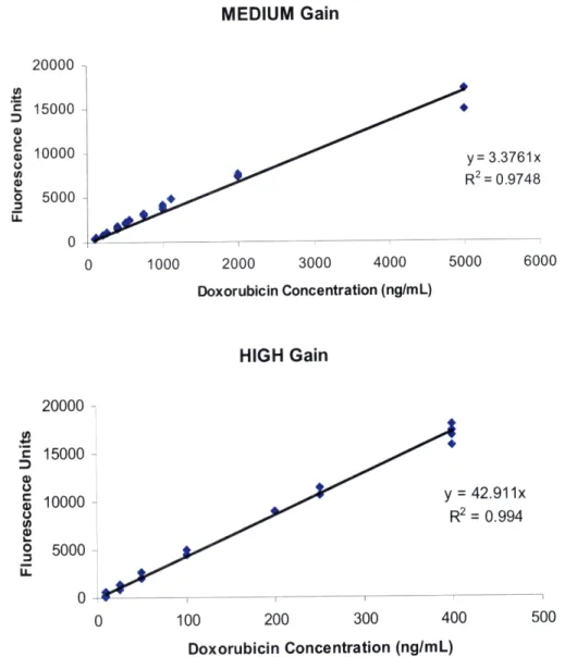
Figure 2-4: Calibration curves for fluorometric measurements of doxorubicin concentration ... 52
Figure 2-5: Schematic of ultrasound protocol (freq = 1.7 MHz) used to deliver doxorubicin to the rat brain at human therapeutic levels ... 57
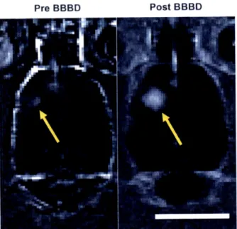
Figure 2-6: Contrast-enhanced T 1-weighted magnetic resonance images of the rat brain before (left) and after (right) ultrasound-induced BBB disruption around the tumor (arrows) ... 65
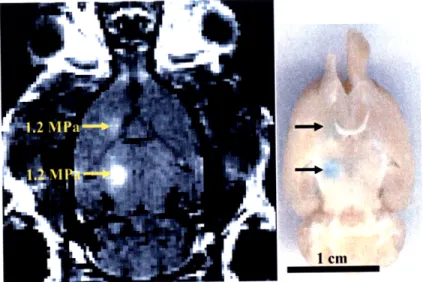
Figure 3-1: Confirmation of ultrasound-induced localized BBB disruption in the rat brain by MRI and by trypan blue ... 69
Figure 3-2: Threshold of BBB disruption induced by transcranial focused ultrasound ... 70
Figure 3-3: Location dependence of threshold for ultrasound-induced BBB disruption. ... 71
Figure 3-4: Normalized MRI signal enhancement (mean ± SD) in the sonicated region after injection of MR contrast agent as a function of pressure amplitude ... 72
Figure 3-5: H&E-stained rat brain exposed in 4 focal locations to pressure amplitudes ranging from 0.5 MPa to 1.7 MPa...73
Figure 4-1: Concentration of DOX delivered to the brain as a function of Optison dose ... 78
Figure 4-2: Correlation of MRI signal enhancement and DOX delivered to the targeted brain parenchyma... . ... 80
Figure 4-3: Transversal H&E-stained sections of rat brains harvested 4 h after sonication with 0.1, 0.2, or 0.5 mL/kg Optison injections ... 81
Figure 4-4: Transversal H&E-stained sections of brains of rats sacrificed 1 week after treatment with FUS+DOX ... ... ... ... 82
Figure 4-5: Fluorescence images showing localized distribution of DOX in sonicated region of rat brain ... 84
Figure 5-1: Fraction of survival (Kaplan-Meier plot) of rats with intracranially implanted 9L gliosarcoma after treatment ... 88
Figure 5-2: T2-weighted magnetic resonance images of a rat brain with implanted 9L gliosarcoma (outlined) before and 1, 2, and 3 weeks after treatment with focused ultrasound and i.v. liposomal doxorubicin (FUS+DOX; top row) or treatment with i.v. liposomal doxorubicin (DOX only; bottom row). ... 90
Figure 5-3: Average tumor volume doubling time in rats with intracranially implanted 9L gliosarcoma after treatment ... 91
Figure 5-4: H&E-stained histologic sections of rat brains implanted with 9L gliosarcoma, harvested 48 h after treatment...93
Table 2-1: Acoustic efficiency of air-backed single-element transducer. ... 46 Table 2-2: Experimental parameters for ultrasound-mediated delivery of doxorubicin to the
norm al rat brain... ... .... ... ... 56 Table 4-1: Mean doxorubicin concentration (ng/g tissue) accumulated in sonicated and control
BBB blood-brain barrier deg C degree Centigrade cm centimeter
CNS central nervous system CT computed tomography
Da Dalton, unit of molecular weight DOX doxorubicin
ETL echo train length FA flip angle
FOV field of view FSE fast spin echo FUS focused ultrasound
g gram
h hour
Hz Hertz, unit of frequency i.p. intraperitoneal
i.v. intravenous m meter
MHz megaHertz, unit of frequency
min minute mg milligram pL microliter mL milliliter pm micrometer (micron) mm millimeter
MPa megaPascal, unit of pressure MRI magnetic resonance imaging ms millisecond
NEX number of excitations Pgp P-glycoprotein
Np Neper, natural logarithmic unit of ratio ppm part per million
RARE rapid acquisition with relaxation enhancement RF radio frequency
ROI region of interest
s second
SD standard deviation
T Tesla, unit of magnetic field strength TE echo time
TJ tight junction TR repetition time W Watt, unit of power
1.1
Motivation
With all the scientific and medical advances over the last century, the brain remains one area of which our understanding has advanced greatly but our ability to treat has been sluggish. While imaging and diagnostics have grown by leaps and bounds, the evolution of therapeutic interventions in the central nervous system (CNS) has not enjoyed such rapid progress. Current standard clinical approaches to malignant brain tumors, including surgical resection in combination with radio- and/or chemotherapy, have met with limited success (1). Many other neurological or neurodegenerative disorders do not yet have effective therapies, despite the rapid growth of the pharmaceutical industry in recent years.
One major reason for the apparent bottleneck is the presence of the blood-brain barrier (BBB). Formed by the endothelial cells that line the microvasculature of the brain, the BBB prevents the entry of most blood-borne substances into the CNS. Its selective permeability, based on lipid solubility, molecular size, and charge, protects the brain from potential toxins but also limits the access of many prospective therapeutics (2). Potential therapeutic agents are prohibited from passing from systemic circulation into brain parenchyma (3, 4) or are unable to accumulate at sufficient therapeutic concentrations
(5). Although tumor vasculature is often malformed and the integrity of its BBB compromised, the complex problem of drug delivery to the brain persists. Because systemic chemotherapeutic agents are not able to penetrate solid tumors homogenously (6), portions of the tumor are often left untreated or partially treated after traditional
intravenous chemotherapy. Additionally, malignant cells may infiltrate the margin beyond the visible tumor where the BBB is intact. Invisible to the surgeon and unreachable by pharmacological interventions, these infiltrating cells are to blame in 78-90% of cases of recurrent glioma (7, 8). Thus, the BBB remains a formidable obstacle in the treatment of patients with brain malignancies. Even with aggressive surgical resection and radiotherapy, the prognosis for the most common and most aggressive form of glioma in adults is associated with a median survival of less than one year from the time of diagnosis (1, 9).
Current strategies to circumvent the BBB are less than ideal. Methods of diffuse BBB disruption allow widespread cytotoxic drug penetration to non-targeted brain tissue (10) and can thus have dose-limiting side effects. Other methods which provide localized drug delivery may increase the drug concentration at the target location while reducing systemic toxicity effects, but they typically require invasive, high-risk neurosurgical procedures (11, 12).
A novel approach to the problem of drug delivery to the brain uses focused ultrasound to temporarily disrupt the BBB in a noninvasive and localized manner (13). High-frequency acoustic energy penetrates soft tissue to induce effects deep below the surface, thus allowing a noninvasive approach to therapy. When applied to the brain in the presence of gas-filled microbubbles, ultrasound has been shown to stimulate active vesicular transport and transiently disassemble tight junctional complexes to allow the passage of molecules which would not otherwise penetrate the BBB (14, 15). As the energy is tightly focused to diameters as small as <1 mm, its effects on the BBB can be confined to a limited volume of tissue to enable targeted therapy (16-18). The high spatial
resolution and noninvasive nature of ultrasound-induced BBB disruption make it an advantageous technique for targeted drug delivery to the brain. In addition, the use of magnetic resonance imaging (MRI) to guide and monitor the procedure enables precise targeting and repeated application without ionizing radiation. Trans-BBB delivery by MRI-guided focused ultrasound has now been demonstrated for numerous agents, including liposomal doxorubicin (17), imaging fluorophores (19), Herceptin (16), Alzheimer's disease immunotherapeutics (20), and other antibodies (21).
Focused ultrasound-induced BBB disruption addresses the limitations of other drug delivery methods and shows great potential to have a positive impact on patients with a variety of neurological disorders. However, further evidence of the therapeutic benefit of ultrasound-enhanced trans-BBB chemotherapy is needed to advance this technology toward clinical trials. Doxorubicin (DOX) is a prime candidate with which to demonstrate the therapeutic potential of ultrasound-mediated trans-BBB drug delivery. It is a highly effective cytotoxic agent with ubiquitous clinical use in the treatment of a wide range of cancer types, but because it does not readily penetrate the intact BBB, it is typically ineffective in treating intracranial lesions. Cancer patients who demonstrate partial or complete response to DOX chemotherapy for extracranial lesions may no longer enjoy such positive results if their lesions metastasize to the brain. Even in high-grade brain tumors, in which the vasculature is abnormally permeable, the BBB often prevents cytotoxic levels of DOX from being achieved in glioma tissue and in intracranial metastases (5). If DOX were allowed to accumulate at sufficient concentrations in the brain, its clinical impact on the treatment of patients with both primary and metastic tumors could be significant. To advance the development of
MRI-guided focused ultrasound for targeted drug delivery applications in the brain, this thesis aimed to develop a protocol using MRI-guided focused ultrasound for the targeted delivery of doxorubicin to the brain and to demonstrate its therapeutic impact in a disease model.
1.2
The Blood-Brain Barrier
1.2.1 Role
Comprised of the brain and spinal cord, the central nervous system (CNS) needs a chemically stable environment to function properly. Neurons communicate through the propagation of action potentials induced by the flow of ions into and out of the cells. In the brain, their complex synaptic connections require that the surrounding extracellular concentrations of sodium, potassium, and calcium be maintained within a very narrow range. The hypersensitivity of neurons to the ionic balance of their environment necessitates a mechanism of strict regulation of access to the brain microenvironment from the vascular compartment. Such regulation exists in the form of the blood-brain barrier (BBB). The BBB is a selectively permeable barrier between the brain parenchyma and its blood supply. It regulates ionic balance within the brain and facilitates the transport of nutrients from systemic circulation, while protecting the brain from potentially harmful blood-borne molecules, which may otherwise be innocuous in the peripheral organ system (4, 22).
1.2.2 History
The restrictive permeability of the brain was first observed by Paul Ehrlich in 1885. While studying staining in animals, he discovered that some injected dyes diffused rapidly into most organs, whereas the brain showed very little uptake. His student Edwin Goldmann further observed in 1913 that a dye injected directly into the surrounding cerebrospinal fluid (CSF) was contained within the CNS, while the rest of the body remained unstained (23). These observations provided the first documented evidence of a
barrier which separates the cerebral microenvironment from its vasculature. Lina Stern later proposed the existence of a "hemato-encephalic barrier" in 1921 (24).
Electron microscopy studies in the 1960's helped to elucidate the physical structure of the BBB. They revealed that the actual barrier lay in the endothelial lining of the brain (25). It is now recognized that the BBB is just one of several blood-CNS barriers. Other blood barriers include the blood-CSF barrier and the blood-spinal cord barrier (26, 27).
1.2.3 Blood supply to the brain
The brain is perfused by an extremely dense and extensive microvascular network. Virtually all neurons and supporting glial cells are within 20 microns of a capillary so that nutrients and oxygen carried by the blood can be delivered to the brain in a quick and efficient manner. Since each brain cell has an almost direct connection to the circulatory system, the vascular route would be an extremely effective way to deliver an agent to the brain, if the molecule were able to enter the brain from circulation. However, the presence of the BBB makes it very difficult for all but a few molecules to do so (28).
1.2.4 Physiology
Multiple mechanisms contribute to the functionality of the BBB. The primary mechanism is the physical barrier between the blood and the brain's extracellular space formed by the endothelial lining of the brain microvasculature. In addition, capillary pericytes directly adjacent to the endothelium share a common basement membrane with the endothelium and help to regulate endothelial metabolism. Astrocytes, one type of glial cell, have extensions of the main cell body which terminate on the vessel wall,
called foot processes; they both provide structural support and contribute to BBB regulation (29). A secondary defense, in the form of an enzymatic barrier, serves to inactivate drugs that may be able to passively diffuse through the endothelial cells. In addition, an active efflux barrier causes many penetrating molecules to be transported from the brain back into the blood. This multifunctionality of the BBB ensures that the neuronal and glial environment is kept stable and that entry into the brain parenchyma is strictly enforced (10).
1.2.4.1 Physical barrier
Endothelial cells which line the capillary walls serve as the physical interface between the brain parenchyma and its blood supply. Unlike the loosely connected endothelial cells of capillaries in peripheral organs, endothelial cells in brain capillaries are tightly connected to each other by specialized proteins which form intercellular tight junctions. Tight junctions are complexes of transmembrane proteins expressed by endothelial cells and perivascular glia, including junctional adhesion molecules (30), occludins (31), and claudins (32). Characterized by very high electrical resistance, on the order of 1000 Ohms/cm2, which is several orders of magnitude higher than intercellular
resistance of endothelial cells outside of the brain, tight junctions play a primary role in forming the highly impermeable physical barrier of the BBB (33). Because the gaps between the endothelial cells are well-occluded by the tight junctions, the paracellular pathway across the endothelium is blocked in the brain.
The transcellular pathway across the endothelium is likewise severely limited in brain capillaries in comparison to peripheral capillaries. Pinocytosis, a process by which molecules are enveloped within a vesicle and transported across the endothelial cell
membrane, is markedly reduced in the brain microvasculature, making it more difficult for nonspecific molecules to diffuse freely across its membrane into the brain parenchyma (34). As molecules can neither pass between nor through the endothelial cells of the blood vessel wall due to the combination of the high-resistance endothelial tight junctions and the restricted pinocytosis, these components both contribute to the physical barrier of the BBB.
1.2.4.2 Enzymatic barrier
The endothelial cells produce a variety of enzymes which inactivate or degrade molecules that are able to get past the physical barrier of the endothelial cells. In addition, neighboring pericytes and astrocytic foot processes also produce such enzymes to contribute to the functionality of the BBB (22, 35, 36). Conversely, the brain also produces specialized enzymes which activate molecules that would otherwise be inactive in the brain. For example, L-DOPA is an amino acid used in the treatment of Parkinson's disease which is inactive in the peripheral blood system but which is converted to its pharmacologically active form, dopamine, once it crosses the BBB (34).
1.2.4.3 Efflux barrier
A third mechanism exists for the undesired influx molecules which may penetrate the other barrier mechanisms. The endothelial cells, pericytes, and astrocytic foot processes of the BBB also produce efflux transporter proteins to escort undesired molecules out of the brain. If a molecule which enters the brain from the blood happens to be a substrate for one of the many active efflux proteins, it will bind to its transporter counterpart and be carried across the endothelial cell membrane back into the vascular compartment. P-glycoprotein (Pgp) is one particularly effective efflux pump which acts
on multiple substrates and is highly active on the interior (lumenal) plasma membrane of the capillary endothelium (37). Its location, multispecificity, and potency make Pgp a critical barrier to therapeutic drug entry into the CNS, and its overexpression in tumor cells confers multidrug resistance (38, 39).
1.2.5 Endogenous transport across the BBB
Selective transcellular access to the brain for circulating molecules is possible via one of two transport mechanisms: free diffusion of small molecules or catalyzed transport of small or large molecules.
1.2.5.1 Free diffusion of small molecules
Certain small molecules are able to cross BBB by free diffusion across the phospholipid bilayer of the endothelial wall. In order to freely diffuse through the endothelial cell membrane, molecules must meet stringent criteria, based on size, charge, and lipid solubility. In general, molecules must be neutral, lipid-soluble, and less than 400-500 Dalton in molecular mass to be able to enter the brain parenchyma from circulation by passive diffusion (2, 28, 34).
1.2.5.2 Catalyzed transport of small or large molecules
Endogenous transport mechanisms exist to accelerate the passage of certain essential molecules which do not meet the criteria for free diffusion through the endothelial wall. Small water-soluble vitamins and nutrients, such as glucose and neutral amino acids, are brought into the brain from circulation through carrier-mediated transport. For certain large-molecule peptides or plasma proteins, receptors present on the endothelial cell membrane recognize specific molecules, such as insulin, leptin, and
transferrin, which are transported through the endothelial cytoplasm by receptor-mediated transcytosis (10).
1.2.6 Strategies for drug delivery to the brain
Multiple strategies to circumvent the BBB for the delivery of potential therapeutic agents have been developed, including catheter-based interventions, pharmacological manipulation of drugs, and alternative nonvascular routes. These techniques have demonstrated potential, but each has major limitations: they are invasive procedures, have toxic side effects and low efficiency, or are not sufficiently safe.
Intracranial drug delivery is further complicated in the case of CNS tumors. Although the integrity of the BBB is often compromised in tumors, granting access to blood-bourne agents which would not normally be able to enter the neural tissue, the disruptions are localized and non-homogeneous (40). In addition, the distribution of the microvasculature in solid tumors is itself hetereogeneous, leading to greater diffusional requirements for drugs to reach neoplastic cells and spatially inconsistent drug delivery. Furthermore, high interstitial pressure within the tumor and edema surrounding the tumor often contribute to an increase in hydrostatic pressure in the normal brain parenchyma adjacent to the tumor, making these regions even less permeable to drugs than normal brain endothelium (34).
1.2.6.1 Catheter-based BBB disruption
Osmotic opening of the BBB is possible by injection of a hypertonic solution, such as mannitol or arabinose, into a catheter placed in the carotid artery. The introduction of the hyperosmolar solution causes a difference in osmotic pressure between the intravascular space and the endothelial wall of the blood vessel. To balance
the osmotic pressure, water rushes out of the endothelial cells, causing their shrinkage and expansion of the intercellular spaces. In this way, the paracellular pathway through the BBB, normally occluded by tight junctions, is transiently opened, allowing small and large molecular agents to enter the brain interstitium over the period of a few hours (2, 41-43). Chemotherapy accompanied by osmotic BBB disruption has yielded moderate augmentation of the delivery of antitumor agents to the brain (44, 45). One major limitation of this method is that it affects the entire volume of tissue supplied by the injected artery, allowing diffuse cytotoxic drug penetration to non-targeted brain tissue. In addition, it has been associated with an increased risk of altered glucose uptake, microembolism, and abnormal neuronal function (3, 34, 46).
A method thought to be potentially safer than osmotic BBB disruption is biochemical BBB disruption by intracarotid infusion of agents such as leukotrienes, bradykinin, nitric oxide, or analogs (47-49). In contrast to osmotic disruption methods, these methods appeared to selectively affect brain tumor capillaries due to the down-regulation of the enzymatic barrier in tumor endothelial cells, while leaving normal brain capillaries unaffected (49, 50). Pre-clinical data in glioma-bearing rats showing enhanced tumor drug delivery and survival led to clinical trials (51, 52), but the trials have since been abandoned due to similar risks shown in osmotic BBB disruption (53).
Convection-enhanced delivery is another technique that utilizes a catheter-based approach (11, 54-56). It is based on maintaining a pressure gradient by delivering a continous interstitial infusion of a drug via intracranial catheters. The resultant bulk fluid convection forces the aqueous solution through the brain interstitium to distribute the drug over large volumes of target tissue (57). An alternative approach to increasing the
pressure gradient is to increase the diffusion gradient by maximizing the concentration of the the infused agent; such an approach has been shown to increase the volume of distribution (58, 59). Clinical trials of convection-enhanced delivery of intratumoral chemotherapeutic agents have shown some significant antitumor response rates with varying degrees of clinical impact (60-63). However, the technique is not without its limitations. Preferential flow of the forced fluid along white matter tracts can result in unintentional distribution patterns of the drug, resulting in harmful consequences such as diffuse astrogliosis (64, 65). Also, as with other transcranial catheter-based approaches, it is an invasive intracranial procedure with significant risk of morbidity and mortality. 1.2.6.2 Pharmacological modification
Without altering the BBB permeability, pharmacological modification is another approach which is being explored to increase the ability of agents to penetrate the BBB. Since lipophilic drugs cross the BBB much more easily than their hydrophilic counterparts, lipidization of small-molecule drugs can facilitate their passage through the phosholipid bilayer of the endothelial cell membrane. By conjugation to a lipid carrier or by reduction of the strength of its hydrogen bonding, lipidization increases the transcellular transport of a molecule into the brain interstitium (66, 67). In another process of pharmacological modification called cationization, proteins can be attached to a charged molecule, which induces transcellular uptake as a result of interactions with anionic groups on the cell membrane (68). However, while uptake in the brain is increased by these processes, uptake is also increased in peripheral tissues, reducing the plasma concentration and availability of the circulating agent and increasing the toxicity to non-targeted tissues. In addition, efflux processes can also be enhanced by these
processes, resulting in poor retention. Thus, poor selectivity and poor retention are major limiting factors in the applicability of these approaches (34).
Other carrier-mediated or receptor-mediated approaches take advantage of the endogenous transport mechanisms of the BBB by conjugating small- or large- molecule drugs to a known BBB transport vector (69-71). These transport vectors have been termed "molecular Trojan horses" because molecules which would not normally cross the BBB are essentially disguised by the attached carrier molecule to gain entry to the brain in pharmacologically significant amounts (72-74). For example, large molecules, such as recombinant proteins, neuropeptides, and therapeutic genes, can be bound to an endogenous peptide or monoclonal antibody which undergoes receptor-mediated transcytosis, to form a chimeric peptide, enabling the attached agent to benefit from the native BBB transport process (28). While this approach may be effective in small doses, the technique is limited by the number of available receptors expressed on the brain endothelium and by the finite carrying capacity of a given carrier (53). Furthermore, in spite of the target selectivity and therapeutic potential of the technique demonstrated in animal models (74), molecularly targeted therapies are often beleaguered by systemic toxicity (75).
Biodegradable polymer-based nanoparticles, liposomes, and micelles are also being developed for drug and gene delivery (76-78). The viability of these carrier-based approaches is increased by the modification of their surfaces with poly(ethylene glycol), a process known as pegylation, which increases their stability and increases their circulation time in the body (79, 80). Nanoparticles have been shown to improve both penetration and retention of a bound drug in the brain (81-83) and to improve therapeutic
efficacy in glioma-bearing rats (76, 84). While these approaches show great promise, they too are limited by their finite carrying capacity and considerable burden for chemical conjugation.
Yet another strategy is to inhibit the drug efflux transporters expressed in the BBB. The use of pharmacological modulators known as poloxamers blocks the activity of efflux pumps such as Pgp, allowing increased transport of their substrates (85-87). These modulators have been shown to effectively enhance the transport of intravenously administered drugs to the brain in animal models (88, 89). However, a major limitation of disabling the powerful, multispecific efflux barriers is that it allows the passage of all substrates normally blocked by the transporter pumps, resulting in loss of protection everywhere in the brain and dramatic neurotoxicity (37).
1.2.6.3 Other nonvascular routes to CNS drug delivery
Direct administration of an agent into the CSF is one way to bypass the BBB altogether by using delivery routes which do not involve the vascular system. Drugs can be infused into CSF through the brain ventricles (intraventricular route) or by lumbar puncture (intrathecal route). Since the drug would be contained within the CSF by the blood-CSF barrier, and since there is free molecular exchange between the CSF and brain interstitial fluid, high CSF drug concentrations should theoretically translate into therapeutic CNS drug concentrations with minimal systemic toxicity (34). In practice, however, the rate of drug distribution within the CSF is very slow. Drug diffusion through the brain parenchyma is also very slow and inversely proportional to the molecular weight of the drug (90, 91). Furthermore, this approach is complicated by
increased intracranial pressure and high clinical incidence of hemorrhage, neurotoxicity, and CNS infections (34).
Localized drug delivery in the brain is possible with methods such as direct intratumoral injection (92, 93) and controlled release from polymer implants (12, 94, 95). Such approaches can deliver therapeutic molecules at a defined rate over a specific period of time. Implantable devices can be positioned by stereotaxy in precisely defined targeted areas and the procedure can be repeated if necessary (96). This technique has been shown to prolong survival in patients with recurrent aggressive glioma and has proven its clinical impact (95). However, the limited diffusion of drugs into the brain from the implanted source and their rapid elimination by active efflux transport present weaknesses to this approach to CNS drug delivery (94). In addition, due to the invasive intracranial surgery required, this approach can only be used in a limited number of patients and carries significant risk of morbidity and mortality with only modest benefit (97).
1.3 Focused Ultrasound (FUS)
A novel approach to deliver drugs to the brain uses focused ultrasound to temporarily disrupt the BBB in a noninvasive and localized manner. In this technique, acoustic energy is concentrated in a focal spot to induce localized biological effects deep below the tissue surface with minimal effects to surrounding nontargeted tissue, even in the path of the ultrasound beam (98-101). Advances in acoustic technology have enabled the use of ultrasound in the brain, in spite of strong energy attenuation by the skull bone (102, 103). Because acoustic energy can be applied in a completely noninvasive manner, has the ability to precisely target tissue of interest while leaving other structures unaltered, and enables the passage of pre-existing drugs through the BBB, focused ultrasound offers distinct advantages over other diffuse or invasive methods of drug delivery to the CNS.
1.3.1 Background
Ultrasound is generated by applying an electrical voltage to a piezoelectric material, such as certain crystals or ceramics, which responds with mechanical deformation in proportion to the applied voltage. The expansion or contraction of the material causes the compression or rarefaction of its surrounding medium, such as air or water. An oscillating voltage then produces pressure waves; at frequencies in excess of -18 kHz, the upper limit of human hearing, the resultant pressure wave is termed ultrasound. The effect works both ways, so mechanical stress will conversely induce a voltage across the material. Thus piezoelectric transducers can be used both to generate and receive ultrasonic signals.
Ultrasound propagates as a mechanical wave through tissue with attenuation of its pressure amplitude P(z) described by:
P(z) = Poe-"z (1)
where p is the frequency-dependent attenuation coefficient per unit path length z due to energy scattering and absorption in tissue, and Po is the incident peak rarefactional pressure amplitude at the surface. Ultrasound has a relatively low absorption rate in soft
tissue; at 1.0 MHz, ultrasound has an approximate wavelength of 1.5 mm and its focal penetration depth can reach up to 10 cm (104). As with any wave, reflection, refraction, and diffraction at media interfaces of vastly different acoustic impedance, due to differences in density and sound speed, severely reduce energy transmission. Much of the mechanical energy lost from the propagating wave is converted to heat and absorbed in the body. Thus, the use of ultrasound has been limited in areas of the body which include interfaces between soft tissue and gas or bone, such as in the lungs, digestive tract, and brain, where energy losses can cause unwanted heating and severe tissue damage (105).
The ability of ultrasound to be focused has made it practical for therapeutic use. The size of the focal region is limited by the wavelength and its sharpness is determined by the ultrasound frequency and the geometry of the source transducer (105). Thus, higher frequencies with smaller wavelengths can achieve tighter foci, while lower frequencies with greater wavelengths produce wider focal regions. Therapeutic applications of focused ultrasound can be achieved with both thermal and mechanical effects. In tissue ablation, the induced temperature is raised high enough over a short period of time (-seconds) to cause cell death by protein denaturization and coagulative necrosis, while in hyperthermia, an induced temperature change of only a few degrees for
an extended period of time (-minutes) can sensitize tissue to radiation and chemotherapy. In addition to thermal effects, focused ultrasound can induce mechanical effects in biological tissue. In cavitation, the interaction of a gas bubble with the acoustic field,
whether by radial oscillation (stable cavitation) or violent collapse (inertial cavitation) (106), can significantly enhance absorption and heating effects in tissue (107). Gas bubbles can form spontaneously in tissue during exposure to high intensity focused ultrasound, or pre-formed gas bubbles, such as those found in ultrasonographic contrast agents, can be introduced into the acoustic field by intravenous injection. The variety of bioeffects induced by focused ultrasound at therapeutic frequencies ranging from 0.5 to 10 MHz has prompted its investigation for diverse medical applications, including tumor and tissue ablation (108-121), hemostasis (122-125), vessel occlusion (126-128), thrombolysis (129-134), and BBB disruption for drug and gene delivery (16, 17, 21, 135-140), in multiple organ systems.
1.3.2 History of therapeutic ultrasound in the brain
The use of ultrasound for therapeutic applications in the brain has been studied for over half a century. In the early part of the 2 0th Century, it was demonstrated that high
frequency sound waves could induce biological effects in tissue (141). In the 1940's, other investigators attempted to use focused ultrasound to induce permanent changes in animal brains for therapeutic applications but could not do so without undesired damage due to the attenuation and distortion caused by the skull bone in the path of the ultrasound (142, 143). Removal of a section of the skull bone by craniotomy in subsequent studies enabled the use of focused ultrasound to produce discrete lesions deep within the brains of animals and humans (144-149). In the 1960's, animal experiments with
micro-thermocouples implanted in the brain revealed that tissue damage was caused by temperature elevation at the acoustic focus resulting from the use of high intensity focused ultrasound (150-154). Other studies demonstrated the capacity of ultrasound to induce changes in the permeability of the BBB (155-157). In addition, ultrasound-induced hyperthermia in the brain has been studied extensively to sensitize tissue and enhance the therapeutic impact of radiation or chemotherapy (158-162). Thus, ultrasound has been used to produce both structural and functional changes within the brain.
1.3.2.1 Development of ultrasound applications through the skull
Since the skull was viewed as a barrier to therapeutic applications in the brain, early clinical studies for the treatment of patients with Parkinson's disease and malignant brain tumors were performed by applying focused ultrasound to the brain through a cranial window (163-166). By the late-1970's, the concept of focusing the acoustic energy through the skull had emerged. Studies demonstrated that ultrasound at frequencies less than 1 MHz could be focused through the skull with some distortion and shifting of the foci (167-169). Phased transducer arrays were later suggested as a method to compensate for the distortion caused by the skull. By adjusting the driving phase of each transducer element in the array, the acoustic focus distorted by the skull could be restored (170). This strategy, along with distributing the acoustic energy over a larger surface area of the skull and active cooling of the scalp, can also address problems of unwanted heating in and around the skull due to energy absorption in the bone (171,
172). The requisite phase and amplitude corrections factors for each individual transducer elements can be derived from x-ray computed tomography (CT) scans (102, 173). A hemispherical MRI-guided phased array system has been designed for thermal ablation of
malignant brain tumors (174, 175), which has led to a phase I clinical trial (101). Since the acoustic intensity used for BBB disruption is at least two orders of magnitude less than for thermal ablation, it follows that such a system could also be used for noninvasive BBB disruption through the human skull. Alternative methods of transcranial ultrasound propagation, using lower frequencies (250-300 kHz) (176) or shear-mode transmission (177, 178), which distort to a lesser extent in the bone, are being investigated to eliminate the need for patient-specific CT corrections. Thus, several methods exist to compensate for distortions in beam propagation induced by the skull bone to allow the ultrasound to be applied completely noninvasively in the brain.
1.3.2.2 Development of targeting and monitoring methods
The development of methods to visualize diseased tissue and to monitor the effects of focused ultrasound has been critical to its application in the brain. Crude targeting in early clinical applications used a stereotactic frame based on x-ray images of bony landmarks (164). A more advanced targeting system using CT guidance was later proposed but not clinically tested (179). However, these methods did not offer the ability to monitor the effects of ultrasound in the brain. More recently, MRI has been demonstrated to provide both an effective means of both targeting and monitoring of ultrasound in the brain (13, 21, 180-185). Its excellent soft-tissue contrast and high temporal resolution confer the ability to distinguish diseased tissue from normal tissue in many cases with unprecedented precision. Temperature-sensitive image sequences can provide information on relative temperature changes with an accuracy of +0.5 deg C (186-188). Furthermore, MR contrast enhancement during MRI-guided ultrasound-mediated delivery of molecules across the BBB has been demonstrated to correlate with
the amount of drug or antibody accumulated in the brain, indicating its potential to provide important feedback during ultrasound-mediated CNS drug delivery (16, 17). Finally, the combination of functional MRI and focused ultrasound may enable precise focal stimulation of brain activity in targeted regions. The ability to steer the ultrasound beam to a desired target and to closely monitor its effects with MRI has enabled this technology to move beyond experimental status into clinical adoption. MRI-guided
focused ultrasound earned approval by the U.S. Food and Drug Administration for the thermal ablation of benign leiomyomata, more commonly known as uterine fibroids, in 2004 (189).
1.3.3 Ultrasound-induced BBB disruption
1.3.3.1 BBB disruption using ultrasound alone
It has long been known that ultrasound is capable of disrupting the BBB (155-157). In the 1950's, deposition of trypan blue and radioactive phosphate tracers in the brain after exposure to focused ultrasound was used to demonstrate increased permeability of brain capillaries without visible structural changes (155). BBB disruption was later observed in the periphery of thermal lesions resulting from ultrasound-induced
tissue coagulation (135, 190). However, under further investigation, thermally induced BBB disruption has always been associated with tissue damage (190). Disruption of the BBB was also observed after high-intensity ultrasound exposures above the cavitation threshold, sometimes without tissue damage, indicating that the effect may be related to an interaction between the acoustic field and gas bubbles. However, the unintentional BBB disruption was sporadic and unpredictable, and its association with tissue damage was unclear (191, 192). In 1990, it was proposed that the phenomenon of
ultrasound-induced BBB disruption could be used with antineoplastic agents and ultrasound-ultrasound-induced thermal ablation to enhance the treatment of brain tumors (135). However, molecular delivery into the brain by ultrasound-mediated BBB disruption would not be pratical without a controlled reversible process which does not induce tissue damage.
1.3.3.2 Microbubble-enhanced ultrasound for BBB disruption
A method to induce focal BBB disruption in a predictable and reproducible manner without obvious permanent damage to the brain tissue was demonstrated in rabbits when pulses of focused ultrasound were applied through a cranial window in the presence of an ultrasonographic contrast agent containing gas-filled microbubbles (13). Unlike in thermal ablation applications which use high-intensity focused ultrasound (193), the intravenous administration of gas-filled microbubbles allows the desired BBB opening to be achieved with powers approximately two orders of magnitude lower than those required in the absence of microbubbles and largely confines the induced bioeffects within the walls of the blood vessel.
Successful focal BBB disruption has been demonstrated using ultrasound frequencies ranging from 260 kHz to 2.04 MHz (13, 194-196), allowing the passage of MRI contrast agents Magnevist® (gadopentatate dimeglumine, molecular weight: 938 Da) (194) and MION (monocrystalline iron oxide nanoparticles, molecular weight: 10,000 Da) (197), trypan blue (molecular weight: 961 Da), horseradish peroxidase (molecular weight: 40,000 Da) (195), and antibodies (molecular weight: 150,000 Da) (16, 21, 140) into the brain. The size of the focal region demonstrated by contrast-enhanced MRI decreased with increasing frequency, with focal diameters smaller than 1 mm possible at the higher frequencies (196). The transient nature of the induced BBB disruption was
confirmed by contrast-enhanced MRI, which showed that the BBB was mostly restored 5-6 h after exposure to ultrasound (sonication) and fully intact 2-5 days and 4 weeks after sonication (195, 198). Focal BBB disruption was sometimes accompanied by minor extravasation of erythrocytes from affected capillaries in the sonicated region, but no regions of ischemia or apoptosis which would indicate a compromised blood supply were detected, nor were delayed effects observed by MRI or histology up to 4 weeks after sonication (198); the incidence of erythrocyte extravasation per unit area decreased with lower frequencies (196). Thus, MRI-guided focused ultrasound can induce transient, localized BBB disruption in a noninvasive manner to enable the passage of diagnostic or therapeutic molecules into the brain and is a relatively safe alternative to invasive or diffuse methods currently available. Furthermore, it can be achieved using ultrasound frequencies suitable for transcranial delivery (195).
The influence of various sonication parameters on the induced BBB disruption has been extensively explored since the introduction of the method. Most work cited has used ultrasound exposures at low pressure amplitudes (<1 MPa) with 10-ms pulses repeated at a frequency of 1 Hz for a duration of 20-30 s. One study showed that BBB disruption was not affected by variation in the pulse repetition frequency but was influenced by burst length (199) and peak negative pressure amplitude (16, 17, 21, 195). The pressure threshold for BBB disruption has been shown to be dependent on both frequency and burst length, increasing with the former (200) and decreasing with the latter (199). However, when expressed in terms of mechanical index, defined as pressure amplitude in MPa divided by the square root of frequency in MHz, the mechanical index threshold appeared to be constant at 0.46 (95% confidence intervals: 0.42 to 0.50) when
other sonication parameters are kept constant (196). Investigations of the impact of the concentration of microbubbles on BBB disruption have been inconclusive (17, 199, 201).
1.3.3.3 Physical mechanisms
Ultrasound-induced BBB disruption appears to be mechanically, rather than thermally, mediated; one study in rabbits using 1.63-MHz sonications concluded that the temperature elevation induced during BBB disruption was approximately 0.025 deg C (13). Although it is believed that ultrasound-induced BBB disruption is achieved by using the microbubbles as cavitation sites (98, 99), the exact physical mechanism by which the ultrasound-microbubble interaction induces disruption of the BBB is not fully understood. Multiple biological effects may arise from the interaction of microbubbles with a propagating acoustic wave in tissue (202). In stable cavitation, a bubble expands and contracts at the frequency of the oscillating pressure field. It may grow in size by rectified diffusion, when more gas diffuses into the bubble from the surrounding medium during expansion than diffuses out of the bubble during the compression phase of the pressure wave (203). In inertial cavitation, which occurs at high acoustic pressures, the inertia of the surrounding medium can cause the violent collapse of the bubble during the positive pressure cycle, producing shock waves and high-velocity jets (107), free radicals (204), and high local temperatures (205-207). Another process known as acoustic streaming occurs when the oscillation of bubbles causes their surrounding medium to flow and is often associated with large shear stresses (208). In addition, the acoustic field exerts a steady radiation force on the bubbles along the direction of the ultrasound beam, pushing the bubbles against the blood vessel wall (209).
significant effect due to the large energy concentrations in the region of the collapsing bubbles. However, ultrasound-induced BBB disruption has been demonstrated over a range of acoustic intensities at 260 kHz without wideband acoustic emission (200), a known signature of inertial cavitation in vivo (150), indicating that inertial cavitation is not necessary for the blood-brain barrier disruption. Bubble collapse detected by wideband acoustic emission was associated with small regions of erythrocyte extravasation, while bubble oscillations were detected when BBB disruption was induced without vascular damage (200).
1.3.3.4 Biological mechanisms
Immunoelectron microscopy studies have provided some insight into the biological mechanisms by which microbubble-enhanced focused ultrasound enables the trans-BBB transport of macromolecules. One study showed that passage through the BBB after treatment with microbubble-enhanced ultrasound occurs via both paracellular and transcellular routes, including open endothelial cell tight junctions, enhanced active vesicular transport, endothelial cell fenestration and channel formation, and free passage through injured endothelium (14). Specifically, the redistribution and loss of immunosignals for TJ-specific proteins occludin, claudin-5, and ZO-1 provide direct evidence of the disassembling of the TJ molecular structure and associated functional loss of the BBB immediately following ultrasound exposure; six hours after sonication, the protein immunosignals and BBB function are both restored (15). Active vesicular transport following ultrasound-stimulated BBB disruption was preferentially demonstrated in brain arterioles, rather than venules or capillaries (210). Furthermore, an in vivo study in mice using multiphoton microscopy through a cranial window revealed
arteriolar vasoconstriction during the ultrasound pulses, followed shortly by leakage of a tracer, suggesting that the increase in BBB permeability might be related to temporary vessel spasm (19).
1.4
Doxorubicin (DOX)
1.4.1 Clinical use
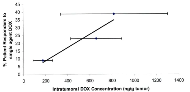
Doxorubicin (molecular weight: 580 Da) is one of the most commonly used cytotoxic drugs in both single-agent and multi-agent chemotherapy regimens. It is used to treat many solid forms of cancer, including breast (211), ovarian (212), bone (213), lung (214), thyroid (215), and gastrointestinal carcinomas (216), as well as blood-derived cancers, such as lymphoma and multiple myeloma (217-219) and soft-tissue sarcomas (220). Because it has demonstrated antineoplastic efficacy against such a variety of cancer types, it has enjoyed widespread clinical use for over 30 years (221). Figure 1-1 shows that intratumoral DOX concentration is strongly correlated (R2 = 0.90) with patient response rate, irrespective of the type of cancer (222).
45 40 -35 - 30 Mc 25 m 20 0 15 I. 10 5 0 . .... r -I F . . .. --1 0 200 400 600 800 1000 1200 1400
Intratumoral DOX Concentration (ng/g tumor)
Figure 1-1: Correlation of intratumoral DOX concentration and patient response rate. DOX concentration measured in excised tumors is linearly proportional to the response rate of patients with breast, gastric, or colorectal carcinoma (R2 = 0.90). Data taken from (222).
Despite its extensive clinical use, DOX is not typically used to treat intracranial tumors because, as a substrate for the powerful efflux pump Pgp, it cannot cross the intact BBB (84). Systemic administration of DOX has seldom been effective in patients with brain tumors due to poor accumulation in glioma tissue (5). However, it has been shown to arrest cell growth and induce apoptosis in malignant glioma cell lines (223). Moreover, direct intratumoral infusion of DOX has been shown to improve survival of glioma patients (92). Thus, the antineoplastic efficacy of DOX against glioma is not in doubt. The evidence indicates that if the accumulation of DOX could be increased to therapeutic levels within the brain, it could be effective in the treatment of malignant brain tumors, whether primary glioma or metastatic brain tumors which originated elsewhere in the body.
1.4.2 Mechanism of action
Formerly known as adriamycin, doxorubicin belongs to the family of anthracycline antibiotics. Like many chemotherapeutic drugs, it combats the uncontrolled proliferation of cancerous cells by binding to DNA to inhibit nucleic acid synthesis and block cell reproduction (224). Cell structure studies have demonstrated that DOX rapidly penetrates cells and binds to perinuclear chromatin (225). It is thought to inhibit the action of topoisomerase II, which is responsible for the unwinding of DNA during the transcription process. The interference of DOX during the process of DNA transcription and gene replication induces chromosomal aberrations and interrupts the continuous cycle of cell proliferation, leading to cell death (226). The bioreactivity of DOX with iron, oxygen, or free electrons in the body can also produce free radicals, such as the
highly reactive hydroxyl radical (OH-), which themselves induce DNA damage (227,
228).
These multiple mechanisms of DNA damage make DOX a highly effective cytotoxic agent. Cancerous cells are strongly impacted by the interference of DOX during the cell reproductive cycle due to their high turnover rate. However, as with any chemotherapeutic drug, DOX also causes unwanted cell death in normal cells as well, especially in those which proliferate quickly, such as hematopoietic cells, gastrointestinal cells and hair follicles. In addition, DOX can cause cardiomyopathy and loss of cardiac function (229, 230), which is associated with the production of free radicals (231). The encapsulation of DOX within microscopic (- 100 nm) phospholipids vesicles, known as liposomes, has been shown to reduce the cardiotoxic effects associated with DOX while prolonging circulation time (232, 233). Due to these benefits, this liposomal form of DOX is most commonly used clinically.
1.4.3 Spectroscopy
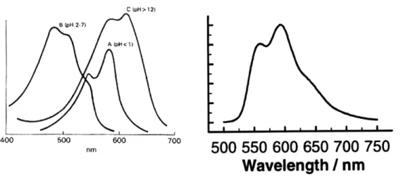
Doxorubicin absorbs and emits light according to the spectra shown in Figure 1-2, with peak absorption at 480 nm and peak emission near 595 nm (234). Because the amount of light emitted is directly proportional to the number of molecules, the fluorescent properties of doxorubicin allow us to make quantitative measurements.
C ipH > 12) 8 (pH 2-7) A IpH< 1) 400 500 600 700 nm 500 550 600 650 700 750
Wavelength
I/
nm
Figure 1-2: Fluorescence spectra for doxorubicin in aqueous solution. A, Absorption spectrum show maximum absorption at 480 nm (curve B, pH 2-7) (235). B, Emission spectrum with excitation at 479 nm shows maximum emission near 595 nm (234).