CASE REPORT
Giant lipoma of the thumb
Valérie Decrouy-Duruz&Daniel Felix Kalbermatten&Pilipp Honigmann
Received: 31 May 2012 / Accepted: 30 July 2012 / Published online: 15 August 2012 # Springer-Verlag 2012
Abstract While MRI is recognized to be the gold standard examination to diagnose giant lipomas and exclude liposar-comas, there is insufficient knowledge about how to clarify the degree of malignancy of intermediate lesions. We report here the case of a digital giant lipoma, where MRI showed a benign polylobulated lipomatous tumor, but that presented nuclear atypia on conventional histological examination suggestive for an atypical lipoma or well-differentiated lip-osarcoma. To exclude such a lipomatous tumor necessitating a more aggressive surgical excision, complementary cyto-genetics with fluorescence in situ hybridization study for MDM2 and CDK4 genes was required. A literature review of the diagnostic approach for lipomatous tumors of the extremities, including MR images, histological examination, and new cytogenetic techniques, is performed.
Level of Evidence: Level V, diagnostic study. Keywords Giant lipoma . Thumb . Atypical lipoma . Liposarcoma . Cytogenetics
Introduction
Ordinary lipomas are benign tumors that can arise every-where on the body within subcutaneous tissue or deep soft tissues. They represent the most frequent benign tumors in the extremities. However, only 5 % of the lipomas of the upper extremity are found on the hand and 1 % on the digits [1]. Diagnosis of a digital lipomatous tumor is clinical and can be easily supported by MR imaging. It is nonetheless of primordial importance to be able to distinguish an ordinary
lipoma from an atypical lipoma or well-differentiated lip-osarcoma, which is of intermediate malignancy.
Giant lipomas are characterized by a size exceeding 5 cm [2,3], which is considered to be the critical size making any soft tissue mass suspect to be malignant [4]. Here, we report the diagnostic and therapeutic approach of a giant lipoma of the thumb. A review of the literature is performed and the importance of histological and cytogenetic studies to dis-criminate benign from atypical lipoma is discussed.
Case report
A retired 65-year-old man with history of valve replacement was referred to our institution because of a semicircular subcutaneous mass at the palmar aspect of his right thumb and extending from the distal first metacarpal to the inter-phalangeal joint (Fig. 1a, b). The patient reported that the mass had slowly grown over the last 2 years without any history of trauma. Normal peripheral sensibility was found and thumb function was preserved.
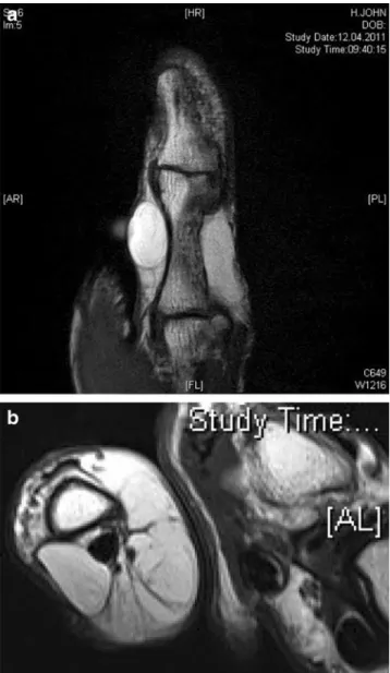
Conventional radiography of the thumb showed no inva-sion of the bone. MR images confirmed the presence of a polylobulated lipomatous tumor with hyperintense signal in T1 and T2 sequences and reduced signal after fat suppression technique. MR images showed that the lesion surrounded the neurovascular bundles without infiltrating them (Fig.2).
As both clinical and radiological examinations supported the diagnosis of lipoma, we planned a marginal excision of the lesion. We performed a Brunner-shaped incision in the palmar aspect of the thumb under regional anesthesia and with the use of tourniquet. A subcutaneous polylobulated lipomatous mass was dissected and completely removed without damaging the bilateral neurovascular bundles (Fig.1c). The 60×40×10 mm tumor was sent for histolog-ical workup (Fig.1d).
V. Decrouy-Duruz (*)
:
D. F. Kalbermatten:
P. Honigmann Department of Plastic, Reconstructive, Aesthetic and Hand Surgery, University Hospital Basel,4031 Basel, Switzerland
e-mail: valerie.decrouy@gmail.com Eur J Plast Surg (2013) 36:331–334 DOI 10.1007/s00238-012-0755-6
Although histological examination showed a well-differentiated mature adipose tissue in the majority, a small fibrous area with discrete nuclear atypia was identified (Fig.3). Additional cytogenetic fluorescence in situ hybrid-ization (FISH) studies were performed. As they revealed no amplification of the MDM2 and CDK4 genes, a well-differentiated liposarcoma was excluded and the diagnosis of ordinary lipoma confirmed.
No complications occurred post-operatively. Three months after the surgery, local examination showed a soft scar. Active and passive mobility of the thumb was normal and the sensibility completely preserved (Fig. 1e). There was no recidive of the tumor within a 1-year follow-up.
Discussion
Ordinary lipoma is the most common soft tissue tumor and may appear at any site. It occurs mainly in the fifth to seventh decade of life, commonly in obese people. Approx-imately 5 % of patients have multiple lipomas [5]. Histo-logically, the lipoma is composed of lobules of mature fat cells, which vary slightly in size and shape. Ordinary lipo-mas present as painless, slowly growing soft tissue lipo-masses. Although common in the upper extremities, lipomas are very rare over the palm and fingers. In 1959, Stein [6] first reported a patient with a lipoma of the finger. Since then, 17
cases of digital lipomas have been reported in the literature, mostly located over the index and the middle fingers. In these reports, patients were rarely symptomatic. A few had decreased motion of the finger or reduced grip strength. Boussouga et al. [7] reported the case of a thenar lipoma with altered sensation in the territory of the adjacent digital nerves, a symptom that is more frequent with lipomas over the distal forearm or arising deep in the palm within Guy-on’s canal or carpal tunnel [1,2]. Deep lipomas (e.g., intra-muscular or interintra-muscular lipomas) are larger and less well defined than their subcutaneous counterparts and can mimic atypical lipomatous tumors or well-differentiated liposarcomas.
According to the World Health Organization’s classifica-tion of Soft Tissue Tumors [5], lipomatous tumors are categorized into three entities: benign, intermediate, and malignant adipocytic tumors. The“intermediate malignan-cy” label groups together atypical lipomatous tumors and well-differentiated liposarcomas, which account for about 40–45 % of all liposarcomas and occur most frequently in deep soft tissue of the limb, especially the thigh, followed by the retroperitoneum and the paratesticular region of middle-aged and older individuals.
Magnetic resonance imaging is the gold standard radio-logical examination for tumors within soft tissues. MR images can specify the nature of the lesion, its local exten-sion and relationship with the neurovascular bundles. In Fig. 1 a, b Giant lipoma of the
thumb, c lipoma excision, d giant lipoma 60×40×10 mm, e 3 months after lipoma excision
case of benign lipoma, MR images reveal tissue that is isointense relative to subcutaneous fat, regardless of pulse
sequences. During fat suppression technique sequences, the signal will be reduced. In less than 50 % of cases, MR images reveal intrinsic thin septa (<2 mm). This is considered as patho-gnomonic for the diagnosis of lipoma. When gadolinium is injected, the mass does not enhance except for its capsule and septas. On a series of 134 MRIs of tumors and pseudotumors of the wrist and the hand, the preoperative diagnosis by MRI of benign lipoma was confirmed by histological investigation in 94 % of the cases [8]. However, distinction between ordinary lipoma and atypical lipoma is difficult to make on MRI.
On a histological level, atypical lipomatous tumors and well-differentiated liposarcomas may also be indistinguish-able from benign adipocytic tumors and inadequate samples can lead to misdiagnosis. Hence, the use of cytogenetic stud-ies, such as FISH and comparative genomic hybridization (CGH), is imperative. The former involves the detection of specific DNA sequences by hybridization with complimenta-ry DNA probes while the latter permits the analysis of DNA sequence copy number changes across the genome. Nishio [9] recently reviewed the contribution of cytogenetics to the di-agnosis of adipocytic tumors. Clonal cytogenetic aberrations have been identified in nearly 60 % of ordinary lipomas, the 12q13–15 region being the most commonly involved. Yet, a CGH study has indicated that no copy number changes are found in ordinary lipomas [10]. In contrast, atypical lipoma-tous tumors and well-differentiated liposarcomas are charac-terized by the presence of supernumerary ring and/or giant marker chromosomes. FISH and CGH studies have shown that ring and giant marker chromosomes are composed mainly of amplified sequences from the 12q14–15 region, including the MDM2 and CDK4 genes. This 12q14–15 amplification is not observed in benign adipocytic tumors and its detection can, therefore, be used as an ancillary diagnostic technique for the diagnosis of atypical lipomatous tumor/well-differentiated liposarcoma [11].
Malignant adipocytic lipomatous tumors may also affect the upper limb. These are easily recognized by radiological and conventional histological analysis. Liposarcomas are characterized on MR images by fat content lower than 75 %. Septas appear thick and enhanced after gadolinium injection [1]. In addition, routine histological examination reveals the presence of nonlipogenic components.
To conclude, lipoma is a benign tumor of the soft tissues that may involve all the fingers. This tumor must be distin-guished from atypical lipoma or liposarcoma, especially when its size exceeds 5 cm. While MRI achieves accurate diagnosis in case of liposarcoma, cytogenetics is compulsory in the case of well-differentiated lipomatous tumors with nonetheless discrete nuclear atypia, where ordinary histological analysis may be insufficient to discriminate a benign from an atypical lipoma or a well-differentiated liposarcoma.
Marginal excision is the treatment of choice for benign lipoma of the hand and fingers. However, atypical lipoma Fig. 2 Giant lipoma of the thumb on MRI. a, T1-tse coronal sequence
b, T2-tse transverse sequence
Fig. 3 Histologic finding showing small fibrous area with discrete nuclear atypia (200×, HE)
and well-differentiated liposarcoma require a wide-margin excision because of their high propensity for local recur-rence. No recurrence of digital lipoma after surgical excision has been reported in the literature to date.
Acknowledgments The authors wish to thank Martin Stoeckli who performed the histological examination and provided the related picture.
Conflict of interest The authors had full control of study design and manuscript preparation and received no financial support for the au-thorship or publication of this article.
References
1. Fnini S, Hassoune J, Garche A, Rahmi M, Largab A (2010) Giant lipoma of the hand: case report and literature review. Chir Main 29 (1):44–47
2. Cribb GL, Cool WP, Ford DJ, Mangham DC (2005) Giant lipo-matous tumours of the hand and forearm. J Hand Surg Br 30 (5):509–512
3. Abkari I, Abidi AE, Latifi M (2011) Giant lipoma of the third finger: a case report. Chir Main 30(2):152–154
4. Hsu CS, Hentz VR, Yao J (2007) Tumors of the hand. Lancet Oncol 8(2):157–166
5. Fletcher CDM, Unni KK, Mertens F (2002) Pathology and Genet-ics of Tumours of Soft Tissue and Bone. IARC World Health Organization Classification of Tumours, IARC Press, Lyon, France 6. Stein AH Jr (1959) Benign neoplastic and non-neoplastic destruc-tive lesions in the long bones of the hand. Surg Gynecol Obstet 109:189–197
7. Boussouga M, Bousselmame N, Lazrak KH (2006) Lipome com-pressif de la loge thénar. A propos d’une observation. Chir Main 25(3–4):156–158
8. Capelastegui A, Astigarraga E, Fernandez-Canton G, Saralegui I, Larena JA, Merino A (1999) Masses and pseudomasses of the hand and wrist: MR findings in 134 cases. Skeletal Radiol 28:498–507
9. Nishio J (2011) Review article: Contributions of cytogenetics and molecular cytogenetics to the diagnosis of adipocytic tumors. J Biomed Biotechnol
10. Szymanska J, Virolainen M, Tarkkanen M, Wiklund T, Asko-Seljavaara S, Tukiainen E et al (1997) Overrepresentation of 1q21–23 and 12q13–21 in lipoma-like liposarcomas but not in benign lipomas: a comparative genomic hybridization study. Can-cer Genet Cytogenet 99(1):14–18
11. Weaver J, Downs-Kelly E, Goldblum JR, Turner S, Kulkarni S, Tubbs RR et al (2008) Fluorescence in situ hybridization for MDM2 gene amplification as a diagnostic tool in lipomatous neo-plasms. Mod Pathol 21(8):943–949