HAL Id: hal-03093950
https://hal.archives-ouvertes.fr/hal-03093950
Submitted on 7 Jan 2021HAL is a multi-disciplinary open access archive for the deposit and dissemination of sci-entific research documents, whether they are pub-lished or not. The documents may come from teaching and research institutions in France or
L’archive ouverte pluridisciplinaire HAL, est destinée au dépôt et à la diffusion de documents scientifiques de niveau recherche, publiés ou non, émanant des établissements d’enseignement et de recherche français ou étrangers, des laboratoires
Pyroglutamide-Based P2X7 Receptor Antagonists
Targeting Inflammatory Bowel Disease
Germain Homerin, Davy Baudelet, Pierrick Dufrénoy, Benoît Rigo, Régis
Millet, Xavier Dezitter, Christophe Furman, Guillaume Ragé, Emmanuelle
Lipka, Amaury Farce, et al.
To cite this version:
Germain Homerin, Davy Baudelet, Pierrick Dufrénoy, Benoît Rigo, Régis Millet, et al.. Pyroglutamide-Based P2X7 Receptor Antagonists Targeting Inflammatory Bowel Disease. Jour-nal of MediciJour-nal Chemistry, American Chemical Society, 2020, 63 (5), pp.2074-2094. �10.1021/acs.jmedchem.9b00584�. �hal-03093950�
Pyroglutamide-Based P2RX7 Receptor Antagonists
Targeting Inflammatory Bowel Disease
Germain Homerin1,2, Davy Baudelet1,2, Pierrick Dufrénoy1,2, Benoît Rigo1,2, Régis Millet1,3, Xavier Dezitter1,3, Christophe Furman1,3, Guillaume Ragé1,3, Emmanuelle Lipka1,4, Amaury Farce1,3, Nicolas
Renault1,3, Boualem Sendid1, Samir Jawhara1, Rogatien Charlet1, Jordan Leroy1, Melodie Phanithavong5, Camille Richeval5, Jean-François Wiart5, Delphine Allorge5, Sahil Adriouch6,7,
Valérie Vouret-Craviari8,9, and Alina Ghinet1,2,10,*
1Inserm U995, LIRIC, Université de Lille, CHRU de Lille, Faculté de médecine – Pôle recherche,
Place Verdun, F-59045 Lille Cedex, France
2Hautes Etudes d’Ingénieur (HEI), Groupe Yncréa Hauts-de-France, UCLille, Laboratoire de
pharmacochimie, 13 rue de Toul, F-59046 Lille, France
3 Institut de Chimie Pharmaceutique Albert Lespagnol, IFR114, 3 rue du Pr Laguesse, F-59006
Lille, France
4 Faculté des Sciences Pharmaceutiques et Biologiques de Lille, Laboratoire de Chimie
Analytique, F-59006 Lille Cedex, France
5 CHRU de Lille, Centre de Biologie Pathologie, Laboratoire de Toxicologie & Génopathies,
Bd du Pr J. Leclercq, CS 70001, F-59037 Lille, France
6 INSERM U905, F-76183 Rouen, France
7 Normandie University, Institute for Research and Innovation in Biomedicine, F-76183 Rouen,
France
8 Institute for Research on Cancer and aging (IRCAN), F-06100 Nice, France 9 University of Nice Cote d’azur (UCA), F-06100 Nice, France
ABSTRACT.
This report deals with the design, the synthesis and the pharmacological evaluation of pyroglutamide-based P2RX7 antagonists. A dozen were shown to possess improved properties, among which inhibition of YO-PRO-1/TO-PRO-3 uptake and IL1β release upon BzATP activation of the receptor and dampening signs of DSS-induced colitis on mice, in comparison with reference antagonist GSK1370319A. Docking study and biological evaluation of synthesized compounds has highlighted new SAR, and low toxicity profiles of pyroglutamides herein described are clues for the finding of a usable h-P2RX7 antagonist drug. Such a drug would raise the hope for a cure to many P2RX7-dependant pathologies, including inflammatory, neurological and immune diseases.
INTRODUCTION
The P2RX7 receptor (P2RX7) belongs to the purinergic receptor family. As all P2X receptors (P2XR), P2RX7 is a ligand-dependent ionic channel activated by ATP.1 The target receptor
possesses some particularities regarding the other P2XR of his sub-family. It has dissimilarities in terms of structure,2 and sensitivity to P2X ligands. P2RX1-P2RX6 receptors have a structure with
379-472 amino acids, while P2RX7 contains 595 amino acids due to a longer COOH-terminal chain. P2RX7 is 100 to 1000 times less sensitive to ATP than the other P2XR.3 Indeed, the different
subtypes of P2XR are distinguished by their sensitivities to ATP. For example, millimolar concentrations are necessary to activate the P2RX7 receptor, whereas for the other sub-types these concentrations are in the micromolar range. The extracellular physiological concentrations of ATP being nanomolar, it is necessary that the ATP is released to reach sufficient concentrations to induce activation. Moreover, P2RX7 has two different behaviors, depending on the duration of its stimulation.4 Over a brief time, the receptor acts as an ion channel permitting a K+ efflux and a Ca2+
and Na+ influx. These ions crossing the membrane are modifying the cell’s potential, and activate
different transduction mechanisms. In the case of a prolonged or repeated stimulation, or in presence of high concentrations of agonist, a large pore is formed. This pore enables molecules up to 900 Da to enter the cell, a process that eventually leads to apoptosis.
The main characteristic of P2RX7 is the opening of the channel that leads to a K+ efflux, a key
step in the inflammatory process. Studies have shown that macrophages pre-treated with KCl do not process or release cytokines when ATP is added, suggesting that channel opening precedes large pore opening that is mandatory for IL1β and IL18 maturation. The Ca2+ influx has been shown
calcium is very important in the proper functioning of neurons, defects in Ca2+ homeostasis being
involved in the pathogenesis of neurodegerative disorders.
IL1β is known to be a mediator of immunity and a cause of inflammation. Lipopolysaccharides (LPS) are the first inducer of IL1β. Nonetheless, their stimulus is not complete; it only induces an accumulation of pro-IL1β within the cell that must be cleaved into mature IL1β. Extracellular ATP was shown to be a strong inducer of pro-IL1β cleavage, via P2RX7 activation.6 P2RX7 implication
in the production and release of IL1β was reported in many studies.7 The potassium efflux induces
the formation of free radicals by the activation of Nicotinamide Adenine Dinucleotide Phosphate (NADPH) oxydase, which facilitate the inflammasome assembly. The inflammasome is a protein assembly where the activation of caspase-1 occurs. Then caspase-1 induces the cleavage of pro-IL1β into active pro-IL1β.7b It is to be noted that NADPH oxydase also induces the death of T-regulatory
cells.8 Moreover, P2RX7 is implicated in the production of other IL1 family cytokins and
chemokins (such as IL18),7a,b known for their role in many chronic inflammation diseases.9 Besides
its role in inflammation, P2RX7 has been shown to be involved in cell proliferation and cell death.10
Another well-known characteristic of P2RX7 is to form a large pore (like P2RX2 and P2RX4 do) when it is over-activated. This pore was the object of many studies.11 After several seconds of
activation, the large pore able molecules up to 900 Da to cross the membrane and to enter the cell.12
It was reported that one or two ATP molecules binding to a “naïve” P2RX7 will induce a transition to desensitization, and a third ATP molecule binding will favor the transition to a dilated form of the receptor.13 The pore formation generally leads to cell death via apoptosis, conferring to P2RX7
a cytolytic activity.14 Many mechanisms were proposed to explain the pore formation but, to date,
Following these observations, many studies were carried out to understand the P2RX7 physiological implications, and its potential uses in medicine. The potency of P2RX7 has been thus highlighted in numerous inflammation-related pathologies, among which neurodegenerative diseases (Parkinson’s disease, Alzheimer’s disease (AD), neurotrauma, multiple sclerosis, Huntington’s disease, spinal cord injury and epilepsy), rheumatoid arthritis, and inflammatory bowel diseases (IBD).15 As mentioned before, P2RX7 is implicated in the production and release
of pro-inflammatory cytokines, such as IL1β and IL18.16 In this context, ATP is a danger signal,
secreted at high concentrations in inflammation loci.17 The proof of concept regarding the reduction
of inflammation by the inactivation of P2RX7 was given in rodent models,18 and some P2RX7R
antagonists were evaluated in clinical trials.19 Unfortunately, none of the trialed drugs succeeded
phase II so far, mostly due to a lack of efficiency. However, P2RX7 modulators (agonists or antagonists) are still of great interest for therapeutic applications.20
Our group is interested in pyroglutamic acid derivatives synthesis as drug-candidates.21 The
development of pyroglutamide-based P2RX7 antagonists by other groups,22 attracted our attention
(Figure 1). This report focuses on the design, the synthesis, and the pharmacological evaluation of analogues of GSK1370319A (compound 1a) and GSK1482160 (compound 1b) as P2RX7 potential antagonists.
N O O N H Cl Cl N O O N H Cl CF3 GSK1482160: IC50 = 3.2 nM 1b: IC50 = 119 nM GSK1370319A: IC50 = 3.2 nM 1a: IC50 = 474 nM O N O O N H Cl CF3 IC50 = 16 nM N O O N H Cl CF3 IC50 = 25 nM N N O O N H Cl Cl N IC50 = 10 nM N H N O O N H Cl Cl IC50 = 10 nM N O O N R R R R 1 2 3 4 O N N O R N R2 4 Pharmacomodulations Rigidification
Structures of target compounds
R1 = H, Me, Ar, Bz, Het
R2 = Me, Ar, Bz, Het R3 = H, Me
R4 = H, enamines, carbonyle
Figure 1. Structures and activities on h-P2RX7 in vitro reported in the literature (black) and
activities observed in our biological assay (green) of reference P2RX7 antagonists GSK1370319A (compound 1a) and GSK1482160 (compound 1b), structures and activities reported in the literature of analog P2RX7 antagonists and general structures of target compounds.
RESULTS AND DISCUSSION
Pharmacophore perception of P2RX7 antagonists.
Since this study began previously to the recent crystallization of the panda (pd-) P2RX7,23 a
ligand-based approach was privileged. We first engineered a pharmacophore model based on 3D-structural similarities of published antagonists (see Supporting Information for the structure of selected antagonists of the literature).24 The pharmacophore model was selected as fitting at best
the 9 molecules of the reference dataset (Figure 2), which was the closest to the one described in the literature.19b Exhibiting two hydrogen bond acceptors and two lipophilic features, this
pharmacophore query was used to filter a database of potential compounds; the best fitting ones being selected for synthesis (see Supporting Information for the structure and scores of designed compounds).
Figure 2. Representation of P2RX7 antagonists’ pharmacophore (pink sphere: predicted
hydrogen-bond acceptor location (A1 and A2); brown sphere: predicted lipophilic centers (L1 and L2)) and potentially active compounds A1, C2, D1, D2, E2 and F2 (see Supporting Information for
The investigations were firstly interested in the modulation of the positions 1 and 5 of the lactam ring. Three series were designed regarding the substitution on position 1 of the pyrrolidone scaffold: Series I (substitution by a methyl); Series II (introduction of (hetero-) aromatic units); Series III (N-benzyl and N-benzhydryl substituents). In these series, the amide in position 5 was substituted with various (hetero-)aryls, benzyls and alkyls groups.
In a second time, series IV was designed to explore the conformational space available in position 3 of the lactam. During the synthesis of this last series, we also unexpectedly observed the formation of pyrroloimidazolediones.21j,k These constrained analogues are gathered in series V.
Furthermore, other cycles replacing the pyroglutamide scaffold were investigated, such as oxazolidinone, pyridine and piperidone. The compounds bearing these moieties constitute series
VI.
Chemistry.
The syntheses of pyroglutamides series I-VI are reported in Schemes 1 to 6. A large part was synthesized from natural amino-acids ˗ L-pyroglutamic acid 2 (PGA), L-glutamic acid 3 (Glu), and L-serine 4 (Ser).
Esterification of PGA 2 was first conducted, leading to methyl pyroglutamate 5 (PGM) in quantitative yield.21a Synthesis of iminoether 6 and following rearrangement to methyl N-methyl
pyroglutamate 7 was realized using dimethylsulfate.21b Aminolysis of ester 7 with corresponding
Scheme 1. Synthesis of N-Methyl Pyroglutamide Derivatives 1a-da (Series I) 1a R2 = CH2-(2,4-diCl)phenyl (77 %) 1b R2 = CH2-(2-Cl-3-CF3)phenyl (54 %) 1c R2 = CH2-(2-Cl-5-CF3)phenyl (16 %) 1d R2 = CH(CH3)-(2,4-diCl)phenyl (51 %) N O O N H I R2 a b N H O O OH 2 N H O O O 5 (>99 %) N O O O 6 (74 %) N O O O 7 (67 %) d c
a Reagents and conditions. (a) CH
3SO3H, MeOH/CHCl3, MS 3Å, reflux, 24 h, quantitative yield;
(b) (i) Me2SO4, 60°C, 12 h; (ii) Et3N, 0°C–r.t., 1 h, 74%; (c) Me2SO4, THF, 80°C, 4 h, 67%; (d)
amine, catalyst (PTSA or ZrCl4), r.t.–120°C, 5–96 h, 16–77%.
N-aryl PGM 8a-l were next obtained by a copper initiated method we recently developed.21c
Aminolyses of esters 5, 7 and 8a-l furnished pyroglutamides 9-13, 15, 16a, 16e, 16g-o, 18 and 22.21d
N-(hetero-)aryl derivatives 14, 16a-d, 16f, 16p-q , 17, 19-21 and 23 were obtained by copper initiated coupling from pyroglutamides 10j, 11e, 11a, 11g-h, 12b and 13 (Scheme 2).21c
Scheme 2. Synthesis of N-Aryl Pyroglutamide Derivatives 14 to 23a (Series II) a b b a R2 = CH2-cyclohexyl, R3 = H 17 R1 = phenyl (b : 13 %) R2 = CH2-(2-Cl)phenyl, R3 = H 18 R1 = phenyl (a : 70 %) R2 = CH2-(2-Cl-3-CF3)phenyl, R3 = H 19 R1 = 2-thienyl (b : 84 %) R2 = CH2-(2-Cl-5-CF3)phenyl, , R3 = H 20 R1 = 2-thienyl (b : 89 %) R2 = CH(CH3)-(2,4-diCl)phenyl, R3 = H 21 R1 = 2-thienyl (a : 16 %) R2 = R3 = Me 22 R1 = phenyl (a : 91 %) R2 = CH2-CH2-(2,4-diCl)phenyl, R3 = H 23 R1 = phenyl (b : 79 %) R2 = (2,4-diCl)phenyl, R3 = H 14 R1 = phenyl (b : 5 %) R2 = 4-tolyl, , R3 = H 15 R1 = phenyl (a : 35 %) R2 = CH2-(2,4-diCl)phenyl, R3 = H 16a R1 = phenyl (a : 60 % ; b : 71 %) 16b R1 = (2-Cl)phenyl (b : 62 %) 16c R1 = (2,4-diCl)phenyl (b : 34 %) 16d R1 = (3-CF3)phenyl (b : 75 %) 16e R1 = (3,5-diCl)phenyl (a : 25 %) 16f R1 = (4-Cl)phenyl (b : 39 %) 16g R1 = (4-CN)phenyl (a : 16 %) 16h R1 = (4-MeO)phenyl (a : 20 %) 16i R1 = 2-thienyl (a : 50 %) 16j R1 = 3-thienyl (a : 12 %) 16k R1 = 2-furanyl (a : 59 %) 16l R1 = 3-furanyl (a : 44 %) 16m R1 = 2-thiazolyl (a : 15 %) 16n R1 = 4-thiazolyl (a : 42 %) 16o R1 = 5-thiazolyl (a : 15 %) 16p R1 = 4-pyridyl (b : 93 %) 16q R1 = 5-quinolinyl (a : 10 %) N O O N II R1 R3 R2 8a R1 = phenyl (95 %) 8b R1 = (2-Cl)phenyl (30 %) 8c R1 = (2,4-diCl)phenyl (45 %) 8d R1 = (3-CF3)phenyl (90 %) 8e R1 = (3,5-diCl)phenyl (65 %) 8f R1 = (4-Cl)phenyl (60 %) 8g R1 = (4-CN)phenyl (70 %) 8h R1 = (4-MeO)phenyl (85 %) 8i R1 = 2-thienyl (65 %) 8j R1 = 3-furanyl (56 %) 8k R1 = 4-pyridyl (75 %) 8l R1 = 5-quinolinyl (65 %) N O O O R1 N H O O O 5 9a R2 = R3 = H (95 %) 9b R2 = R3 = Me (98 %) R3 = H, 10a R2 = n-hexyl (29 %) 10b R2 = cyclohexyl (16 %) 10c R2 = 2-norbornyl (84 %) 10d R2 = phenyl (40 %) 10e R2 = (2,4-diCl)phenyl (50 %) 10f R2 = (2,4-diMeO)phenyl (18 %) 10g R2 = (3,4-diMeO)phenyl (17 %) 10h R2 = (3,4,5-triMeO)phenyl (38 %) 10i R2 = (3-F-4-MeO)phenyl (53 %) 10j R2 = (4-Me)phenyl (90 %) 10k R2 = (4-MeO)phenyl (47 %) 10l R2 = 4-pyridyl (38 %) 11a R2 = CH2-cyclohexyl (70 %) 11b R2 = CH2-phenyl (95 %) 11c R2 = CH2-(2-Cl)phenyl (85 %) 11d R2 = CH2-(2-MeO)phenyl (43 %) 11e R2 = CH2-(2,4-diCl)phenyl (90 %) 11f R2 = CH2-(2,4-diMeO)phenyl (35 %) 11g R2 = CH2-(2-Cl-3-CF3)phenyl (54 %) 11h R2 = CH2-(2-Cl-5-CF3)phenyl (42 %) 11i R2 = CH2-(4-MeO)phenyl (42 %) 11j R2 = CH2-(3,4,5-triMeO)phenyl (59 %) 11k R2 = CH2-4-pyridyl (77 %) 12a R2 = CH2-CH2-phenyl (83 %) 12b R2 = CH2-CH2-(2,4-diCl)phenyl (70 %) (R)13 R2 = CH(CH3)-phenyl (60 %) (S)13 R2 = CH(CH3)-phenyl (59 %) N H O O N R3 R2
a Reagents and conditions. (a) R
1-X (X = Br, I), CuI, N,N’-DMEDA, Cs2CO3, dioxane, 60°C–
PGM 5 was N-silylated to furnish compound 24,21f then reacted on the one hand with silylated
benzhydrols leading to N-benzhydryl PGM 25a and 25b21g and with 4-nitrobenzyl chloride at 150°C
for 40 h to provide N-(4-nitrobenzyl) PGM 26.21f Other N-benzyl PGM 27a, 27b and 27d-g were
synthesized from Glu 3 according to our reductive amination method.21h Ester 27c was synthesized
from PGM 5 using NaH and 2,4-dichlorobenzyl chloride.21i Amidation of esters 25-27 provided
N-benzyl PGAm 28-43 (Scheme 3).21d
Scheme 3. Synthesis of N-Benzyl and N-Benzhydryl Pyroglutamide Derivatives 28 to 43a (Series
III) N O O SiMe3 O N H O O O 5 25a R1 = R2 = phenyl (95 %) 25b R1 = R2 = (4-Cl)phenyl (96 %) 3 27a R1 = phenyl (f : 90 %) 27b R1 = (2-Cl)phenyl (f : 86 %) 27c R1 = (2,4-diCl)phenyl (e : 75 %) 27d R1 = (4-Cl)phenyl (f : 89 %) 27e R1 = (4-Br)phenyl (f : 87 %) 27f R1 = (4-Me)phenyl (f : 85 %) 27g R1 = (4-MeO)phenyl (f : 82 %) N O O O O2N 26 (71 %) NH2 CO2H HO2C a b d c c c 24 (85 %) f n = 0, R1 = (2-Cl)phenyl, R2 = R3 = H 28 R4 = (2,4-diCl)phenyl (38 %) n = 1, R1 = phenyl, R2 = R3 = H 29a R4 = phenyl (90 %) 29b R4 = (2-Cl)phenyl (82 %) 29c R4 = (2,4-diCl)phenyl (73 %) n = 1, R1 = (2-Cl)phenyl, R2 = R3 = H 30a R4 = (2-Cl)phenyl (28 %) 30b R4 = (2,4-diCl)phenyl (47 %) n = 1, R1 = (4-Cl)phenyl, R2 = R3 = H 31a R4 = (2-Cl)phenyl (20 %) 31b R4 = (2,4-diCl)phenyl (55 %) n = 1, R1 = (2,4-diCl)phenyl, R2 = R3 = H 32a R4 = phenyl (52 %) 32b R4 = (2-Cl)phenyl (66 %) 32c R4 = (2,4-diCl)phenyl (71 %) 32d R4 = (2,4-diMeO)phenyl (74 %) 32e R4 = 2-furanyl (60 %) n = 1, R1 = (4-NO2)phenyl, R2 = R3 = H 33 R4 = (2.4-diCl)phenyl (60 %) n = 1, R1 = (2,4-diCl)phenyl, R2 = H, R3 = Me 34 R4 = phenyl (67 %) n = 1, R1 = R2 = phenyl, R3 = H 35 R4 = (2,4-diCl)phenyl (68 %) n = 1, R1 = R2 = (4-Cl)phenyl, R3 = H 36 R4 = (2,4-diCl)phenyl (66 %) n = 2, R1 = phenyl, R2 = R3 = H 37 R4 = phenyl (6 %) n = 2, R1 = (4-Cl)phenyl, R2 = R3 = H 38 R4 = phenyl (28 %) n = 2, R1 = (4-Br)phenyl, R2 = R3 = H 39 R4 = phenyl (11 %) n = 2, R1 = (4-Me)phenyl, R2 = R3 = H 40 R4 = phenyl (42 %) n = 2, R1 = (4-MeO)phenyl, R2 = R3 = H 41 R4 = phenyl (31 %) n = 2, R1 = (2,4-diCl)phenyl, R2 = R3 = H 42 R4 = phenyl (54 %) n = 2, R1 = (4-NO2)phenyl, R2 = R3 = H 43 R4 = phenyl (66 %) N O O O N O O O N O O N n III e R1 R1 R1 R2 R2 R3 R4
a Reagents and conditions. (a) Me
3SiCl, Et3N, toluene, 80°C, 6 h, 85%; (b) Me3SiOCH(Ph)2,
TfOH, 130°C, 2–5 h, 95–96%; (c) amine, catalyst (PTSA or ZrCl4), r.t.–120°C, 24–72 h, 6–90%;
The introduction of a dimethylenamine moiety in position 3 of the lactam ring can be realized using Bredereck’s reagent (BR).25 This transformation and further modifications of the obtained
β-enaminones furnished compounds 44-48 constituting series IV (lactams modulated in position 3). Moreover, we have shown in two previous studies that BR was able to provide pyrroloimidazolediones 49 and 50 (compounds of series V), which can be next processed to synthesize β-enaminones 51-54 (series IV) and pyrroloimidazoletriones 55.21j,k Thus, we enriched
series IV with NH-free lactams, and we obtained series V of pyrroloimidazoles (Scheme 4). Interestingly, the Vilsmeier-Haak reaction (equivalent to Bredereck’s reaction) on the substrate
11e, provided the N-formyl lactam 56, which was attached to series I for its bulkiness, while the
Scheme 4. Synthesis of Pyroglutamide Derivatives 44 to 55 (Series IV and V) and Pyroglutamide Derivatives 56a (Series I) 48 R2 = H, R3 = CH2-(2,4-diCl)phenyl (65 %) 52a NR2R3 = morpholine (21 %) 52b NR2R3 = N-methylpiperazine (67 %) 52c R2 = H, R3 = benzo-(15-crown-5)-yl (53 %) 52d R2 = H, R3 = CH2-(3,4,5-triMeO)phenyl (82 %) 53 (77 %) 54 (40 %) N H O O N H Cl Cl O H f g N H O O N H Cl Cl HON N H O O N H Cl Cl N O N N O Cl O Me2N Cl 55 (34 %) b h IV a N O O N H Cl Cl S 16i N O O N H Cl Cl S NMe2 44 (45 %) b N O O N H Cl Cl S HON 45 (1 %) N O NMe2 CO2Me CO2Me 46 (90 %) d N O CO2Me CO2Me N H Cl Cl 47 (11 %) + N H O N H Cl Cl O N H Cl Cl 48 (15 %) IV N H O O N H O N N O NMe2 Me2N a O N N O Me2N N H O O N H NMe2 e 49 (20 %) 50a R1 = n-hexyl (14 %) 50b R1 = (3,4,5-triMeO)phenyl (55 %) 50c R1 = (4-MeO)phenyl (6 %) 50d R1 = CH2-phenyl (47 %) 50e R1 = CH2-(2-Cl)phenyl (47 %) 50f R1 = CH2-(2-MeO)phenyl (29 %) 50g R1 = CH2-(2,4-diCl)phenyl (75 %) 50h R1 = CH2-(2,4-diMeO)phenyl (52 %) (S)50i R1 = CH(CH3)-phenyl (14 %) (R)50i R1 = CH(CH3)-phenyl (61 %) 50j R1 = CH2-CH2-phenyl (7 %) 50k R1 = C(CHNMe2)-(4-pyridyl) (47 %) 51a R1 = (4-MeO)phenyl (30 %) 51b R1 = CH2-(2-MeO)phenyl (59 %) 51c R1 = CH2-(2,4-diCl)phenyl (72 %) (S)51d R1 = CH(CH3)-phenyl (67 %) (R)51d R1 = CH(CH3)-phenyl (71 %) N O O N H O Cl Cl i 56 (5 %) V IV 10a R1 = n-hexyl 10d R1 = phenyl 10h R1 = (3,4,5-triMeO)phenyl 10j R1 = (4-MeO)phenyl 11b R1 = CH2-phenyl 11c R1 = CH2-(2-Cl)phenyl 11d R1 = CH2-(2-MeO)phenyl 11e R1 = CH2-(2,4-diCl)phenyl 11f R1 = CH2-(2,4-diMeO)phenyl 11k R1 = CH2-4-pyridyl 12b R1 = CH2-CH2-phenyl (S)13 R1 = C(CH3)-phenyl (R)13 R1 = C(CH3)-phenyl I N H O CO2Me 5 c R1 R1 n-hexyl R1 R2 R3 +
a Reagents and conditions. (a) t-BuOCH(NMe
Oxazolidinone 57 was synthesized from Ser 4 according the method reported in the literature.26
Methyl ester 58 was further obtained upon esterification of oxazolidinone 57,21a and then treated
with 2,4-dichlorobenzylamine to provide the amide 59.21d Afterward, the 2-iodothiophene was
reacted with the resulting amide under copper-coupling conditions and provided N-(thienyl)oxazolidinone 60 and side products (Scheme 5).21c
To introduce the amide linker in position 4 of the lactam, a synthetic strategy starting from biosourced itaconic acid 61 was privileged. Further esterification to dimethyl itaconate 62 and cyclisation using ammonia provided lactam 63.27 Aminolysis and copper-coupling conditions were
then applied to lactam 63 and provided PGAm 64 and 65.
Some other analogues of the lactam moiety were investigated, such as pyridines 67 and 68, synthesized through previously described route. The extension of the pyrrolidone ring to piperidone was also realized. Compound 70 was obtained by peptide coupling, and used as the substrate of BR to furnish β-enaminone 71.
Scheme 5. Synthesis of Pyroglutamide Analogues 59, 60, 64, 65, 67, 68, 70 and 71a (Series VI) a b c d O N O O N H Cl Cl S 60 (29 %) O N H O O N H Cl Cl 59 (78 %) O N H O O O 58 (36 %) O N H O O OH 57 (22 %) O H NH2 O OH 4 VI N H O OH O N H O N H Cl Cl O g 69 70 (62 %) O N H N H O Cl Cl NMe2 h 71 (16 %) VI IV N O O N O N H Cl Cl N+ O N H Cl Cl I c f 66 67 (80 %) 68 (28 %) VI d 64 (77 %) 65 (85 %) VI S N O N H O Cl Cl N H O N H O Cl Cl b e O H O OH O 61 62 (90 %) 63 (10 %) N H O O O c O O O O
a Reagents and conditions. (a) ClCO
2Ph, NaOH, toluene/H2O, <30°C, 3 h, 22%; (b) MeOH,
SOCl2, reflux, 48 h, 36–90%; (c) 2,4-dichlorobenzylamine, PTSA, r.t., 24 h, 77–80%; (d)
2-iodothiophene, CuI, N,N’-DMEDA, Cs2CO3, dioxane, r.t., 24 h, 25–85%; (e) NH3, H2O, rt, 10%;
(f) MeI, reflux, 24 h, 28%; (g) 2,4-dichlorobenzylamine, EDCI, CH2Cl2, r.t., 24 h, 62%; (h)
t-BuOCH(NMe2)2, 120°C, 3 h, 16%.
In order to evaluate the affinity of our antagonists with the receptor, a radioligand was synthesized from aminoquinoleine 72. Thiocyanate 73 and aminoquinoleines 74 were synthesized according to
the radiolabeled P2RX7-ligand 75, which was further used as the reference in the competitive binding assay further described.
Scheme 6. Synthesis of P2RX7 ligands 74 and 75a
N NH2 N NCS N N H N N H NC N N H N N H T NC 72 73 (97 %) 74a R1 = H (80 %) 74b R1 = Br (56 %) 75 a b c R1
a Reagents and conditions. (a) CSCl
2, NaHCO3, CH2Cl2, H2O, N2, 0°C, 2h, 97%; (b) (i) amine,
THF, r.t., 1h, (ii) Hg(OAc)2, (iii) NaNHCN, r.t., 15h, 56–80%; (c) 3T labelling outsourced.29
Because harsh chemical conditions were used for aminolyses, compound 16i was selected for supercritical fluid chiral chromatography (Figure S15 of the Supporting Information) to verify the retention of configuration. Only one isomer was identified, confirming the retention of configuration along the syntheses we previously described.21c,d,j,k
SAR of Pyroglutamide Derivatives as P2RX7 Antagonists.
Pyroglutamide derivatives and precursors were tested by flow cytometry using (4-benzoyl)benzoyl-adenosine-5'-triphosphate (BzATP) as activator of the P2RX7 and AZD11645373 as reference (see the Experimental Section for material and methods, see Table S1 to S6 in the Supporting Information file for detailed data of the flow cytometry study of series I-VI, see Table 1 for structure and activities of the most active compounds). Precursors (NH-free lactam 9-13, and esters 5-8, 24-27, 46, 57, 58 and 63) have shown no significant activity on the P2RX7 (data not shown). Thus, the amide in position 4 or 5 of the lactam, and the substitution of position 1 of the heterocycle seemed important for the activity.
Benzyl and benzhydryl substitution of series III provided no active compounds (Table S3). These substitutions may be too bulky for the receptor’s binding site. This affirmation was sustained by
arylated compounds (Table S2). Indeed, a 5-member heterocycle is, most of the time, better than a methyl (compound 1a vs compounds 16i-n, and compound 1b vs compound 19), but 6- or more membered (hetero-)aromatic ring in position 1 provided less active compounds (compound 16a-h,
16p and 16q vs compound 1a, 1b and 16i-n). We thus hypothesized that the binding pocket size
corresponding to position 1 is limited, but it needs to be occupied in order to obtain a P2RX7 antagonist. Interestingly, N-formyl lactam 56 was not active, either because the specific conformation issued from carbonyls repulsion, or due to an unfavorable charge distribution.
The substitution of the amide in position 5 was then studied. In series I of N-methylated compounds, only references 1a and 1b (respectively 2,4-dichlorobenzyl and 2-chloro-3-trifluorobenzyl substitution on the amine) were active. Changing the position of the trifluoromethyl group from position 3 to position 5 (in compound 1c) was deleterious for the activity, such as the introduction of a methyl between the amide and the aromatic ring (compound 1d). The same observations were made in series II of N-arylated PGAm, and the only acceptable substitutions for the amide seemed to be 2,4-dichlorobenzyl and 2-chloro-trifluoromethylbenzyl. Moreover, the 3-trifluoromethyl group provided a better activity than a 4-chloro modulation.
The activities of rigidified compounds of series IV and activities of compounds substituted in position 3 of series V are very low (Tables S4 and S5). We reported, in a previous work,21k that the
substitution in position 3 of the lactam and the rigidification between the pyrrolidone nitrogen and amide nitrogen decreased the activity on P2RX7. This was once again confirmed with more compounds in the current study.
Finally, modulation of the pyrrolidin-2-one moiety of series VI was evaluated, with the hope of changing the lactam paradigm (Table S6). The introduction of an oxygen in position 3 (compounds
diminished the biological activity. The modification of the pyrrolidone to a pyridine or to a piperidone (compounds 67 and 68, and compounds 70 and 71, respectively) was deleterious for the activity. Finally, the initial lactam seemed better than the other exemplified scaffolds.
Regarding their structures and activities (Table 1), 8 hits were selected for advanced biological studies and comparison with references 1a and 1b (Figure 3). Seven N-hetero-aryl pyroglutamides
16 and 19 (IC50 from 100 to 813 nM), were selected for their improved activities regarding 1a and
1b activities (respectively 474 nM and 119 nM). Oxazolidinone 60 was also chosen for further
biological investigation as lactam closely related compound in spite of low antagonist activity P2RX7 is low (IC50 = 3168 nM). This choice was strategic for the comparison of pharmacokinetic
parameters of oxazolidinone compared to active lactams. The antagonistic activity on P2RX7R of these compounds ˗ first measured by flow cytometry ˗ was assessed in a competitive binding study with radiolabeled [3H]A-804598 75 (Table 1). The results of the assay were concordant with flow
cytometry results. Thus, the binding site of pyroglutamides should be the same as the binding site of A-804598 74a.
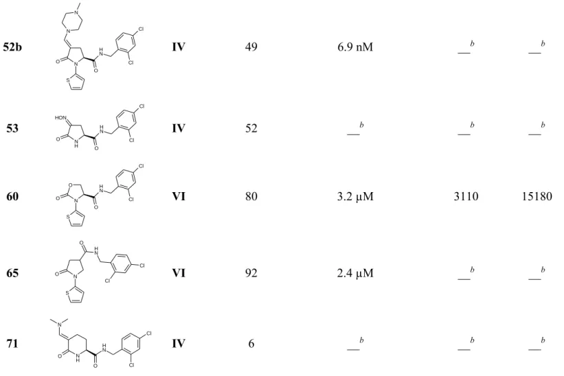
Table 1. Inhibition of P2RX7 Activity by References 1a and 1b, and Most Active Compounds.
Flow cytometry Competitive radioligand
binding study with 75
compd Structure Series
Inhibition of h-P2RX7 at 10-5M (%)a IC50 ± SDa Ki (nM)a IC50 (nM)a 1a (ref)c N O O N H Cl Cl I 100 474.0 ± 12.0 nM 176 862 1b (ref)d N O O N H Cl CF3 I 98 119.3 ± 7.0 nM 68 332
16a O N O N H Cl Cl I 50 10.0 µM __b __b 16i O N O N H Cl Cl S II 98 229.0 ± 4.5 nM 147 721 16j O N O N H Cl Cl S II 78 813.2 ± 56.7 nM 256 1252 16k O N O N H Cl Cl O II 94 244.0 ± 105.0 nM 118 575 16l O N O N H Cl Cl O II 85 199.5 ± 15.5 nM 86 424 16m O N O N H Cl Cl S N II 83 415.0 ± 85.0 nM 489 2386 16n O N O N H Cl Cl N S II 98 256.4 ± 1.0 nM 78 384 19 O N O N H Cl S CF3 II 100 100.0 ± 10.1 nM 44 218
29c O N O N H Cl Cl III 100 1.1 µM __b __b 30b O N O N H Cl Cl Cl III 85 3.5 µM __b __b 45 O N O N H Cl Cl S HON IV 76 3.7µM __b __b 47 N O O O NH O O Cl Cl IV 99 4.3 µM __b __b 50g O N N O NMe2 Me2N Cl Cl V 24 __b __b __b 51c N H O O N H Cl Cl N IV 53 __b __b __b 52a N H O O N H Cl Cl N O IV 56 9.6 nM __b __b
52b N O O N H Cl Cl S N N IV 49 6.9 nM __b __b 53 N H O O N H Cl Cl HON IV 52 __b __b __b 60 N O O O N H Cl Cl S VI 80 3.2 µM 3110 15180 65 O N S N H O Cl Cl VI 92 2.4 µM __b __b 71 O N H Cl Cl N H O N IV 6 __b __b __b
aMean value of n ≥ 2 in the presence of a positive control. b Not determined. cGSK1370319A. dGSK1482160.
Figure 3. Structures of References 1a and 1b and of the Eight Selected Hits
1a N O O N H Cl Cl 16i N O O N H Cl Cl S 16j N O O N H Cl Cl S 16l N O O N H Cl Cl O 16m N O O N H Cl Cl S N 60 N O O O N H Cl Cl S 1b N O O N H Cl CF3 19 N O O N H Cl S CF3 16k N O O N H Cl Cl O 16n N O O N H Cl Cl N S
Structure-based insights for P2RX7 ligand binding
Thanks to the recent crystallization of pd-P2RX7,23 the binding pocket of P2RX7-antagonists was
shown to be allosteric, one of the best fitting compounds was JNJ47965567 (Figure 4).30 Some
P2RX7-ligands complex models were generated using docking experiments with the most suitable PDB template co-crystallized with JNJ47965567.
Figure 4. Structure of JNJ47965567 O S N N H O N N
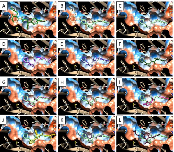
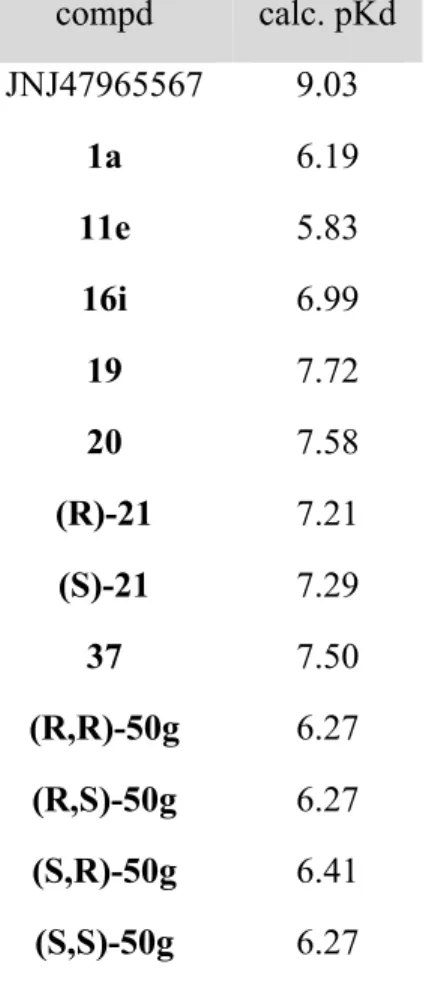
Some of the synthesized pyroglutamides and analogues were selected for a docking study in which pKd was estimated (Table 2) for the best predicted pose of each ligand (see Figure 5, see Figure S16 to Figure S27 of the Supporting Information for zooms).
Figure 5. Docking poses predicted for selected compounds with JNJ47965567 as reference (A:
JNJ47965567 experimental (grey) vs JNJ47965567 predicted (green); B: JNJ47965567 experimental (grey) vs 16i (light green); C: 16i (light green) vs 19 (white); D: 19 (white) vs 20 (pink); E: 19 (white) vs (R)-21 (violet); F: 19 (white) vs (S)-21 (green); G: 16i (light green) vs 16a (light pink); H: 16i (light green) vs 1a (orange); I: 16i (light green) vs 11e (pink); J: 16i (light green) vs 37 (yellow); K: 16i (light green) vs 50g 1st best fitting pose (grey) ; L: 16i (light green) vs 50g
The docking protocol confirmed that the predicted pose of JNJ47965567 fitted with the experimental pose (Figure 5.A). The poses of compounds 1a, 11e, 16i, 19, 20, 21 ((R)- and (S)- isomers), 37 and 50g ((R,R)-, (S,R)-, (R,S)- and (S,S)- isomers) were calculated. All best poses fitted into the allosteric binding site of JNJ47965567. Antagonist 16i (N-(2,4-dichlorobenzyl)-PGAm) pose was compared to JNJ47965567 experimental pose, with almost all positions fitting the pocket (Figure 5.B). N-(2-chloro-3-triofluorobenzyl)-PGAm 19 and N-(2-chloro-5-triofluorobenzyl)-PGAm 20 are in the same position as 16i (Figure 5.C and Figure 5.D). Introduction of a methyl on the amide substituent in compounds (R)-21 and (S)-21 (N-[1-(2,4-dichlorophenyl)ethyl]-PGAm) seemed to change the position of the aromatic. These findings were in accordance with the results observed in the flow cytometry assay: i) the introduction of a methyl between the amide and the aromatic is deleterious foe the activity; ii) the 4-chlorine may be changed to a trifluoromethyl group. On this last point, our in silico model doesn’t predict the loss of activity observed for compound 20 (N-(2-chloro-5-triofluorobenzyl)-PGAm).
Now substitution in position 1 of the lactam was studied (NH, methyl, (hetero-)aryl and N-benzyl lactams) and seemed to be of great importance for the position of this ring in the binding pocket: (hetero-)aryl-PGAm 16a, 16i, 19, and 20 were not in a similar pose to the ones of N-methyl-PGAm 1a, NH-free lactam 11e and N-benzyl-PGAm 37 (Figure 5.C to Figure 5.J). We hypothesize that the interaction of (hetero-)aryl groups with the receptor is responsible of the positioning of the lactam ring and induces the better activity of N-thienyl-PGAm 16i and 19 compared to N-methyl-PGAm 1a and 1b. For constrained analogs, the model showed a steric clash between the receptor and the pyrroloimidazoledione 50g (Figure 5.K and Figure 5.L), as expected regarding the low activity of this compound.
Table 2. Calculated pKd of selected compounds for docking study. compd calc. pKd JNJ47965567 9.03 1a 6.19 11e 5.83 16i 6.99 19 7.72 20 7.58 (R)-21 7.21 (S)-21 7.29 37 7.50 (R,R)-50g 6.27 (R,S)-50g 6.27 (S,R)-50g 6.41 (S,S)-50g 6.27
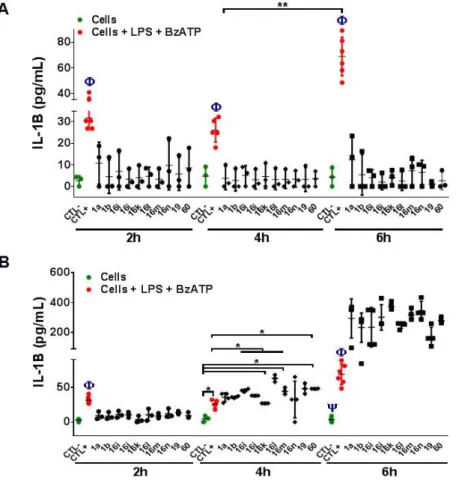
Impact of P2RX7 antagonists on IL1 production and ROS generation
IL1β is a major pro-inflammatory cytokine that is rapidly released via P2RX7 receptor activation. We analyzed the effect of ten P2RX7 antagonists on the modulation of IL1β production from human THP-1 macrophages. As expected, IL1β level gradually increased when macrophages were treated with LPS and BzATP, at 2, 4, and 6-hour post-incubation. The macrophages were pretreated with P2RX7 antagonist molecules, with GSK1370319A (compound 1a) and GSK1482160 (compound
1b) as references, at a concentration of 10-5 mol/L and 10-6 mol/L, and then stimulated with both
IL1β production remained at a basal level at 2h, 4h and 6h and was like unstimulated macrophages ones. Besides, the pretreatment of the macrophages with the ten antagonists at a concentration of 10-5 mol/L, and following addition of LPS and BzATP showed a significantly lower level of IL1β
when compared to that from unstimulated macrophages, whereas these antagonists at a concentration of 10-6 mol/L did not inhibit IL1 production after 6-hour incubation (Figure 6). In
this test, N-heteroaryl-PGAm were as good as references 1a and 1b (N-methyl-PGAm). Compound
19, bearing thiophene and 2-chloro-3-trifluoromethyl moiety was the best at 6h.
Figure 6. Effect of P2RX7 antagonists on IL1 production from macrophages. Analysis of IL1
level in the supernatant of macrophages pretreated with P2RX7 antagonist molecules at a decreasing concentration (A: 10-5 and B: 10-6 mol/L). CTL- corresponds to macrophages, CTL+ corresponds
The generation of reactive oxygen species (ROS) after activation of P2RX7 receptor by ATP is well known in macrophages. We observed that the percentage of ROS significantly increased in the macrophages incubated with LPS and BzATP whereas the pretreatment of macrophages with the ten molecules at a concentration of 10-5 mol/L significantly reduced the ROS production. We next
used a decreasing concentration of antagonists and we found that PGAm 16m, 16n and 16k at a concentration of 10-6 mol/L did not inhibit the ROS production when compared to other antagonist
molecules (Figure 7). All tested PGAm were good inhibitors of the ROS production, and compounds 16i and 19, both bearing a thienyl moiety, were comparable to references 1a and 1b. 2,4-Dichlorobenzylamide 16i being easier to afford in large quantity, the further assays described in this study were realized using it preferentially to 2-chloro-3-trifluoromethylbenzylamide 19 or other N-aryl-PGAm of formula 16.
concentration (A: 10-5 and B: 10-6 mol/L). CTL- corresponds to macrophages. CTL+ corresponds
to macrophages + LPS + BzATP. **P<0.01 (CTL+ vs compounds). *P<0.05 (Compounds vs CTL-, H2O2).
Selectivity and ADME-Tox evaluation.
Cytotoxicity was evaluated on wild-type HEK293 and P2RX7-transfected HEK293 cell lines for the 8 selected hits. No toxicity was found after 72 hours compared to control (data not shown).
Platelet aggregation assay was implemented to reveal an eventual interaction, notably with P2RX1, a receptor expressed on platelets membrane at a high concentration. No effect was observed (see Supporting Information), indicating the probable absence of interaction of these compounds with P2RX1 and influence on the platelets aggregation.
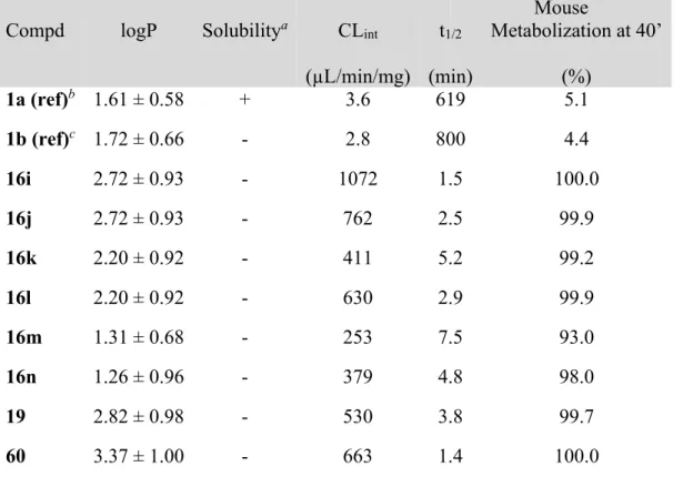
The pharmacokinetic (PK) profiles of the ten selected hits were investigated (Table 3). Lipophilicity was predicted to be low (calculated logP from 1.5 to 3.5)31, and none of the tested
compounds, but reference 1a, was soluble in physiological saline at a concentration of 1 mg/mL. Moreover, while references 1a and 1b were very stable, N-aryl pyroglutamides were rapidly metabolized by h-hepatocytes. We determined that the main metabolite of N-aryl pyroglutamide
16i using human hepatic microsomes was NH-free pyroglutamide 10e (see Experimental Section
for detailed protocol,32 see Supporting Information for results). We hypothesize that the reduced
activity of compound 16i compared to 1a – later described in the in vivo assays – comes from rapid metabolization of 16i into inactive 10e. Thus, modulations of N-aryl pyroglutamide P2RX7 antagonists to improve the PK profile and metabolic stability is to be investigated and will be reported in a further study.
Table 3. Pharmacokinetic Parameters for Selected Hits
Mouse
Compd logP Solubilitya CL
int (µL/min/mg) t1/2 (min) Metabolization at 40’ (%) 1a (ref)b 1.61 ± 0.58 + 3.6 619 5.1 1b (ref)c 1.72 ± 0.66 - 2.8 800 4.4 16i 2.72 ± 0.93 - 1072 1.5 100.0 16j 2.72 ± 0.93 - 762 2.5 99.9 16k 2.20 ± 0.92 - 411 5.2 99.2 16l 2.20 ± 0.92 - 630 2.9 99.9 16m 1.31 ± 0.68 - 253 7.5 93.0 16n 1.26 ± 0.96 - 379 4.8 98.0 19 2.82 ± 0.98 - 530 3.8 99.7 60 3.37 ± 1.00 - 663 1.4 100.0
aThe solubility was measured by a method as follows33: 1.0 mg of the appropriate compound
dissolved in 1.0 mL of physiological saline (containing 2.5% ethanol and 2.5% Tween-80); + , completely dissolved; - , not all dissolved. bGSK1370319A. cGSK1482160.
Furthermore, 65 synthesized drug-candidates were selected by the National Cancer Institute (NCI) for evaluation of their antiproliferative activity against 60 cancer cell lines, including several multidrug-resistant (MDR) tumor cell lines (HCT-15: human colon Duke’s type D, colorectal adenocarcinoma; UACC-62: human malignant melanoma; NCI/ADR-RES: human ovary adenocarcinoma; RXF 393: human kidney poorly differentiated hypernephroma; UO-31: human kidney carcinoma; MCF7: human breast adenocarcinoma; SF268: human CNS anaplastic astrocytoma and SF539: human CNS glioblastoma).
low toxicity on human cancer cells also. This result raises good hope for further development of pyroglutamide series as anti-inflammatory drugs.
The N-thienyl-PGAm 16i molecule ameliorates acute colitis
Having shown that the N-thienyl-PGAm 16i molecule (need to explain, why 16i in particular) inhibits IL1β production from human THP-1 macrophages, we tested this molecule in mice, using the dextran sulfate sodium (DSS)-induced colitis protocol (Elson Charles O et al, 1995
Gastroenterology 109:1344-1367). Indeed, among experimental models of IBD, this model is one
of the most popular for drug screening studies as reviewed by Valatas and colleagues (Valatas V.
et al, European journal of pharmacology, 2015).
This model induces reproducible mucosal colonic inflammation manifested by bloody diarrhea, weight loss, mucosal ulceration and neutrophilic infiltration within colon. These features led to measurable clinical symptoms after 5 days of DSS treatment (Cooper HS et al, Lab Invest 1993). Mice treated daily with compound 16i lost 50% less weight than mice treated with placebo (figure 8.A) and showed milder signs of colitis, characterized by less diarrhea and less rectal bleeding. Indeed, the DAI score of mice treated with molecule 16i, is decreased by 31% (figure 8.B). These clinical symptoms started at day 9 and were statistically significant between days 11 and 12, i.e. when re epithelization occurs. Of interest, compound 16i is as efficient as the reference compound
1a to dampen signs of colitis.
To evaluate the degree of inflammation, we performed histological analysis at day 14. Representative image of mice treated with placebo showed mucosal edema, transmural immune cell infiltration and the presence of large areas of erosion (Figure 8C, left panel). In mice treated with
compounds 16i and 1a, areas of normal colonic mucosa with crypts being straight, well defined and sitting on the muscularis mucosa are larger. Scoring of inflammation confirmed that both antagonists decreased by 42 to 50% the level of inflammation (Figure 8C, right panel).
The inhibition observed with 16i or the reference compound is within the range of what is classically observed with the DSS-colitis protocol (Greten F et al, Cell, 2004, Gong Z et al, 2018 Molecular
Immunology, Melgar S. et al, International immunopharmacology, 2008). Of interest, analysis of
clinical scores and inflammatory index, revealed that mice treated with the antagonist compounds recovered more rapidly from DSS-induced colitis than mice treated with placebo. Strikingly, a faster re-epithelization of the digestive mucosa was also observed in P2rx7-/- mice during the recovery phase (Hofman et al, Cancer Research, 2015). This observation suggests that 16i compound phenocopies the loss of P2RX7 activity.
I n f l a m m a t o r y i n d e x a r b i t a r y u n i t s 2 4 6 8 * * * C Placebo 16i 1a DSS DSS A B D a y s % R e la t iv e B o d y W e ig h t 0 5 1 0 1 5 - 3 0 - 2 0 - 1 0 0 1 0 1 6 i 1 a p la c e b o ** ** * * ** ** * ** *** ***** ** D a y s D is e a s e A c ti v it y S c o r e 2 4 6 8 1 0 1 2 1 4 0 2 4 6 * * *
Figure 8. Compound 16i ameliorates DSS-induced colitis. Analyses were performed from cohort
of 10 WT mice treated with placebo, compound 16i and 1a. A. Body weight loss. B. Disease activity score. C. Tissue sections were stained with hematoxylin and eosin and analyzed microscopically (scale bar, 1 mm). Histologic scoring of colon tissue sections was calculated as indicated in Materials and Methods section. Data are presented as means SEM; *p<0.05, **p<0.01
CONCLUSION
Novel series of PGAm were designed following a ligand-based approach. They were synthesized using previously described methods and compared to references 1a and 1b. Improved properties were obtained regarding P2RX7 antagonism and decrease of IL1β and ROS production. New SAR have been proposed: 1) An hetero-aryl moiety in position 1 of the lactam was the best modulation for the antagonistic properties on P2RX7, but showed decreased metabolic stability compared to a methyl; 2) the amide substitution was limited to 2,4-dichlorobenzyle and 2-chloro-3-trifluoromethylbenzyle; 3) rigidification, such as modulation on position 3, of PGAm are deleterious to activity; 4) the lactam moiety can be replaced by an oxazolidinone, but resulted in diminished activity, and other analogues of the lactam, such as pyridine or piperidone had no antagonistic potential; 5) the amide can be transferred from position 5 to position 4 of the lactam ring, but slightly decreased the activity.
Ten hits were selected for further studies. They were effectively reducing IL1β and ROS production and were allosteric modulators of P2RX7 (binding site of A-804598). The ADME-Tox profile and selectivity for P2RX7 were encouraging for further development as low toxic drugs. Lastly, in vivo study in a rodent model of bowel inflammation showed a real potential of
Finally, better understanding of P2RX7 antagonist interactions with P2RX7 have been provided, and PGAm have been shown as potential drug-candidates for inflammation-related diseases, such as IBD.
EXPERIMENTAL SECTION Chemistry.
Starting materials were commercially available and were used without further purification. Melting points were measured on a MPA 100 OptiMelt® apparatus and are uncorrected. Nuclear
magnetic resonance (NMR) spectra were acquired at 400 MHz for 1H NMR, 100 MHz for 13C NMR
and 376 MHz for 19F NMR on a Varian 400-MR spectrometer with tetramethylsilane (TMS) as
internal standard, at room temperature. Chemical shifts (δ) are expressed in ppm relative to TMS. Splitting patterns are designed: s, singlet; d, doublet; dd, doublet of doublet; t, triplet; q, quadruplet; quint, quintuplet; m, multiplet; br s, broaden singlet; br t, broaden triplet. Coupling constants (J) are reported in Hertz (Hz). Thin layer chromatography (TLC) was realized on Macherey Nagel silica gel plates with fluorescent indicator and were visualized under a UV-lamp at 254 nm and 365 nm. Column chromatography was performed with a CombiFlash Rf Companion (Teledyne-Isco System) using RediSep packed columns. IR spectra were recorded on a Varian 640-IR FT-IR Spectrometer. Elemental analyses (C, H, N, S) of new compounds were determined on a Thermo Electron apparatus by ‘Pôle Chimie Moléculaire-Welience’, Faculté de Sciences Mirande, Université de Bourgogne, Dijon, France.
(S)-N-[2-chloro-5-(trifluoromethyl)benzyl]-1-methyl-5-oxopyrrolidine-2-carboxamide (1c). A mixture of 7 (1.00 g, 6.4 mmol), 2-chloro-5-(trifluoromethyl)benzylamine (1.33 g, 6.4 mmol), and PTSA (0.05 g, 0.3 mmol) was stirred under a nitrogen atmosphere at 110°C for 24 hours. It was
white solid. mp 139-141°C (CH2Cl2); Rf (CH2Cl2:MeOH = 95:5) = 0.4; 1H NMR (CDCl3, 400
MHz) δ ppm 2.00-2.11 (m, 1H, CH2CH2CH), 2.21-2.32 (m, 1H, CH2CH2CH), 2.34-2.52 (m, 2H,
CH2CH2CH), 2.80 (s, 3H, NCH3), 4.04 (dd, J = 9.3, 3.9 Hz, 1H, CH2CH2CH), 4.59 (dd, J = 15.5,
6.1 Hz, 1H, NHCH2), 4.64 (dd, J = 15.5, 6.1 Hz, 1H, NHCH2), 7.28 (br t, J = 6.6 Hz, 1H, NHCH2),
7.50 (s, 1H, ArH), 7.51 (s, 1H, ArH), 7.61 (s, 1H, ArH); 13C NMR (CDCl
3, 100 MHz) δ ppm 23.4
(CH2), 29.1 (CH2), 29.4 (CH3), 41.1 (CH2), 64.0 (CH), 123.6 (q, J = 272.0 Hz, CF3), 125.7 (q, J =
3.7 Hz, CH), 126.5 (q, J = 3.7 Hz, CH), 129.5 (q, J = 33.0 Hz, C), 130.2 (CH), 136.5 (C), 137.2 (C), 171.6 (C), 175.9 (C); 19F NMR (CDCl
3, 376 MHz) δ (ppm) -62.7 (CF3); IR ν (cm-1): 3289, 1683,
1656, 1326, 1112, 1081, 845; Anal. Calcd for C14H14ClF3N2O2: C, 50.24; H, 4.22; N, 8.37. Found:
C, 50.02; H, 3.84; N 8.39%.
(S)-N-(3,4-Dimethoxyphenyl)-5-oxopyrrolidine-2-carboxamide (10g). Amide 10g was synthesized from 5 with ZrCl4 as catalyst, following the procedure described for 1c, and crystalized
in CH3CN. Yield 17.1%. Dark blue solid. mp 145-146°C (Et2O); Rf (CH2Cl2:MeOH = 95:5) = 0.1; 1H NMR (CDCl 3, 400 MHz) δ ppm 2.27-2.56 (m, 3H, CH2CH2CH), 2.60-2.72 (m, 1H, CH2CH2CH), 3.87 (s, 3H, OCH3), 3.89 (s, 3H, OCH3), 4.27-4.32 (m, 1H, CH2CH2CH), 6.08 (br s, 1H, NHCH), 6.82 (d, J = 8.6 Hz, 1H, ArH), 6.97 (dd, J = 2.3, 8.6 Hz, 1H, ArH), 7.33 (d, J = 2.3 Hz, 1H, ArH), 7.76 (br s, 1H, NHAr); 13C NMR (CDCl 3, 100 MHz) δ ppm 26.3 (CH2), 29.1 (CH2), 56.0 (CH3), 56.1 (CH3), 57.4 (CH), 104.8 (CH), 111.3 (CH), 111.9 (CH), 130.4 (C), 146.4 (C), 149.1 (C), 169.7 (C), 179.1 (C); IR ν (cm-1): 3358, 3244, 1669, 1513, 1237, 1131, 1020; Anal. Calcd for C13H16N2O4: C, 59.08; H, 6.10; N, 10.60. Found: C, 59.11; H, 6.13; N, 10.59%.
(S)-N-(3-Fluoro-4-methoxyphenyl)-5-oxopyrrolidine-2-carboxamide (10i). Amide 10i was synthesized from 5 following the procedure described for 1c, and crystalized in CH3CN. Yield
MHz) δ ppm 2.25-2.55 (m, 3H, CH2CH2CH), 2.60-2.71 (m, 1H, CH2CH2CH), 3.88 (s, 3H, OCH3),
4.27-4.32 (m, 1H, CH2CH2CH), 6.36 (br s, 1H, NHCH), 6.91 (t, J = 9.0 Hz, 1H, ArH), 7.19 (m, 1H,
ArH), 7.50 (dd, J = 12.6, 2.4 Hz, 1H, ArH), 7.96 (br s, 1H, NHAr); 13C NMR (DMSO-d6, 100 MHz)
δ ppm 25.2 (CH2), 29.1 (CH2), 56.0 (CH), 56.3 (CH3), 107.7 (d, J = 22.4 Hz, CH), 113.9 (d, J = 2.6
Hz, CH), 115.2 (d, J = 3.4 Hz, CH), 132.1 (d, J = 9.2 Hz, C), 143.0 (d, J = 11.1 Hz, C), 150.7 (d, J = 241.8 Hz, C), 171.0 (C), 177.3 (C); 19F NMR (CDCl
3, 376 MHz) δ (ppm) -132.6 (ArF); IR ν (cm -1): 3301, 3084, 2937, 1683, 1656, 1514, 1226, 1122, 1025; Anal. Calcd for C
12H13FN2O3: C, 57.14;
H, 5.19; N, 11.11. Found: C, 57.14; H, 5.18; N, 11.07%.
(S)-N-(2-Chloro-3-(trifluoromethyl)benzyl)-5-oxopyrrolidine-2-carboxamide (11g). Amide 11g was synthesized from 5 following the procedure described for 1c, and crystalized in CH3CN. Yield
62.4%. Yellow solid. mp 143-145°C (Et2O); Rf (CH2Cl2:MeOH = 95:5) = 0.2; 1H NMR (CDCl3,
400 MHz) δ ppm 2.12-2.22 (m, 1H, CH2CH2CH), 2.26-2.41 (m, 2H, CH2CH2CH), 2.49-2.60 (m, 1H, CH2CH2CH), 4.19 (dd, J = 4.8, 9.1 Hz, 1H, CH2CH2CH), 4.57 (dd, J = 6.0, 14.6 Hz, 1H, NHCH2), 4.63 (dd, J = 6.0, 14.6 Hz, 1H, NHCH2), 6.65 (br s, 1H, NHCH2), 6.81 (br t, J = 5.7 Hz, 1H, NHCH), 7.36 (t, J = 8.1 Hz, 1H, ArH), 7.57 (d, J = 7.7 Hz, 1H, ArH), 7.66 (d, J = 7.7 Hz, 1H, ArH); 13C NMR (CDCl 3, 100 MHz) δ ppm 26.0 (CH2), 29.1 (CH2), 41.6 (CH2), 56.9 (CH), 125.5 (q, J = 273.5 Hz, CF3), 126.9 (CH), 127.2 (q, J = 5.6 Hz, CH), 129.2 (q, J = 30.8 Hz, C), 131.7 (C), 133.7 (CH), 137.6 (C), 172.1 (C), 179.2 (C); 19F NMR (CDCl 3, 376 MHz) δ (ppm) -62.5 (CF3); IR
ν (cm-1): 3237, 2956, 1736, 1685, 1437, 1204, 1153, 1041; Anal. Calcd for C
13H12ClF3N2O2: C,
48.69; H, 3.77; N, 8.74. Found: C, 48.55; H, 3.74; N, 8.70%.
(S)-N-(2-Chloro-5-(trifluoromethyl)benzyl)-5-oxopyrrolidine-2-carboxamide (11h). Amide 11h was synthesized from 5 following the procedure described for 1c, and crystalized in CH3CN. Yield
MHz) δ ppm 2.14-2.24 (m, 1H, CH2CH2CH), 2.28-2.45 (m, 2H, CH2CH2CH), 2.53-2.64 (m, 1H, CH2CH2CH), 4.21 (dd, J = 5.0, 9.1 Hz, 1H, CH2CH2CH), 4.59 (d, J = 5.9 Hz, 2H, NHCH2), 6.08 (br s, 1H, NHCH), 6.61 (br s, 1H, NHCH2), 7.51-7.52 (m, 2H, ArH), 7.61 (s, 1H, ArH); 13C NMR (CDCl3, 100 MHz) δ ppm 26.0 (CH2), 29.1 (CH2), 41.2 (CH2), 57.0 (CH), 125.9 (q, J = 3.9 Hz, CH), 126.3 (q, J = 272.0 Hz, CF3), 126.7 (q, J = 3.8 Hz, CH), 129.6 (q, J = 33.1 Hz, C), 130.2 (CH), 136.2 (C), 137.3 (C), 172.2 (C), 179.4 (C); 19F NMR (CDCl 3, 376 MHz) δ (ppm) -62.6 (CF3); IR ν
(cm-1): 3279, 1690, 1665, 1551, 1326, 1264, 1170, 1112, 1083, 824; Anal. Calcd for
C13H12ClF3N2O2: C, 48.69; H, 3.77; N, 8.74. Found: C, 48.44; H, 3.61; N, 8.78%.
(S)-N-(4-Methoxybenzyl)-5-oxopyrrolidine-2-carboxamide (11i). Amide 11i was synthesized from 5 with PTSA as catalyst, following the procedure described for 1c, and purified on a silica column (silica gel, CH2Cl2:MeOH = 95:5). Yield 42.1%. White solid. mp 122-123°C (Et2O); Rf
(CH2Cl2:MeOH = 95:5) = 0.1; 1H NMR (CDCl3, 400 MHz) δ ppm 2.07-2.17 (m, 1H, CH2CH2CH), 2.19-2.30 (m, 2H, CH2CH2CH), 2.38-2.49 (m, 1H, CH2CH2CH), 3.77 (s, 3H, OCH3), 4.11 (dd, J = 4.5, 9.1 Hz, 1H, CH2CH2CH), 4.30 (dd, J = 14.5, 5.8 Hz, 1H, NHCH2), 4.36 (dd, J = 14.5, 5.8 Hz, 1H, NHCH2), 6.80-6.85 (m, 2H, ArH), 6.96 (br t, J = 6.1 Hz, 1H, NHCH2), 7.14-7.19 (m, 3H, NHCH and ArH); 13C NMR (CDCl 3, 100 MHz) δ ppm 25.8 (CH2), 29.3 (CH2), 43.0 (CH2), 55.3 (CH3), 57.1 (CH), 114.1 (2CH), 129.2 (2CH), 130.0 (C), 159.1 (C), 172.0 (C), 179.5 (C); IR ν (cm -1): 3401, 3241, 1651, 1513, 1245; Anal. Calcd for C
13H16N2O3: C, 62.89; H, 6.50; N, 11.28. Found:
C, 62.91; H, 6.53; N, 11.27%.
(S)-5-Oxo-N-(3,4,5-trimethoxybenzyl)pyrrolidine-2-carboxamide (11j). Amide 11j was synthesized from 5 with PTSA as catalyst, following the procedure described for 1c, and purified on a silica column (silica gel, CH2Cl2:MeOH = 95:5). Yield 58.6%. White solid. mp 181-183°C
CH2CH2CH), 2.25-2.41 (m, 2H, CH2CH2CH), 2.48-2.58 (m, 1H, CH2CH2CH), 3.82 (s, 3H, OCH3),
3.83 (s, 6H, OCH3), 4.18 (dd, J = 9.0, 4.9 Hz, 1H, CH2CH2CH), 4.30 (dd, J = 14.5, 5.6 Hz, 1H,
NHCH2), 4.41 (dd, J = 14.5, 5.6 Hz, 1H, NHCH2), 6.48 (s, 2H, ArH), 6.70 (br t, J = 5.7 Hz, 1H, NHCH2), 6.77 (br s, 1H, NHCH); 13C NMR (CDCl3, 100 MHz) δ ppm 26.0 (CH2), 29.2 (CH2), 43.9
(CH2), 56.2 (2CH3), 57.0 (CH), 60.9 (CH3), 105.1 (2CH), 133.4 (C), 137.5 (C), 153.4 (2C), 171.8
(C), 179.1 (C); IR ν (cm-1): 3350, 3290, 1695, 1650, 1131; Anal. Calcd for C
15H20N2O5: C, 58.43;
H, 6.54; N, 9.09. Found: C, 58.40; H, 6.56; N, 9.07%.
(S)-N-(2,4-dichlorophenyl)-5-oxo-1-phenylpyrrolidine-2-carboxamide (14). Amide 14 was synthesized from 8a with PTSA as catalyst and toluene as solvent, following the procedure described for 1c, and crystalized in CH3CN. Yield 7.9%. Dark-blue solid. mp 175-184°C (CH3CN);
Rf (CH2Cl2:MeOH = 95:5) = 0.8; 1H NMR (CDCl3, 400 MHz) δ ppm 2.28-2.38 (m, 1H,
CH2CH2CH), 2.59-2.72 (m, 2H, CH2CH2CH), 2.73-2.88 (m, 1H, CH2CH2CH), 4.83-4.89 (m, 1H,
CH2CH2CH), 7.18 (t, J = 1.3 Hz, 2H, ArH), 7.21 (dd, J = 2.0, 9.0 Hz, 1H, ArH), 7.39 (tt, J = 2.4,
7.4 Hz, 2H, ArH), 7.53-7.59 (m, 2H, ArH), 7.97 (br s, 1H, NH), 8.19 (d, J = 9.1 Hz, 1H, ArH); 13C
NMR (CDCl3 100 MHz) δ ppm 23.9 (CH2), 30.9 (CH2), 63.5 (CH), 121.4 (2CH), 122.6 (CH), 124.1
(C), 126.1 (CH), 127.9 (CH), 128.8 (CH), 129.4 (2CH), 130.1 (C), 132.2 (C), 137.4 (C), 169.4 (C), 174.5 (C); IR ν (cm-1): 3238, 1702, 1671, 1581, 1524, 1499; Anal. Calcd for C
17H14Cl2N2O2: C,
58.47; H, 4.04; N, 8.02. Found: C, 58.49; H, 4.06; N 8.22%.
(S)-N-(2,4-dichlorobenzyl)-5-oxo-1-(2,4-dichlorophenyl)pyrrolidine-2-carboxamide (16c). Amide 16c was synthesized from 8c with PTSA as catalyst and CH3CN as solvent, following the
procedure described for 1c, and crystalized in CH3CN. Yield 34.1%. Yellow solid. mp 123-155°C
NHCH2), 4.53 (dd, J = 4.7, 8.3 Hz, 1H, CH2CH2CH), 6.23 (t, J = 5.5 Hz, 1H, ArH), 7.03 (d, J = 8.2
Hz, 1H, ArH), 7.13 (dd, J = 8.2, 2.0 Hz, 2H, ArH), 7.18 (d, J = 2.0 Hz, 1H, ArH), 7.34 (m, 1H, ArH), 7.42 (m, 1H, ArH); 13C NMR (CDCl
3, 100 MHz) δ ppm 23.8 (CH2), 29.7 (CH2), 41.1 (CH2),
63.0 (CH), 127.3 (CH), 128.2 (CH), 129.4 (CH), 130.1 (CH), 131.1 (C), 131.1 (CH), 132.6 (C), 133.2 (C), 133.3 (CH), 134.2 (C), 134.5 (C), 134.7 (C), 170.2 (C), 175.1 (C); IR ν (cm-1): 3290,
3073, 2937, 1674, 1549, 1479, 1384, 1227, 1101, 1049, 813; Anal. Calcd for C18H14Cl4N2O2: C,
50.03; H, 3.27; N, 6.48. Found: C, 49.60; H, 3.20; N, 6.42%.
(S)-N-(2,4-dichlorobenzyl)-5-oxo-1-(4-chlorophenyl) pyrrolidine-2-carboxamide (16f). Amide
16f was synthesized from 8f with CH3SO3H as catalyst and CH3CN as solvent, following the
procedure described for 1c, and crystalized in CH3CN. Yield 38.7%. White solid. mp 165-168°C
(CH3CN); Rf (CH2Cl2:MeOH = 97:3) = 0.4; 1H NMR (CDCl3, 400 MHz) δ ppm 2.13-2.24 (m, 1H,
CH2CH2CH), 2.47-2.61 (m, 2H, CH2CH2CH), 2.64-2.75 (m, 1H, CH2CH2CH), 4.34 (dd, J = 15.2,
5.6 Hz, 1H, NHCH2), 4.46 (dd, J = 15.2, 5.6 Hz, 1H, NHCH2), 4.61 (dd, J = 8.8, 4.4 Hz, 1H, CH2CH2CH), 6.35 (t, J = 5.6 Hz, 1H, NHCH2), 6.97 (d, J = 8.0 Hz, 1H, ArH), 7.12 (dd, J = 8.0, 2.4
Hz, 1H, ArH), 7.23 (d, J = 9.2 Hz, 2H, ArH), 7.31 (d, J = 2.4 Hz, 1H, ArH), 7.35 (d, J = 9.2 Hz, 2H, ArH); 13C NMR (CDCl
3, 100 MHz) δ ppm 23.5 (CH2), 31.0 (CH2), 41.0 (CH2), 63.1 (CH),
122.3 (2CH), 127.3 (CH), 129.2 (2CH), 129.4 (CH), 130.9 (CH), 131.0 (C), 133.3 (C), 134.0 (C), 134.4 (C), 136.2 (C), 170.8 (C), 174.5 (C); LC-MS (APCI+) m/z: 397.0 (MH+), tr 3.77 min; IR ν
(cm-1): 3278, 2923, 1698, 1661, 1557, 1493, 1354, 1229, 1098, 1053, 1011, 822; Anal. Calcd for
C18H15Cl3N2O2: C, 54.36; H, 3.80; N, 6.68. Found: C, 54.60; H, 4.08; N, 6.36%.
(S)-N-(2,4-dichlorobenzyl)-5-oxo-1-(4-cyanophenyl)pyrrolidine-2-carboxamide (16g). Amide
16g was synthesized from 8g with CH3SO3H as catalyst and CH3CN as solvent, following the
(CH3CN); Rf (CH2Cl2:MeOH = 97:3) = 0.5; 1H NMR (CDCl3, 400 MHz) δ ppm 2.13-2.22 (m, 1H,
CH2CH2CH), 2.45-2.64 (m, 2H, CH2CH2CH), 2.67-2.82 (m, 1H, CH2CH2CH), 4.37 (dd, J = 6.2,
14.9 Hz, 1H, NHCH2), 4.45 (dd, J = 6.2, 14.9 Hz, 1H, NHCH2), 4.62-4.68 (m, 1H, CH2CH2CH),
6.32 (br t, J = 6.3 Hz, 1H, NHCH2), 7.15 (d, J = 8.4 Hz, 1H, ArH), 7.16 (dd, J = 8.4, 2.0 Hz, 1H,
ArH), 7.33 (d, J = 2.0 Hz, 1H, ArH), 7.56 (d, J = 9.0 Hz, 2H, ArH), 7.59 (d, J = 9.0 Hz, 2H, ArH);
13C NMR (CDCl
3, 100 MHz) δ ppm 23.5 (CH2), 31.1 (CH2), 41.3 (CH2), 62.5 (CH), 108.3 (CN),
118.3 (C), 120.2 (2CH), 127.4 (CH), 129.4 (CH), 131.4 (CH), 133.1 (C), 133.1 (2CH), 134.2 (C), 134.7 (C), 141.8 (C), 170.3 (C), 174.8 (C); LC-MS (APCI+) m/z: 388.1 (MH+), tr 3.50 min; IR ν
(cm-1): 3280, 2922, 2851, 1702, 1660, 1558, 1494, 1352, 1221, 821; Anal. Calcd for C
19H15Cl2N3O2:
C, 58.78; H, 3.89; N, 10.82. Found: C, 59.06; H, 4.08; N, 10.66%.
(S)-N-(2,4-dichlorobenzyl)-5-oxo-1-(4-methoxyphenyl)pyrrolidine-2-carboxamide (16h). Amide
16h was synthesized from 8h with PTSA as catalyst and CH3CN as solvent, following the procedure
described for 1c, and crystalized in CH3CN. Yield 19.9%. White solid. mp 153-158°C (CH3CN);
Rf (CH2Cl2:MeOH = 98:2) = 0.4; 1H NMR (CDCl3, 400 MHz) δ ppm 2.12-2.22 (m, 1H,
CH2CH2CH), 2.43-2.58 (m, 2H, CH2CH2CH), 2.62-2.74 (m, 1H, CH2CH2CH), 3.79 (s, 3H, OCH3),
4.31 (dd, J = 14.7, 5.7 Hz, 1H, NHCH2), 4.43 (dd, J = 14.7, 5.7 Hz, 1H, NHCH2), 4.56-4.61 (m, 1H, CH2CH2CH), 6.52 (br t, J = 5.7 Hz, 1H, ArH), 6.77 (d, J = 6.6 Hz, 2H, ArH), 6.85 (d, J = 8.2
Hz, 1H, ArH), 7.04 (dd, J = 8.2, 2.0 Hz, 1H, ArH), 7.27 (d, J = 2.0 Hz, 1H, ArH), 7.29 (d, J = 6.6 Hz, 2H, ArH); 13C NMR (CDCl
3, 100 MHz) δ ppm 23.5 (CH2), 30.9 (CH2), 40.8 (CH2), 55.4 (CH3),
63.7 (CH), 114.3 (2CH), 123.4 (2CH), 127.1 (CH), 129.2 (CH), 130.5 (CH), 130.5 (C), 133.5 (2C), 134.0 (C), 157.5 (C), 171.3 (C), 174.6 (C); IR ν (cm-1): 3270, 2924, 1696, 1647, 1514, 1249, 1228,
(S)-N-(2,4-dichlorobenzyl)-5-oxo-1-(thiophen-3-yl)pyrrolidine-2-carboxamide (16j). A stirred mixture of 11e (5.00 g, 17 mmol), 3-iodothiophene (3.66 g, 17 mmol), cuprous iodide (CuI) (1.66 g, 8 mmol), cesium carbonate (11.33 g, 34 mmol), and N,N’-dimethylethylene-1,2-diamine (1.53 g, 17 mmol) in dioxane was heated at reflux for 5hours under a nitrogen atmosphere. The resulting solution was filtered, then the solvent was evaporated and the resulting crude was solved in CH2Cl2.
It was washed two times with water and dried with MgSO4. The organic phase is evaporated,
resulting in a brown solid. The solid was purified on a silica column (silica gel, EtOAc:n-Heptane = 7:3) to give 16j (0.73 g, 11.6% yield) as a pink solid. mp 156-159°C (Et2O); Rf (CH2Cl2:MeOH
= 95:5) = 0.6; 1H NMR (CDCl 3, 400 MHz) δ ppm 2.18-2.27 (m, 1H, CH2CH2CH), 2.50-2.72 (m, 3H, CH2CH2CH), 4.34 (dd, J = 14.9, 5.5 Hz, 1H, NHCH2), 4.51 (dd, J = 14.9, 6.8 Hz, 1H, NHCH2), 4.60 (m, 1H, CH2CH2CH), 6.29 (br t, J = 6.7 Hz, 1H, NHCH2), 7.02 (d, J = 8.1 Hz, 1H, ArH), 7.13 (dd, J = 8.2, 2.0 Hz, 1H, ArH), 7.24 (dd, J = 3.9, 1.2 Hz, 1H, ArH), 7.26 (dd, J = 2.7, 2.0 Hz, 1H, ArH), 7.29-7.31 (m, 2H, ArH); 13C NMR (CDCl 3, 100 MHz) δ ppm 23.7 (CH2), 30.5 (CH2), 41.1 (CH2), 63.4 (CH), 110.9 (CH), 120.2 (C), 125.6 (CH), 127.3 (CH), 129.4 (CH), 130.7 (CH), 133.3 (CH), 134.1 (C), 134.3 (C), 136.1 (C), 171.2 (C), 173.5 (C); IR ν (cm-1): 3278, 1687, 1647, 1537,
1455; Anal. Calcd for C16H14Cl2N2O2S: C, 52.04; H, 3.82; N, 7.59; S, 8.68. Found: C, 51.31; H,
4.10; N, 7.70; S, 7.18%.
(S)-N-(2,4-dichlorobenzyl)-1-(furan-2-yl)-5-oxopyrrolidine-2-carboxamide (16k). Amide 16k was synthesized from 11e following the procedure described for 16j. Yield 58.6%. White solid. mp 189-191°C (Et2O); Rf (CH2Cl2:MeOH = 95:5) = 0.4; 1H NMR (CDCl3, 400 MHz) δ ppm 2.22-2.35
(m, 1H, CH2CH2CH), 2.45-2.58 (m, 2H, CH2CH2CH), 2.62-2.75 (m, 1H, CH2CH2CH), 4.43 (dd, J
= 15.4, 6.1 Hz, 1H, NHCH2), 4.51 (dd, J = 15.4, 6.1 Hz, 1H, NHCH2), 4.66 (dd, J = 9.3, 3.5 Hz, 1H, CH2CH2CH), 6.31 (br t, J = 6.0 Hz, 1H, NHCH2), 6.39 (dd, J = 3.6, 1.6 Hz, 1H, ArH), 6.41
(dd, J = 3.6, 1.2 Hz, 1H, ArH), 7.03 (dd, J = 2.0, 1.2 Hz, 1H, ArH), 7.13 (d, J = 8.2 Hz, 1H, ArH), 7.15 (dd, J = 8.2, 1.9 Hz, 1H, ArH), 7.36 (d, J = 1.9 Hz, 1H, ArH); 13C NMR (CDCl
3, 100 MHz) δ
ppm 23.8 (CH2), 30.0 (CH2), 41.1 (CH2), 61.3 (CH), 96.8 (CH), 111.8 (CH), 127.4 (CH), 129.4
(CH), 130.7 (CH), 133.5 (C), 134.1 (C), 134.2 (C), 136.5 (CH), 144.8 (C), 170.9 (C), 172.7 (C); IR ν (cm-1): 3285, 1698, 1646, 1542, 1412, 710; Anal. Calcd for C
16H14Cl2N2O3: C, 54.41; H, 4.00; N,
7.93. Found: C, 54.06; H, 3.95; N, 7.95%.
(S)-N-(2,4-dichlorobenzyl)-1-(furan-3-yl)-5-oxopyrrolidine-2-carboxamide (16l). Amide 16l was synthesized from 11e following the procedure described for 16j and purified on a silica column (silica gel, EtOAc:n-Heptane = 8:2). Yield 44.5%. White solid. mp 174-178°C (Et2O); Rf
(CH2Cl2:MeOH = 95:5) = 0.5; 1H NMR (CDCl3, 400 MHz) δ ppm 2.20-2.31 (m, 1H, CH2CH2CH),
2.49-2.69 (m, 3H, CH2CH2CH), 4.38-4.45 (m, 2H, NHCH2 and CH2CH2CH), 4.48 (dd, J = 14.8,
6.7 Hz, 1H, NHCH2), 6.25 (br t, J = 6.3 Hz, 1H, NHCH2), 6.43 (dd, J = 2.0, 0.8 Hz, 1H, ArH),
7.14-7.21 (m, 2H, ArH), 7.31 (t, J = 2.0 Hz, 1H, ArH), 7.33 (t, J = 1.2 Hz, 1H, ArH), 7.89 (q, J = 0.8 Hz, 1H, ArH); 13C NMR (CDCl
3, 100 MHz) δ ppm 24.2 (CH2), 29.9 (CH2), 41.2 (CH2), 62.4 (CH),
103.6 (CH), 125.0 (C), 127.3 (CH), 129.4 (CH), 131.1 (CH), 132.1 (CH), 133.3 (C), 134.3 (C), 134.4 (C), 142.3 (CH), 171.0 (C), 173.4 (C); IR ν (cm-1): 3278, 1689, 1645, 1542, 1229, 868, 770;
Anal. Calcd for C16H14Cl2N2O3: C, 54.41; H, 4.00; N, 7.93. Found: C, 54.04; H, 3.87; N, 7.94%.
(S)-N-(2,4-dichlorobenzyl)-5-oxo-1-(1,3-thiazol-2-yl)pyrrolidine-2-carboxamide (16m). Amide
16m was synthesized from 11e following the procedure described for 16j and purified on a silica
column (silica gel, EtOAc:n-Heptane = 6:4). Yield 15.3%. White solid. mp 200-209°C (Et2O); Rf
(CH2Cl2:MeOH = 95:5) = 0.8; 1H NMR (CDCl3, 400 MHz) δ ppm 2.27-2.40 (m, 1H, CH2CH2CH),