HAL Id: inserm-02440891
https://www.hal.inserm.fr/inserm-02440891
Submitted on 15 Jan 2020
HAL is a multi-disciplinary open access
archive for the deposit and dissemination of
sci-entific research documents, whether they are
pub-lished or not. The documents may come from
teaching and research institutions in France or
abroad, or from public or private research centers.
L’archive ouverte pluridisciplinaire HAL, est
destinée au dépôt et à la diffusion de documents
scientifiques de niveau recherche, publiés ou non,
émanant des établissements d’enseignement et de
recherche français ou étrangers, des laboratoires
publics ou privés.
New autoantibodies in early rheumatoid arthritis
Caroline Charpin, Fanny Arnoux, Marielle Martin, Eric Toussirot, Nathalie
Lambert, Nathalie Balandraud, Daniel Wendling, Elisabeth Diot, Jean
Roudier, Isabelle Auger
To cite this version:
Caroline Charpin, Fanny Arnoux, Marielle Martin, Eric Toussirot, Nathalie Lambert, et al.. New
autoantibodies in early rheumatoid arthritis. Arthritis Research and Therapy, BioMed Central, 2013,
15 (4), pp.R78. �10.1186/ar4255�. �inserm-02440891�
R E S E A R C H A R T I C L E
Open Access
New autoantibodies in early rheumatoid arthritis
Caroline Charpin
1,2, Fanny Arnoux
1, Marielle Martin
1, Eric Toussirot
3, Nathalie Lambert
1, Nathalie Balandraud
1,2,
Daniel Wendling
3, Elisabeth Diot
4, Jean Roudier
1,2and Isabelle Auger
1*Abstract
Introduction: Rheumatoid arthritis (RA) is a chronic inflammatory joint disease causing articular cartilage and bone destruction. Since irreversible joint destruction can be prevented by intervention at the early stages of disease, early diagnosis of RA is important. In this study, we identified new autoantibodies in the sera of patients with early (less than one year) RA.
Methods: We screened the sera of 20 RA patients with disease duration less than one year, 19 RA patients with disease duration more than five years and 23 controls on 8,268 human protein arrays. We confirmed the validity of protein array detection by ELISA assays. We then performed epitope mapping with overlapping 15-mers to analyze RA sera reactivity.
Results: WIBG (within BGCN homolog (Drosophila)), GABARAPL2 (GABA(A) receptor associated protein like 2) and ZNF706 (zinc finger protein 706) proteins are preferentially recognized by autoantibodies from early RA patients. Of interest, autoantibodies to WIBG are very specific for early RA. Indeed, 33% of early RA patients’ sera recognize WIBG versus 5% of RA patients with disease duration more than 5 years and 2% of controls. We identified three linear peptides on WIBG GABARAPL2 and ZNF706 that are preferentially recognized by sera of early RA patients.
Conclusions: We identified new autoantibodies associated with RA with disease duration less than one year. These autoantibodies could be used as diagnosis markers in RA patients.
Keywords: rheumatoid arthritis, autoantibodies, early biomarkers
Introduction
Rheumatoid arthritis (RA) is a chronic autoimmune dis-ease affecting 0.5% of the world population. It is character-ized by inflammation of joints that results in cartilage and bone destruction, joint deformity and loss of mobility. Although RA has been extensively studied, its cause is unknown. Treatment is directed towards reducing inflam-mation and stopping joint destruction. Since joint destruc-tion can be stopped by intervendestruc-tion at the early stages of the disease, early diagnosis of RA is important. However, diagnosis of RA can be difficult. Immunologic tests that can be performed for the diagnosis of RA include detec-tion of anti-citrullinated protein antibodies (ACPA) [1]. ACPA identify 65% of RA patients. Negative ACPA testing does not exclude RA.
To identify new autoantibodies in RA, we selected sera from 20 RA patients with disease duration less than one year, 19 RA patients with disease duration more than five years and 23 controls, to screen 8,268 human protein arrays. We identified 25 autoantigens recognized by the sera of early RA patients.
To confirm the validity of protein array detection, we used the 25 purified proteins in ELISAs. We tested the sera of 124 RA patients with disease duration less than 1 year and 40 RA patients with disease duration more than 5 years. We also tested 186 controls (81 patients with ankylosing spondylitis (AS), 30 patients with psor-iatic arthritis (PsA), 19 patients with systemic lupus erythematosus (SLE), 16 patients with systemic sclerosis (SSc) and 40 healthy subjects). We validated three pro-teins that are significantly recognized by autoantibodies from patients with early RA. These proteins are: within BGCN homolog (Drosophila) (WIBG), GABA(A) recep-tor-associated protein-like 2 (GABARAPL2) and zinc
* Correspondence: [email protected]
1
INSERM UMRs 1097, Aix Marseille Université, 163 Avenue de Luminy, 13288 Marseille, France
Full list of author information is available at the end of the article
© 2013 Charpin et al.; licensee BioMed Central Ltd. This is an open access article distributed under the terms of the Creative Commons Attribution License (http://creativecommons.org/licenses/by/2.0), which permits unrestricted use, distribution, and reproduction in any medium, provided the original work is properly cited.
finger protein (ZNF706). Of interest, autoantibodies to WIBG are very specific for early RA.
Epitope mapping on WIBG, GABARAPL2 and ZNF706 allowed us to identify peptide targets of autoan-tibodies, which may prove interesting in the diagnosis of early RA.
Materials and methods
Patients and controls
Informed consent was obtained from all patients and controls. The study protocol was approved by the Ethics Committee of Marseille, France (DC2008-327). The characteristics of patients and controls are shown in Table 1. ACPA were detected using the anti-CCP2 kit from Eurodiagnostica (Malmö, Sweden) for all RA patients and controls. Rheumatoid factor (RF) was detected by ELISA for all RA patients using the Orgen-tec Kit (Mainz, Germany).
Serum samples for protein arrays
We analyzed the sera of 39 RA patients from the rheu-matology unit at Sainte Marguerite Hospital in Marseille. Twenty RA patients had disease duration less than one year and 19 more than five years (time elapsed since first diagnosis by a physician). All RA patients fulfilled the 2010 American College of Rheumatology (ACR)/ European League Against Rheumatism (EULAR) revised criteria [2]. Controls were seven patients with AS, two with SLE and four with SSc, from the rheumatology unit at Sainte Marguerite Hospital in Marseille. Ten healthy controls were recruited among laboratory staff volunteers.
Protein arrays
We used Invitrogen (Carlsbad, California, USA) ProtoAr-rays that contain 8,268 human proteins. All proteins have been expressed as glutathione-S-transferase (GST) fusion proteins, purified under native conditions and spotted on nitrocellulose-coated glass slides [3]. Slides were blocked with 1% BSA/phosphate-buffered saline/Tween (PBST). Sera were added to the arrays. After washing, anti-human
immunoglobulin G (IgG) conjugated to Alexa Fluor 647 dye was added. Arrays were washed and dried (Partner-ship, Evry, France). Arrays were scanned with a GenePix 4000B Fluorescent Scanner, GenePix.Molecular Devices, Sunnyvale, California, USA. Data were acquired with GenePix Pro software and processed using ProtoArray Prospector 2.0 (Invitrogen). A panel of values was calcu-lated for each protein array, including theZ-score, the Chebyshev inequality precision (CIP) value and the coeffi-cient of variation (CV) value as previously described [3]. AZ-score >3.0, a CIP value <0.05 and a CV <0.5 define a positive spot.
Sera samples for ELISA analysis
RA patients were chosen from the Rheumatology Ward at Hospital Sainte Marguerite, Marseille and from the Rheumatology Ward at Hospital Jean Minjoz, Besançon. AS patients and PsA patients were chosen from the Rheu-matology Ward at Hospital Sainte Marguerite, Marseille. SLE patients were chosen from the Rheumatology Ward at Hospital Sainte Marguerite, Marseille and from the Internal medicine Ward at Hospital Bretonneau, Tours. Patients with SSc were from Hospitals Saint Antoine, Saint Louis, Paris; Hospital Claude Huriez, Lille and Hos-pital Sainte Marguerite, Marseille. Volunteers from the laboratory staff and the Marseille Blood Transfusion Cen-ter staff served as normal controls.
Detection of autoantibodies by ELISA
Plates were coated with purified proteins and blocked with PBS containing 5% milk. Sera diluted to 1:100 in PBS were incubated for 3 h. After washing with 0.1% Tween 20, peroxydase-conjugated anti-human IgG (Sigma, Saint-Quentin Fallavier, France) was added. Optical density (OD) was read at 405 nm. Background OD was obtained by adding each serum to a well with-out protein. Positive sera were defined by an OD value more than twice the background OD [3]. Moreover, positive sera were defined by an OD value more than twice the mean OD of the control groups (AS, PsA,
Table 1 Patients and controls for protein arrays and ELISA
Number Age at diagnosis Disease duration Anti-CCP-positive RF-positive
(years) (years) (%) (%) RA <1 year 144 54 0.8 70 68 RA >5 years 59 48 11 87 75 AS 88 34 10 1 nd PsA 30 41 6 3 nd SLE 21 37 8 5 nd SSC 20 51 0.5 5 nd Healthy 50 nd nd 0 nd
ELISA, enzyme-linked immunosorbent assay; CCP, cyclic citrullinated peptide; RF, rheumatoid factor; RA, rheumatoid arthritis; AS, ankylosing spondylitis; PsA, psoriatic arthritis; SLE, systemic lupus erythematosus; SSC, systemic sclerosis; nd, not determined.
Charpinet al. Arthritis Research & Therapy 2013, 15:R78 http://arthritis-research.com/content/15/4/R78
SLE, SSC and healthy subjects). Data were similar using both methods. P-values were calculated using the chi squared test.
Synthetic peptides
15-mer peptides encompassing residues from WIBG (locus NM_032345.1) GABARAPL2 (locus NM_007285.5) and ZNF706 (locus NM_016096.1) overlapping on seven amino acids were synthesized using the solid-phase sys-tem, and purified (Neosyssys-tem, Strasbourg, France).
Epitope mapping
Plates were coated overnight with 10 μg/well peptides diluted in PBS, pH 7.4. Plates were blocked with 5% milk PBS. Sera, diluted to 1:100 in PBS, were incubated for 2 h. After washing with 0.1% Tween 20, peroxydase-conjugated anti-human IgG, (Sigma, France) was added. OD was read at 405 nm. Background OD was obtained by adding each serum to a well without peptide. Positive sera were defined by an OD value more than twice the background OD. P-values were calculated using the chi squared test.
Results and discussion
Autoantibody pattern associated with RA patients and controls
We selected sera from 20 RA patients with disease dura-tion less than 1 year and compared their reactivity pat-tern on protein arrays with that of sera from 19 RA patients with disease duration more than 5 years, and with sera from 23 controls. The control group included seven AS patients, two SLE patients, four SSc patients and ten healthy subjects. Autoantibodies were detected by anti-human IgG antibody.
The sera of RA patients with disease duration more than 5 years bound, on average, 101 proteins. AS sera bound 112 proteins, SLE sera bound 91 proteins, SSc sera bound 103 proteins and healthy controls’ sera bound 89 proteins (data not shown). Sera from patients with early RA bound, on average, 58 proteins. Among these proteins, we identi-fied 25 that were recognized by 30% to 60% of RA patients with disease duration less than 1 year and less than 10% of controls (Table 2). These proteins were recognized by 0% to 37% of RA patients with disease duration more than 5 years.
Autoantibodies specific for RA patients
To confirm the validity of protein array detection, we developed ELISAs using the 25 purified proteins. We tested sera from 124 RA patients with disease duration less than 1 year, 40 RA patients with disease duration more than 5 years, 81 AS patients, 30 PsA patients, 19 SLE patients, 16 SSC patients and 40 healthy subjects. Four proteins were significantly recognized by early RA
patients and less than 10% of controls (Figure 1A,B). These proteins are WIBG, GABARAPL2, ZNF706 and PAD4 (peptidyl arginine deiminase 4).
Autoantibodies to GABARAPL2, ZNF706 and PAD4 were less sensitive and specific for early RA than autoanti-bodies to WIBG. Indeed, autoantiautoanti-bodies to PAD4 identi-fied 38% of RA patients with disease duration more than 5 years. Moreover, autoantibodies to GABARAPL2, ZNF706 and PAD4 were also found in SLE and SSC patients. Autoantibodies to WIBG were strongly asso-ciated with early RA. Indeed, 33% of RA patients’ sera recognized WIBG versus 5% of RA patients with disease duration more than 5 years (P = 0.0004), 0% of AS patients (P < 10-7), 0% of PsA patients (P = 0.0002), 10% of SLE
patients (P = 0.05), 6% of SSc patients (P = 0.03) and 0% of healthy individuals (P = 0.00003).
In combination, WIBG, GABARAPL2 and ZNF706 proteins identified 48% of patients with early RA versus 10% of RA patients with disease duration more than 5 years and 6% of controls (Table 3). Among patients with early AS, PsA, SLE and SSC with disease duration less than one year, only 2/34 were positive for GABAR-APL2 and WIBG (data not shown). Of interest, the combination of WIBG, GABARAPL2 and ZNF706 pro-teins identified 43% of early RA patients negative for anti-CCP, 44% of early RA patients negative for RF and 39% of early RA patients negative for anti-CCP and RF.
Epitope mapping on WIBG, GABARAPL2 and ZNF706
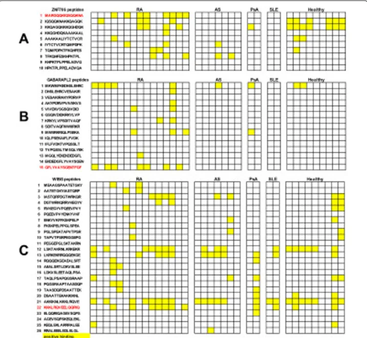
To identify B cell epitopes on ZNF706, GABARAPL2 and WIBG, we synthesized overlapping 15-mer peptides encompassing the entire sequence of these proteins. As a first step, we screened these peptides with the sera of 15 RA patients known to contain autoantibodies to ZNF706, GABARAPL2 and WIBG and 20 controls. Among the ten peptides from ZNF706, eight were recognized by at least one of the tested sera (Figure 2). One peptide, P1, was specifically recognized by sera from RA patients. Indeed, 8/15 RA sera recognized P1 versus 0/20 controls (Figure 2A).
Among the 15 peptides from GABARAPL2, 8 were recognized by at least one of the tested sera (Figure 2B). One peptide, P15, was specifically recognized by sera from RA patients. Indeed, 7/15 RA sera recognized P15 versus 0/20 controls.
Among the 26 peptides from WIBG, 18 were recog-nized by at least one of the tested sera (Figure 2C). One peptide, P22, was preferentially recognized by sera from RA patients. Indeed, 7/15 RA sera recognized P22 ver-sus 4/20 controls.
To confirm these reactivities, we tested by ELISA sera from 60 patients with early RA and 106 controls (20 AS, 24 PsA and 22 SLE patients, and 40 healthy subjects) on ZNF706 P1, GABARAPL2 P15 and WIBG P22 (Table 4).
Autoantibodies to ZNF706 P1, GABARAPL2 P15 and WIBG P22 were respectively found in 35%, 37% and 30% of patients with early RA, with a specificity ranging from 95% to 100%. In combination, these three peptides identi-fied 45% of patients with early RA versus 8% of controls (P <10-7
for 60 patients with early RA versus 106 con-trols). Of interest, the combination of these peptides identified 41% of early RA patients negative for anti-CCP, 35% of early RA patients negative for RF and 42% of early RA patients negative for anti CCP and RF.
Discussion
To get an insight into the early immunological events leading to the development of RA and to develop diagnos-tic tools for early RA, we screened 8,268 protein arrays with sera from patients with early RA (less than one year) RA with disease duration more than 5 years, and controls. We selected 25 proteins that could constitute specific ser-ologic markers of early RA because they identified 30% to
60% of patients with early RA and less than 10% of controls.
To confirm the validity of protein array detection, we used the same proteins in ELISAs to screen more sera from patients and controls. Among these proteins, only one, PAD4, the enzyme that converts arginine into citrul-line, was already known to be a target for RA autoantibo-dies [4-8]. Autoantiboautoantibo-dies to PAD4 are more frequent in established than in early RA. In a previous study, we have identified peptide targets of anti-PAD4 autoantibodies [8]. Most anti-PAD4 positive sera recognized peptides located both in the N-terminal domain (amino acids 211 to 290) and the C-terminal domain (amino acids 601 to 650) of PAD4. Peptide recognition in the substrate-bind-ing domain of PAD4 influences the enzymatic activity of PAD4. Indeed, most autoantibodies to PAD4 inhibit PAD4-mediated citrullination.
By using protein arrays and ELISA assays, we validated three new autoantibodies significantly associated with
Table 2 Protein array analysis
Percentage of positive sera Protein Protein name Protein
abbreviation Reference RA <1 year (20) RA >5 years (19) Controls (23)
P1 IMMUNOGLOBULIN (CD79A) BINDING PROTEIN 1 IGBP1 NM_001551.1 60 0 0 P2 PEPTIDYL ARGININE DEIMINASE, TYPE IV PAD4 NM_012387.1 45 37 0 P3 FK506 binding protein FKBP3 NM_002013.2 45 5 10 P4 TROPOMYOSIN 2 (BETA) TPM2 NM_003289.3 45 0 0 P5 ZINC FINGER PROTEIN 706 ZNF706 NM_016096.1 45 2 10 P6 COILED-COIL DOMAIN CONTAINING 72 CCDC72 NM_015933.1 40 0 0 P7 Hypothetical protein MGC17403 (MGC17403), Transcription elongation
factor A (SII) N-terminal and central domain containing (TCEANC)
MGC17403 NM_152634.1 40 0 0 P8 ELG PROTEIN C17orf85 NM_018553.1 40 0 0 P9 HYPOTHETICAL PROTEIN MGC11257 C7orf50 NM_032350.3 40 0 0 P10 T complex mouse like TCP10L NM_144659.1 40 5 5 P11 ELONGATION FACTOR 1 HOMOLOG (S. CEREVISIAE) ELOF1 NM_032377.2 40 5 0
P12 FGF12 FGF12 NM_004113.3 40 0 0
P13 SYNAPTOTAGMIN I SYT1 NM_005639.1 40 0 0 P14 WITHIN BGCN HOMOLOG (DROSOPHILA) WIBG NM_032345.1 40 10 0 P15 YY1 TRANSCRIPTION FACTOR YY1 NM_003403.3 40 0 0 P16 Eucaryotic transl factor 1A EIF1AX NM_001412.2 40 5 5 P17 DOUBLECORTEX LISSENCEPHALY, X-LINKED (DOUBLECORTIN) DCX NM_178151.1 35 0 0
P18 LAMIN A/C LMNA BC033088.1 35 0 0
P19 TROPOMYOSIN 4 TPM4 BC002827.1 30 0 0 P20 ACIDIC (LEUCINE-RICH) NUCLEAR PHOSPHOPROTEIN 32 FAMILY, MEMBER
E
ANP32E NM_030920.1 30 0 0 P21 HYPOTHETICAL PROTEIN MGC20255 CCDC97 NM_052848.1 30 5 0 P22 SPANX-N3 PROTEIN SPANXN3 NM_001009609.1 30 0 0 P23 THYMIC STROMAL LYMPHOPOIETIN TSLP NM_138551.1 30 0 0 P24 GABA(A) RECEPTOR-ASSOCIATED PROTEIN-LIKE 2 GABARAPL2 NM_007285.5 30 0 0
P25 SCY1like SCYL1 BC009967.1 30 0 5
RA, rheumatoid arthritis.
Charpinet al. Arthritis Research & Therapy 2013, 15:R78 http://arthritis-research.com/content/15/4/R78
early RA. These autoantibodies recognize ZNF706, GABARAPL2, and WIBG. ZNF706 belongs to the C2H2-type zinc-finger protein family (Additional file 1: Table S1) [9]. GABARAPL2 is involved in autophagy, the process by which proteins and organelles are sequestered in autophagosomal vesicles and delivered to the lysosome for degradation [10]. WIBG is a ribosome-associated pro-tein involved in the disassembly of exon junction com-plexes (EJCs). EJCs, assembled during mRNA splicing, transport mRNA during nuclear export into the cyto-plasm and are removed during translation [11]. WIBG enhances translation of mRNA. WIBG has also been shown to enhance the translation of viral genes, acting as a so-called chaperone.
WIBG is the protein recognized by the highest number of sera from patients with early RA. The percentage of anti-WIBG-positive RA patients decreases with disease duration. Indeed, WIBG is recognized by 33% of RA patients with disease duration less than 1 year, 12% of RA patients with disease duration between 1 and 5 years and only 5% of RA patients with disease duration more than 5 years (data not shown).
The level of many RA-associated autoantibodies is already known to decrease with time. For example, anti-nuclear antibodies (ANA) are positive in 20% to 30% of patients with early RA. However, a few months after disease onset, the ANA test may turn and remain negative [12]. This is also true for anti-Sa and RF [13].
Figure 1 Autoantibodies to within BGCN homolog (WIBG), GABA(A) receptor-associated protein-like 2 (GABARAPL2), zinc finger protein 706 (ZNF706) and peptidyl arginine deiminase 4 (PAD4) proteins are significantly detected in patients with early rheumatoid arthritis (RA). Sera from RA patients and controls were tested on the 25 purified proteins by ELISA. (A) Optical density (OD) read at 405 nm. The cutoff, marked as a horizontal line, defines positive sera with an OD value higher than twice the mean OD of the control groups (patients with ankylosing spondylitis (AS), psoriatic arthritis (PsA), systemic lupus erythematosus (SLE), systemic sclerosis (SSC) and healthy subjects). (B) Percentage of positive sera. P-values were obtained by comparing the group with early RA to the control groups (AS, PsA, SLE, SSC and healthy subjects).
Table 3 Autoantibodies to WIBG, GABARAPL2, ZNF706 and PAD4 proteins in patients with early RA
WIBG, ZNF706 and GABARAPL2
Anti-CCP RF PAD4 WIBG ZNF706 GABARAPL2 combination
RA <1 year (n = 124) vs 186 controls (AS, PsA, SLE, SSc, and healthy) Sensitivity (%) 70 68 18.5 33 18.5 23 48
Specificity (%) 98 nd 91 98 98 96 94
PPV (%) 96 nd 57 93 85 80 84
NPV (%) 81 nd 63 69 64 65 73
RA <1 year negative for anti-CCP (n = 37) vs 186 controls (AS, PsA, SLE, SSc, and healthy) Sensitivity (%) 0 24 11 16 11 27 43
Specificity (%) nd nd 91 98 98 96 94
PPV (%) nd nd 19 67 50 59 59
NPV (%) nd nd 84 86 85 87 89
RA <1 year negative for RF (n = 39) vs 186 controls (AS, PsA, SLE, SSc, and healthy) Sensitivity (%) 23 0 10 15 15 28 44
Specificity (%) 98 nd 91 98 98 96 94
PPV (%) 69 nd 19 67 60 61 61
NPV (%) 84 nd 83 85 85 86 89
RA <1 year negative for anti-CCP and RF (n = 28) vs 186 controls (AS, PsA, SLE, SSc, and healthy) Sensitivity (%) 0 0 4 7 11 32 39
Specificity (%) nd nd 91 98 98 96 94
PPV (%) nd nd 5 40 43 56 50
NPV (%) nd nd 86 87 88 90 91
WIBG, within BGCN homolog (Drosophila); GABARAPL2, GABA(A) receptor-associated protein-like 2; ZNF706, zinc finger protein 706; PAD4, peptidyl arginine deiminase 4; RA: rheumatoid arthritis; CCP, cyclic citrullinated peptide; RF, rheumatoid factor; AS, ankylosing spondylitis; PsA, psoriatic arthritis; SLE, systemic lupus eryth ematosus; SSc, systemic sclerosis; PPV, positive predictive value; NPV, negative predictive value; nd, not determined. Charpin et al .Arthritis Research & Therapy 2013, 15 :R78 http://arthr itis-research.com /content /15/4/R78 Page 6 of 9
Disease-modifying therapies could explain the autoanti-body decrease in RA patients. Alternatively, the early recog-nition of autoantigens in RA could give an insight into the early mechanisms that lead to disease development. In this respect, it is remarkable that proteins associated with DNA or RNA are often initial targets of immune responses. Later on, disease development is accompanied by narrowing of the immune response to specific targets. The best example of this phenomenon is the evolution of Sharp’s syndrome into RA, scleroderma or lupus, which is accompanied by
loss of anti-ribonucleoprotein (RNP) antibodies and devel-opment of disease-specific autoantibodies [14]. Of interest, the combination of autoantibodies to WIBG, GABARAPL2 and ZNF706 may be used to help diagnosis of early RA, especially in patients without anti-cyclic citrillunated pro-tein (CCP) antibodies and RF.
Finally, we analyzed the epitopes recognized by autoanti-bodies to ZNF706, GABARAPL2, and WIBG in a direct ELISA, using a set of synthetic peptides and sera from patients with early RA. Fine epitope mapping on ZNF706,
Figure 2 Epitope mapping on zinc finger protein (706ZNF706), GABA(A) receptor-associated protein-like 2 (GABARAPL2) and within BGCN homolog (WIBG). Sera from patients with early RA, and from patients with ankylosing spondylitis (AS), psoriatic arthritis (PsA), systemic lupus erythematosus (SLE), and healthy controls were tested for binding to ZNF706, GABARAPL2 and WIBG peptides by ELISA. After washing, peroxydase-conjugated anti-human immunoglobulin G was added. Optical density (OD) was read at 405 nm. Background OD was obtained by adding each serum to a well without peptide. Positive sera were defined by an OD value higher than twice the background OD (yellow).
Table 4 Autoantibodies to WIBG, GABARAPL2, ZNF706 peptides in patients with early RA WIBG P22, ZNF706 P1 and GABARAPL2 P15 combination Anti-CCP RF WIBG P22 ZNF706 P1 GABARAPL2 P15
RA <1 year (n = 60) vs 106 controls (AS, PsA, SLE, and healthy) Sensitivity (%) 72 70 30 35 37 45
Specificity (%) 100 nd 95 100 95 92
PPV (%) 100 nd 78 100 81 75
NPV (%) 86 nd 71 73 73 75
RA <1 year negative for anti-CCP (n = 17) vs 106 controls (AS, PsA, SLE, and healthy) Sensitivity (%) 0 29 12 6 23 41
Specificity (%) nd nd 95 100 95 92
PPV (%) nd nd 18 10 31 44
NPV (%) nd nd 87 86 88 91
RA <1 year negative for RF (n = 17) vs 106 controls (AS, PsA, SLE, and healthy) Sensitivity (%) 29 0 12 12 18 35
Specificity (%) 100 nd 95 100 95 92
PPV (%) 100 nd 29 100 37 40
NPV (%) 90 nd 87 88 88 90
RA <1 year negative for anti-CCP and RF (n = 12) vs 106 controls (AS, PsA, SLE, and healthy) Sensitivity (%) 0 0 8 8 25 42
Specificity (%) nd nd 95 100 95 92
PPV (%) nd nd 17 100 37 36
NPV (%) nd nd 90 91 92 93
WIBG, within BGCN homolog (Drosophila); GABARAPL2, GABA(A) receptor-associated protein-like 2; ZNF706, zinc finger protein 706; RA, rheumatoid arthritis; CCP, cyclic citrullinated peptide; RF, rheumatoid factor; AS, ankylosing spondylitis; PsA, psoriatic arthritis; SLE, systemic lupus eryth ematosus; SSc, systemic sclerosis; PPV, positive predictive value; NPV, negative predictive value; nd, not determined.
Charpin et al .Arthritis Research & Therapy 2013, 15 :R78 http://arthr itis-research.com /content /15/4/R78 Page 8 of 9
GABARAPL2, and WIBG enabled us to identify three lin-ear peptides that may prove interesting diagnostic tools in the early stages of RA. The next step will be to include more patients and controls to identify the best marker combination for diagnosis of early RA.
Conclusions
There is a great need for new biological markers of RA to permit early intervention to potentially prevent inflamma-tion and joint destrucinflamma-tion. To identify new autoantibody signatures in RA patients, we have analyzed autoantibodies in the sera of patients with early RA (duration less than one year) on protein arrays containing 8,000 human pro-teins. We found and validated three new autoantigens sig-nificantly associated with early RA. These autoantigens (ZNF706, GABARAPL2 and WIBG) and peptides derived from these autoantigens can be used to identify RA at the early stage of the disease.
Additional material
Additional file 1: Table S1 Characteristics of the early RA autoantigens. Description of early autoantigens (WIBG, ZNF706 and GABARAPL2 proteins).
List of abbreviations
ACPA: anti-citrullinated protein antibodies; ACR: American College of Rheumatology; ANA: anti-nuclear antibodies; AS: ankylosing spondylitis; BSA: bovine serum albumin; CIP: Chebyshev inequality precision; CV: coefficient of variation; EJC: exon junction complexes; ELISA: enzyme-linked
immunosorbent assay; EULAR: European League Against Rheumatism; GABARAPL2: GABA(A) receptor-associated protein-like 2; GST: glutathione-S-transferase; IgG: immunoglobulin G; OD: optical density; PAD4: peptidyl arginine deiminase 4; PBS: buffered saline; PBST: phosphate-buffered saline/Tween; PsA: psoriatic arthritis; RA: rheumatoid arthritis; RF: rheumatoid factor; RNP: ribonucleoprotein; SLE: systemic lupus erythematosus; SSc: systemic sclerosis; WIBG: within BGCN homolog (Drosophila); ZNF706: zinc finger protein 706.
Conflicts of interest
A patent was submitted by INSERM TRANSFERT in May 2011, submission number: 11305584; Receiving Office, European Patent Office, The Hague. Authors’ contributions
IA and JR contributed to the conception and design of the study and drafted the manuscript. CC, ET, NL, NB, DW, ED and JR collected clinical samples and data. CC, FA, MM and IA performed the experiments. All authors were involved in the acquisition of data and the revision of the manuscript. All authors read and approved the final manuscript. Acknowledgements
This study was supported by grants from INSERM, Arthritis Fondation Courtin, GFRS.
Authors’ details
1INSERM UMRs 1097, Aix Marseille Université, 163 Avenue de Luminy, 13288
Marseille, France.2Rheumatology, IML, APHM, 270 Boulevard de Sainte
Marguerite, 13009 Marseille, France.3Service de Rhumatologie, CHU Jean Minjoz, 2 Boulevard Fleming, 25030 Besançon, France.4Service de Médecine
Interne, CHU Bretonneau, 2 Boulevard Tonnellé, 37000 Tours, France.
Received: 18 December 2012 Revised: 19 March 2013 Accepted: 25 July 2013 Published: 25 July 2013 References
1. Schellekens GA, Visser H, de Jong BA, van den Hoogen FH, Hazes JM, Breedveld FC, van Venrooij WJ: The diagnostic properties of rheumatoid arthritis antibodies recognizing a cyclic citrullinated peptide. Arthritis Rheum 2000, 43:155-163.
2. Aletaha D, Neogi T, Silman AJ, Funovits J, Felson DT, Bingham CO, Birnbaum NS, Burmester GR, Bykerk VP, Cohen MD, Combe B,
Costenbader KH, Dougados M, Emery P, Ferraccioli G, Hazes JM, Hobbs K, Huizinga TW, Kavanaugh A, Kay J, Kvien TK, Laing T, Mease P, Ménard HA, Moreland LW, Naden RL, Pincus T, Smolen JS, Stanislawska-Biernat E, Symmons D, et al: 2010 Rheumatoid arthritis classification criteria: an American College of Rheumatology/European League Against Rheumatism collaborative initiative. Arthritis Rheum 2010, 62:2569-2581. 3. Auger I, Balandraud N, Rak JM, Lambert NC, Martin M, Roudier J: New
autoantigens in rheumatoid arthritis: screening 8268 protein arrays with RA patients’ sera. Ann Rheum Dis 2009, 68:591-594.
4. Takizawa Y, Sawada T, Suzuki A, Yamada R, Inoue T, Yamamoto K: Peptidylarginine deiminase 4 (PADI4) identified as a conformation-dependent autoantigen in rheumatoid arthritis. Scand J Rheumatol 2005, 3:212-215.
5. Roth EB, Stenberg P, Book C, Sjöberg K: Antibodies against transglutaminases, peptidylarginine deiminase and citrulline in rheumatoid arthritis: new pathways to epitope spreading. Clin Exp Rheumatol 2006, 1:12-18.
6. Halvorsen EH, Pollmann S, Gilboe IM, van der Heijde D, Landewé R, Ødegård S, Kvien TK, Molberg Ø: Serum IgG antibodies to
peptidylarginine deiminase 4 in rheumatoid arthritis and associations with disease severity. Ann Rheum Dis 2008, 67:414-417.
7. Zhao J, Zhao Y, He J, Jia R, Li Z: Prevalence and significance of anti-peptidylarginine deiminase 4 antibodies in rheumatoid arthritis. J Rheumatol 2008, 35:969-974.
8. Auger I, Martin M, Balandraud N, Roudier J: RA specific autoantibodies to PAD4 inhibit citrullination of fibrinogen. Arthritis Rheum 2010, 62:126-131. 9. Luchi S: Three classes of C2H2 zinc finger proteins. Cell Mol Life Sci 2001,
58:625-635.
10. Behrends C, Sowa ME, Gygi SP, Harper JW: Network organization of the human autophagy system. Nature 2010, 46:68-76.
11. Gehring NH, Lamprinaki S, Kulozik AE, Hentze MW: Disassembly of exon junction complexes by PYM. Cell 2009, 137:536-548.
12. Goldbach-Mansky R, Lee J, McCoy A, Hoxworth J, Yarboro C, Smolen JS, Steiner G, Rosen A, Zhang C, Ménard HA, Zhou ZJ, Palosuo T, Van Venrooij WJ, Wilder RL, Klippel JH, Schumacher HR Jr, El-Gabalawy HS: Rheumatoid arthritis associated autoantibodies in patients with synovitis of recent onset. Arthritis Res 2000, 3:236-243.
13. Guzian MC, Carrier N, Cossette P, de Brum-Fernandes AJ, Liang P, Ménard HA, Boire G: Outcomes in recent-onset inflammatory polyarthritis differ according to initial titers, persistence over time, and specificity of the autoantibodies. Arthritis Care Res 2010, 62:1624-1632.
14. Gendi N, Welsh K, Van Venrooij W, Vancheeswaran R, Gilroy J, Black C: HLA type as a predictor of mixed connective tissue differentiation. Arthritis Rheum 1995, 38:259-266.
doi:10.1186/ar4255
Cite this article as: Charpin et al.: New autoantibodies in early rheumatoid arthritis. Arthritis Research & Therapy 2013 15:R78.

![Table S1) [9]. GABARAPL2 is involved in autophagy, the process by which proteins and organelles are sequestered in autophagosomal vesicles and delivered to the lysosome for degradation [10]](https://thumb-eu.123doks.com/thumbv2/123doknet/14656037.552844/6.892.89.811.134.716/gabarapl-involved-autophagy-organelles-sequestered-autophagosomal-delivered-degradation.webp)