HAL Id: hal-02325860
https://hal.archives-ouvertes.fr/hal-02325860
Submitted on 22 Oct 2019
HAL is a multi-disciplinary open access
archive for the deposit and dissemination of
sci-entific research documents, whether they are
pub-lished or not. The documents may come from
teaching and research institutions in France or
abroad, or from public or private research centers.
L’archive ouverte pluridisciplinaire HAL, est
destinée au dépôt et à la diffusion de documents
scientifiques de niveau recherche, publiés ou non,
émanant des établissements d’enseignement et de
recherche français ou étrangers, des laboratoires
publics ou privés.
Histone variants: essential actors in male genome
programming
Naghmeh Hoghoughi, Sophie Barral, Alexandra Vargas, Sophie Rousseaux,
Saadi Khochbin
To cite this version:
Naghmeh Hoghoughi, Sophie Barral, Alexandra Vargas, Sophie Rousseaux, Saadi Khochbin. Histone
variants: essential actors in male genome programming. Journal of Biochemistry, Oxford University
Press (OUP), 2018, 163 (2), pp.97-103. �10.1093/jb/mvx079�. �hal-02325860�
JB Special Review—Chromatin Structure and Function:
Biological Implications in Epigenetics
Histone variants: essential actors in male genome programming
Received September 6, 2017; accepted October 13, 2017; published online November 20, 2017
Naghmeh Hoghoughi, Sophie Barral,
Alexandra Vargas, Sophie Rousseaux and
Saadi Khochbin*
CNRS UMR 5309, Inserm, U1209, Universite´ Grenoble Alpes, Institut Albert Bonniot, Grenoble F-38700, France
*Saadi Khochbin, Chromatin and Cell Signaling, Institute for Advanced Bioscience, Domaine de la Merci, La Tronche 38706, France. Tel: +33 4 76 54 95 83, Fax: 33 4 76 54 95 95, email: [email protected]
Prior to its transmission to the offspring, the male genome has to be tightly compacted. A genome-scale histone eviction and the subsequent repackaging of DNA by protamines (Prms) direct this essential genome condensation step. The requirement for male germ cells to undergo such a dramatic and unique genome reorganization explains why these cells express the largest number of histone variants, including many testis-specific ones. Indeed, an open chromatin, nucleo-some instability and a facilitated process of histone dis-assembly are direct consequences of the presence of these histone variants in the chromatin of male germ cells. These histone-induced changes in chromatin first control a stage-specific gene expression program and then dir-ectly mediate the histone-to-Prm transition process. This review aims at summarizing and discussing a series of recent functional studies of male germ cell histone vari-ants with a focus on their impact on the process of his-tone eviction and male genome compaction.
Keywords: H2A.L.2; H3.3; protamines; TH2B; tran-sition proteins.
Spermatogenesis is a differentiation process which pro-duces mature spermatozoa, which will deliver the male genome to the oocytes in the female organism. Spermatogenic cells are continuously produced by an asymmetrical division of progenitor cells, named sper-matogonia. These cells on the one hand maintain the stem cell population and on the other hand generate meiotic cells, or spermatocytes, which themselves undergo two successive meiotic divisions. The second meiotic division produces haploid cells known as sperm-atids. The early post-meiotic spermatids are called round spermatids. These cells activate a specific gene expression program that will direct their transformation into spermatozoa, through a series of reorganizations of their various cellular constituents. These reorganiza-tions mostly take place in the subsequent stages of post-meiotic differentiation, in cells known as elongating and
condensing spermatids. Large-scale histone post-trans-lational modifications (PTMs), essentially histone acetylation, signal and regulate the initiation of the process of histone-to-protamine (Prm) replacement in elongating spermatids (13).
Although still largely unexplored, this genome-wide replacement of histones by Prms is the most dramatic of the known processes involved in chromatin remodelling, genome reorganization and reprogramming (4). In add-ition to Prms, early investigations also identified other basic non-histone proteins that appear at the time of histone replacement but, in contrast to Prms, totally disappear afterwards (5). These proteins have been named transition proteins (TPs). The early studies of sperm and testis histones in various species also revealed the existence of histones that are only expressed in male germ cells (6). Indeed testis-specific histones were among the first histone variants identified. Additionally, spermatogenic cells not only express a large number of various testis-specific histones, but are also characterized by the presence of some histone vari-ants, which almost entirely replace their canonical coun-terparts (Fig. 1). Therefore, prior to histone replacement, spermatogenic cell chromatin accumulates unique germline histones in replacement of their canon-ical counterparts. Histone variants, including the testis-specific ones, will eventually be replaced by Prms.
An open question is whether and how this large number of histone variants, individually and collectively, are involved in directing the spermatogenic gene expres-sion program and in preparing chromatin for the final histone-to-Prm transition. Additionally, it is critical to decipher the individual contribution of each of the spe-cific or generalist histone variants to these unique events. Another related question is the interplay between his-tones, TPs and Prms in the process of histone eviction. Most of these questions have remained obscure until recently. It is only during the last few years, a long time after the discovery of the first testis-specific histone variants that several pilot studies have started to shed light on the role of histone variants in the stepwise pro-graming of the male genome, in the eviction of histones and in the final genome compaction.
Here, we summarize these recent advances in our knowledge of the role of histone variants, specifically testis-specific histone variants, in spermatogenesis, with a specific focus on the process of genome-wide histone eviction and Prm assembly.
TH2B
One of the first histone variants discovered belongs to the H2B family and was named TH2B (TSH2B, according
to the new nomenclature). Indeed, the protein was
Featured
reported in 1975 (7) as a H2B histone expressed in the testes of several mammalian species.
The first insight into a potential specific function of this H2B variant came from a structural study showing that a histone octamer containing TH2B presents an increased instability compared to a canonical octamer (8). Additional studies showed that TH2B is also pre-sent during late spermatogenesis and at the time of histone removal, and that it remains in the transitional structures that are generated during the transform-ation of nucleosomes into nucleoprotamines (9).
The first functional studies of TH2B in its physio-logical setting, during spermatogenic cell differentiation, were published only 38 years after its identification, fol-lowing the generation of different mouse models (10). These studies show that, from the onset of meiosis, TH2B massively replaces H2B, and that most of the genome becomes covered by TH2B-containing nucleo-somes. However, surprisingly, in the total absence of TH2B, spermatogenesis seems to occur completely nor-mally, and there is no effect on male fertility.
The investigation of spermatogenesis in TH2B-less mice revealed that, in fact, in the absence of TH2B, a compensatory mechanism leads to the accumulation of H2B, thereby physically compensating the lack of TH2B by an increased production of H2B and allow-ing the formation of nucleosomes at the genome scale. This observation raised the question of the functional utility of TH2B during spermatogenesis. Indeed, if H2B can efficiently replace TH2B, then what could be the specific role of TH2B?
The answer to this question came from the compara-tive quantitacompara-tive proteomic analysis of histone PTMs between TH2B-less and wild-type spermatogenic cells. These data show that, in the absence of TH2B, nucleo-somes undergo an epigenetic reprogramming to become less stable. Indeed, in TH2B-less nucleosomes, histones gain several chemical modifications that, without any
exception, are all located at structurally strategic pos-itions and present the potential to weaken the overall stability of the nucleosomes. These are H3K122 and H4K77 crotonylation. These two lysines are in contact with DNA and the addition of a bulky crotonyl group with its charge neutralizing capacity is able to affect nucleosome stability. The other PTMs found in TH2B-less nucleosomes are methylations at H4R35, H4R55, H4R67 and H2BR72. Remarkably all these residues are involved in histoneDNA and histonehis-tone interactions and their methylation has the poten-tial to weaken these intranucleosomal interactions.
Hence, these investigations indicate that the com-pensatory mechanism that is activated after the loss of TH2B encompasses not only an over-expression of the canonical H2B, but also specific modifications of histones with nucleosomal destabilizing effects. In other words, the primary function of TH2B could be to generate less stable nucleosomes at the whole genome scale, which would lower the energy necessary for the cells to undertake chromosome-wide (sex chromosome inactivation) and genome-wide histone exchanges in later stages.
The nucleosome destabilizing property of TH2B was later confirmed through a series of structural studies. Compared to H2B, the presence of TH2B in a nucleo-some leads to the loss of a hydrogen bound with H4 (11). Other structural works considering TH2B and TH2A (an H2A variant co-expressed with TH2B) showed even more dramatic effects on the nucleosomes stability, with significantly reduced histone-DNA con-tacts and modified histonehistone interactions (12, 13). TH2B was also found expressed in oocytes and to be assembled again on the paternal genome after fertiliza-tion before its total disappearance during later devel-opment (Fig. 2) (10, 14, 15). The use of TH2BTH2A in cell reprograming showed that their ectopic expres-sion stimulates the reprograming of somatic cells into
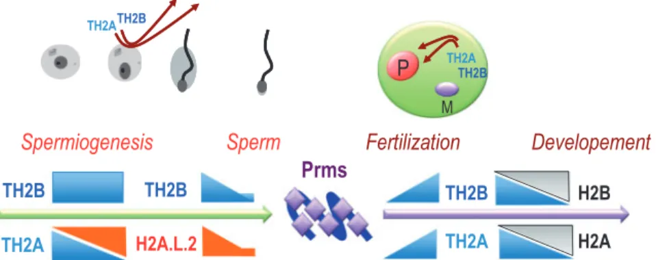
Fig. 1 Differentiation-dependent expression of testis-specific histone variants during spermatogenesis. Detailed studies of the indicated histone variants established the precise timing of their expression during the spermatogenic cells differentiation. A fraction of TH2B and H2A.L.2 escape the general histone-to-Prm transition and remains in the mature sperm nuclei.
N. Hoghoughi et al.
98
Downloaded from https://academic.oup.com/jb/article-abstract/163/2/97/4642838 by guest
induced pluripotent stem cells, a property that could be related to the ability of these histones to create more accessible chromatin (14).
In contrast to TH2B-less situation, the knock-out of both TH2B and TH2A leads to an arrest of spermato-genesis during meiosis (16). However in this model, al-though the lack of TH2B induced a compensatory accumulation of H2B, as reported in the TH2B-less model (10), no H2A accumulation was observed (16). Since TH2A is also the main H2A in spermatogenic cells, in Th2a/Th2b KO spermatogenic cells, the absence of compensatory gain of H2A expression suggests a sig-nificant H2A under-dosage in these cells. The defects observed could therefore be due to an inappropriate chromatin structure and subsequent genome disorgan-ization, rather than to a specific action of TH2A or TH2B (16). In agreement with this hypothesis, two recent reports showed that TH2A could be phosphory-lated at the TH2A-specific Thr127 and that this phos-phorylation is dispensable for normal spermatogenesis and male fertility (17, 18).
The animal models used to decipher TH2B functions also included mice that expressed a modified endogenous TH2B, bearing three tags at its C-terminal (10). Remarkably, although in these mice all the investigated events at the cellular and molecular levels, i.e. TH2B-tag assembly, transcription, meiosis, sex-chromosome inacti-vation, H2A.X phosphorylation and histone exchange, etc., occur normally in spermatogenic cells up to the ap-pearance of elongating spermatids, an arrest of sperm-atogenesis is observed at the time of histone-to-Prm transition. This study demonstrated that TH2B-tag ex-pressed in female animals could also sustain oocyte mat-uration and embryonic development with no detectable defects and leading to the generation of healthy adult mice (10).
Overall, these data show that the presence of a tag at the C-terminal end of TH2B, interferes very specifically and exclusively with defined and particular events that take place in elongating-condensing spermatids (10). Detailed observations indicated, that in these TH2B-tag expressing male germ cells, although TP and Prms
are produced, Prms are unable to replace histones lead-ing to the accumulation of transitional structures fol-lowing nucleosome disassembly. Further investigations are required to decipher the exact role of TH2B, and more specifically of its C-terminal region, in the final histone eviction.
H2A.L.2
In an attempt to identify histones that could facilitate the process of histone-to-Prm transition, surviving his-tones from spermatids undergoing histone replacement were purified and identified (9). This led to the identi-fication of 5 new histone variants, two H2Bs and three H2As, which were respectively named H2B.L.1, H2B.L.2, H2A.L.1, H2A.L.2 and H2A.L.3 [please see their new nomenclature in (19, 20)]. Among these histones, H2A.L.2 presented all the criteria to be con-sidered as a candidate histone variant involved in his-tone removal. Indeed, the protein is first detected in spermatids at the same time as TPs (9, 21). Additionally, H2A.L.2 along with TH2B was found in the H3/H4-less transitional structures, which were evidenced in condensing spermatids after extensive micrococcal nuclease (MNase) digestion. Finally, a fraction of H2A.L.2 remains in mature spermatozoa associated with the pericentric regions (9).
The generation of an H2A.L.2-less mouse model proved its critical role in the process of histone-to-Prm replacement. The molecular studies of spermatogenic cells lacking H2A.L.2 revealed its role in the incorpor-ation of TPs onto chromatin. Additionally, a series of in vitro nucleosome reconstitution assays demonstrated that the incorporation of H2A.L.2 can drastically modify the structure of the corresponding nucleosome. A more ‘open’ H2A.L.2-containing nucleosome is able to load TPs, which invade the nucleosome without releasing the DNA. These TPs in turn buffer the incom-ing Prms and allow their assembly and the final histone eviction. TPs also mediate the processing of Prm2, which is produced as a pre-Prm2 protein (21). In fact the phenotype of H2A.L.2-less spermatogenic cells is very
Fig. 2 The packaging cycles of the male genome with TH2A/TH2B. The histone variants TH2A and TH2B become the major histones in spermatogenic cells, until the replacement of TH2A by H2A.L.2, at the time of histone-to-Prm transition. A fraction of TH2BH2A.L.2 dimer survives histone replacement. After fertilization, the removal of Prms is associated with the re-assembly of maternal TH2ATH2B on the male genome. These histones will be entirely replaced by their somatic counterparts during early development. The schemes respectively represent maturing spermatids (left) and a fertilized egg with paternal (P) and maternal (M) genomes (right).
close to the phenotype of TP-less cells (2224) suggest-ing that, in the absence of H2A.L.2, although TPs are produced, they remain non-functional.
Altogether, these observations fully support the idea that the incorporation of H2A.L.2 changes the struc-ture of the nucleosomes allowing chromatin loading of TPs, which in turn buffer and regulate Prm processing and assembly (Fig. 3).
Prms are the true histone displacers. Indeed, they can displace histones even in the absence of TPs (or H2A.L.2). However, although not required for histone removal, TPs are necessary for an efficient Prm-depend-ent genome compaction.
The major outcome of these investigations is also a new vision of the process of histone-to-Prm transition. Indeed, the general belief was that histones were first replaced by TPs, which were later replaced by Prms to fully compact the genome. The new data described above demonstrate that the full picture of the process requires a histone variant that ‘prepares’ the nucleo-somes to undergo disassembly (Fig. 1). In this context, TPs and Prms work together, not successively, to fully transform the nucleosomes into nucleosprotamine structures (Fig. 3).
H2A.B.3
In an attempt to identify and functionally characterize H2A histone variants that lack the characteristic acidic patch, which is present in most H2As, including canon-ical H2A, the mouse genome was screened in search for H2A genes with an altered acidic patch (25). The acidic patch is typically composed of a series of six acidic amino acids of H2A, present at the surface of a nu-cleosome, which mediates the nucleosomenucleosome interaction and hence chromatin compaction (26, 27). This approach led to the identification of four H2A variants with an altered acidic patch, which were named H2A.Lap1-4, ‘Lap’ standing for Lack of
Acidic Patch. A further analysis showed that some of them had already been identified by Govin and col-leagues: H2A.L.2 actually is H2A.Lap3, H2A.L.1 is H2A.Lap2 and H2A.L.3 is H2A.Lap4 (20). H2A.Lap1, now known as H2A.B.3, was uniquely identified in this study and functionally characterized. This H2A variant shows similar structural features as H2A.L.2 but its expression pattern is drastically differ-ent (Fig. 1). H2A.B.3 first appears in late pachytene spermatocytes and particularly accumulates in post-meiotic round spermatids but disappears from the elongating spermatids nucleus (25), when H2A.L.2 starts to accumulate (21). The protein was then found enriched at the transcriptional start sites (TSS) of highly active genes in spermatocytes as well as in round spermatids. In these latter cells, H2A.B.3 be-comes associated with the X-linked genes that escape sex chromosome transcriptional inactivation (25). Further studies demonstrated that H2A.B.3 has an RNA-binding motif, binds RNA and a series of RNA processing factors and co-localizes with splicing speckles in highly active nuclear subdomains (28). These studies highlight the possibility that H2A.B.3 incorporation at exonintron boundaries facilitates the recruitment of splicing factors from the splicing speckles.
It is of note that the studies on H2A.B.3 also re-vealed a specific functional interplay between this his-tone and the widely-studied H2A.Z in spermatogenic cells. Indeed, several years ago, a microscopic analysis of H2A.Z histone variants revealed a post-meiotic accumulation of H2A.Z on the sex chromosomes (29). A high resolution mapping of H2A.Z and H2A.B.3 revealed an interesting relationship between these two H2A variants. The replacement of H2A.Z by H2A.B.3 at the exonintron boundaries in spermato-genic cells may play a role in the regulation of splicing at active genes. Additionally, in the surround of genes TSSs, a combination of H2A, H2A.Z and H2A.B.3 could define specific gene categories in terms of expres-sion timing and functional classifications of their prod-ucts (30). These studies extend the role of histone variants from a specific action on particular chromatin regions to a possibly direct regulation of RNA process-ing and splicprocess-ing.
Testis specific H3
A testis-specific histone H3 was also first reported almost 40 years ago (31). However, similarly to TH2B, it is structural biology that brought hints on the possible functions of this variant, first for the human H3T (32) and then for the mouse H3t variant (33). These studies showed that the alteration of sev-eral residues in H3T compared to H3.1, perfectly ex-plains the instability of nucleosomes bearing H3T, observed in vitro and in vivo. In the case of H3t, only one H3t-specific amino acid, H42, was found to be responsible for creating an ‘open’ nucleosome with flexible DNA at the entryexit of the nucleosomes (33). The H3t-encoding gene becomes active in differ-entiating spermatogonia, and its product gradually replaces most of the somatic-type H3s.
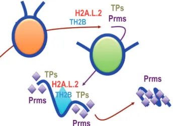
Fig. 3 Coordinate actions of histone variants, TPs and Prms in the process of histone-to-Prm replacement. The exchange of TH2B TH2A by TH2BH2A.L.2 opens the nucleosomes, allowing the loading of TPs that in turn direct a controlled replacement of his-tones, Prm assembly and genome compaction.
N. Hoghoughi et al.
100
Downloaded from https://academic.oup.com/jb/article-abstract/163/2/97/4642838 by guest
In the absence of H3t, a significant reduction of the number of differentiating c-Kit positive spermatogonia was observed, leading to complete male infertility. Further investigations showed a drastic decrease in the number of post-meiotic cells and suggested a possible role for this histone in the entry of cells into meiosis. However, since there was no compensatory increase of H3 gene expression to maintain cellular H3 levels, the observed defects in the absence of H3t could also be explained by histone underdosage.
H3.3
Besides the mouse models harbouring the inactivation of genes encoding testis-specific histone variants, the targeted inactivation of H3.3-encoding genes in the mouse testis was also performed. H3.3 is a replica-tion-independent H3 that is required for chromatin as-sembly whenever the replication-dependent pathway is not operational, such as post-fertilization chromatin assembly on the paternal genome (3436).
In the mouse two independent H3.3 encoding genes, H3f3a and H3f3b, generate identical H3.3. However individual targeted KO of these genes have different impact on spermatogenesis. The knock-out of H3f3a leads to the generation of abnormal spermatozoa (37), while the KO of H3f3b has a much more dramatic phenotype including a spermatogenesis arrest at the stage of round spermatid (37, 38).
These functional analyses of H3.3 had been awaited since the interesting observation by de Boer’s labora-tory of the massive replacement of H3 by H3.3 on the sex chromosomes in meiotic cells (39). During meiosis the unsynapsed X and Y chromosomes create a specific domain in the nucleus of spermatocytes known as the sex body, which undergoes a total transcriptional inactivation during meiosis known as meiotic sex chromosome inactivation (MSCI).
A consequence of this selective incorporation of H3.3 on the sex chromosomes is the creation of interesting combinations of histone variants. In spermatocytes, the macroH2A histone variant was known to accumulate on the sex body (40), while in post-meiotic cells it is H2A.Z that becomes enriched on these chromosomes (29). The presence of both H3.3 and macroH2A in spermatocytes and of H3.3 and H2A.Z in round sperm-atids increases the probability for these combinations to be present within the same nucleosome. While macroH2A is associated with less active and more stable chromatin domains (41), the H3.3H2A.Z com-bination is known to create a particularly unstable nu-cleosome (42, 43), which could be important for the post-meiotic reactivation of the sex-linked genes.
The generation of these H3.3 KO models therefore provided the possibility to investigate more specifically the importance of H3.3 on the sex chromosome biol-ogy during spermatogenesis. However, at least in spermatocytes, no global defects were observed after a reduction in H3.3 expression (37), and the question of the function of H3.3 in MSCI and in the post-mei-otic partial reactivation of sex chromosomes at the molecular levels remains open.
Concluding Remarks
In addition to almost all the histone variants present in somatic cells, spermatogenic cells express a number of unique histone variants of the H2A, H2B and H3 types (Fig. 1).
The structural studies of all these testis-specific vari-ants revealed their capacity to induce nucleosome in-stability. Remarkably, short H2As, H2A.L.2 and H2A.B.3 as well as H3t are able to generate flexible DNA ends at the entry and exit of nucleosomes. Although these variants induce open chromatin fea-tures, these histones are expressed at quite different timings during spermatogenesis. H3t is expressed early in differentiating spermatogonia, while H2A.B.3 is expressed in pachytene spermatocytes and to a greater extent in round spermatids and is eventually replaced by H2A.L.2 in elongating spermatids. Therefore, the configuration of chromatin seems to gradually open from the early stage of commitment of cells into meiotic divisions to the late post-meiotic stage of histone-to-Prm transition. Additionally, spermatocytes express TH2B and TH2A that also form unstable and programmable chromatin. TH2BTH2A remains in spermatogenic cells until the time of histone replacement, when H2A.L.2 accu-mulates and becomes the privileged partner of TH2B (Fig. 2).
Therefore, due to the action of these histone variants, the chromatin of spermatogenic cells becomes potentially ‘prepared’ to undergo histone-to-Prm re-placement. Consequently, it is possible to speculate that it is the timing of TPs and Prms expression that decides histones replacement. However, the generation of an H2A.L.2-less mouse model demonstrated that the incorporation of this variant is indispensable for TPs to be efficiently loaded onto chromatin and that TPs incorporation controls the efficiency of the Prm-dependent replacement of histones (21).
Additionally, a genome-wide occurrence of histone hyperacetylation and of other histone acylations also seems necessary for histone replacement (13). It can therefore be concluded that a change in the structure of nucleosomes due to the incorporation of testis-specific histone variants is only one facet of the mechanisms involved in general histone eviction and that other mechanisms, mostly histone PTM-based events, are equally important.
Therefore, the future challenge will be to decipher the molecular links between histone variants and his-tone PTMs in the process of coupled hishis-tone eviction and Prm assembly.
Funding
This work is supported by ANR Episperm 3 program, by a grant from ‘Fondation pour la Recherche Medicale (FRM)’ ‘analyse bio-informatique pour la recherche en biologie’ program as well as INCa libre program (RPT13001CCA). Additional supports were from: Fondation ARC ‘Canc’air’ project (RAC16042CLA), Plan Cancer (CH7-INS15B66) and Plan Cancer (ASC16012CSA) and the ‘Universite´ Grenoble Alpes’ ANR-15-IDEX-02.
Conflict of Interest
References
1. Goudarzi, A., Shiota, H., Rousseaux, S., and Khochbin, S. (2014) Genome-scale acetylation-dependent histone eviction during spermatogenesis. J. Mol. Biol. 426, 33423349
2. Goudarzi, A., Zhang, D., Huang, H., Barral, S., Kwon, O.K., Qi, S., Tang, Z., Buchou, T., Vitte, A.L., He, T., Cheng, Z., Montellier, E., Gaucher, J., Curtet, S., Debernardi, A., Charbonnier, G., Puthier, D., Petosa, C., Panne, D., Rousseaux, S., Roeder, R.G., Zhao, Y., and Khochbin, S. (2016) Dynamic competing histone H4 K5K8 acetylation and butyrylation are hallmarks of highly active gene promoters. Mol. Cell 62, 169180 3. Liu, S., Yu, H., Liu, Y., Liu, X., Zhang, Y., Bu, C.,
Yuan, S., Chen, Z., Xie, G., Li, W., Xu, B., Yang, J., He, L., Jin, T., Xiong, Y., Sun, L., Liu, X., Han, C., Cheng, Z., Liang, J., and Shang, Y. (2017) Chromodomain protein CDYL acts as a crotonyl-CoA hydratase to regulate histone crotonylation and sperm-atogenesis. Mol. Cell 67, 853866
4. Bao, J., and Bedford, M.T. (2016) Epigenetic regulation of the histone-to-protamine transition during spermio-genesis. Reproduction 151, R55R70
5. Meistrich, M.L., Mohapatra, B., Shirley, C.R., and Zhao, M. (2003) Roles of transition nuclear proteins in spermiogenesis. Chromosoma 111, 483488
6. Govin, J., Caron, C., Lestrat, C., Rousseaux, S., and Khochbin, S. (2004) The role of histones in chromatin remodelling during mammalian spermiogenesis. Eur. J. Biochem. 271, 34593469
7. Shires, A., Carpenter, M.P., and Chalkley, R. (1975) New histones found in mature mammalian testes. Proc. Natl. Acad. Sci. U S A 72, 27142718
8. Li, A., Maffey, A.H., Abbott, W.D., Conde e Silva, N., Prunell, A., Siino, J., Churikov, D., Zalensky, A.O., and Ausio´, J. (2005) Characterization of nucleosomes consist-ing of the human testis/sperm-specific histone H2B vari-ant (hTSH2B). Biochemistry 44, 25292535
9. Govin, J., Escoffier, E., Rousseaux, S., Kuhn, L., Ferro, M., Thevenon, J., Catena, R., Davidson, I., Garin, J., Khochbin, S., and Caron, C. (2007) Pericentric hetero-chromatin reprogramming by new histone variants during mouse spermiogenesis. J. Cell Biol. 176, 283294 10. Montellier, E., Boussouar, F., Rousseaux, S., Zhang, K., Buchou, T., Fenaille, F., Shiota, H., Debernardi, A., Hery, P., Curtet, S., Jamshidikia, M., Barral, S., Holota, H., Bergon, A., Lopez, F., Guardiola, P., Pernet, K., Imbert, J., Petosa, C., Tan, M., Zhao, Y., Gerard, M., and Khochbin, S. (2013) Chromatin-to-nucleoprotamine transition is controlled by the histone H2B variant TH2B. Genes Dev. 27, 16801692
11. Urahama, T., Horikoshi, N., Osakabe, A., Tachiwana, H., and Kurumizaka, H. (2014) Structure of human nucleo-some containing the testis-specific histone variant TSH2B. Acta Crystallogr. F Struct. Biol. Commun. 70, 444449 12. Padavattan, S., Shinagawa, T., Hasegawa, K.,
Kumasaka, T., Ishii, S., and Kumarevel, T. (2015) Structural and functional analyses of nucleosome com-plexes with mouse histone variants TH2a and TH2b, involved in reprogramming. Biochem. Biophys. Res. Commun. 464, 929935
13. Padavattan, S., Thiruselvam, V., Shinagawa, T., Hasegawa, K., Kumasaka, T., Ishii, S., and Kumarevel, T. (2017) Structural analyses of the nucleo-some complexes with human testis-specific histone vari-ants, hTh2a and hTh2b. Biophys. Chem. 221, 4148
14. Shinagawa, T., Takagi, T., Tsukamoto, D., Tomaru, C., Huynh, L.M., Sivaraman, P., Kumarevel, T., Inoue, K., Nakato, R., Katou, Y., Sado, T., Takahashi, S., Ogura, A., Shirahige, K., and Ishii, S. (2014) Histone variants enriched in oocytes enhance reprogramming to induced pluripotent stem cells. Cell Stem Cell 14, 217227 15. Iuso, D., Czernik, M., Toschi, P., Fidanza, A., Zacchini,
F., Feil, R., Curtet, S., Buchou, T., Shiota, H., Khochbin, S., Ptak, G.E., and Loi, P. (2015) Exogenous expression of human protamine 1 (hPrm1) remodels fibroblast nuclei into spermatid-like structures. Cell Rep. 13, 17651771
16. Shinagawa, T., Huynh, L.M., Takagi, T., Tsukamoto, D., Tomaru, C., Kwak, H.G., Dohmae, N., Noguchi, J., and Ishii, S. (2015) Disruption of Th2a and Th2b genes causes defects in spermatogenesis. Development 142, 12871292
17. Hada, M., Kim, J., Inoue, E., Fukuda, Y., Tanaka, H., Watanabe, Y., and Okada, Y. (2017) TH2A is phos-phorylated at meiotic centromere by Haspin. Chromosoma 126, 769780
18. Hada, M., Masuda, K., Yamaguchi, K., Shirahige, K., and Okada, Y. (2017) Identification of a variant-specific phosphorylation of TH2A during spermiogenesis. Sci. Rep. 7, 46228
19. Talbert, P.B., Ahmad, K., Almouzni, G., Ausio, J., Berger, F., Bhalla, P.L., Bonner, W.M., Cande, W.Z., Chadwick, B.P., Chan, S.W., Cross, G.A., Cui, L., Dimitrov, S.I., Doenecke, D., Eirin-Lopez, J.M., Gorovsky, M.A., Hake, S.B., Hamkalo, B.A., Holec, S., Jacobsen, S.E., Kamieniarz, K., Khochbin, S., Ladurner, A.G., Landsman, D., Latham, J.A., Loppin, B., Malik, H.S., Marzluff, W.F., Pehrson, J.R., Postberg, J., Schneider, R., Singh, M.B., Smith, M.M., Thompson, E., Torres-Padilla, M.E., Tremethick, D.J., Turner, B.M., Waterborg, J.H., Wollmann, H., Yelagandula, R., Zhu, B., and Henikoff, S. (2012) A unified phyl-ogeny-based nomenclature for histone variants. Epigenetics Chromatin 5, 7
20. El Kennani, S., Adrait, A., Shaytan, A.K., Khochbin, S., Bruley, C., Panchenko, A.R., Landsman, D., Pflieger, D., and Govin, J. (2017) MS_HistoneDB, a manually curated resource for proteomic analysis of human and mouse histones. Epigenetics & Chromatin 10, 2
21. Barral, S., Morozumi, Y., Tanaka, H., Montellier, E., Govin, J., de Dieuleveult, M., Charbonnier, G., Coute, Y., Puthier, D., Buchou, T., Boussouar, F., Urahama, T., Fenaille, F., Curtet, S., Hery, P., Fernandez-Nunez, N., Shiota, H., Gerard, M., Rousseaux, S., Kurumizaka, H., and Khochbin, S. (2017) Histone variant H2A.L.2 guides transition protein-dependent protamine assembly in male germ cells. Mol. Cell 66, 89101.e8
22. Zhao, M., Shirley, C.R., Hayashi, S., Marcon, L., Mohapatra, B., Suganuma, R., Behringer, R.R., Boissonneault, G., Yanagimachi, R., and Meistrich, M.L. (2004) Transition nuclear proteins are required for normal chromatin condensation and functional sperm development. Genesis 38, 200213
23. Zhao, M., Shirley, C.R., Mounsey, S., and Meistrich, M.L. (2004) Nucleoprotein transitions during spermio-genesis in mice with transition nuclear protein Tnp1 and Tnp2 mutations. Biol. Reprod. 71, 10161025 24. Shirley, C.R., Hayashi, S., Mounsey, S., Yanagimachi,
R., and Meistrich, M.L. (2004) Abnormalities and reduced reproductive potential of sperm from Tnp1-and Tnp2-null double mutant mice. Biol. Reprod. 71, 12201229
N. Hoghoughi et al.
102
Downloaded from https://academic.oup.com/jb/article-abstract/163/2/97/4642838 by guest
25. Soboleva, T.A., Nekrasov, M., Pahwa, A., Williams, R., Huttley, G.A., and Tremethick, D.J. (2011) A unique H2A histone variant occupies the transcriptional start site of active genes. Nat. Struct. Mol. Biol. 19, 2530 26. Chodaparambil, J.V., Barbera, A.J., Lu, X., Kaye,
K.M., Hansen, J.C., and Luger, K. (2007) A charged and contoured surface on the nucleosome regulates chro-matin compaction. Nat. Struct. Mol. Biol. 14, 11051107 27. Zhou, J., Fan, J.Y., Rangasamy, D., and Tremethick, D.J. (2007) The nucleosome surface regulates chromatin compaction and couples it with transcriptional repres-sion. Nat. Struct. Mol. Biol. 14, 10701076
28. Soboleva, T.A., Parker, B.J., Nekrasov, M., Hart-Smith, G., Tay, Y.J., Tng, W.-Q., Wilkins, M., Ryan, D., Tremethick, D.J., and Schneider, R. (2017) A new link between transcriptional initiation and pre-mRNA spli-cing: the RNA binding histone variant H2A.B. PLoS Genet. 13, e1006633
29. Greaves, I.K., Rangasamy, D., Devoy, M., Marshall Graves, J.A., and Tremethick, D.J. (2006) The X and Y chromosomes assemble into H2A.Z-containing [cor-rected] facultative heterochromatin [cor[cor-rected] following meiosis. Mol. Cell. Biol. 26, 53945405
30. Soboleva, T.A., Nekrasov, M., Ryan, D.P., and Tremethick, D.J. (2014) Histone variants at the tran-scription start-site. Trends Genet. 30, 199209
31. Franklin, S.G., and Zweidler, A. (1977) Non-allelic variants of histones 2a, 2b and 3 in mammals. Nature 266, 273275 32. Tachiwana, H., Kagawa, W., Osakabe, A., Kawaguchi, K., Shiga, T., Hayashi-Takanaka, Y., Kimura, H., and Kurumizaka, H. (2010) Structural basis of instability of the nucleosome containing a testis-specific histone vari-ant, human H3T. Proc. Natl. Acad. Sci. U S A 107, 1045410459
33. Ueda, J., Harada, A., Urahama, T., Machida, S., Maehara, K., Hada, M., Makino, Y., Nogami, J., Horikoshi, N., Osakabe, A., Taguchi, H., Tanaka, H., Tachiwana, H., Yao, T., Yamada, M., Iwamoto, T., Isotani, A., Ikawa, M., Tachibana, T., Okada, Y., Kimura, H., Ohkawa, Y., Kurumizaka, H., and Yamagata, K. (2017) Testis-specific histone variant H3t gene is essential for entry into sperm-atogenesis. Cell Rep. 18, 593600
34. Filipescu, D., Szenker, E., and Almouzni, G. (2013) Developmental roles of histone H3 variants and their chaperones. Trends Genet. 29, 630640
35. Lin, C.J., Koh, F.M., Wong, P., Conti, M., and Ramalho-Santos, M. (2014) Hira-mediated H3.3 in-corporation is required for DNA replication and ribo-somal RNA transcription in the mouse zygote. Dev. Cell. 30, 268279
36. Inoue, A., and Zhang, Y. (2014) Nucleosome assembly is required for nuclear pore complex assembly in mouse zygotes. Nat. Struct. Mol. Biol. 21, 609616
37. Tang, M.C.W., Jacobs, S.A., Mattiske, D.M., Soh, Y.M., Graham, A.N., Tran, A., Lim, S.L., Hudson, D.F., Kalitsis, P., O’Bryan, M.K., Wong, L.H., Mann, J.R., and Tremethick, D. (2015) Contribution of the two genes encoding histone variant h3.3 to viability and fer-tility in mice. PLoS Genet. 11, e1004964
38. Yuen, B.T., Bush, K.M., Barrilleaux, B.L., Cotterman, R., and Knoepfler, P.S. (2014) Histone H3.3 regulates dynamic chromatin states during spermatogenesis. Development 141, 34833494
39. van der Heijden, G.W., Derijck, A.A., Posfai, E., Giele, M., Pelczar, P., Ramos, L., Wansink, D.G., van der Vlag, J., Peters, A.H., and de Boer, P. (2007) Chromosome-wide nucleosome replacement and H3.3 incorporation during mammalian meiotic sex chromo-some inactivation. Nat. Genet. 39, 251258
40. Hoyer-Fender, S., Costanzi, C., and Pehrson, J.R. (2000) Histone macroH2A1.2 is concentrated in the XY-body by the early pachytene stage of spermatogenesis. Exp Cell Res. 258, 254260
41. Talbert, P.B., and Henikoff, S. (2017) Histone variants on the move: substrates for chromatin dynamics. Nat. Rev. Mol. Cell Biol. 18, 115126
42. Jin, C., and Felsenfeld, G. (2007) Nucleosome stability mediated by histone variants H3.3 and H2A.Z. Genes Dev. 21, 15191529
43. Jin, C., Zang, C., Wei, G., Cui, K., Peng, W., Zhao, K., and Felsenfeld, G. (2009) H3.3/H2A.Z double variant-containing nucleosomes mark ?nucleosome-free regions? of active promoters and other regulatory regions. Nat. Genet. 41, 941945