HAL Id: inserm-00384487
https://www.hal.inserm.fr/inserm-00384487
Submitted on 15 May 2009
HAL is a multi-disciplinary open access
archive for the deposit and dissemination of
sci-entific research documents, whether they are
pub-lished or not. The documents may come from
teaching and research institutions in France or
abroad, or from public or private research centers.
L’archive ouverte pluridisciplinaire HAL, est
destinée au dépôt et à la diffusion de documents
scientifiques de niveau recherche, publiés ou non,
émanant des établissements d’enseignement et de
recherche français ou étrangers, des laboratoires
publics ou privés.
Function of retinoic acid receptors during embryonic
development.
Manuel Mark, Norbert Ghyselinck, Pierre Chambon
To cite this version:
Manuel Mark, Norbert Ghyselinck, Pierre Chambon. Function of retinoic acid receptors during
em-bryonic development.. Nucl Recept Signal, 2009, 7, pp.e002. �10.1621/nrs.07002�. �inserm-00384487�
Function of retinoic acid receptors during
embryonic development
Manuel Mark, Norbert B. Ghyselinck and Pierre Chambon
Corresponding Author: [email protected] [email protected]
Institut de Génétique et de Biologie Moléculaire et Cellulaire (IGBMC), Département de Biologie Cellulaire and Développement (MM and NG) and Département de Génomique Fonctionnelle (PC), and Collège de France (PC), Strasbourg, France
Retinoids, the active metabolites of vitamin A, regulate complex gene networks involved in vertebrate morphogenesis, growth, cellular differentiation and homeostasis. Studies performed in vitro, using either acellular systems or transfected cells, have shown that retinoid actions are mediated through heterodimers between the RAR and RXR nuclear receptors. However, in vitro studies indicate what is possible, but not necessarily what is actually occurring in vivo, because they are performed under non-physiological conditions. Therefore, genetic approaches in the animal have been be used to determine the physiological functions of retinoid receptors. Homologous recombination in embryonic stem cells has been used to generate germline null mutations of the RAR- and RXR-coding genes in the mouse. As reviewed here, the generation of such germline mutations, combined with pharmacological approaches to block the RA signalling pathway, has provided genetic evidence that RAR/RXR heterodimers are indeed the functional units transducing the RA signal during prenatal development. However, due to (i) the complexity in “hormonal” signalling through transduction by the multiple RARs and RXRs, (ii) the functional redundancies (possibly artefactually generated by the mutations) within receptor isotypes belonging to a given family, and (iii) in utero or early postnatal lethality of certain germline null mutations, these genetic studies have failed to reveal all the physiological functions of RARs and RXRs, notably in adults. Spatio-temporally-controlled somatic mutations generated in given cell types/tissues and at chosen times during postnatal life, will be required to reveal all the functions of RAR and RXR throughout the lifetime of the mouse.
Received February 6th, 2009; Accepted March 13th, 2009; Published April 3rd, 2009 | Abbreviations: AD: activating domain; AF: activation function; BA: branchial arche; CYP: cytochrome P450 hydroxylase; DORV: double outlet right ventricle; E: embryonic day; INZ: interdigital necrotic zone; NCC: neural crest cells; NR: nuclear receptor; PHPV: persistent and hyperplastic vitreous body; POM: periocular mesenchyme; R1 to R7:
rhombomeres 1 to 7; RA: retinoic acid; RALDH: retinaldehyde dehydrogenase; RAR: retinoic acid receptor; RBP: retinol-binding protein; RDH: retinol dehydrogenase; RXR: retinoid X receptor; STRA: stimulated by retinoic acid; VAD: vitamin A-deficiency | Copyright © 2009, Mark et al. This is an open-access article distributed under the terms of the Creative Commons Non-Commercial Attribution License, which permits unrestricted non-commercial use, distribution and reproduction in any medium, provided the original work is properly cited.
Cite this article: Nuclear Receptor Signaling (2009) 7, e002
Introduction
Both clinical findings and experimental approaches have revealed that vitamin A (retinol) and its active derivatives (i.e., the retinoids) exert a wide variety of effects on vertebrate embryonic body shaping and organogenesis, tissue homeostasis, cell proliferation, differentiation and apoptosis (reviewed in [Blomhoff, 1994; Kastner et al., 1995; Mark et al., 2006; Morriss-Kay and Ward, 1999; Sporn et al., 1994]). Following Hale’s initial demonstration that vitamin A-deficiency (VAD) induces congenital ocular malformations [Hale, 1933], Warkany and his
collaborators showed that a large array of congenital malformations affecting the ocular, cardiac, respiratory and urogenital systems (collectively referred as to the fetal VAD syndrome) occurred in fetuses from vitamin A-deficient (VAD) rats (reviewed in [Wilson et al., 1953]). It was shown, much later, that retinoic acid (RA) could replace vitamin A during embryogenesis, at least at certain stages and in certain organs [Dickman et al., 1997; White et al., 1998].
How RA can exert such pleiotropic effects was a long-standing question, which found its solution with the discovery of two classes of RA-binding transcriptional regulators, the retinoic acid receptors (the multiple isoforms of the RARα, β and γ isotypes), and the retinoid
X receptors (the multiple isoforms of the RXRα, β and γ isotypes) (reviewed in [Chambon, 1996; Leid et al., 1992]. The present review focuses on three main questions: (i) are RARs and RXRs involved in the transduction of RA signals in vivo? (ii) to what extent do the observations made in vivo support the molecular mechanisms deduced from in vitro studies? (iii) what are the developmental events controlled by RARs and RXRs? We summarize and discuss in this review the developmental phenotypes induced by mutations that have been introduced at loci encoding RARs and RXRs, and compare them to the phenotypes resulting either from VAD or from
administration of pharmacological doses of antagonistic ligands for RARs in vertebrates, placing emphasis on results obtained in mammals.
RARs and RXRs are instrumental to
retinoic acid signalling during
embryonic development
Signalling through RARs is indispensable for
embryonic patterning and organogenesis
Rara, Rarb, and Rarg null mutant mice are viable. They
display some aspects of the fetal (and postnatal) VAD syndromes, as well as a few additional congenital
malformations (Table 1). However, their abnormalities are restricted to a subset of tissues normally expressing these receptors, probably reflecting the existence of functional redundancies between RARs (discussed in [Kastner et al., 1995; Kastner et al., 1997a; Mark and Chambon, 2003; Mark et al., 1995; Mascrez et al., 1998]). To test this hypothesis, mutants lacking two RAR isotypes (Rara/b-, Rara/g- and Rarb/g-null mutants), or several isoforms belonging to distinct isotypes were generated. For the sake of clarity, only abnormalities displayed by double null mutants lacking a couple of RAR isotypes (all isoforms deleted) are listed in Table 2 and Table 3. Similar abnormalities, albeit often less penetrant, which are displayed by "isoform-specific" double null mutants are listed in [Kastner et al., 1995] (mutants lacking
RARα1/RARβ2/4, RARα1/RARγ, RARα1α2+/-/RARγ and RARβ2/4/RARγ), in [Subbarayan et al., 1997] (mutants lacking RARα/RARγ1 and RARα/RARγ2), in [Luo et al., 1996] (mutants lacking RARα1/RARβ), in [Ghyselinck et al., 1998] (mutants lacking RARα/RARβ1/3 and
RARβ1/3/RARγ), and in [Grondona et al., 1996] (mutants lacking RARβ2/RARγ2).
Rara/b-, Rara/g- and Rarb/g-null mutants die in utero or
at birth because of severe developmental defects that altogether include the complete spectrum of
malformations belonging to the fetal VAD-induced syndrome reported by Warkany’s group 55 years ago [Wilson et al., 1953] (Table 2). As with Rar-single-null mutants (see Table 1), Rar-double-null mutants (Table 3) also exhibit congenital abnormalities that were not described in Hale’s and Warkany’s pioneering fetal VAD studies [Hale, 1933; Wilson et al., 1953], encompassing ageneses of the Harderian glands, skeletal defects of the skull, face, vertebrae, limbs and forebrain [Ghyselinck et al., 1997; Lohnes et al., 1993; Lohnes et al., 1994; Luo et al., 1996; Mendelsohn et al., 1994; Subbarayan et al., 1997]. The occurrence of these “non-VAD” defects in Rar-single and double-null mutant mice is most probably accounted for by the difficulty to achieve, by dietary deprivation, a state of profound VAD compatible with pregnancy. In fact, almost all these “non-VAD” defects have been subsequently produced in rodent embryos (i) deficient in vitamin A, but supplemented with RA [Dickman et al., 1997; White et al., 2000; White et al., 1998]; (ii) deficient in both RBP (retinol-binding protein) and vitamin A [Quadro et al., 2005]; (iii) lacking the retinaldehyde synthesising enzyme RDH10 (retinol dehydrogenase 10) [Sandell et al., 2007]; (iv) lacking the RA synthesising enzymes RALDH2 (retinaldehyde dehydrogenase 2) [Halilagic et al., 2007; Molotkova et al., 2007; Niederreither et al., 2002a; Niederreither et al., 1999; Niederreither et al., 2001; Niederreither et al., 2000; Niederreither et al., 2002b; Ribes et al., 2006] or RALDH3 [Dupe et al., 2003; Halilagic et al., 2007] or (v) treated with synthetic retinoids possessing panRAR antagonistic activities [Kochhar et al., 1998; Wendling et al., 2000; Wendling et al., 2001]. In addition, it was recently found that the Matthew-Wood syndrome, which consists of a spectrum of congenital abnormalities also observed in
Rara/b, Rara/g and Rarb/g-null mutants, is caused by
mutations in the RBP receptor gene, STRA6 [Golzio et
al., 2007; Pasutto et al., 2007]. As STRA6 loss-of-function mutations most probably yield a state of RA insufficiency in embryonic tissues [Kawaguchi et al., 2007], this discovery provides evidence that RAR signalling performs similar functions during embryonic development in mice and humans.
The comparison of the Rar-null mutant phenotypes with those of rodents and humans carrying the aforementioned blocks in RA signal transduction, demonstrate that liganded RARs play crucial roles at many distinct stages of the development of numerous organs ([Kastner et al., 1995] and references therein). For example, the severe malformations found in Rara/g-null embryos [Wendling et al., 2001] are similar to those of Aldh1a2 (formerly Raldh2)-null embryos [Niederreither et al., 1999], and reflect early roles of RAR signalling in axial rotation, segmentation and closure of the hindbrain, formation of otocysts, pharyngeal arches and forelimb buds, as well as in closure of the primitive gut. RARs are also indispensable for the ontogenesis of (almost) all the anatomical structures that are derived from
mesectodermal cells, i.e. the cranial neural crest cells (NCC) that give rise to mesenchymal derivatives (reviewed in [Kastner et al., 1995; Mark et al., 1998; Mark et al., 1995]). RARs are involved in antero-posterior patterning of the somitic mesoderm and hindbrain neurectoderm [Dupe et al., 1999b; Ghyselinck et al., 1997; Lohnes et al., 1993; Lohnes et al., 1994; Wendling et al., 2001], notably through controlling expression of homeobox genes [Allan et al., 2001; Dupe et al., 1997; Dupe et al., 1999b; Houle et al., 2000; Oosterveen et al., 2003; Serpente et al., 2005]. RARs are also involved in the establishment of the antero-posterior axis of the limbs [Dupe et al., 1999a; Lohnes et al., 1994; Mascrez et al., 1998]. RARs are required for the development of a large number of eye structures (Table 2 and Table 3) and for histogenesis of the retina [Ghyselinck et al., 1997; Grondona et al., 1996; Lohnes et al., 1994], cardiomyocyte differentiation [Kastner et al., 1994; Kastner et al., 1997b], as well as for the control of physiological apoptosis in the retina [Grondona et al., 1996], the frontonasal and interdigital mesenchymes [Crocoll et al., 2002; Dupe et al., 1999a; Ghyselinck et al., 1997; Lohnes et al., 1994], the conotruncal segment of the embryonic heart [Ghyselinck et al., 1998] and the embryonic nephric duct [Batourina et al., 2005]. In the embryonic urogenital tract, RARs control
epithelial-mesenchymal interactions in the kidney through expression of the receptor tyrosine kinase Ret [Batourina et al., 2002; Batourina et al., 2001; Mendelsohn et al., 1999], as well as the formation of the genital ducts and ureters [Batourina et al., 2001; Batourina et al., 2005; Ghyselinck et al., 1997; Mendelsohn et al., 1994]. In the developing respiratory tract, RA-liganded RARs are necessary for the morphogenesis of the nasal cavities and for their communication with the more caudal airways [Dupe et al., 2003; Ghyselinck et al., 1997] (Table 2 and Table 3). RA-liganded RARs are also required for the partitioning of the primitive foregut into oesophagus and trachea, and regulate lung branching morphogenesis [Chen et al., 2007; Desai et al., 2006; Malpel et al., 2000;
Table 1. Postnatal manifestations of germline ablation of Rar and Rxr genes. CD: congenital defects, PnVAD: abnormalities present in postnatal
vitamin A-deficiency (Wolbach and Howe, 1925); fetal VAD: abnormalities present in vitamin A-deficiency during pregnancy (Wilson et al., 1953). #: these abnormalities are completely penetrant. Rara1, Rara2, Rarb1/3, Rarb2/4, Rarg1 and Rarg2 refer to isoform-specific ablations.
Mollard et al., 2000; Wang et al., 2006], as well as lung alveoli septation [Massaro and Massaro, 2003; Massaro et al., 2003; Massaro et al., 2000; McGowan et al., 2000] (Table 1, Table 2, and Table 3).
Altogether, the developmental abnormalities of Rar-null mutant mice faithfully recapitulate those of rodents lacking the RAR ligand either through dietary vitamin A
deprivation or genetically-impaired RA synthesis. There are, however, two notable exceptions to this rule which are related to the control, through RA signalling, of (i) the
symmetry of somite patterning and (ii) germ cell differentiation in the embryonic ovary. Concerning somitogenesis, it has been firmly established, in several vertebrate species, that RA is instrumental to the generation of bilaterally symmetrical pairs of somites [Echeverri and Oates, 2007; Kawakami et al., 2005; Sirbu and Duester, 2006; Vermot and Pourquie, 2005]. It was also recently demonstrated that signalling by RA is required for expression of Stra8 (stimulated by retinoic acid 8) which, in turn, triggers meiosis in the embryonic ovary [Baltus et al., 2006; Bowles et al., 2006; Koubova
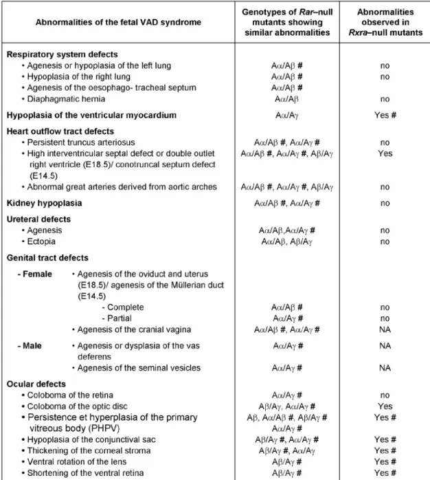
Table 2. Abnormalities of the fetal vitamin A deficiency (VAD) syndrome (Wilson et al., 1953) present in Rarb-null mutants (Aβ), Rxra-null mutants and in compound Rara/b-, Rara/g- and Rarb/g-null mutants. (Aα/Aβ, Aα/Aγ and Aβ/Aγ, respectively). #, this abnormality is completely penetrant. NA, not applicable, as the corresponding structure is normally not found at E14.5, the time around which Rxra-null mutants die. From references (Ghyselinck et al., 1997; Lohnes et al., 1994; Kastner et al., 1994). Note that most of the abnormalities seen in Rara/b-null mutants occur at similar frequencies in Rara/b2-mutants (Mendelsohn et al., 1994).
et al., 2006]. However, defects in the symmetry of somite derivatives, such as vertebrae and muscles, or in the histology of the ovaries, were never observed in Rara/b-,
Rara/g- and Rarb/g-null mutant mice. As mentioned
above, the apparent lack of phenotypic convergence between the animal models lacking RA and those lacking RARs is probably a consequence of the artificial compensation of the functions of the RARs that are missing in the knockout mice by the remaining one.
RARs have been instrumental to the
phylogenesis of mesectodermal derivatives
In addition to the dramatic craniofacial skeletal
deficiencies affecting Rara/g-null mutants [Lohnes et al., 1994], subtle defects which often alter the shape of a single skeletal piece are observed in several Rar-null mice, including: a cartilaginous or osseous connection between the incus middle ear bone and the alisphenoid bone (the pterygoquadrate element), a cartilage separating the trigeminal ganglion from the brain (the pila antotica) and an agenesis of the rostral ethmoturbinate
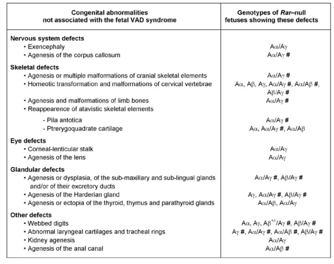
Table 3. Abnormalities absent from the fetal vitamin A deficiency (VAD) syndrome are found in Rara-, Rarb- and Rarg-null mutants (Aα, Aβ, Aγ), and in compound Rara/b-, Rara/g- and Rarb/g-null mutants (Aα/Aβ, Aα/Aγ and Aβ/Aγ). #: this abnormality is completely penetrant. From
references (Ghyselinck et al., 1997; Lohnes et al., 1994; Kastner et al., 1994). Note that most of the abnormalities seen in Rara/b-null mutants occur at similar frequencies in Rara/b2-mutants (Mendelsohn et al., 1994).
and maxillary sinus (Table 3) [Ghyselinck et al., 1997; Lohnes et al., 1994; Mark et al., 1998]. The
pterygoquadrate element and the pila antotica, which were lost during evolution from reptiles to mammals, represent atavistic features (discussed in [Mark et al., 1998; Mark et al., 1995]). Along the same lines, ethmoturbinate bones and paranasal sinuses (such as the maxillary sinus) are typical mammalian features not present in reptiles [Novacek, 1993]. It is thus conceivable that agenesis of these nasal structures in Rar-null mutants also mimics an atavistic condition. The presence of atavistic characteristics in Rar-null mutants supports the possibility that changes in the temporal or spatial patterns of expression of Rar genes has provided a general mechanism for modifying the number and shape of individual cranial skeletal elements during vertebrate evolution. Interestingly, recent teratogenic experiments using RA in excess have led to similar conclusions [Vieux-Rochas et al., 2007].
The persistent and hyperplastic vitreous body (PHPV) seen in Rarb-null mutants is comparable to the pecten oculi, a normal vascular and pigmented projection from
the optic disk found in some reptiles, which is thought to function in the nutrition of the retina [Mann, 1937]. The shortening of the ventral retina observed in Rarb/g-null mutants might also be interpreted as a modification towards an ancestral condition, since comparative embryology of the retina shows that the ventral retinal field increases in size as one ascends the vertebrate scale from fishes to amphibia, reptiles and mammals [Mann, 1937]. Thus, RARβ and RARγ might have been instrumental in the expansion of the ventral retinal field during vertebrate evolution.
RXR
α
is the main RXR isotype involved in
embryogenesis
Rxra-null mutants all display a hypoplasia of the compact
layer of the ventricular myocardium (Table 2), which appears to be the main cause of mutant death occurring by cardiac failure around E14.5 [Kastner et al., 1994; Kastner et al., 1997b; Ruiz-Lozano et al., 1998; Sucov et al., 1994]. That a similar myocardium defect is observed in VAD and Rar-null fetuses [Mendelsohn et al., 1994; Wilson et al., 1953], suggests that RXRα is involved in the transduction of the RA signal required for myocardial
growth. This requirement is unlikely to be cell-autonomous, as over-expressing RXRα in
cardiomyocytes by means of trangenesis does not prevent the Rxra-null mutation-induced hypoplasia of the ventricular myocardium [Subbarayan et al., 2000]. Other data further indicate that Rxra expression in the epicardium is required for triggering a paracrine signal necessary for myocardial growth [Chen et al., 2002; Kang and Sucov, 2005; Merki et al., 2005].
About one third of the Rxra-null mutants lack the conotruncal septum, which normally divides the embryonic heart outflow tract (or conotruncus) into the intracardiac portions of the aorta and the pulmonary trunk [Kastner et al., 1994]. Interestingly, deficiencies of this septum represent both a classical VAD defect in rodents and a leading cause of human congenital heart defects, ranging from high interventricular septal defects to double outlet right ventricle (DORV). In Rxra-null mutants, the agenesis of the conotruncal septum appears secondary to an enhanced rate of cell death in both the mesenchymal cells of the conotruncal ridges and the parietal conotruncal cardiomyocytes, therefore indicating that RXRα is required for the transduction of the RA signal that controls apoptosis in the conotruncal segment of the embryonic heart [Ghyselinck et al., 1998].
In addition to heart defects, all Rxra-null fetuses display a characteristic ocular syndrome characterized by a PHPV, closer eyelid folds, a thickened ventral portion of the corneal stroma, a ventral rotation of the lens, an agenesis of the sclera and a shortening of the ventral retina (Table 2) [Kastner et al., 1994]. As similar defects are present in VAD fetuses and in Rarb/g-null mutants (Table 2) [Ghyselinck et al., 1997; Wilson et al., 1953], RXRα appears to play an essential role in the transduction of the RA signals required for several ocular
morphogenetic processes, notably the formation of the ventral retinal field.
Importantly, the fact that mice lacking both RXRβ and RXRγ (Rxrb/g-null mutants) do not display any obvious morphogenetic defects, even when additionally lacking one allele of Rxra, clearly indicates that RXRα is functionally the most important RXR during
morphogenesis of the embryo proper [Krezel et al., 1996].
The AF-1-containing A/B domain and the
ligand-dependent AF-2 of RXR
α
are differentially
involved in development
The role played by RXRs as either “active” or “silent” heterodimerization partners in the transcription of target genes, inferred from in vitro studies, has been a controversial issue. To determine the transcriptional role of RXRαin vivo, mouse mutants were engineered that
express truncated RXRα proteins lacking (i) the N-terminal activation function (AF)-1-containing A/B region (Rxraaf1o mutants) [Mascrez et al., 2001], (ii) the AF-2 activating domain (AD) core-containing helix 12 located at the C-terminus of the E region (Rxraaf2o mutants) [Mascrez et al., 1998] and (iii) both AF-1 and AF-2 (Rxraafo mutants) [Mascrez et al., 2009].
The Rxraaf2o mutants display the myocardium hypoplasia and the ocular syndrome that are hallmarks of the
Rxra-null phenotype [Kastner et al., 1994], but at low
frequency [Mascrez et al., 1998]. This may reflect a functional compensation by RXRβ, as (i) the frequency of the myocardium hypoplasia increases from 5% in
Rxraaf2o mutants to 50% upon the additional inactivation of Rxrb (which, on its own, has no effect) [Kastner et al., 1996], and (ii) the frequency of the ocular syndrome increases from about 15% in Rxraaf2o mutants to 100% upon further inactivation of Rxrb [Mascrez et al., 1998]. The full penetrance of the Rxra-null ocular syndrome observed in Rxraaf2o/Rxrb-null mutants [Mascrez et al.,
1998], as well as in Rxraafo mutants [Mascrez et al., 2009], supports the view that the AF-2 of RXRα (and thus possibly a RXR agonistic ligand) is indispensable for ocular morphogenesis. On the other hand, the rare or modest penetrance of the myocardium hypoplasia in
Rxraaf2o (5%), Rxraaf2o/Rxrb-null (50%) and
Rxraaf2o/Rxrb/g-null mutants (50%) suggests that the
transcriptional activity of RXRα becomes necessary for myocardial growth only in “unfavourable” genetic backgrounds. Accordingly, we recently found that the heart histology is normal in 80% of Rxraafo fetuses [Mascrez et al., 2009], indicating that a transcriptionally “silent” RXRα can promote myocardial growth.
Involution of the primary vitreous body represents the developmental process which likely requires the highest concentration of RA-liganded retinoid receptors, since both VAD [Wilson et al., 1953], Rarb ablation [Ghyselinck et al., 1997], Rxra ablation [Kastner et al., 1994], deletion of RXRα AF-2 [Mascrez et al., 1998] and deletion of RXRα AF-1 and AF-2 [Mascrez et al., 2009] frequently yield a PHPV. Apart from an occasional PHPV, the
Rxraaf1o mutants never display any of the Rxra-null developmental defects [Mascrez et al., 2001]. This near-absence of defects does not reflect a functional compensation by RXRβ or RXRγ, as Rxraaf1o/Rxrb-null
and Rxraaf1o/Rxrb/g-null mutants display no other
developmental defect than a PHPV. However, the frequency of this PHPV increases from 10% in Rxraaf1o mutants to 100% in Rxraaf1o/Rxrb/g-null mutants [Mascrez
et al., 2001]. Altogether, these observations indicate that involution of the primary vitreous body requires both RXRα AF-1 and AF-2, while the other RA-dependent ocular morphogenetic events require RXRα AF-2 only, as they normally take place in the absence of the RXRα AF-1-containing A/B domain.
Thus, these data support the view that the activation functions of RXRα are differentially required for eye morphogenesis and that they can be dispensable for heart development. They also indicate that, due to functional redundancy, the role played by each activation function can be revealed only in genetic backgrounds impaired for RA-signalling (or under RA-insufficiency conditions). Assuming that the frequent requirement of RXRα AF-2 for developmental events reflects the binding of an agonistic ligand, this raises the question of the possible existence and nature of a physiological RXR ligand(s) in
vivo. The fact that 9-cis RA is undetectable in rodent
embryos [Horton and Maden, 1995; Matt et al., 2005b] makes it doubtful that this RXR physiological ligand could be 9-cis RA (reviewed in [Wolf, 2006]; see also [Calleja et al., 2006]).
The RXR
α
AF-1-containing A/B domain has a
specific function in the involution of the
interdigital mesenchyme
The AF-2 of RXRα appears functionally more important during development than its AF-1, as (i)
Rxraaf2o/Rxrb/g-null fetuses all display a large array of
congenital defects and die in utero, while
Rxraaf1o/Rxrb/g-null fetuses display only a few congenital
defects and are often viable; and (ii) transcription of a RA-responsive lacZ reporter transgene in the mouse requires RXRα AF-2, but not AF-1 [Mascrez et al., 1998; Mascrez et al., 2001]. However, the RXRα
AF-1-containing A/B region has a unique role in the RA-dependent disappearance of the interdigital mesenchyme.
The first evidence implicating RA in the involution of the interdigital mesenchyme was provided by organ culture experiments using whole limb in a RA-deprived medium [Lussier et al., 1993]. Subsequently, it was shown that mice lacking both alleles of either Rara or Rarg, as well as mice heterozygous for the Rxra-null mutation occasionally exhibit mild forms of interdigital webbing (i.e., soft tissue syndactyly) (Table 1) [Ghyselinck et al., 1997; Kastner et al., 1994; Lohnes et al., 1993; Lufkin et al., 1993]. Surprisingly, this defect was absent in Rarb-null mutants, even though Rarb is strongly and specifically expressed in the interdigital necrotic zones (INZs) [Ghyselinck et al., 1997]. However, disruption of one (or both) allele(s) of Rarb in a Rarg-null genetic background consistently yields interdigital webbing [Ghyselinck et al., 1997]. The persistence of the fetal interdigital
mesenchyme responsible for digit webbing is caused by a marked decrease in programmed cell death, as well as by an increase of cell proliferation in the mutant INZs [Dupe et al., 1999a]. As Rarb and Rarg are not co-expressed in the INZs, interdigital mesenchyme involution must involve paracrine interactions between this mesenchyme, which expresses Rarb, and either the cartilaginous blastema of the digits or the surface epidermis, which both express Rarg (discussed in [Dupe et al., 1999a]).
The RXRα AF-1-containing A/B region is indispensable for the function of RXRα/RARβ and/or RXRα/RARγ heterodimers involved in interdigital mesenchyme involution, since the majority of Rxraaf1o mutants and all
Rxraaf1o/Rxrb/g-null mutants display a soft tissue
syndactyly [Mascrez et al., 2001]. In contrast, Rxraaf2o and Rxraaf2o/Rxrb/g-null mutants never display this defect
[Mascrez et al., 1998], indicating a specific requirement of the RXRα AF-1-containing A/B region in the involution of the interdigital mesenchyme. Interestingly,
phosphorylation of RXRα at a specific serine residue located in the A domain is necessary for the
anti-proliferative response of F9 teratocarcinoma cells to RA [Bastien et al., 2002; Rochette-Egly and Chambon,
2001]. Phosphorylation of the RXRα A domain may therefore play an important function in the cascade of molecular events that, in vivo, leads to the normal disappearance of the interdigital mesenchyme.
Retinoic acid signals are transduced by specific
RXR
α
/RAR heterodimers during development
Compound mutants, in which a null mutation of a given RAR isotype is associated either with a Rxra-null, a
Rxraaf1o or a Rxraaf2o mutation, altogether recapitulate the abnormalities exhibited by Rar-null mutants (Table 4) [Kastner et al., 1994; Kastner et al., 1997a; Mascrez et al., 1998; Mascrez et al., 2001]. This synergism between Rar and Rxra loss-of-function mutations supports the conclusion that RXRα/RARα, RXRα/RARβ and RXRα/RARγ heterodimers are the functional units transducing RA signals during embryogenesis. Moreover, that Rxrb/g-null mutants develop normally indicates that RXRα is functionally the most important RXR isotype during development [Krezel et al., 1996]. This conclusion is further supported by a lack of synergism, during embryogenesis, between Rara-, Rarb- or Rarg-null mutations and Rxrb- or Rxrg-null mutations [Kastner et al., 1997a].
Comparing the severity and penetrance of a given abnormality between various mutants led to the identification of heterodimers preferentially involved in transducing RA signals in a given developmental process. For instance, myocardium hypoplasia is found in none of the Rxraaf1o mutants and in 5% of the Rxraaf2o mutants. It is observed in 45% of the Rxraaf1o/Rara-null and in 80%
of the Rxraaf2o/Rara-null mutants, but in none of the Rxraaf1o/Rarb-null or Rxraaf1o/Rarg-null mutants and in
only 20% of the Rxraaf2o/Rarb-null or Rxraaf2o/Rarg-null
mutants [Mascrez et al., 1998; Mascrez et al., 2001]. Together, these genetic data indicate that RXRα/RARα heterodimers are preferentially transducing the RA signal acting on myocardial growth.
Similarly, although all three RARs are expressed in developing ocular structures [Mori et al., 2001], several lines of evidence indicate the involvement of RXRα/RARβ and RXRα/RARγ but not of RXRα/RARα heterodimers during eye morphogenesis. Firstly, there is a strong synergism between Rxra- and Rarb- or Rarg-null mutations, which is manifested by a marked increase in the severity of the Rxra-null ocular defects, whereas no synergism is observed with the Rara-null mutation (Table 4) [Kastner et al., 1997a]. Secondly, there is also a strong synergism between the Rarb- or Rarg-null mutations and ablation of either the AF-1-containing A/B domain or the AF-2 of RXRα for the generation of ocular defects (Table 4) [Mascrez et al., 1998; Mascrez et al., 2001]. In fact, apart from the PHPV discussed above, the characteristic
Rxra-null ocular syndrome is never observed in Rxraaf1o
mutants and is found in less than 15% of the Rxraaf2o mutants. On the other hand, this ocular syndrome is observed in 100% of the Rxraaf1o/Rarb-null,
Rxraaf1o/Rarg-null, Rxraaf2o/Rarb-null and Rxraaf2o/Rarg-null
mutants, while it is absent in all Rxraaf1o/Rara-null and Rxraaf2o/Rara-null mutants [Mascrez et al., 1998; Mascrez
Table 4. Evidence that RXRα and RAR act synergistically in embryonic development. Similar congenital defects absent (or very rare) in
Rxra-null, Rxraaf1o , Rxraaf2o
, and Rara-, Rarb-, Rarg-null mutants are observed in compound Rxra/Rara-, Rxra/Rarb-, and Rxra/Rarg-null mutants (Xα/Aα, Xα/Aβ and Xα/Aγ), in compound Rxraaf2o
/Rara-, Rxraaf2o
/Rarb-, and Rxraaf2o
/Rarg-null mutants (Xαaf2o/Aα, Xαaf2o/Aβ and Xαaf2o/Aγ), in compound Rxraaf1o/Rara-, Rxraaf1o/Rarb-, and Rxraaf1o/Rarg-null mutants (Xαaf1o/Aα, Xαaf1o/Aβ and Xαaf1o/Aγ), as well as in compound Rara/b-, Rara/g- and Rarb/g-null mutants (Aα/Aβ, Aα/Aγ and Aβ/Aγ). *: this abnormality is present in a majority of the mutants. #: this abnormality is fully penetrant. VAD, these abnormalities belong to the fetal vitamin A deficiency syndrome (Wilson et al., 1953). From references (Kastner et al., 1997a and 1997b; Mascrez et al., 1998; Kastner et al., 1994; Mascrez et al., 2001).
et al., 2001]. Altogether, these genetic data indicate that RXRα/RARβ and RXRα/RARγ are the heterodimers which are instrumental to ocular morphogenesis.
Pharmacological and somatic
mutagenesis approaches provide clues
on RAR-controlled mechanisms
operating in the head region during
development
The endoderm of branchial arches is the target
of RA action mediated by RAR
α
and/or RAR
β
Rara/b-null mutants, analysed at fetal stages of gestation,
display the complete set of defects that can be generated in the chick by surgical ablation of post-otic NCC, namely thymus and parathyroid gland ageneses or ectopias, aberrant patterning of cephalic arteries, absence of pulmonary arteries and aorticopulmonary septum ([Ghyselinck et al., 1997; Mark et al., 1998; Mendelsohn et al., 1994]and references therein). These defects are
also present in the CATCH22 syndrome, which is an archetype of human neurocristopathy (i.e., congenital malformations of NCC-derived structures). These and other observations led to the proposal that cranial NCC giving rise to mesenchymal derivatives (i.e., the mesectodermal cells) are major targets of RA action (reviewed in [Mark et al., 2004]).
Unexpectedly, Rara/b-null mutants analysed at embryonic stages of gestation display very small caudal branchial arches (BA), but do not show NCC alterations [Dupe et al., 1999b]. BA are transient bulges of the embryonic head and neck, partially filled with NCC, and separated from one another by evaginations of the endoderm, the pharyngeal pouches. Caudal BA and pouches give rise to the adult organs affected in the aforementioned NCC ablation experiments. As the BA defects observed in
Rara/b-null embryos are less severe than those displayed
by embryos lacking RALDH2 (i.e., embryos devoid of RA) [Niederreither et al., 1999], they do not reflect a complete block in RA signal transduction. To analyse NCC
migration, as well as formation of BA and pharyngeal pouches, in a situation where the degree of the block in RA signal transduction could be precisely controlled, a culture system was designed in which wild-type embryos are exposed, at selected times, to the panRAR antagonist BMS493 [Wendling et al., 2000].
Treatment with BMS493, induces a lack of caudal BA and pharyngeal pouches, and slightly disturbs the paths of post-otic NCC migration, without affecting the amount of NCC. Moreover, and most interestingly, this treatment inhibits caudal BA development only during a narrow window of time, which does not correspond to the period of post-otic NCC migration. Thus, contrary to what was expected from the set of abnormalities displayed by
Rara/b-null fetuses [Dupe et al., 1999b], migrating NCC
destined for the caudal BA do not represent exclusive primary targets of RA action. On the other hand, BMS493-induced alterations in endodermal expression of “patterning” genes and of genes encoding peptides involved in paracrine signalling pathways indicate that RA signalling (i) is required to specify the pharyngeal endoderm, and (ii) may provide a permissive environment for NCC migration through secretion of specific paracrine factors by the pharyngeal endoderm [Mark et al., 2004; Wendling et al., 2001]. These data also raise the possibility that genes responsible for the human CATCH22 syndrome are actually expressed in the endoderm, under the control of RA during the fourth and fifth weeks of gestation.
RARs act on top of a genetic cascade controlling
hindbrain segmentation
The embryonic hindbrain is transiently divided into segments (rhombomeres), of which seven (R1 to R7) are visible in mammals. Although early and transient, hindbrain segmentation is instrumental to organize adult structures such as cranial nerves. The hindbrain of
Rara/g-null embryos shows a posterior expansion of R3
and R4 markers, but fails to express kreisler, a specific marker of R5 and R6 [Wendling et al., 2001]. In contrast, the neurectodermal territory corresponding to R5 and R6 is markedly enlarged in Rara/b-null embryos [Dupe et al., 1999b]. Treating embryonic day (E)7.0 wild-type embryos with the panRAR antagonist BMS493 duplicates the abnormal hindbrain phenotype of Rara/g-null embryos, while BMS493 administration started at E8.0 results in a
Rara/b-null-like phenotype [Wendling et al., 2000].
Therefore, the distinct phenotypes in Rara/b- and
Rara/g-null embryos are related to RA actions during
different windows of time. At E7.5 (when RA synthesis begins in the embryo) [Niederreither et al., 1999], RARγ and RARα transduce a signal required to specify the R5/R6 territory. At E8.0, RARβ and RARα mediate a local increase in RA signalling in the posterior portion of the hindbrain to control the position of the R6 caudal boundary, thus allowing the next caudal rhombomere, R7, to be specified [Mark et al., 2004; Serpente et al., 2005; Wendling et al., 2000].
That the expression domains of several important hindbrain patterning genes are altered in Rara/b- and
Rara/g-null embryos provides evidence that RA acts on
top of the genetic hierarchy controlling hindbrain patterning [Wendling et al., 2001]. Moreover, generation of a graded embryonic block in RA signal transduction through varying the concentrations of the panRAR antagonist in the cultures, demonstrates that individual rhombomeres are specified by distinct thresholds of RA signalling, and support the view that RA acts as a posteriorizing signal for the patterning of the embryonic hindbrain ([Dupe and Lumsden, 2001; Serpente et al., 2005; Wendling et al., 2001]and references therein). Threshold levels of RA signalling can be set up through modulations of the expression levels of RAR or of RA-synthesising and catabolising enzymes (RALDHs and CYP26s, respectively) [Abu-Abed et al., 2001; MacLean et al., 2001; Niederreither et al., 2000; Tahayato et al., 2003; Uehara et al., 2007].
Retinoic acid-dependent eye morphogenesis is
orchestrated by the neural crest
As mentioned above, Rarb/g-, Rxra/Rarb- and
Rxra/Rarg-null mutants, but not Rxra/Rara-null mutants,
display similar, albeit much more severe ocular defects than Rxra-null mutants, indicating that RXRα/RARβ and RXRα/RARγ heterodimers are instrumental to the morphogenesis of eye structures, including the ventral retina (Table 2, Table 3, and Table 4). Both Rarb and
Rarg are expressed in the periocular mesenchyme
(POM), but not in the retina [Ghyselinck et al., 1997; Mori et al., 2001]. Therefore, RA signalling in the POM (a NCC-derived tissue) may be instrumental to the morphogenesis of the retina (a neuroectoderm-derived tissue). Using a somatic mutagenesis approach (reviewed in [Metzger and Chambon, 2001]), we have recently demonstrated that selective excision of both Rarb and
Rarg genes in POM precursor cells (Rarb/gNCCα/α mutants) recapitulates the eye defects generated upon germline ablation of the same receptor genes. In POM cells, RXRα/RARβ and RXRα/RARγ heterodimers appear to control the extent of cell-death involved in POM
remodelling and the expression of Foxc1 and Pitx2 genes [Matt et al., 2005a], which play crucial roles the
development of the anterior eye segment in mice and humans [Cvekl and Tamm, 2004]. Interestingly, the POM does not express any RA-synthesizing enzymes (RALDHs), and is therefore unable to synthesise RA. Instead, the neural retina, the retinal pigment epithelium and the corneal ectoderm express both RALDH1 and RALDH3 ([Matt et al., 2005a] and references therein). The fact that in mutants lacking both RALDH1 and RALDH3 (i) the activity of a RA-sensitive reporter transgene is abolished in the POM and (ii) the ocular defects that are generated recapitulate those observed in Rarb/g-null mutants establishes that these two RA-synthesizing enzymes provide the RA required to activate RXRα/RARβ and RXRα/RARγ heterodimers in the POM. Therefore, in the developing eye, RA acts as a paracrine signal: it is synthesised by epithelial compartments (i.e., the retina, retinal pigment epithelium and corneal ectoderm), but exerts its effect on the mesenchymal compartment (i.e., the POM). Conversely,
the mesenchymal compartment appears to respond to the RA signal by synthesising a yet unknown paracrine factor required for the growth of the ventral retina [Matt et al., 2005a; Matt et al., 2008].
Conclusions and perspectives
The phenotypic analyses of mutants lacking retinoid receptors have provided compelling evidence that RA is actually the metabolite of vitamin A, which is active during early embryogenesis, organogenesis, as well as postnatally. This conclusion was subsequently
strengthened by the demonstration that RA, synthesized by the retinaldehyde dehydrogenases (RALDH1, RALDH2 and RALDH3), acts as an indispensable developmental hormone [Dupe et al., 2003; Matt et al., 2005a; Mic and Duester, 2003; Mic et al., 2002; Mic et al., 2003; Mic et al., 2004; Niederreither et al., 2002a; Niederreither et al., 1999; Niederreither et al., 2001; Niederreither et al., 2000; Niederreither et al., 2002b; Vermot et al., 2003]. Furthermore, the results from genetic and
pharmacological studies conducted in the mouse have proven that the molecular mechanisms underlying transduction of the RA signal by retinoid receptors, which were suggested from in vitro studies, are actually instrumental to retinoid signalling under physiological conditions. They have also conclusively established that the teratogenic effects resulting from administration of exogenous RA to embryos do not reflect the physiological role of endogenous RA in the corresponding
developmental processes (discussed in [Mark et al., 2006]).
RXRα/RAR heterodimers are clearly the main functional units transducing RA signals during development, and specific heterodimers are involved in given developmental processes (reviewed in [Mark and Chambon, 2003]). This strongly supports the initial proposal that the pleiotropic effects of RA reflect sophisticated combinatorial mechanisms, through which multiple RXR/RAR heterodimers differentially transduce retinoid signals to selectively control the expression of numerous sets of RA target genes [Leid et al., 1992]. Secondly, in vivo, the RXR partner can be either transcriptionally active (thus acting in synergy with its RAR partner) or inactive within RXR/RAR heterodimers, depending on the developmental event under consideration. Thirdly, the transcriptional activity of RXR is subordinated to ligand binding to the RAR partner in vivo [Elmazar et al., 2001; Matt et al., 2003; Mic et al., 2003], as is the case in cultured cells in
vitro [Durand et al., 1994; Rochette-Egly and Chambon,
2001; Roy et al., 1995], and when RXRα is
transcriptionally active, either one or both activation functions (AF-1 and AF-2) can be involved, their activity depending on the nature of the RA-controlled
developmental event [Mascrez et al., 2009; Mascrez et al., 1998; Mascrez et al., 2001].
The genetic studies summarized in this review have revealed an extensive functional redundancy within the members of each family (RARs or RXRs), although each of these members appears to individually exert at least one specific physiological function. Since the members
of each family share a common ancestor [Escriva et al., 2004], such a redundancy is not surprising. However, it raises the question as to whether it is physiologically relevant or artefactually generated when a given receptor is missing, as is the case in cultured F9 cells
[Rochette-Egly and Chambon, 2001; Taneja et al., 1995; Taneja et al., 1996]. In this respect, note that the existence of two fully redundant genes is, in an evolutionary sense, unlikely [Brookfield, 1992; Thomas, 1993]. It is also noteworthy that the occurrence of a given morphological defect in Rar double-null mutants contrasting with its absence in Rar single-null mutants should not be taken as a definite proof of a
cell-autonomous functional redundancy. Another possible explanation could imply the action of distinct RARs in different tissues, which may independently direct the making of a given structure. Such a possibility may apply to the case of the interdigital soft tissue, which involutes normally in all Rarb- and in almost all Rarg-null mutants, but persists in all Rarb/g-null mutants yielding webbed digits (Table 1 and Table 3). In this instance, a functional cell-autonomous redundancy between RARβ and RARγ is hardly conceivable, as Rarb and Rarg exhibit
non-overlapping expression patterns in the limbs [Ghyselinck et al., 1997].
Importantly, there is much less functional redundancy in RXRα/RAR compound mutants (Table 2, Table 3, and Table 4). As RXRα/RAR heterodimers are the functional transducing units, the easiest way to interpret these observations is to postulate that redundancy occurs only when a single partner of the “physiological heterodimer” is missing [Kastner et al., 1997a; Mascrez et al., 1998; Mascrez et al., 2001]. In other words, the activity of the “alternative heterodimer” may still be above the functional threshold level when either the RXR or the RAR partner of the “physiological heterodimer” is missing, but not when both are deleted. According to this interpretation, the selective involvement of a given RAR or RXR could be revealed only under conditions where the functional threshold level is not reached. This would notably account for the observations that the role of the RXRα AF-1 or AF-2 cannot be fully revealed, unless the activity of the “physiological heterodimer” is altered by the additional ablation of either the RAR partner or the redundant RXR isotypes [Mascrez et al., 1998; Mascrez et al., 2001]. Thus, any conditions which would lower the activity of RXR/RAR heterodimers (for instance a decreased availability of RA), may reduce or abrogate functional redundancy. As the supplies in vitamin A, the precursor of RA, could be more limiting in wild-life than in the context of an animal facility, the functional redundancies between RAR and RXR may therefore be much less prominent in natural environments.
Quite surprisingly, RXR loss-of-function mutants do not display defects in morphogenesis other than those observed in the fetal VAD syndrome or upon ablation of the RA-synthesizing enzymes. This suggests that RXRs are involved in morphogenesis solely through their heterodimerization with RARs. Accordingly, mice harbouring null mutations for the other nuclear receptors
(NRs) dimerizing with RXRs and for which a ligand is known (i.e., PPARα, PPARβ, TRα, TRβ, VDR, LXRα, LXRβ, FXR, PXR, CAR) do not display morphological abnormalities [Flamant and Samarut, 2003; Kato, 2000; Lee and Gonzalez, 1996; Peet et al., 1998; Peters et al., 2000; Sinal et al., 2000; Staudinger et al., 2001; Wei et al., 2000; Xie et al., 2001], with the exception of PPARγ-null mice [Barak et al., 1999]. In this latter case, the embryonic heart defect is clearly secondary to a severe placental hypoplasia [Barak et al., 1999]. Thus, amongst the multiple “hormone-like” signals that RXR/NR heterodimers can integrate, RA appears to be the most crucial, if not the only one involved in morphogenesis of the embryo proper.
The genetic approach summarized in this review has provided valuable insights on the functions of RA receptors during development. However, this strategy has intrinsic limitations, which are mostly due to the introduction of mutations in the germline. First, the effect of a germline mutation may be functionally compensated for during development, thus precluding the appearance of a defect. On the other hand, the mutation can be lethal
in utero (e.g., the Rxra knockout and the RAR compound
null mutants), thus preventing analysis of the functions of the gene at postnatal stages. The mutation can also arrest the development of a given organ at an early stage, thus precluding further analysis of the gene functions at a later stage. Moreover, introducing mutations in germline makes it difficult to distinguish cell-autonomous from non-cell autonomous functions of genes belonging to families, such as RARs and RXRs that are involved in pleiotropic signalling pathways. In many instances, these limitations may actually prevent the determination of the function of a given gene product in a defined cell type/tissue and/or at a given time of the life of the animal. This is obviously the case for RARs and RXRs.
To overcome all these limitations, strategies for spatio-temporally-controlled somatic mutagenesis of RARs and RXRs in mice have been designed, which are based on the cell type-specific expression of a
tamoxifen-inducible form of the Cre recombinase (called Cre-ERT and Cre-ERT2) (reviewed in [Metzger and Chambon, 2001; Metzger et al., 2003]). The combined use of transgenic mice expressing chimeric
tamoxifen-inducible Cre recombinases in specific cell types and of mouse lines harbouring loxP-flanked conditional alleles for RAR and RXR genes will provide invaluable models to elucidate the postnatal functions of retinoid receptors.
References
Abu-Abed, S., Dolle, P., Metzger, D., Beckett, B., Chambon, P. and Petkovich, M. (2001) The retinoic acid-metabolizing enzyme, CYP26A1, is essential for normal hindbrain patterning, vertebral identity, and development of posterior structures Genes Dev 15, 226-40.
Allan, D., Houle, M., Bouchard, N., Meyer, B. I., Gruss, P. and Lohnes, D. (2001) RARgamma and Cdx1 interactions in vertebral patterning Dev Biol 240, 46-60.
Baltus, A. E., Menke, D. B., Hu, Y. C., Goodheart, M. L., Carpenter, A. E., de Rooij, D. G. and Page, D. C. (2006) In germ cells of mouse
embryonic ovaries, the decision to enter meiosis precedes premeiotic DNA replication Nat Genet 38, 1430-4.
Barak, Y., Nelson, M. C., Ong, E. S., Jones, Y. Z., Ruiz-Lozano, P., Chien, K. R., Koder, A. and Evans, R. M. (1999) PPAR γ is required for placental, cardiac, and adipose tissue development Mol Cell 4, 585-95.
Bastien, J., Adam-Stitah, S., Plassat, J. L., Chambon, P. and
Rochette-Egly, C. (2002) The phosphorylation site located in the A region of retinoic X receptor α is required for the antiproliferative effect of retinoic acid (RA) and the activation of RA target genes in F9 cells J Biol Chem
277, 28683-9.
Batourina, E., Tsai, S., Lambert, S., Sprenkle, P., Viana, R., Dutta, S., Hensle, T., Wang, F., Niederreither, K., McMahon, A. P., Carroll, T. J. and Mendelsohn, C. L. (2005) Apoptosis induced by vitamin A signaling is crucial for connecting the ureters to the bladder Nat Genet 37, 1082-9.
Batourina, E., Choi, C., Paragas, N., Bello, N., Hensle, T., Costantini, F. D., Schuchardt, A., Bacallao, R. L. and Mendelsohn, C. L. (2002) Distal ureter morphogenesis depends on epithelial cell remodeling mediated by vitamin A and Ret Nat Genet 32, 109-15.
Batourina, E., Gim, S., Bello, N., Shy, M., Clagett-Dame, M., Srinivas, S., Costantini, F. and Mendelsohn, C. (2001) Vitamin A controls
epithelial/mesenchymal interactions through Ret expression Nat Genet
27, 74-8.
Blomhoff, R. (1994) Transport and metabolism of vitamin A Nutr Rev 52, S13-23.
Bowles, J., Knight, D., Smith, C., Wilhelm, D., Richman, J., Mamiya, S., Yashiro, K., Chawengsaksophak, K., Wilson, M. J., Rossant, J., Hamada, H. and Koopman, P. (2006) Retinoid signaling determines germ cell fate in mice Science 312, 596-600.
Brookfield, J. (1992) Can genes be truly redundant? Curr Biol 2, 553-4.
Calleja, C., Messaddeq, N., Chapellier, B., Yang, H., Krezel, W., Li, M., Metzger, D., Mascrez, B., Ohta, K., Kagechika, H., Endo, Y., Mark, M., Ghyselinck, N. B. and Chambon, P. (2006) Genetic and pharmacological evidence that a retinoic acid cannot be the RXR-activating ligand in mouse epidermis keratinocytes Genes Dev 20, 1525-38.
Chambon, P. (1996) A decade of molecular biology of retinoic acid receptors Faseb J 10, 940-54.
Chen, T. H., Chang, T. C., Kang, J. O., Choudhary, B., Makita, T., Tran, C. M., Burch, J. B., Eid, H. and Sucov, H. M. (2002) Epicardial induction of fetal cardiomyocyte proliferation via a retinoic acid-inducible trophic factor Dev Biol 250, 198-207.
Chen, F., Desai, T. J., Qian, J., Niederreither, K., Lu, J. and Cardoso, W. V. (2007) Inhibition of Tgf β signaling by endogenous retinoic acid is essential for primary lung bud induction Development 134, 2969-79.
Crocoll, A., Herzer, U., Ghyselinck, N. B., Chambon, P. and Cato, A. C. (2002) Interdigital apoptosis and downregulation of BAG-1 expression in mouse autopods Mech Dev 111, 149-52.
Cvekl, A. and Tamm, E. R. (2004) Anterior eye development and ocular mesenchyme: new insights from mouse models and human diseases Bioessays 26, 374-86.
Desai, T. J., Chen, F., Lu, J., Qian, J., Niederreither, K., Dolle, P., Chambon, P. and Cardoso, W. V. (2006) Distinct roles for retinoic acid receptors α and β in early lung morphogenesis Dev Biol 291, 12-24. Dickman, E. D., Thaller, C. and Smith, S. M. (1997) Temporally-regulated retinoic acid depletion produces specific neural crest, ocular and nervous system defects Development 124, 3111-21.
Dupe, V., Matt, N., Garnier, J. M., Chambon, P., Mark, M. and Ghyselinck, N. B. (2003) A newborn lethal defect due to inactivation of retinaldehyde dehydrogenase type 3 is prevented by maternal retinoic acid treatment Proc Natl Acad Sci U S A 100, 14036-41.
Dupe, V., Ghyselinck, N. B., Thomazy, V., Nagy, L., Davies, P. J., Chambon, P. and Mark, M. (1999b) Essential roles of retinoic acid signaling in interdigital apoptosis and control of BMP-7 expression in mouse autopods Dev Biol 208, 30-43.
Dupe, V. and Lumsden, A. (2001) Hindbrain patterning involves graded responses to retinoic acid signalling Development 128, 2199-208.
Dupe, V., Davenne, M., Brocard, J., Dolle, P., Mark, M., Dierich, A., Chambon, P. and Rijli, F. M. (1997) In vivo functional analysis of the Hoxa-1 3' retinoic acid response element (3'RARE) Development 124, 399-410.
Dupe, V., Ghyselinck, N. B., Wendling, O., Chambon, P. and Mark, M. (1999a) Key roles of retinoic acid receptors α and β in the patterning of the caudal hindbrain, pharyngeal arches and otocyst in the mouse Development 126, 5051-9.
Durand, B., Saunders, M., Gaudon, C., Roy, B., Losson, R. and Chambon, P. (1994) Activation function 2 (AF-2) of retinoic acid receptor and 9-cis retinoic acid receptor: presence of a conserved autonomous constitutive activating domain and influence of the nature of the response element on AF-2 activity Embo J 13, 5370-82.
Echeverri, K. and Oates, A. C. (2007) Coordination of symmetric cyclic gene expression during somitogenesis by Suppressor of Hairless involves regulation of retinoic acid catabolism Dev Biol 301, 388-403.
Elmazar, M. M., Ruhl, R. and Nau, H. (2001) Synergistic teratogenic effects induced by retinoids in mice by coadministration of a RARalpha-or RARgamma-selective agonist with a RXR-selective agonist Toxicol Appl Pharmacol 170, 2-9.
Escriva, H., Bertrand, S. and Laudet, V. (2004) The evolution of the nuclear receptor superfamily Essays Biochem 40, 11-26.
Flamant, F. and Samarut, J. (2003) Thyroid hormone receptors: lessons from knockout and knock-in mutant mice Trends Endocrinol Metab 14, 85-90.
Ghyselinck, N. B., Wendling, O., Messaddeq, N., Dierich, A., Lampron, C., Decimo, D., Viville, S., Chambon, P. and Mark, M. (1998) Contribution of retinoic acid receptor β isoforms to the formation of the conotruncal septum of the embryonic heart Dev Biol 198, 303-18.
Ghyselinck, N. B., Dupe, V., Dierich, A., Messaddeq, N., Garnier, J. M., Rochette-Egly, C., Chambon, P. and Mark, M. (1997) Role of the retinoic acid receptor β (RARbeta) during mouse development Int J Dev Biol 41, 425-47.
Golzio, C., Martinovic-Bouriel, J., Thomas, S., Mougou-Zrelli, S., Grattagliano-Bessieres, B., Bonniere, M., Delahaye, S., Munnich, A., Encha-Razavi, F., Lyonnet, S., Vekemans, M., Attie-Bitach, T. and Etchevers, H. C. (2007) Matthew-Wood syndrome is caused by truncating mutations in the retinol-binding protein receptor gene STRA6 Am J Hum Genet 80, 1179-87.
Grondona, J. M., Kastner, P., Gansmuller, A., Decimo, D., Chambon, P. and Mark, M. (1996) Retinal dysplasia and degeneration in
RARbeta2/RARgamma2 compound mutant mice Development 122, 2173-88.
Hale, F. (1933) Pigs born without eyeballs J Hered 24, 105-6.
Halilagic, A., Ribes, V., Ghyselinck, N. B., Zile, M. H., Dolle, P. and Studer, M. (2007) Retinoids control anterior and dorsal properties in the developing forebrain Dev Biol 303, 362-75.
Horton, C. and Maden, M. (1995) Endogenous distribution of retinoids during normal development and teratogenesis in the mouse embryo Dev Dyn 202, 312-23.
Houle, M., Prinos, P., Iulianella, A., Bouchard, N. and Lohnes, D. (2000) Retinoic acid regulation of Cdx1: an indirect mechanism for retinoids and vertebral specification Mol Cell Biol 20, 6579-86.
Kang, J. O. and Sucov, H. M. (2005) Convergent proliferative response and divergent morphogenic pathways induced by epicardial and endocardial signaling in fetal heart development Mech Dev 122, 57-65.
Kastner, P., Mark, M., Leid, M., Gansmuller, A., Chin, W., Grondona, J. M., Decimo, D., Krezel, W., Dierich, A. and Chambon, P. (1996) Abnormal spermatogenesis in RXR β mutant mice Genes Dev 10, 80-92. Kastner, P., Grondona, J. M., Mark, M., Gansmuller, A., LeMeur, M., Decimo, D., Vonesch, J. L., Dolle, P. and Chambon, P. (1994) Genetic analysis of RXR α developmental function: convergence of RXR and RAR signaling pathways in heart and eye morphogenesis Cell 78, 987-1003.
Kastner, P., Mark, M., Ghyselinck, N., Krezel, W., Dupe, V., Grondona, J. M. and Chambon, P. (1997a) Genetic evidence that the retinoid signal is transduced by heterodimeric RXR/RAR functional units during mouse development Development 124, 313-26.
Kastner, P., Mark, M. and Chambon, P. (1995) Nonsteroid nuclear receptors: what are genetic studies telling us about their role in real life? Cell 83, 859-69.
Kastner, P., Messaddeq, N., Mark, M., Wendling, O., Grondona, J. M., Ward, S., Ghyselinck, N. and Chambon, P. (1997b) Vitamin A deficiency and mutations of RXRalpha, RXRbeta and RARalpha lead to early differentiation of embryonic ventricular cardiomyocytes Development 124, 4749-58.
Kato, S. (2000) The function of vitamin D receptor in vitamin D action J Biochem 127, 717-22.
Kawaguchi, R., Yu, J., Honda, J., Hu, J., Whitelegge, J., Ping, P., Wiita, P., Bok, D. and Sun, H. (2007) A membrane receptor for retinol binding protein mediates cellular uptake of vitamin A Science 315, 820-5.
Kawakami, Y., Raya, A., Raya, R. M., Rodriguez-Esteban, C. and Belmonte, J. C. (2005) Retinoic acid signalling links left-right asymmetric patterning and bilaterally symmetric somitogenesis in the zebrafish embryo Nature 435, 165-71.
Kochhar, D. M., Jiang, H., Penner, J. D., Johnson, A. T. and Chandraratna, R. A. (1998) The use of a retinoid receptor antagonist in a new model to study vitamin A-dependent developmental events Int J Dev Biol 42, 601-8.
Koubova, J., Menke, D. B., Zhou, Q., Capel, B., Griswold, M. D. and Page, D. C. (2006) Retinoic acid regulates sex-specific timing of meiotic initiation in mice Proc Natl Acad Sci U S A 103, 2474-9.
Krezel, W., Dupe, V., Mark, M., Dierich, A., Kastner, P. and Chambon, P. (1996) RXR γ null mice are apparently normal and compound RXR α +/-/RXR β -/-/RXR γ -/- mutant mice are viable Proc Natl Acad Sci U S A
93, 9010-4.
Lee, S. S. and Gonzalez, F. J. (1996) Targeted disruption of the peroxisome proliferator-activated receptor α gene, PPAR αAnn N Y Acad Sci 804, 524-9.
Leid, M., Kastner, P. and Chambon, P. (1992) Multiplicity generates diversity in the retinoic acid signalling pathways Trends Biochem Sci 17, 427-33.
Lohnes, D., Kastner, P., Dierich, A., Mark, M., LeMeur, M. and Chambon, P. (1993) Function of retinoic acid receptor γ in the mouse Cell 73, 643-58. Lohnes, D., Mark, M., Mendelsohn, C., Dolle, P., Dierich, A., Gorry, P., Gansmuller, A. and Chambon, P. (1994) Function of the retinoic acid receptors (RARs) during development (I). Craniofacial and skeletal abnormalities in RAR double mutants Development 120, 2723-48.
Lufkin, T., Lohnes, D., Mark, M., Dierich, A., Gorry, P., Gaub, M. P., LeMeur, M. and Chambon, P. (1993) High postnatal lethality and testis degeneration in retinoic acid receptor α mutant mice Proc Natl Acad Sci U S A 90, 7225-9.
Luo, J., Sucov, H. M., Bader, J. A., Evans, R. M. and Giguere, V. (1996) Compound mutants for retinoic acid receptor (RAR) β and RAR α 1 reveal developmental functions for multiple RAR β isoforms Mech Dev 55, 33-44.
Lussier, M., Canoun, C., Ma, C., Sank, A. and Shuler, C. (1993) Interdigital soft tissue separation induced by retinoic acid in mouse limbs cultured in vitro Int J Dev Biol 37, 555-64.
MacLean, G., Abu-Abed, S., Dolle, P., Tahayato, A., Chambon, P. and Petkovich, M. (2001) Cloning of a novel retinoic-acid metabolizing cytochrome P450, Cyp26B1, and comparative expression analysis with Cyp26A1 during early murine development Mech Dev 107, 195-201.
Malpel, S., Mendelsohn, C. and Cardoso, W. V. (2000) Regulation of retinoic acid signaling during lung morphogenesis Development 127, 3057-67.
Cambridge University PressMann, I. (1937) Developmental abnormalities of the eye. (Cambridge University Press Cambridge)
Mark, M., Ghyselinck, N. B. and Chambon, P. (2006) Function of retinoid nuclear receptors: lessons from genetic and pharmacological dissections of the retinoic acid signaling pathway during mouse embryogenesis Annu Rev Pharmacol Toxicol 46, 451-80.
Mark, M. and Chambon, P. (2003) Functions of RARs and RXRs in vivo: Genetic dissection of the retinoid signaling pathway Pure Appl Chem 75, 1709-1732.
Mark, M., Ghyselinck, N. B., Kastner, P., Dupe, V., Wendling, O., Krezel, W., Mascrez, B. and Chambon, P. (1998) Mesectoderm is a major target of retinoic acid action Eur J Oral Sci 106 Suppl 1, 24-31.
Mark, M., Ghyselinck, N. B. and Chambon, P. (2004) Retinoic acid signalling in the development of branchial arches Curr Opin Genet Dev
14, 591-8.
Mark, M., Lohnes, D., Mendelsohn, C., Dupe, V., Vonesch, J. L., Kastner, P., Rijli, F., Bloch-Zupan, A. and Chambon, P. (1995) Roles of retinoic acid receptors and of Hox genes in the patterning of the teeth and of the jaw skeleton Int J Dev Biol 39, 111-21.
Mascrez, B., Ghyselinck, N. B., Chambon, P. and Mark, M. (2009) A transcriptionally silent RXR{α} supports early embryonic morphogenesis and heart development Proc Natl Acad Sci U S A 106, 4272-4277.
Mascrez, B., Mark, M., Krezel, W., Dupe, V., LeMeur, M., Ghyselinck, N. B. and Chambon, P. (2001) Differential contributions of AF-1 and AF-2 activities to the developmental functions of RXR αDevelopment 128, 2049-62.
Mascrez, B., Mark, M., Dierich, A., Ghyselinck, N. B., Kastner, P. and Chambon, P. (1998) The RXRalpha ligand-dependent activation function 2 (AF-2) is important for mouse development Development 125, 4691-707.
Massaro, G. D., Massaro, D. and Chambon, P. (2003b) Retinoic acid receptor-α regulates pulmonary alveolus formation in mice after, but not during, perinatal period Am J Physiol Lung Cell Mol Physiol 284, L431-3.
Massaro, G. D., Massaro, D., Chan, W. Y., Clerch, L. B., Ghyselinck, N., Chambon, P. and Chandraratna, R. A. (2000) Retinoic acid receptor-β: an endogenous inhibitor of the perinatal formation of pulmonary alveoli Physiol Genomics 4, 51-7.
Massaro, D. and Massaro, G. D. (2003a) Retinoids, alveolus formation, and alveolar deficiency: clinical implications Am J Respir Cell Mol Biol
28, 271-4.
Matt, N., Schmidt, C. K., Dupe, V., Dennefeld, C., Nau, H., Chambon, P., Mark, M. and Ghyselinck, N. B. (2005a) Contribution of cellular retinol-binding protein type 1 to retinol metabolism during mouse development Dev Dyn 233, 167-76.
Matt, N., Ghyselinck, N. B., Pellerin, I. and Dupe, V. (2008) Impairing retinoic acid signalling in the neural crest cells is sufficient to alter entire eye morphogenesis Dev Biol 320, 140-8.
Matt, N., Dupe, V., Garnier, J. M., Dennefeld, C., Chambon, P., Mark, M. and Ghyselinck, N. B. (2005b) Retinoic acid-dependent eye
morphogenesis is orchestrated by neural crest cells Development 132, 4789-800.
Matt, N., Ghyselinck, N. B., Wendling, O., Chambon, P. and Mark, M. (2003) Retinoic acid-induced developmental defects are mediated by RARbeta/RXR heterodimers in the pharyngeal endoderm Development
130, 2083-93.
McGowan, S., Jackson, S. K., Jenkins-Moore, M., Dai, H. H., Chambon, P. and Snyder, J. M. (2000) Mice bearing deletions of retinoic acid receptors demonstrate reduced lung elastin and alveolar numbers Am J Respir Cell Mol Biol 23, 162-7.
Mendelsohn, C., Lohnes, D., Decimo, D., Lufkin, T., LeMeur, M., Chambon, P. and Mark, M. (1994) Function of the retinoic acid receptors (RARs) during development (II). Multiple abnormalities at various stages of organogenesis in RAR double mutants Development 120, 2749-71.
Mendelsohn, C., Batourina, E., Fung, S., Gilbert, T. and Dodd, J. (1999) Stromal cells mediate retinoid-dependent functions essential for renal development Development 126, 1139-48.
Merki, E., Zamora, M., Raya, A., Kawakami, Y., Wang, J., Zhang, X., Burch, J., Kubalak, S. W., Kaliman, P., Belmonte, J. C., Chien, K. R. and Ruiz-Lozano, P. (2005) Epicardial retinoid X receptor α is required for myocardial growth and coronary artery formation Proc Natl Acad Sci U S A 102, 18455-60.
Metzger, D. and Chambon, P. (2001) Site- and time-specific gene targeting in the mouse Methods 24, 71-80.
Metzger, D., Indra, A. K., Li, M., Chapellier, B., Calleja, C., Ghyselinck, N. B. and Chambon, P. (2003) Targeted conditional somatic mutagenesis in the mouse: temporally-controlled knock out of retinoid receptors in epidermal keratinocytes Methods Enzymol 364, 379-408.
Mic, F. A., Haselbeck, R. J., Cuenca, A. E. and Duester, G. (2002) Novel retinoic acid generating activities in the neural tube and heart identified by conditional rescue of Raldh2 null mutant mice Development 129, 2271-82.
Mic, F. A. and Duester, G. (2003a) Patterning of forelimb bud myogenic precursor cells requires retinoic acid signaling initiated by Raldh2 Dev Biol 264, 191-201.
Mic, F. A., Sirbu, I. O. and Duester, G. (2004) Retinoic acid synthesis controlled by Raldh2 is required early for limb bud initiation and then later as a proximodistal signal during apical ectodermal ridge formation J Biol Chem 279, 26698-706.
Mic, F. A., Molotkov, A., Benbrook, D. M. and Duester, G. (2003b) Retinoid activation of retinoic acid receptor but not retinoid X receptor is sufficient to rescue lethal defect in retinoic acid synthesis Proc Natl Acad Sci U S A 100, 7135-40.
Mollard, R., Ghyselinck, N. B., Wendling, O., Chambon, P. and Mark, M. (2000) Stage-dependent responses of the developing lung to retinoic acid signaling Int J Dev Biol 44, 457-62.
Molotkova, N., Molotkov, A. and Duester, G. (2007) Role of retinoic acid during forebrain development begins late when Raldh3 generates retinoic acid in the ventral subventricular zone Dev Biol 303, 601-10.
Mori, M., Ghyselinck, N. B., Chambon, P. and Mark, M. (2001) Systematic immunolocalization of retinoid receptors in developing and adult mouse eyes Invest Ophthalmol Vis Sci 42, 1312-8.
Morriss-Kay, G. M. and Ward, S. J. (1999) Retinoids and mammalian development Int Rev Cytol 188, 73-131.
Niederreither, K., Subbarayan, V., Dolle, P. and Chambon, P. (1999) Embryonic retinoic acid synthesis is essential for early mouse post-implantation development Nat Genet 21, 444-8.
Niederreither, K., Vermot, J., Messaddeq, N., Schuhbaur, B., Chambon, P. and Dolle, P. (2001) Embryonic retinoic acid synthesis is essential for heart morphogenesis in the mouse Development 128, 1019-31.
Niederreither, K., Vermot, J., Schuhbaur, B., Chambon, P. and Dolle, P. (2002b) Embryonic retinoic acid synthesis is required for forelimb growth and anteroposterior patterning in the mouse Development 129, 3563-74.