HAL Id: hal-02359343
https://hal.archives-ouvertes.fr/hal-02359343
Submitted on 12 Nov 2019
HAL is a multi-disciplinary open access
archive for the deposit and dissemination of
sci-entific research documents, whether they are
pub-lished or not. The documents may come from
teaching and research institutions in France or
abroad, or from public or private research centers.
L’archive ouverte pluridisciplinaire HAL, est
destinée au dépôt et à la diffusion de documents
scientifiques de niveau recherche, publiés ou non,
émanant des établissements d’enseignement et de
recherche français ou étrangers, des laboratoires
publics ou privés.
Monitoring of enterovirus diversity in wastewater by
ultra-deep sequencing: An effective complementary tool
for clinical enterovirus surveillance
Maxime Bisseux, Didier Debroas, Audrey Mirand, Christine Archimbaud,
Hélène Peigue-Lafeuille, Jean-Luc Bailly, Cécile Henquell
To cite this version:
Maxime Bisseux, Didier Debroas, Audrey Mirand, Christine Archimbaud, Hélène Peigue-Lafeuille, et
al.. Monitoring of enterovirus diversity in wastewater by ultra-deep sequencing: An effective
com-plementary tool for clinical enterovirus surveillance. Water Research, IWA Publishing, 2020, 169,
pp.115246. �10.1016/j.watres.2019.115246�. �hal-02359343�
Monitoring of enterovirus diversity in wastewater by ultra-deep
sequencing: An effective complementary tool for clinical enterovirus
surveillance
Maxime Bisseux
a,b,*, Debroas Didier
a, Mirand Audrey
a,b, Archimbaud Christine
a,b,
Peigue-Lafeuille H!
el"
ene
a,b, Bailly Jean-Luc
a,b, Henquell C!
ecile
a,baUniversit!e Clermont Auvergne, CNRS, Laboratoire Microorganismes: Genome et Environnement, F-63000, Clermont-Ferrand, France b
CHU Clermont-Ferrand, 3 IHP, Centre National de R!ef!erence des ent!erovirus et parechovirus - Laboratoire Associ!e, Laboratoire de Virologie, F-63000,
Clermont-Ferrand, France
a r t i c l e
i n f o
Article history:
Received 24 June 2019 Received in revised form 7 October 2019 Accepted 26 October 2019 Available online 31 October 2019
Keywords: Environmental surveillance Public health Communicable diseases Community infections Wastewater effluents Enteroviruses
a b s t r a c t
In a one-year (October 2014eOctober 2015) pilot study, we assessed wastewater monitoring with sus-tained sampling for analysis of global enterovirus (EV) infections in an urban community. Wastewater was analysed by ultra-deep sequencing (UDS) after PCR amplification of the partial VP1 capsid protein gene. The nucleotide sequence analysis showed an unprecedented diversity of 48 EV types within the community, which were assigned to the taxonomic species A (n ¼ 13), B (n ¼ 23), and C (n ¼ 12). During the same period, 26 EV types, of which 22 were detected in wastewater, were identified in patients referred to the teaching hospital serving the same urban population. Wastewater surveillance detected a silent circulation of 26 EV types including viruses reported in clinically rare respiratory diseases. Wastewater monitoring as a supplementary procedure can complement clinical surveillance of severe diseases related to non-polio EVs and contribute to the final stages of poliomyelitis eradication.
©2019 The Authors. Published by Elsevier Ltd. This is an open access article under the CC BY-NC-ND license (http://creativecommons.org/licenses/by-nc-nd/4.0/).
1. Introduction
Enteroviruses (EVs) are small, non-enveloped RNA viruses within the Picornaviridae family. EVs involved in human infections include polioviruses (PVs, n ¼ 3), coxsackieviruses (CVs) A (n ¼ 21) and B (n ¼ 6), echoviruses (Es, n ¼ 29) and 49 other types numbered 68e121 which are grouped among four taxonomic species EV-A to EV-D. EV-B types are the most frequent causes of aseptic meningitis outbreaks and have been occasionally reported in other central nervous system syndromes (Mirand et al., 2008). Several EV types have been observed in sporadic encephalitis cases, in particular, infections caused by EV-A71, that are marked by utmost severity (Xing et al., 2014).
The analysis of environmental and wastewater samples collected from defined communities has been used to estimate the
prevalence of specific enteric pathogens in populations for which no clinical data are available, including EVs (Berchenko et al., 2017). The World Health Organization has included environmental PV surveillance in the new Strategic Plan of the Global Polio Eradica-tion Initiative as a supplement to the surveillance of acute flaccid paralysis (AFP) (Polio Eradication & Endgame Strategic Plan, 2013e2018, 2013). Environmental surveillance has detected in-fections with wild or vaccine-derived recombinant PVs without observation of paralysis cases and the early introduction of virulent strains within communities (Cowger et al., 2017; Manor et al., 2014). As circulation of wild PVs has been virtually interrupted, poliomyelitis is becoming a rare cause of AFP worldwide (Tangermann et al., 2017). However, non-polio EVs are increasingly reported in diseases with similar manifestations. In this respect, the EV types EV-A71 and EV-D68 have emerged as a growing cause of concern in children (Casas-Alba et al., 2017;Holm-Hansen et al., 2016). Most EVs replicate in the small intestine mucosa and are excreted in the faeces for 3e4 weeks, regardless of whether fections are symptomatic or not. Health risks to susceptible in-dividuals can stem from poor sanitation and the release of untreated or inadequately treated wastewater effluents, which
*Corresponding author. CHU Clermont-Ferrand, Laboratoire de Virologie, Centre National de R!ef!erence des Ent!erovirus-Parechovirus e Laboratoire associ!e, Clermont-Ferrand, France.
E-mail address:mbisseux@chu-clermontferrand.fr(M. Bisseux).
Contents lists available atScienceDirect
Water Research
j o u r n a l h o m e p a g e :w w w . e l s e v i e r . c o m / l o c a t e / w a t r e s
https://doi.org/10.1016/j.watres.2019.115246
increase the risk of exposure to disease-causing EVs and virus transmission in the community. Accordingly, systematic environ-mental sampling could provide important supplementary surveil-lance data for non-polio EVs (Adeniji and Faleye, 2014).
We previously performed studies combining surveillance of clinical EV manifestations and virological data to investigate the epidemiologic patterns of EV diseases in France (Antona et al., 2016;
Lang et al., 2014;Mirand et al., 2016,2010,2008). In other studies, we recorded changing patterns of several EV types each year among patients finding a wide genetic diversity of virus lineages within a same epidemic EV type and described the circulation of EV strains between countries (Hassel et al., 2017,2015;Henquell et al., 2013;
Othman et al., 2015). The findings suggested that susceptible in-dividuals are exposed to a wide array of EV strains and that the epidemiological patterns of clinical manifestations are driven by constant flows of virus strains from different countries, regardless of whether they are neighbouring or distant countries. We hy-pothesized that yearly outbreaks caused by a given EV type result not only from the transmission of one epidemic virus strain within the community but also from the exposure of individuals to mul-tiple viruses of distinct lineages and geographic origins.
To investigate how environmental surveillance could be implemented to complement clinical surveillance and improve public health monitoring and evaluation, we performed a one-year pilot study of municipal wastewater effluents of the city of Clermont-Ferrand, France, to analyse enteric infections (Bisseux et al., 2018). In the present study, we analysed wastewater sam-ples by ultra-deep sequencing (UDS) to determine the pattern of EV infections of the whole community and compared the data with those derived from clinical surveillance of the same population and the entire French population. We discuss the relevance and limi-tations of the present pilot study and the lessons learnt in terms of epidemiologic surveillance and future applications.
2. Material and methods
2.1. Wastewater sampling and virus concentration
Sampling procedure and virus concentration steps have been previously described (Bisseux et al., 2018). Briefly, from October 2014 to October 2015, we sampled 20 L of raw wastewater every 2 weeks at the wastewater treatment plant (WWTP) of the urban area of Clermont-Ferrand that currently treats the waste of 250,000 people. A total of 27 samples were collected from the influent of WWTP. They were immediately filtered under gravity through a 50
m
m filter and transferred to the laboratory within 1 h aftercollection. After a second gravity filtration stage (20
m
m), sampleswere concentrated by tangential flow ultrafiltration using a hallow fibre filter (molecular cut-off 30 kDa, inner diameter 200
m
m) and atransmembrane pressure of 0.07e0.1 bar. After the addition of 10 mM sodium pyrophosphate decahydrate and sonication, sam-ples were centrifuged at 8,000 x g for 20 min. The pH of superna-tants was neutralised to pH 7. Virus precipitation was further performed with polyethylene glycol (PEG). PEG 8000 and sodium chloride were added to the supernatant at a final concentration of, respectively, 10% and 0.6% (w/v) and incubated at 4"C for 24e48 h.
The white phase containing viruses was centrifuged at 8,000 x g at 4"C for 20 min. Pellets were suspended in buffer (0.1 M NaCl, 8 mM
MsSO-7H2O, 50 mM Tris-HCl and 0.005% (w/v) glycerol, pH 7). After
addition of 1 M KCl, the mixture was incubated on ice for 40 min to precipitate PEG, leaving purified virus particles in suspension. After centrifugation (12,000 x g, 10 min at 4"C), the supernatant was
stored at #20"C. The raw wastewater samples were concentrated
to an average of 60 mL (concentration factor: 333). Total nucleic acids were extracted from 1 mL of viral concentrates with the
NucliSENS® EasyMAG® platform (bioM!erieux, Marcy l’Etoile, France), and eluates were immediately aliquoted and stored at #80"C until analysis.
2.2. Target gene amplification and deep-sequencing
All the 27 samples tested positive for EV ARN (Enter-ovirus@ceeram, bioM!erieux, France) and quantified as previously reported (Bisseux et al., 2018) with an in-house assay (Volle et al., 2012), and were subjected to deep sequencing. Two manipulators in two distant laboratories performed all reactions in duplicate independently following standard precautions to prevent PCR contamination. The partial gene (360e390 nucleotides) encoding the major VP1 capsid protein was amplified with pan-EV RT-nested PCR (Nix et al., 2006). After the first amplification (40 cycles, first set of Nix primers), the second round (40 cycles) used the second set of Nix primers tagged with a single sequence of eight nucleo-tides for each replicate of each sample.
The positive control was a stool collected from a healthy child containing a mixture of echovirus 13 (E#13) and coxsackievirus A2 (CV-A2), identified in a specific Illumina sequencing run. The negative control was DNA- and RNA-free sterile water.
PCR products and controls were checked on standard 1% agarose gel and purified with the MinElute® gel extraction kit (Qiagen, Paris, France). All purified DNA samples were quantified with Agi-lent D1000 Screen Tape system and mixed in the same quantity. Sequencing was performed with the Illumina MiSeq sequencing platform, which uses 2 x 300 bp paired-end chemistry at the GATC platform, Germany. The sequencing run contained 58 tagged amplicons from 27 WWTP samples, the positive and negative controls, all in duplicate.
2.3. Reference databases for EV identification from partial 1DVP1
Two different databases were built for the bio-informatics analysis. The first database, named database I, was built by align-ing sequences from all human and non-human EV reference strains downloaded from the NCBI GenBank database. Sequences of Sapelovirus were added as an outgroup. The second database (database II) was built to span intratypic EV diversity. The partial VP1 gene sequences targeted by the PCR primers used in this study were recovered from all nucleotide sequences for each human EV type available in GenBank (as of March 2015). These published sequences were compiled with those determined in our laboratory. All the sequence data sets were constructed with the BioEdit soft-ware version 7.1.9 (http://www.mbio.ncsu.edu/bioedit/bioedit. html). Assignment and sequence quality were checked with phylogenetic trees reconstructed by the neighbour joining method implemented in MEGA software v5.01 (Tamura et al., 2011). The sequences with assignment error or with suspected sequencing error were discarded. For each type, we selected sequences repre-senting each cluster (defined as sequences with less than 1% of nucleic acid differences). The final database contained 3,256.
2.4. Bioinformatic analysis and quality control of sequence data
The VSEARCH open source tool for metagenomics (https:// github.com/torognes/vsearch) was used for processing and pre-paring sequence data. After assembly of paired-end reads, se-quences were aligned and those containing ambiguous bases (N) or mismatches in primers were removed. Putative chimaeras were identified and removed with the VSEARCH tool. The remaining sequences that shared >98% nucleotide sequence identity were assigned to a single operational taxonomic unit (OTU). The taxo-nomic assignment of the sequence representing each OTU was M. Bisseux et al. / Water Research 169 (2020) 115246
performed by similarity and phylogeny using the two reference databases described above. For each OTU, the closest sequences in database II were selected by similarity for a first assignment. These sequences were then added to the phylogenetic tree reconstructed with FastTree (Price et al., 2010) from sequences of database I for a second phylogenetic assignment. The consistency of the results provided by the two methods was verified. This process was per-formed with a modified version of the pipeline PANAM (Phyloge-netic Analysis of Next-generation AMplicons https://github.com/ panammeb/) (Taib et al., 2013). We checked the data for detecting eventual mistagging effect (Esling et al., 2015;Schnell et al., 2015). By removing sequences representing less 0.5% of the total abun-dance, we eliminated the spurious ones (Technical appendix A, supplementary data). Duplicates were then pooled for the final analysis. We normalised the WWTP samples using the vegan package of R software, which consists in a random selection of the same number of sequences in each sample. This number was defined as the number of sequences present in the sample con-taining the lowest sequence number (16,250 sequences in our run).
2.5. EV sequences from clinical infections over the same period
Between October 2014 and October 2015, 3191 EV infections were reported to the French national laboratory network of enterovirus surveillance (RSE), coordinated by the Agency Sant!e publique France and the National Reference Centre for enterovi-ruses and parechovienterovi-ruses. EVs were detected by molecular typing in 2124 patients, of whom 122 were seen at the teaching hospital of Clermont-Ferrand. Typing was performed prospectively in all clinical samples (cerebrospinal fluid [CSF], blood, respiratory, stool and throat). Nucleotide sequences were obtained after complete VP1 gene amplification with species specific primers (Mirand et al., 2016,2006;Schuffenecker et al., 2016) or with pan-EV primers (Nix et al., 2006) targeting a partial region of the VP1 gene. The ampli-cons were sequenced by the Sanger technique using the BigDye Terminator kit (Applied Biosystems).
2.6. Phylogenetic analysis
Phylogenetic analysis based on the partial VP1 gene sequences was performed with the maximum parsimony method imple-mented in the Bionumerics software (bioM!erieux, Marcy l’Etoile, France). The sequences that shared $98% nucleotide identity were clustered in a single sphere.
2.7. Accession numbers
The partial VP1 gene sequences have been deposited in the GenBank sequence database under accession numbers MK086267 to MK086510 and MK086163 to MK086266 for wastewater (OTUs) and clinical sequences, respectively.
3. Results
3.1. Sequencing data
Of the merged sequences (assembly of forward and reverse reads), 33% were discarded by the quality control analyses. Of the remaining sequences assigned to an EV type, 0.4% were removed because of incorrect or inaccurate assignment. The sequences assigned to rhinovirus types (0.7%) were also excluded, leaving a set of 2,001,179 sequences for further analyses. The cleaning analyses applied to the entire dataset left 1,546,641 sequences in the wastewater samples and 438,750 sequences after the normal-isation of data (Table 1).
3.2. Diversity of EV types in the WWTP samples
OTUs were assigned to 3 taxonomic species EV-A, B and C (Table 1). No sequences were allocated to the EV-D species. A total of 48 EV types were detected in the wastewater samples. Among them, 15 types (CV-A1, A11, A13, A19, A20, and A22, EV-A76, A89, A90, A119, EV-B78, EV-C99, C105, C109, and C116) are rarely re-ported in the clinical setting at a national level. Some, such as EV-A119 and EV-A76, were detected sporadically in the wastewater samples and others, such as CV-A11 and CV-A22, were present over time in several consecutive samples. A set of 16 sequences clustered in a single OTU and shared 99.4% nucleotide identity with the Sabin PV-1 vaccine sequence (accession number V01150.1) and accord-ingly were classified as Sabin-like PV-1.
After cleaning and normalisation, sequences were clustered into 180 OTUs, which were assigned to 33 EV types (Table 1). The se-quences were distributed among EV-B (60.1%), EV-A (26%) and EV-C (13.9%) species. Four EV types accounted for more than 70% of all normalised sequences (Table 1): B5 (43.9% of sequences), CV-A16 (13.3%), CV-A5 (7.4%), and CV-A11 (6.2%). A set of 10 minority EV types accounted for 1.5% of all sequences. Fifteen EV types represented by a handful of sequences (less than 0.5% of the se-quences from a respective sample; 2 to 703 sese-quences by type in the whole sequence set) were removed during mistagging cleaning and were excluded from the temporal analysis because their allo-cation to a given sampling date was unreliable. However, they were included in the analysis of the global diversity of EVs within the community during the study period (Table 1).
3.3. Distribution of EV types in the clinical samples tested over the study period
From October 2014 to October 2015, 126 patients (median age ¼ 3.07 years [range 4 dayse53.8 years]) referred to the teaching hospital of Clermont-Ferrand had an EV infection, mainly menin-gitis (Table 2). The 26 EV types identified in these patients were distributed among A (n ¼ 7), B (n ¼ 15), C (n ¼ 3) and EV-D (n ¼ 1) species (Table 3). The two most frequent EV types were CV-B5 and EV-D68. CV-B5 was detected in 30 patients and was responsible for 15/52 (28.5%) meningitis cases observed during the summer of 2015. EV-D68 (n ¼ 17 patients), a cause of severe res-piratory syndromes, was recorded only in October and November 2014. E#25, the third EV type, was reported in eight patients. The EV types identified in stools during systematic examination for adoption were EV-A120, CV-B5, CV-A13, and EV-C99.
During the same period, members of the EV surveillance na-tional network (excluding the Clermont-Ferrand reporting site) reported 45 types, which were assigned to EV-A (n ¼ 8), EV-B (n ¼ 28), EV-C (n ¼ 8) and EV-D (n ¼ 1) species (unpublished data). The overall EV-B species accounted for 1643 (82%) infections reported in France, followed by A (n ¼ 172 infections, 8.6%), EV-D (n ¼ 165, 8.2%) and EV-C (n ¼ 22, 1%) species.The three most frequent EV types reported in France during 2014e2015 were E6 (18.6%), CV-B5 (14.5%), and EV-D68 (8.2%).
3.4. Comparison of EV types detected by clinical and environmental surveillance
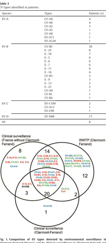
Over the study period, 48 EV types were detected in wastewater and 26 in patients referred to the teaching hospital of Clermont-Ferrand (Fig. 1). Of the 26 types identified in the patients, 22 (85%) were detected in wastewater. More than half (13/22, 60%) were assigned to species B. The EV types EV-A120, EV-D68, E#7 and E#13 were detected only by clinical surveillance and 26 types only by environmental surveillance.
Phylogenetic analysis showed high intra-type genetic diversity, a pattern indicating that multiple viral lineages co-circulated within the community (Fig. 2). High genetic diversity was observed for the CV-B5 and CV-A6 types, both of which were detected in wastewater and patients, and for other types detected only in wastewater (e.g. CV-A11). Most viral sequences (54%) recorded in patients clustered with wastewater OTUs, evidence of close genetic relationships between viruses within the community and those detected in patients. Marked differences were observed among EV types in the genetic clustering rate of clinical sequences with wastewater OTUs. This rate ranged from 0% (CV-A2, E#6, and E#25) to 75% (CV-A6) and 100% (E#11). The CV-B5 sequences
Table 1
Detection of EV types in wastewater and evolution of their abundance in the successive steps of the bio-informatics analysis.
species types before mistagging detection after mistagging cleaning after data normalisation
sequences (n) sequences (%) OTUs (n) sequences (n) sequences (%) OTUs (n) sequences (n) sequences (%) OTUs (n) EV-A CV-A16 365 880 18,3 199 291 124 18,8 19 58 344 13,3 19 CV-A5 163 623 8,2 333 103 424 6,7 14 32 404 7,4 14 EV-A119 72 236 3,6 138 41 054 2,7 6 11 098 2,5 6 CV-A2 30 844 1,5 93 16 641 1,1 4 5 222 1,2 4 CV-A4 25 517 1,3 145 14 866 1,0 4 2 474 0,6 4 CV-A12 18 355 0,9 14 7 576 0,5 3 1 980 0,5 3 EV-A76 9 516 0,5 8 8 215 0,5 1 2 217 0,5 1 CV-A6 703 0,0 14 - - - -EV-A89 676 0,0 4 529 0,0 1 276 0,1 1 CV-A14 490 0,0 11 - - - -CV-A8 252 0,0 4 - - - -EV-A71 62 0,0 5 - - - -EV-A90 22 0,0 2 - - - -total EV-A 688 176 34,4 970 483 429 31,3 52 114 015 26,0 52 EV-B CV-B5 705 235 35,2 868 606 460 39,2 43 192 616 43,9 43 E#9 72 306 3,6 89 56 796 3,7 7 17 021 3,9 7 E#33 46 597 2,3 24 45 525 2,9 4 13 081 3,0 4 CV-B2 36 093 1,8 43 26 971 1,7 5 8 582 2,0 5 E#11 33 361 1,7 72 23 125 1,5 3 5 475 1,2 3 E#6 22 469 1,1 17 19 269 1,2 1 3 875 0,9 1 E#14 21 476 1,1 23 20 180 1,3 4 8 843 2,0 4 CV-A9 20 377 1,0 77 4 774 0,3 6 1 831 0,4 6 CV-B4 14 630 0,7 145 8 055 0,5 2 2 950 0,7 2 CV-B1 12 691 0,6 30 4 403 0,3 4 889 0,2 4 E#16 9 015 0,5 18 2 839 0,2 2 799 0,2 2 CV-B3 6 161 0,3 9 4 937 0,3 1 1 631 0,4 1 E#18 5 277 0,3 25 2 777 0,2 3 623 0,1 3 E#5 5 011 0,3 15 2 170 0,1 1 631 0,1 1 CV-B6 4 771 0,2 6 4 680 0,3 1 4 275 1,0 1 E#1 1 708 0,1 4 1 383 0,1 1 300 0,1 1 E#20 729 0,0 3 705 0,0 1 170 0,0 1 E#25 477 0,0 6 - - - -E#21 80 0,0 3 - - - -E#30 50 0,0 3 - - - -EV-B78 45 0,0 1 - - - -E#3 38 0,0 2 - - - -E#27 2 0,0 1 - - - -total EV-B 1 018 599 50,9 1484 835 049 54,0 89 263 592 60,1 89 EV-C CV-A11 136 481 6,8 186 116 318 7,5 13 27 133 6,2 13 CV-A22 79 259 4,0 72 56 409 3,6 5 15 980 3,6 5 EV-C109 20 189 1,0 65 9 992 0,6 9 2 929 0,7 9 EV-C99 20 085 1,0 52 11 907 0,8 3 8 543 1,9 3 CV-A1 15 606 0,8 32 14 636 0,9 5 3 140 0,7 5 CV-A13 10 597 0,5 15 7 089 0,5 2 1 891 0,4 2 CV-A20 10 133 0,5 5 10 036 0,6 1 931 0,2 1 CV-A24 1 909 0,1 7 1 776 0,1 1 596 0,1 1 EV-C116 122 0,0 6 - - - -Sabin-like-PV-1 16 0,0 1 - - - -EV-C105 4 0,0 1 - - - -CV-A19 3 0,0 1 - - - -total EV-C 294 404 14,7 443 228 163 14,8 39 61 143 13,9 39 total EV 2 001 179 100,0 2897 1 546 641 100,0 180 438 750 100,0 180 Table 2
Clinical manifestations in the 130 hospitalized patients from October 2014 to October 2015.
Symptoms Patients (n)
meningitis/neurological disease 52 infection in pregnant women and neonates 27 respiratory infection 23
febrile syndrome 12
myocarditis/pericarditis 4 examination for international adoption dossier 4 hand foot and mouth disease 2
diarrhea 2
not specified 4
M. Bisseux et al. / Water Research 169 (2020) 115246
detected in patients clustered with OTUs (29/30, 97%) and one major genetic cluster included OTUs collected in MarcheOctober 2015 and clinical sequences detected in AprileSeptember 2015. One OTU detected in December 2014 only in wastewater was
genetically distant from all other sequences and was identified as CV-B5 genogroup A by phylogenetic analysis (data not shown).
3.5. Temporal dynamics of viral diversity
The temporal distribution was performed for wastewater sam-ples after normalisation of data and for clinical data using only local infection in Clermont-Ferrand city. On the basis of the viral di-versity observed, distinct patterns defining four successive periods were distinguished (Fig. 3). Between 28 October, 2014 and 3 March 2015, half of the viral sequences (50.5%) in wastewater samples were assigned to types of the EV-A species. The samples contained a range of 6e12 EV types, of which CV-A16 was predominant (35.2% of the total sequences collected over the period). During the same period, the viral sequences assigned to the EV-C species repre-sented 27.5% of the total sequences. CV-A11 (15.6%) and CV-A22 (7.3%) were, respectively, the second and third most frequent types. The EV-B species accounted for 22% of sequences, and the most frequent types were E#33 (6.3%), CV-B2 (5.2%), and CV-B5 (3.3%). The first CV-B5 detected in December 2014 was identified as belonging to genogroup A while the following sequences detected during this period and after were identified as belonging to genogroup B.
The total viral load determined in the wastewater samples during the four months was the lowest of the whole study period with a mean 1,057 (range 102e1,678) viral genome copies/mL. Clinical surveillance detected few EV infection cases except in October and November 2014, two months marked by 17 respiratory infections associated with EV-D68. CV-A16, the predominant EV type in wastewater, was associated with no clinical cases. EV-C109 was associated with two respiratory infections and was detected in wastewater.
During the second period, 16 March 2015 to 18 August 2015, the CV-B5 sequences accounted for 77.1% of the total sequences and ranged between 40% in a sample collected in May to 100% in a sample collected in June. With 9.1% of the total viral sequences, CV-A5 was the second most frequent type. The total viral load in wastewater was 4,072 (range 290e4072) genome copies/mL. The seasonal peak of clinical EV infections occurred during this second period, in July 2015 (n ¼ 56/130 cases, 43%). CV-B5 was the most frequent type identified in patients (n ¼ 24/56). Environmental surveillance detected CV-B5 in March 2015 before the first clinical case in May 2015 and the summer meningitis outbreak in July. During this period, 16 other EV types were also associated with clinical cases and assigned to three EV species (B, n ¼ 11, A, n ¼ 4 and C, n ¼ 1), but only half were detected in wastewater.
During the third period (September 2015), the mean viral load was the highest determined over the year: 17,046 (range 2769e17046) copies/mL. The proportion of all the EV-B types, including CV-B5, dropped to 64% of the total number of sequences. Two other EV types, EV-C99 and CV-A22, accounted for 21.4% of the total number of sequences. The number of clinical cases dropped to 10, 9 of which were associated with 5 types of EV-B species and 1 with CV-A6 (A species).
The total viral load (mean 2,107 [range 138e4075] genome copies/mL) decreased over the fourth period, from 14 Octobere26 October 2015, and the distribution of EV types shifted again with 89.7% and 10.3% of sequences being assigned to EV species B and A, respectively. CV-B5 was once again predominant in wastewater (74.8% of viral sequences) while the number of clinical cases remained low.
4. Discussion
This one-year pilot study to analyse wastewater sampled from a
Table 3
EV types identified in patients.
Species Types Patients (n)
EV-A CV-A4 4 CV-A6 4 CV-A2 3 CV-A5 1 CV-A8 1 EV-A71 1 EV-A120 1 EV-B CV-B5 30 E#25 8 E#18 7 E#5 6 E#6 6 E#7 6 E#11 6 E#16 4 CV-B3 3 E#9 3 E#13 2 E#21 2 CV-A9 1 CV-B1 1 CV-B4 1 EV-C EV-C109 2 CV-A13 1 EV-C99 1 EV-D EV-D68 17 NT 8
Fig. 1. Comparison of EV types detected by environmental surveillance in Clermont-Ferrand (France) and by clinical surveillance in Clermont-Ferrand and nationwide during the period 2014-2015.
The 48 EV types detected in the WWTP and types associated with patient hospitali-zation in Clermont-Ferrand (n ¼ 26) and France (n ¼ 45) were compared and repre-sented in a Venn diagram. Types in the intersection were detected by both surveillance systems. EV-A species is shown in blue, EV-B species in red, EV-C species in green and EV-D species in black.
WWTP serving an urban community showed a highly dynamic co-circulation of 48 EV types assigned to the taxonomic species A, B, and C. Our results were consistent with those of a previous Amer-ican study (Brinkman et al., 2017) that described a similar seasonal distribution of EV species with an alternative predominance of EV-A and EV-B, a major peak of EV-B during summer and fall and the circulation of EV-C throughout the year. Unlike in our study, the
highest viral concentration in the sewage from the two WWTP analysed was observed in July. To our knowledge, our study yields the first environmental and clinical data on the surveillance of non-polio EV infections without cell culture bias concomitantly ob-tained in a same urban population. The results showed the con-sistency between the two surveillance systems: 22/26 EV types identified in patients referred to the teaching hospital serving the
Fig. 2. Phylogenetic network of EV.
All EV types detected during the 2014e2015 study period are represented in phylogenetic networks for species A (A), B (B) and C (C). Strains detected in wastewater are in green and clinical strains in red. Networks were built with Bionumerics software and maximum parsimony methods. Strains with less than 2% nucleotide differences were grouped into a single sphere with a diameter proportional to the number of strains inside. For CV-B5, the two genogroups A and B, as defined in a previous study (Henquell et al., 2013), are indicated.
M. Bisseux et al. / Water Research 169 (2020) 115246
population under study were also detected in wastewater moni-toring. There were close phylogenetic relationships between virus sequences detected in patients and wastewater. Yet, analysis of the wastewater showed that EVs associated with community-acquired infections in the urban population were much more diverse than those associated with infections reported in the clinical setting during the same period. The net number of EV types (intertype diversity) was higher in the whole urban population than in
patients. Notably, the intertype diversity within the urban popu-lation was actually similar to that reported in patients admitted to hospitals nationwide during the same period. These findings sup-port the conclusion that UDS with a multiplexed amplicon approach can be used in raw urban wastewater to investigate the ecology and transmission of EVs and assess the burden of these enteric viruses in the general population.
The epidemiology of EV infections in 2014e2015 was marked by
Fig. 3. Quantity and diversity of EVs detected in the Clermont-Ferrand area over one year by the two surveillance systems.
(A) Relative abundance of the 33 conserved EV types detected in the WWTP of Clermont-Ferrand during 2014e2015 are given for each sample.(B) The quantity of EV expressed as genome copies/mL. of WWTP water was determined in a previous study (Bisseux et al., 2018).(C) The diversity of EV types associated with patient hospitalization occurring in the same city in the same period is represented for every two weeks preceding the mentioned date.
the epidemic circulation of CV-B5, which accounted for 44% of the sequences in wastewater and 28.5% (15/52) of meningitis cases. The abrupt upsurge of CV-B5 was clearly identified in March 2015 by wastewater analysis. The large circulation of CV-B5 was also observed by the national network for EV surveillance, which recorded this virus type as the second most frequent in France in 2015. Ten other EV-B types were co-detected with CV-B5 during the winter 2014-2015 by environmental surveillance, notably E#11 (3% of all sequences over the winter) and E#6 (2.4%). These viruses were also detected during the meningitis outbreak in the summer. In contrast, EV types E#33 (6.3%) and CV-B2 (5.2%) were only detected in wastewater. Accordingly, the winter pattern of EV di-versity in wastewater was not completely predictive of the pattern during the following seasonal outbreak. Longer follow-up studies including WWTPs serving distinct urban communities are required to provide a complete description of viral circulation dynamics. Striking differences in the distribution pattern of EV-A and EV-C types were also observed. Few infections associated with EV-A types required hospitalization but the viruses circulated widely in the community (26% of reads in wastewater), indicating that most infected individuals had subclinical infections or self-limited ill-nesses managed in ambulatory settings. CV-A16 is an example in this respect. Conversely, while CV-A6 accounted for 0.03% reads in wastewater, the virus was detected in four patients reported with meningitis, myocarditis, and HFMD (including a case of atypical HFMD clinical presentation). Our data are consistent with the emergence of CV-A6 as a cause of worldwide disease concern (Bian et al., 2015). Environmental and clinical surveillance revealed the unexpected epidemiologic pattern of the EV-C types: large circu-lation in the urban community and few disease cases (Apostol et al., 2012;Benschop et al., 2017;Harvala et al., 2014). All the three EV-C types identified in patients were traced back to wastewater. EV-C109 was detected in two patients referred for respiratory dis-eases and clustered close to viruses in wastewater. In contrast, CV-A13 and EV-C99 detected in the stools of two adopted children and in wastewater did not cluster.
Based on the structure of the sequence database and phyloge-netic techniques used in our study, the sequence assignment rate was high (99.6%) and enabled us to detect less abundant viruses. Two EV types with direct public health relevance (EV-A71 and PV-1 Sabin) were detected in wastewater and identified down to the strain level by analysis of sequence data. The low nucleotide vari-ation in the wastewater PV-1 sequence compared with that of the vaccine strain suggested no or limited circulation in the population. Our French data are consistent with earlier results from the United States and other European countries showing the importation of vaccine PV strains into polio-free countries (Benschop et al., 2017;
Delogu et al., 2018). The findings clearly show the need to maintain a high immunization coverage to avoid the occurrence of an outbreak associated with the circulation of vaccine-derived polio-viruses (VDPV), as reported in 2015 in Ukraine (Khetsuriani et al., 2017). CV-A11 and CV-A22 were among the five most abundant types in this study. These unexpectedly prevalent non-polio EV-C types could be a gene reservoir for the emergence of recombinant VDPV (Bessaud et al., 2011). We also evidenced the circulation of rare EV-A (eg -A76, -A89), EV-B (eg -B78) and EV-C (eg -C105, -C116) types previously unreported in the French population and for which few epidemiological and genomic data are available. Notably, our environmental surveillance identified ‘new’ EV-C types asso-ciated with severe respiratory diseases and neurological disease conditions, whose detection in the faeces was considered to be difficult (Holm-Hansen et al., 2016). Our findings suggest that the circulation of these viruses in the community could be higher than anticipated from the number of reported clinical infections and are consistent with suggestions to enhance the surveillance of EV-C
species in respiratory specimens (Barnadas et al., 2017;Van Leer-Buter et al., 2016).
Our study has two limitations. First, although the pan-EV primers used included a high number of degenerate positions, variations in the levels of PCR amplification exist among EV types and could explain why we did not detect EV-D68. This virus was previously detected, however, by type-specific amplification, confirmed by direct Sanger sequencing in 7/27 samples (Bisseux et al., 2018). Second, we showed that incorrect assignment of reads caused the random spread of signals within multiplexed samples and artificially inflated the diversity detected in each sample. This bias, referred to as mistagging (Esling et al., 2015) or tag jump (Schnell et al., 2015), has been rarely documented and taken into account in UDS analyses despite its critical impact on the quality of data collected for ecological and epidemiological studies. Minimising incorrect assignments and obtaining reliable results is a real challenge for metabarcoding studies on Illumina sequencing platforms. Strategies including controls must be incorporated into the experimental design to perform sequence data filtering (Esling et al., 2015;Galan et al., 2016). Despite these limits, amplicon deep-sequencing in wastewater for surveillance and epidemiological studies clearly surpasses earlier techniques used in this field. With the implementation of robust quality controls, monitoring EVs in wastewater can be implemented as a supplementary surveillance system of the community at large to complement clinical surveillance.
5. Conclusion
The combination of temporal wastewater sampling, efficient virus concentration from samples, analysis with amplicon deep-sequencing and robust sequence assignment allow the detection of a large array of EV types while avoiding the bias caused by cell culture. Sustained environmental surveillance is an advanced approach for a better understanding of the diversity of the EV landscape within an urban community. The environmental findings of this study show the circulation of clinically uncommon EVs, including respiratory EV-C species strains and Sabin-like PV-1, and provide a powerful complement to the clinical data.
Declaration of competing interest
The authors declare that they have no known competing financial interests or personal relationships that could have appeared to influence the work reported in this paper.
Acknowledgements
We are grateful to Nathalie Rodde and Emilie Leroy for their excellent technical assistance in enterovirus genotyping. We thank Jeffrey Watts for revision of the English manuscript.
This research did not receive any specific grant from commercial or not-for-profit sectors. The Centre National de R!ef!erence des Ent!erovirus-Parechovirus is supported by an annual public grant from the national Agency Sant!e publique France.
Appendix A. Supplementary data
Supplementary data to this article can be found online at
https://doi.org/10.1016/j.watres.2019.115246. References
Adeniji, J.A., Faleye, T.O.C., 2014. Isolation and identification of enteroviruses from sewage and sewage-contaminated water in Lagos, Nigeria. Food Environ. Virol.
M. Bisseux et al. / Water Research 169 (2020) 115246
6 (2), 75e86.https://doi.org/10.1007/s12560-014-9137-5.
Antona, D., Kossorotoff, M., Schuffenecker, I., Mirand, A., Leruez-Ville, M., Bassi, C., Aubart, M., Moulin, F., L!evy-Bruhl, D., Henquell, C., Lina, B., Desguerre, I., 2016. Severe paediatric conditions linked with EV-A71 and EV-D68, France, May to October 2016. Euro Surveill. 21 (46), 30402.
https://doi.org/10.2807/1560-7917.ES.2016.21.46.30402.
Apostol, L.N.G., Imagawa, T., Suzuki, A., Masago, Y., Lupisan, S., Olveda, R., Saito, M., Omura, T., Oshitani, H., 2012. Genetic diversity and molecular characterization of enteroviruses from sewage-polluted urban and rural rivers in the Philippines. Virus Genes 45 (2), 207e217.https://doi.org/10.1007/s11262-012-0776-z. Barnadas, C., Midgley, S.E., Skov, M.N., Jensen, L., Poulsen, M.W., Fischer, T.K., 2017.
An enhanced Enterovirus surveillance system allows identification and char-acterization of rare and emerging respiratory enteroviruses in Denmark, 2015-16. J. Clin. Virol. 93, 40e44.https://doi.org/10.1016/j.jcv.2017.05.017. Benschop, K.S.M., van der Avoort, H.G., Jusic, E., Vennema, H., van Binnendijk, R.,
Duizer, E., 2017. Polio and measles down the drain: environmental enterovirus surveillance in The Netherlands, 2005-2015. Appl. Environ. Microbiol. 83
https://doi.org/10.1128/AEM.00558-17e00558-17.
Berchenko, Y., Manor, Y., Freedman, L.S., Kaliner, E., Grotto, I., Mendelson, E., Huppert, A., 2017. Estimation of polio infection prevalence from environmental surveillance data. Sci. Transl. Med. 9 (383) https://doi.org/10.1126/
scitranslmed.aaf6786.
Bessaud, M., Joffret, M.-L., Holmblat, B., Razafindratsimandresy, R., Delpeyroux, F., 2011. Genetic relationship between cocirculating human enteroviruses species C. PLoS One 6 (9), e24823.https://doi.org/10.1371/journal.pone.0024823. Bian, L., Wang, Y., Yao, X., Mao, Q., Xu, M., Liang, Z., 2015. Coxsackievirus A6: a new
emerging pathogen causing hand, foot and mouth disease outbreaks world-wide. Expert Rev. Anti Infect. Ther. 13 (9), 1061e1071.https://doi.org/10.1586/
14787210.2015.1058156.
Bisseux, M., Colombet, J., Mirand, A., Roque-Afonso, A.-M., Abravanel, F., Izopet, J., Archimbaud, C., Peigue-Lafeuille, H., Debroas, D., Bailly, J.-L., Henquell, C., 2018. Monitoring human enteric viruses in wastewater and relevance to infections encountered in the clinical setting: a one-year experiment in central France,
2014 to 2015. Euro Surveill. 23 (7), 17-00237.
Brinkman, N.E., Fout, G.S., Keely, S.P., 2017. Retrospective surveillance of wastewater to examine seasonal dynamics of enterovirus infections. mSphere 2 (3).https://
doi.org/10.1128/mSphere.00099-17e00099-17.
Casas-Alba, D., de Sevilla, M.F., Valero-Rello, A., Fortuny, C., García-García, J.-J., Ortez, C., Muchart, J., Armangu!e, T., Jordan, I., Luaces, C., Barrabeig, I., Gonz!alez-Sanz, R., Cabrerizo, M., Mu~noz-Almagro, C., Launes, C., 2017. Outbreak of brainstem encephalitis associated with enterovirus-A71 in Catalonia, Spain (2016): a clinical observational study in a children’s reference centre in Cata-lonia. Clin. Microbiol. Infect.https://doi.org/10.1016/j.cmi.2017.03.016. Cowger, T.L., Burns, C.C., Sharif, S., Gary, H.E., Iber, J., Henderson, E., Malik, F., Zahoor
Zaidi, S.S., Shaukat, S., Rehman, L., Pallansch, M.A., Orenstein, W.A., 2017. The role of supplementary environmental surveillance to complement acute flaccid paralysis surveillance for wild poliovirus in Pakistan - 2011-2013. PLoS One 12 (7), e0180608.https://doi.org/10.1371/journal.pone.0180608.
Delogu, R., Battistone, A., Buttinelli, G., Fiore, S., Fontana, S., Amato, C., Cristiano, K., Gamper, S., Simeoni, J., Frate, R., Pellegrinelli, L., Binda, S., Veronesi, L., Zoni, R., Castiglia, P., Cossu, A., Triassi, M., Pennino, F., Germinario, C., Balena, V., Cicala, A., Mercurio, P., Fiore, L., Pini, C., Stefanelli, P., 2018. Poliovirus and other enteroviruses from environmental surveillance in Italy, 2009-2015. Food Envi-ron. Virol. 10 (4), 333e342.https://doi.org/10.1007/s12560-018-9350-8. Esling, P., Lejzerowicz, F., Pawlowski, J., 2015. Accurate multiplexing and filtering for
high-throughput amplicon-sequencing. Nucleic Acids Res. 43 (5), 2513e2524.
https://doi.org/10.1093/nar/gkv107.
Galan, M., Razzauti, M., Bard, E., Bernard, M., Brouat, C., Charbonnel, N., Dehne-Garcia, A., Loiseau, A., Tatard, C., Tamisier, L., Vayssier-Taussat, M., Vignes, H., Cosson, J.-F., 2016. 16S rRNA amplicon sequencing for epidemiological surveys of bacteria in wildlife. mSystems 1 (4).
https://doi.org/10.1128/mSys-tems.00032-16e00032-16.
Harvala, H., Calvert, J., Van Nguyen, D., Clasper, L., Gadsby, N., Molyneaux, P., Templeton, K., McWilliams Leitch, C., Simmonds, P., 2014. Comparison of diagnostic clinical samples and environmental sampling for enterovirus and parechovirus surveillance in Scotland, 2010 to 2012. Euro Surveill. 19 (15),
20772.
Hassel, C., Mirand, A., Farkas, A., Diedrich, S., Huemer, H.P., Peigue-Lafeuille, H., Archimbaud, C., Henquell, C., Bailly, J.-L., HFMD French Study Network, 2017. Phylogeography of coxsackievirus A16 reveals global transmission pathways and recent emergence and spread of a recombinant genogroup. J. Virol. 91 (18)
https://doi.org/10.1128/JVI.00630-17.
Hassel, C., Mirand, A., Lukashev, A., TerletskaiaLadwig, E., Farkas, A., Schuffenecker, I., Diedrich, S., Huemer, H.P., Archimbaud, C., Peigue-Lafeuille, H., Henquell, C., Bailly, J.-L., 2015. Transmission patterns of human enterovirus 71 to, from and among European countries, 2003 to 2013. Euro Surveill. 20 (34), 30005.https://doi.org/10.2807/1560-7917.ES.2015.20.34.30005.
Henquell, C., Mirand, A., Richter, J., Schuffenecker, I., B€ottiger, B., Diedrich, S., Ter-letskaia-Ladwig, E., Christodoulou, C., Peigue-Lafeuille, H., Bailly, J.-L., 2013. Phylogenetic patterns of human coxsackievirus B5 arise from population
dynamics between two genogroups and reveal evolutionary factors of molec-ular adaptation and transmission. J. Virol. 87 (22), 12249e12259. https://
doi.org/10.1128/JVI.02075-13.
Holm-Hansen, C.C., Midgley, S.E., Fischer, T.K., 2016. Global emergence of entero-virus D68: a systematic review. Lancet Infect. Dis. 16 (5), e64ee75.https://
doi.org/10.1016/S1473-3099(15)00543-5.
Khetsuriani, N., Perehinets, I., Nitzan, D., Popovic, D., Moran, T., Allahverdiyeva, V., Huseynov, S., Gavrilin, E., Slobodianyk, L., Izhyk, O., Sukhodolska, A., Hegazi, S., Bulavinova, K., Platov, S., O’Connor, P., 2017. Responding to a cVDPV1 outbreak in Ukraine: implications, challenges and opportunities. Vaccine 35 (36), 4769e4776.https://doi.org/10.1016/j.vaccine.2017.04.036.
Lang, M., Mirand, A., Savy, N., Henquell, C., Maridet, S., Perignon, R., Labbe, A., Peigue-Lafeuille, H., 2014. Acute flaccid paralysis following enterovirus D68
associated pneumonia, France, 2014. Euro Surveill. 19 (44), 20952.
Manor, Y., Shulman, L.M., Kaliner, E., Hindiyeh, M., Ram, D., Sofer, D., Moran-Gilad, J., Lev, B., Grotto, I., Gamzu, R., Mendelson, E., 2014. Intensified environmental surveillance supporting the response to wild poliovirus type 1 silent circulation
in Israel, 2013. Euro Surveill. 19 (7), 20708.
Mirand, A., Archimbaud, C., Henquell, C., Michel, Y., Chambon, M., Peigue-Lafeuille, H., Bailly, J.-L., 2006. Prospective identification of HEV-B enteroviruses during the 2005 outbreak. J. Med. Virol. 78 (12), 1624e1634.https://doi.org/
10.1002/jmv.20747.
Mirand, A., Henquell, C., Archimbaud, C., Chambon, M., Charbonne, F., Peigue-Lafeuille, H., Bailly, J.-L., 2008. Prospective identification of enteroviruses involved in meningitis in 2006 through direct genotyping in cerebrospinal fluid. J. Clin. Microbiol. 46 (1), 87e96.https://doi.org/10.1128/JCM.01020-07. Mirand, A., le Sage, F.V., Pereira, B., Cohen, R., Levy, C., Archimbaud, C.,
Peigue-Lafeuille, H., Bailly, J.-L., Henquell, C., 2016. Ambulatory pediatric surveillance of hand, foot and mouth disease as signal of an outbreak of coxsackievirus A6 infections, France, 2014-2015. Emerg. Infect. Dis. 22 (11), 1884e1893.https://
doi.org/10.3201/eid2211.160590.
Mirand, A., Schuffenecker, I., Henquell, C., Billaud, G., Jugie, G., Falcon, D., Mahul, A., Archimbaud, C., Terletskaia-Ladwig, E., Diedrich, S., Huemer, H.P., Enders, M., Lina, B., Peigue-Lafeuille, H., Bailly, J.-L., 2010. Phylogenetic evidence for a recent spread of two populations of human enterovirus 71 in European countries. J. Gen. Virol. 91 (Pt 9), 2263e2277.https://doi.org/10.1099/vir.0.021741-0. Nix, W.A., Oberste, M.S., Pallansch, M.A., 2006. Sensitive, seminested PCR
amplifi-cation of VP1 sequences for direct identifiamplifi-cation of all enterovirus serotypes from original clinical specimens. J. Clin. Microbiol. 44 (8), 2698e2704.https://
doi.org/10.1128/JCM.00542-06.
Othman, I., Mirand, A., Slama, I., Mastouri, M., Peigue-Lafeuille, H., Aouni, M., Bailly, J.-L., 2015. Enterovirus migration patterns between France and Tunisia. PLoS One 10 (12), e0145674.https://doi.org/10.1371/journal.pone.0145674. Polio Eradication & Endgame Strategic Plan 2013-2018, 2013
Price, M.N., Dehal, P.S., Arkin, A.P., 2010. FastTree 2–approximately maximum-likelihood trees for large alignments. PLoS One 5 (3), e9490.https://doi.org/
10.1371/journal.pone.0009490.
Schnell, I.B., Bohmann, K., Gilbert, M.T.P., 2015. Tag jumps illuminated–reducing sequence-to-sample misidentifications in metabarcoding studies. Mol. Ecol. Resour. 15 (6), 1289e1303.https://doi.org/10.1111/1755-0998.12402. Schuffenecker, I., Mirand, A., Josset, L., Henquell, C., Hecquet, D., Pilorge, L.,
Petit-jean-Lecherbonnier, J., Manoha, C., Legoff, J., Deback, C., Pillet, S., Lepiller, Q., Mansuy, J.M., Marque-Juillet, S., Antona, D., Peigue-Lafeuille, H., Lina, B., 2016. Epidemiological and clinical characteristics of patients infected with entero-virus D68, France, July to December 2014. Euro Surveill. 21 (19), 30226.https://
doi.org/10.2807/1560-7917.ES.2016.21.19.30226.
Taib, N., Mangot, J.-F., Domaizon, I., Bronner, G., Debroas, D., 2013. Phylogenetic affiliation of SSU rRNA genes generated by massively parallel sequencing: new insights into the freshwater protist diversity. PLoS One 8 (3), e58950.https://
doi.org/10.1371/journal.pone.0058950.
Tamura, K., Peterson, D., Peterson, N., Stecher, G., Nei, M., Kumar, S., 2011. MEGA5: molecular evolutionary genetics analysis using maximum likelihood, evolu-tionary distance, and maximum parsimony methods. Mol. Biol. Evol. 28 (10), 2731e2739.https://doi.org/10.1093/molbev/msr121.
Tangermann, R.H., Lamoureux, C., Tallis, G., Goel, A., 2017. The critical role of acute flaccid paralysis surveillance in the Global Polio Eradication Initiative. Int. Health 9 (3), 156e163.https://doi.org/10.1093/inthealth/ihx016.
Van Leer-Buter, C.C., Poelman, R., Borger, R., Niesters, H.G.M., 2016. Newly identified enterovirus C genotypes, identified in The Netherlands through routine sequencing of all enteroviruses detected in clinical materials from 2008 to 2015. J. Clin. Microbiol. 54 (9), 2306e2314.https://doi.org/10.1128/JCM.00207-16. Volle, R., Nourrisson, C., Mirand, A., Regagnon, C., Chambon, M., Henquell, C.,
Bailly, J.-L., Peigue-Lafeuille, H., Archimbaud, C., 2012. Quantitative real-time RT-PCR assay for research studies on enterovirus infections in the central ner-vous system. J. Virol. Methods 185 (1), 142e148. https://doi.org/10.1016/
j.jviromet.2012.06.019.
Xing, W., Liao, Q., Viboud, C., Zhang, J., Sun, J., Wu, J.T., Chang, Z., Liu, F., Fang, V.J., Zheng, Y., Cowling, B.J., Varma, J.K., Farrar, J.J., Leung, G.M., Yu, H., 2014. Hand, foot, and mouth disease in China, 2008-12: an epidemiological study. Lancet Infect. Dis. 14 (4), 308e318.https://doi.org/10.1016/S1473-3099(13)70342-6.