Leptinand reproduction
11
0
0
Texte intégral
(2) Leptin and reproduction similar to structures from the family of helical cytokines that includes interleukin (IL)-2 and growth hormone (Madej et al., 1995). Further investigations with nuclear magnetic resonance analysis revealed that it is a four-helix bundle. Helix lengths and disulphide patterns suggest that leptin is a member of the shorthelix cytokine family (Kline et al., 1997). Leptin communicates nutritional status to regulatory centres in the brain and circulates in blood, bound to a family of binding proteins (Campfield et al., 1996). Leptin interacts with the proteinase inhibitor, α2 macroglobulin. The leptin–α2 macroglobulin complex is stable and is recognized by the α2 macroglobulin receptor/low density lipoprotein receptor-related protein (Birkenmeir et al., 1998). In lean subjects, with relatively low adipose tissue, the majority of circulating leptin is proteinbound. In obese individuals, the majority of leptin circulates in free form, presumably the bioactive protein, and thus obese individuals are resistant to free leptin (Hoggard et al., 1998). Leptin receptor is the product of the db gene and belongs to the class I cytokine superfamily of receptors. The full-length receptor has the signalling capabilities of the IL-6 type receptor and its helical structure is similar to this cytokine (Baumann et al., 1996; Tartaglia, 1997). Leptin receptor mRNA is expressed in the anterior pituitary, in several areas of the brain and in other tissues (Finn et al., 1998). Leptin receptor mRNA has also been detected in granulosa and theca cells (Zachow and Magoffin, 1997). The cloned leptin receptor contains two homologous segments representing potential ligand binding sites. These domains localized to amino acid residues 323–640 and between amino acid residues 428–635, have been identified as a fibronectin type 3 domain (Fong et al., 1998). In humans and rodents, two major forms of leptin receptors (OB-R) are expressed. The short form (OB-RS) is detected in many organs and is considered to lack signalling capability (Wang et al., 1997) as it has a truncated intracellular domain (Campfield et al., 1996). The long form (OBRL) with the complete intracellular domain, predominates in the hypothalamus, but is expressed in low amounts in peripheral tissues. OB-RL transduces an intracellular signal by activation of the signal transducer and activator of transcription (STAT) proteins, induction of acute-phase plasma proteins, and synergism with IL-1 and tumour necrosis factor (TNF)-α in hepatoma cell lines (Wang et al., 1997). The leptin receptor could also exert a control on its own leptin synthesis in rat adipose tissue (Zhang et al., 1997). Soluble leptin receptor has also been reported in humans (Lewandowski et al., 1999). Leptin receptor activation involves the activation of STAT3, a member of the STAT family of proteins. STAT3 can also be activated by a variety of cytokines, e.g. IL-6, granulocyte-colony stimulating factor (G-CSF) and epidermal growth factor (EGF) (Takeda et al., 1998). The OB-R might exert a signalling effect similar to G-CSF receptor (G-CSFR), leukaemia inhibitory factor receptor (LIFR) or the glycoprotein, gp130, through the activation of receptor-associated kinases of the Janus kinase family. Activation of these kinases phosphorylate and activate in turn DNA binding activity of signal transducers and activators of transcription proteins (STAT1, STAT3 and STAT5) (Baumann et al., 1996).. 291. Neuroendocrine, hormonal and paracrine regulation of leptin Many leptin effects on food intake and energy expenditure are thought to be mediated centrally via neurotransmitters, e.g. neuropeptide Y (Houseknecht et al., 1998). Catecholamine metabolism seems also to have a role in the regulation of leptin. Plasma epinephrine has been negatively correlated with leptin concentrations (Mills et al., 1998). However, using an inhibitor of catecholamine synthesis (α-methyl-para-tyrosine) it was not possible to demonstrate that noradrenaline represents the afferent signal from the central nervous system which modulates leptin release from adipocytes in human (Zimmermann et al., 1998). Glucocorticoids and insulin participate in the regulation of leptin metabolism. Glucocorticoids and insulin act as long-term regulators of leptin in omental (Om) and sub-abdominal (Sc) adipose tissue (Rusell et al., 1998). Obese children treated with a single dose of dexamethasone showed a significant increase in leptin concentration, indicating that glucocorticoids up-regulate human leptin (Kiess et al., 1996). An inverse correlation between cortisol and leptin has been reported in lean or obese subjects, either after food intake or fasting (Korbonits et al., 1997). This study revealed that leptin is a powerful indicator of insulin values and body mass index (BMI); however, leptin concentrations did not change acutely after food administration. Interestingly, insulin did not regulate acute leptin concentrations in lean or obese subjects, nor was leptin secretion different in diabetic obese and lean patients. Basal concentrations of insulin and leptin were positively correlated only in insulin-sensitive individuals (Sinha and Caro, 1998). High concentrations of soluble leptin receptor might be involved in the leptin resistance found in insulindependent diabetic women (Lewandowski et al., 1999). Some experimental data suggest that leptin could regulate the expression of insulin. The molecular mechanisms underlying the effect of leptin on insulin secretion are unknown. It has been suggested that physiological concentrations of leptin in normal rodents modulate a potentiation of glucose-induced insulin secretion involving cyclic AMP or phospholipase C/protein kinase activation (Poitout et al., 1998). In obese mice, leptin inhibits transcription of the proinsulin gene by altering the transcription factor binding (STAT5) element, conferring glucose responsivity and secretion of insulin by activation of ATP-sensitive potassium channels. Leptin may inhibit insulin gene transcription and secretion in pancreatic β-cells by different mechanisms (Seufert et al., 1999). However, in ovariectomized ewes, leptin treatment raised plasma lactate and fatty acid concentrations but had no effect on glucose or insulin concentrations (Henry et al., 1999). Leptin secretion in humans shows gender differences, independent of adiposity. Leptin concentrations are three times higher in women than in men (Sinha and Caro, 1998). Dexamethasone and oestradiol stimulate leptin release only in females. In vitro, Om adipose tissue of women secretes more leptin than that of men. However, regardless of gender, progesterone or oestrone did not modify leptin secretion (Casabiell et al., 1998). The sexual dimorphism of leptin concentrations could be due to the effect of androgens. Obesity in women (opposite to that of men), is associated with increased ovarian androgen production, and this difference might be due to the action of leptin (Conway and Jacobs, 1997). In addition, rapid.
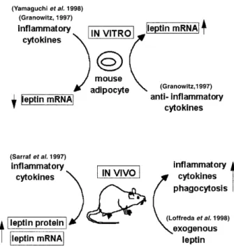
(3) 292. R.R.González et al.. Figure 1. Leptin and inflammatory response. In-vitro mouse adipocytes differentially regulate leptin mRNA expression by actions of inflammatory or anti-inflammatory cytokines. In-vivo response of genetically deficient leptinrelated mouse by cytokines actions is contrary to in-vitro response. Leptin is up-regulated by inflammatory cytokines, i.e. interleukin-1 (IL-1) and tumour necrosis factor-α (TNF-α) and also it could up-regulate macrophage synthesis of these cytokines. Leptin is linked to transforming growth factor-β (TGF-β) action (Granowitz, 1997; Sarraf et al., 1997; Loffreda et al., 1998; Yamaguchi et al., 1998).. fluctuations in plasma concentrations of leptin have been found to be negatively correlated with those of adrenocorticotropic hormone (ACTH) (Licinio et al., 1997).. Leptin is related to inflammatory response Leptin is a key hormone coupling the immune system and energy balance (Finck et al., 1998). Inflammatory cytokines influence leptin concentrations, but leptin may also induce the synthesis of inflammatory cytokines in vivo and in vitro (Figure 1). Mouse 3T3-L cells that differentiated into adipocytes, expressed higher levels of leptin mRNA. In these cells, IL-1β, IL-6, IL-11, TNF-α (inflammatory cytokines) decreased but transforming growth factor (TGF)-β (anti-inflammatory cytokine) increased leptin mRNA (Loffreda et al., 1998). Mouse parametrial adipocytes can produce TNF-α, which could in turn inhibit leptin mRNA expression through TNF receptor (TNF-R1). Similar results have been found with cultures of human adipocytes. The addition of human recombinant TNF-α to adipocytes from the subcutaneous fat of pregnant women inhibited leptin secretion (Yamaguchi et al., 1998). In contrast, other investigators have found a stimulatory effect of TNF-α on leptin secretion from mouse 3T3L1 adipocytes. A rapid stimulation of leptin accumulation in cultures of mouse adipocytes was observed after 6 h of TNF-α treatment and this release of leptin was completely inhibited by brefeldin A, an inhibitor of TNF-α. These findings indicate that TNF-α can act directly on adipocytes, regulating the release of a preformed pool of leptin (Kirchgessner et al., 1997).. Inflammatory cytokines also induce the synthesis of leptin in vivo. In female mice, administration of TNF, IL-1 and LIF increased serum leptin concentrations and mRNA expression in fat (Sarraf et al., 1997). Lethal effects of TNF in leptin-defective mice were reduced by exogenous leptin. Thus, leptin could participate in protective mechanisms against the autoaggressive effects of the immune system (Takahashi et al., 1999). TNF-α and IL-1 are mediators of response to lipopolysaccharide (LPS), inducing anorexia and increasing leptin mRNA expression in hamster adipose tissue. Induction of leptin during the host response to infection may contribute to the anorexia associated with infection (Grunfeld et al., 1997). Injection of LPS or turpentine into IL-1β-deficient (–/–) mice did not elevate plasma leptin concentrations, unlike IL-1β (+/+) mice. This finding demonstrated that IL-1β is essential for leptin induction by both LPS and turpentine in mice (Fanggioni et al., 1998). In humans, administration of recombinant human IL-1α to cancer patients increased serum leptin concentrations (Janik et al., 1997). TNF-α infusion to patients with solid tumours also increased serum leptin concentrations (Zumbach et al., 1997). Studies in rodents with genetic abnormalities in leptin (ob/ob) or leptin receptor (db/db) demonstrated that exogenous leptin upregulated both macrophage phagocytosis and the production of inflammatory cytokines (Loffreda et al., 1998). It has been also reported that leptin enhanced cytokine production and phagocytosis by murine peritoneal macrophages, although human leptin has exhibited no capacity to stimulate cell survival or proliferation in cultures of murine or human marrow cells. These data demonstrate that leptin may also be able to regulate aspects of haemopoiesis and macrophage function (Gainsford et al., 1996). Leptin may be implicated in the regulation of apoptosis in the adipose tissue. Leptin mRNA was higher in Sc than in Om adipocytes (Montague et al., 1997, 1998). This relationship was inverse for cellular inhibitor of apoptosis, the protein-2 (cIAP2) mRNA. Depot-specific differences (in leptin and cIAP2) could play a role in the regulation of apoptosis in adipose tissue (Montague et al., 1998). In addition, reduction of adipose tissue through apoptosis has been observed after intra-cerebroventricular administration of leptin in rats. The adipose tissue of leptin-treated rats showed the characteristic features of apoptosis, e.g. internucleosomal fragmentation of genomic DNA, increased amounts of DNA strand breaks and a reduction in total DNA content and cell volume (Qian et al., 1998). However, the effect of leptin on apoptosis differs in other cell types. In myeloid leukaemia cell lines, leptin had proliferative and anti-apoptotic properties. Leptin reduced apoptosis induced by cytokines in these cell lines (Konopleva et al., 1999).. Leptin as a signal of nutritional status linked to the reproductive process The amount of body fat stored is known to influence fertility, indicating a link between adipose tissue and the reproductive system (Frisch, 1990). An interesting hypothesis is that leptin is a peripheral signal indicating the adequacy of nutritional status for reproductive function (Tataranni et al., 1997). Therefore, it seems possible that low leptin concentrations indicate a status of inadequate nutritional stores and could prevent an unwanted.
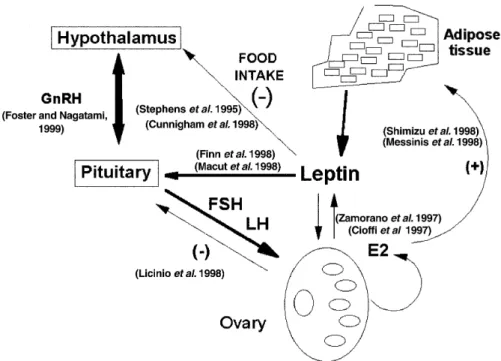
(4) Leptin and reproduction. 293. Figure 2. Hypothetical model of relationships between leptin and the hypothalamus–pituitary–ovary axis. Leptin is a molecular signal from adipose tissue that regulates the food intake, presumably through neuropeptide Y actions. Recent data support the idea that leptin binds to the short form of its receptor to transport itself into the arcuate hypothalamic region. Neuropeptide Y synthesis is decreased, diminishing the appetite and increasing whole-body energy expediture and weight loss. Therefore, leptin communicates the size of the adipose reserve to the hypothalamus. Leptin is also synthesized in the reproductive tissues and it is related to the hypothalamus–pituitary–ovary axis function. Gonadotrophin-releasing hormone (GnRH) and LH pulses can be related to leptin actions. Leptin may directly regulate the function of the reproductive organs and, via paracrine effects, may regulate oestradiol synthesis. In addition, oestradiol concentrations could also influence leptin synthesis. Leptin may be a signal of metabolic status to the reproductive system (Stephens et al., 1995; Cioffi et al., 1997; Shimizu et al., 1997; Zamorano et al., 1997; Finn et al., 1998; Licinio et al., 1998; Macut et al., 1998; Messinis et al., 1998; Cunningham et al., 1999; Foster and Nagatami, 1999).. pregnancy which demands additional energy to support a growing fetus. However, leptin is not a sensitive marker of nutritional status (Korbonits et al., 1997). In starved mice, the lack of reproductive function coincides with a fall of plasma leptin concentrations and several neuroendocrine changes. Exogenous leptin injections to these mice restore fertility (Ahima et al., 1997). In addition, leptin infusions are able to restore ovulatory function in an animal model of starvation. However, fasting-induced anoestrus was not reversed by leptin when glucose oxidation was blocked with 2-deoxy-D-glucose and fatty acid oxidation with methyl palmoxirate (Schneider et al., 1998). Nevertheless, differences in leptin plasma concentrations in long-term conditions need not indicate different fertility rates. Clark and Henry (1999) have recently discussed whether chronic undernutrition or differences in the long-term maintenance of body fat could affect the reproductive function. At the present time, the mechanism and reproductive consequences of low leptin status remain unknown (Miller et al., 1998). On the other hand, human obesity is not characterized by leptin deficiency. An attractive idea is that obesity could be a state of leptin resistance, but evidence for this assumption is limited (Conway and Jacobs, 1997). A few cases of congenital leptin deficiency have been reported in humans. These were due either to low leptin synthesis (Strobel et al., 1998), or to splice-site mutation in the leptin receptor, which leads to a truncated form of the receptor with no signalling function (Clément et al., 1998). The ob/ob mutant female mouse does not produce an active form of leptin, and is acyclic and sterile. This sterility is reversed. by treatment with recombinant leptin, but not by food restriction, suggesting that leptin is required for normal reproductive function. Moreover, impaired reproductive function of ob/ob male mice was corrected only with leptin treatment (Mounzih et al., 1997). Leptin could be a regulator of hypothalamic–pituitary–ovarian function (Figure 2). It has been suggested that leptin is a metabolic signal to the reproductive axis in primates, where leptin increases the plasma concentrations of LH and follicle stimulating hormone (FSH) (Finck et al., 1998; Cunningham et al., 1999), and LH pulse frequency and amplitude (Licinio et al., 1998). Different degrees of subnormal gonadotrophin secretion with lower LH compared with FSH secretion have been found in patients suffering from diverse degrees of severity of weight loss (Conway and Jacobs, 1997). Severely food-restricted animals have reduced circulating concentrations of leptin, which are associated with markedly reduced secretion of LH and FSH (Cunningham et al., 1999). Reducing the amount of nutrition during adulthood can lead to infertility, primarily through reduction of gonadotrophin-releasing hormone (GnRH) secretion (Foster and Nagatami, 1999). Furthermore, patients with hypothalamic amenorrhoea are characterized by lower leptin concentrations than in eumenorrhoeic controls (Licinio et al., 1998). FSH administration induces a parallel increase in serum oestrogen and leptin concentrations (Mannucci et al., 1998; Messinis et al., 1998). To date, the mechanism linking leptin, LH and oestradiol concentrations has not yet been elucidated. It has been suggested that LH and oestradiol oscillations are under the regulation of.
(5) 294. R.R.González et al.. leptin (Licinio et al., 1998), and these authors have proposed that the nocturnal rise of leptin determines the change in nocturnal LH in the mid- to late follicular phase. Thus, these data could explain the altered hypothalamic–pituitary–ovarian function in anorexia nervosa and cachexia, when leptin is decreased (Licinio et al., 1998). However, an important role for oestradiol as regulator of leptin production by the adipocyte has also been suggested (Shimizu et al., 1997; Messinis et al., 1998). Most recent data demonstrate a significant reduction in leptin concentrations in normal women following bilateral ovariectomy (Messinis et al., 1999). A significant reduction in leptin values was observed in both phases of the cycle during the week following the operation. This reduction was preceded by a rapid increase during the first 24 h after the operation. However, the precise mechanism leading to the temporal increase in leptin values is unknown. These authors reported that in these patients BMI seems to be the predominant factor related to leptin concentrations and also suggested that oestradiol and progesterone may participate in the control of leptin production during the human menstrual cycle. Overall, leptin seems to be a signal to the neuroendocrine reproductive system and, when low concentrations of leptin are secreted due to inadequate energy reserves, this protein acts as a metabolic gate to inhibit the neuroendocrine reproductive axis in both sexes (Cunninhgan et al., 1999). Initiation of puberty in girls may occur when sufficient leptin concentrations are reached (Palmert et al., 1998). Genetic studies have shown that leptin is needed for the initiation of puberty (Clément et al., 1998; Strobel et al., 1998). Earlier menarche in obese girls as compared with normal girls could depend on leptin action. Leptin can serve as a metabolic cue in the neuronal activation of GnRH at the end of the prepubertal period (Macut et al., 1998). Initiation of sexual maturation is associated with body growth of chronological age. Longevity and growth is reduced in many species by energy restriction. These organisms may not even attain puberty before they die (Foster and Nagatami, 1999). However, whether leptin is a primary stimulus of the reproductive axis or acts as a permissive factor, remains an open debate (Clarke and Henry, 1999; Cunningham et al., 1999). Many studies have demonstrated that leptin has physiological fluctuations during the menstrual cycle, and its concentrations were significantly lower in the early follicular phase (Hardie et al., 1997; Shimizu et al., 1997; Lukaszuk et al., 1998; Mannucci et al., 1998; Messinis et al., 1998, 1999). However, other investigators have reported a different pattern for serum leptin concentrations during the menstrual cycle. Leptin concentrations were found to be similar during the early and late follicular and late secretory phases (Teirmaa et al., 1998). Moreover, leptin concentrations were not influenced by oral contraceptives, indicating that oestrogen and/or progesterone did not influence peripheral serum leptin concentrations. These women however, showed an association between leptin and LH (Teirmaa et al., 1998). Menstrual abnormalities in young, healthy women are notably related to adiposity and leptin. Adiposity <15% and leptin <3 ng/ml have been associated with impaired reproductive function. Even the degree of alteration is correlated with leptin concentrations: amenorrhoeic < anovulatory < eumenorrheic women (Tataranni et al., 1997). In addition, effects of leptin in obese women have been associated with increased ovarian androgen production. (Conway and Jacobs, 1997). Although leptin metabolism has been related to PCOS, its potential contribution to the pathogenesis of PCOS is unknown at the present time. Serum leptin concentrations are elevated in anovulatory women with PCOS (Conways and Jacobs, 1997). It has been suggested that high leptin values may contribute to infertility in PCOS by counteracting the sensitizing effects of insulin-like growth factor-I (IGF-I) in dominant follicles (Zachow and Magoffin, 1997). However, other investigators did not confirm the hypothesis that leptin is closely related to PCOS. Circulating leptin concentrations patients with PCOS did not differ from those in age and weight-matched controls (Mantzoros et al., 1997; Gennarelli et al., 1998). Furthermore, oestrogens did not affect leptin concentrations in young oral contraceptive users or post-menopausal women receiving hormone replacement therapy. Young women have higher leptin concentrations than men, but similar concentrations to post-menopausal women and, in all groups, leptin values are related to BMI (Castracane et al., 1998). However, other investigators have found lower concentrations of leptin in postmenopausal women compared with premenopausal women, but still higher than those in men (Rosenbaum et al., 1996; Shimizu et al., 1997). The mechanism(s) whereby leptin modulates reproductive function are unknown, however it is possible that, in addition to its action on GnRH, leptin may directly regulate the function of reproductive organs (Conway and Jacobs, 1997; Zamorano et al., 1997). Using the reverse transcription–polymerase chain reaction (RT– PCR) in humans, leptin mRNA has been found in granulosa and cumulus cells, and protein has been detected in mature oocytes (Cioffi et al., 1997). However, other investigators have not been able to detect leptin mRNA in ovary using the same technique (Karlsson et al., 1997). Granulosa and theca cells express leptin receptor mRNA (Zachow and Magoffin, 1997; Agarwal et al., 1999). Follicular fluid contains similar concentrations of leptin as serum (Karlsson et al., 1997; Agarwal et al., 1999). Highly specialized sub-populations of granulosa and cumulus oophorus cells can accumulate and sequester leptin, TGF-β and vascular endothelial growth factor (VEGF). Interestingly, the release of leptin and growth factors from these cells may be pulsatile. These findings suggested a novel apocrine-like mechanism within the human ovarian follicle (Antczak et al., 1997). In-vitro studies have revealed some effects of leptin on the regulation of steroidogenesis in ovary. Leptin effects seem to be dependent on LH. In human granulosa cell cultures, LH production was induced by oestradiol and this effect was inhibited by leptin. However, no effect on oestradiol production was found when LH was not present (Karlsson et al., 1997). Similarly, Agarwal et al. (1999) have found that oestradiol production was not influenced by leptin in granulosa cells in either the presence or absence of FSH. Furthermore, leptin did not effect androstenedione synthesis in granulosa or theca cells. These investigators found that leptin could also directly inhibit IGF-I action in theca cells at concentrations commonly present in obese women (Agarwal et al., 1999). In agreement with these findings, Zachow and Magoffin (1997) had previously reported that leptin could directly inhibit IGF-I action in rat ovarian granulosa cells. Moreover, leptin can directly impair the IGF-I-mediated increase.
(6) Leptin and reproduction of FSH stimulation of oestradiol synthesis in rat granulosa cells, but progesterone synthesis was unchanged (Zachow and Magoffin, 1997). Furthermore, leptin can directly influence insulin-induced steroidogenesis of bovine ovarian theca cells and stimulates proliferation. In these cells, the inhibitory effect of leptin on insulin action appears to be mediated through leptin binding to its own receptor (Spicer and Francisco, 1998). Recent studies have demonstrated that leptin time- and dose-dependently inhibited human chorionic gonadotrophin (HCG)-stimulated progesterone production by human luteinized granulosa cells, but did not alter basal steroidogenesis. This inhibitory effect was only observed when granulosa cells were cultured in the presence of insulin, apparently by antagonizing insulin action. Leptin suppression of insulin-supported steroidogenesis was also timeand dose-dependent (Brannian et al., 1999). In-vivo studies showed that leptin treatment of ob/ob mice increased cholesterol side chain cleavage and 17α hydroxylase mRNA. Expression of these genes in reproductive tissues can be regulated by leptin through a direct or indirect mechanism. (Zamorano et al., 1997). Taken together, these data suggest an apparent autocrine mechanism involving leptin in the human ovary that may influence pre- and post-ovulatory follicular development.. Embryonic development and implantation An elegant study (Antczak and Van Blerkom, 1997) demonstrated that leptin and STAT3 proteins are immunolocalized in mouse and human oocytes, and preimplantation embryos. Both leptin and STAT3 were found in a polarized manner in the oocyte, and differences in allocation of these proteins between blastomeres occurred after the first cell division (2–4-cell stage). These authors observed a unique pattern of cellular domains consisting of leptin/ STAT3-rich and leptin/STAT3-poor populations of cells. A cellborne concentration gradient of these proteins extended along the surface of the embryo at morula stage. A potential role of these proteins in early development has been suggested for the morula stage where inner blastomeres contain little leptin/STAT3, while outer cells contain both leptin/STAT3-rich and -poor cells. This pattern was observed through to the hatched blastocyst stage (Antczak and Van Blerkom, 1997). In addition, mouse oocytes in the metaphase II stage (MII) expressed leptin receptor mRNA and protein (Matsuoka et al., 1999). Moreover, leptin at physiological concentrations caused tyrosine phosphorylation of STAT3 in mouse MII stage oocytes. Therefore, leptin could have a role in several aspects of oocyte maturation through the activation of STAT3 signal transduction (Matsuoka et al., 1999). Mutant ob/ob mice, characterized by obesity and sterility, synthesize a truncated version of leptin (Zhang et al., 1994), and had their fertility restored by exogenous leptin (Chehab et al., 1996). Embryos from STAT3-deficient mice can implant, but the embryos degenerate after implantation (Takeda et al., 1997) and disruption of STAT3 in mice produced embryonic death prior to gastrulation (Akira, 1998). Fertilized oocytes from these STAT3deficient mice could still contain the protein of maternal origin, as they were generated from STAT3+/– parents. Thus, the presence and polarized distribution of regulatory proteins (leptin/STAT3) in mammalian oocytes and embryos may be derived from the. 295. oocyte itself or from a maternal source (Antczak and Van Blerkom, 1997). Although leptin is present in the mature oocyte, an association between follicular fluid leptin concentration and embryo development has not been observed. It has been postulated that a post-ovulatory increase in serum leptin concentration could be associated with implantation potential (Cioffi et al., 1997). However, Mounzih et al. (1998) reported from studies with ob/ob mice pre-treated with exogenous leptin to restore fertility that leptin does not seems to be an important factor for implantation in mice. The relevance of leptin for implantation can not be deduced from this investigation, since leptin is present in the oocyte and preimplantation embryo (Antczak and Van Blerkom, 1997). More recent data suggest a role for leptin during the preimplantation phase in humans. By using a human in-vitro model to study interactions between the human embryo and endometrial epithelial cells (De los Santos et al., 1996; Simón et al., 1999), it was observed that leptin was present in conditioned media from human blastocyst whether or not co-cultured with endometrial epithelial cells (González et al., 1999a). Endometrial epithelial cell cultures and arrested blastocysts cultured alone secreted similar amounts of leptin. Moreover, significant differences were found between arrested and competent embryos cultured in vitro. Hatched blastocysts cultured alone secreted significantly higher concentrations of leptin than arrested blastocyst alone or endometrial epithelial cells alone. Nevertheless, when competent blastocysts were co-cultured with endometrial epithelial cells, leptin concentrations were lower than when arrested blastocysts were co-cultured with endometrial epithelial cells. This finding suggests two possibilities: (i) leptin produced by a competent blastocyst may bind to endometrial epithelial cells, or (ii) these cells can inhibit the secretion of leptin by the human blastocyst. The higher leptin secretion found in competent blastocyst cultures, compared with arrested blastocysts, suggests that this molecule may be marker of cell viability. Differences between arrested and competent blastocysts suggest autocrine/paracrine regulation of leptin secretion during the cross-talk between endometrial epithelial cells and preimplantation embryos (González et al., 1999a). Leptin and IL-1 system actions might be related during the early phases of human embryo implantation. It has been suggested that the IL-1 system could play an important role in the cross-talk established between the preimplantation embryo and the receptive endometrium during the early phase of human implantation process (Simón et al., 1994). Endometrial epithelial cells express maximal IL-1 receptor (at protein and mRNA levels) during the implantation window, in the mid-secretory phase of the menstrual cycle, where the highest endometrial receptivity to embryo implantation is expected (Simón et al., 1993). The addition of IL-1 receptor antagonist decreased the implantation rate of mouse embryos (Simón et al., 1994). IL-1 synthesis is higher in competent human embryos co-cultured with endometrial epithelial cells, compared with arrested embryos. Moreover, IL-1 secreted by preimplantatory embryos co-cultured with endometrial epithelial cells induces the expression of β3 integrin in endometrial epithelial cells (Simón et al., 1997). The expression of β3 epithelial integrin has been proposed as a reliable marker of endometrial receptivity (Lessey et al., 1995; González et al.,.
(7) 296. R.R.González et al.. 1999b). These data underline the putative role of this cytokine system as paracrine/autocrine mediator in local intercellular interaction during embryo implantation (Simón and Polan, 1994; De los Santos et al., 1996). An inverse association between concentrations of leptin and IL-1 in conditioned media from arrested and competent human blastocyst could indicate a sophisticated molecular dialogue among the maternal endometrium and human embryo preceding and during the implantation process. In addition, IL-1 system and leptin have evident relationships during the trophoblastic invasion. This subject will be further discussed.. Fetal development Leptin is synthesized and secreted by placental trophoblast (Mazusaki et al., 1997). From in-vitro studies, it has been proposed that leptin plays an important role in the regulation of HCG production by cytotrophoblastic cells (CTB) (Chardonnens et al., 1999). In BeWo cells (a choriocarcinoma cell line), the trophoblast-specific transcription of the human leptin gene involved the promoter activity in the 208 bp region (Ebihara et al., 1997). In these cells, leptin secretion is increased by forskolin (an inductor of CTB differentiation into syncytium) (Mazusaki et al., 1997). In rodents, leptin does not seem to be a critical molecule for implantation, gestation and parturition. The pregnant rat placenta is not a major source of leptin. The total amount of leptin mRNA is only significantly increased in rat maternal adipose tissue during pregnancy (Kawai et al., 1997; Tomimatsu et al., 1997). Leptin receptor mRNA increased in the uterus of pregnant rats concomitantly with serum leptin (Chien et al., 1997). Female ob/ob mice, previously treated with exogenous leptin and mated to similarly treated ob/ob males, developed a normal pregnancy without further administration of leptin. Interestingly, in most of these pregnant mice, a prolonged parturition was observed (Mounzih et al., 1998). In contrast, other investigators have reported high levels of gene expression (mRNA) for leptin, the leptin receptor, and the long splice variant (OB-RL) in the murine placenta and fetus (Hoggard et al., 1997). Amico et al. (1998) found a significant increase of serum leptin concentrations from days 14 to 21 of gestation. This rise in leptin concentration was concomitant with an increase (4–5-fold) of placental leptin mRNA. The human situation is probably different. Leptin can be considered as a novel placental hormone (Masuzaki et al., 1997); its concentration increases throughout gestation, especially in the second trimester, and is correlated with oestradiol and HCG (Hardie et al., 1997) concentrations. RT–PCR and immunohistochemical analysis showed the presence of leptin in the cytoplasm of syncytiotrophoblast but not in the villous core, indicating that the human placenta is a source of leptin (Senaris et al., 1997). A role in intrauterine fetal development has been proposed for this placental leptin (Hassink et al., 1997). During the last weeks of gestation, higher leptin concentrations have been observed in the female fetus, compared with males (Tamura et al., 1998), suggesting that sexual differentiation may be involved in leptin regulation (Jaquet et al., 1998). At time of delivery, cord plasma. leptin concentrations were not influenced by gender difference. During the early postnatal period, leptin concentrations decreased in both sexes and higher concentrations were found in female newborns (Hytinantti et al., 1999). In addition, preterm neonates had significantly lower serum leptin concentration than full-term neonates. Thus, leptin derived either from placenta or fetal adipose tissue may regulate fetal growth and development. It has been suggested that testosterone could be a suppresser of leptin synthesis in preterm male infants (Ertl et al., 1999). Leptin has been also related to pre-eclampsia, a hypertensive disorder of late pregnancy. Placental production of leptin is increased in severe pre-eclampsia, suggesting that leptin is a possible marker of placental hypoxia (Mise et al., 1998). Increased leptin mRNA and protein was found in placentas from insulin-treated diabetic women. Insulin is likely to play a critical role in leptin regulation (Lepercq et al., 1998).. Trophoblast invasion Secretion of proteases by maternal and fetal tissues and changes of the integrin repertoire in both maternal and fetal tissues characterize the invasion phase of embryo implantation. Gelatinase B (92 kDa) or matrix metalloproteinase 9 (MMP-9) is a major protease secreted by CTB cultured in vitro (Bischof et al., 1995a). Invasive CTB express the integrin α6β4 (a laminin receptor) in a non-polarized manner. A switch of integrin expression in CTB is associated with the invasion of trophoblast into decidual tissue. MMP-9 secretion is higher in α6β4-positive CTB (Bischof et al., 1995b). Trophectodermal cells, once they reach the endometrial basement membrane, express α6β4 integrin and induce secretion of gelatinases. These proteases digest the basement membrane, allowing the embryo to make contact with the stromal extracellular matrix (ECM). Integrin α5β1 (a fibronectin receptor) anchors the embryo into the ECM, and induces secretion of collagenases that digest the ECM allowing the embryo to burrow into the endometrium. Invasive trophoblast cells are thus characterized by protease secretion and α6β4 integrin expression This process is under the paracrine control of endometrial cytokines and ECM glycoproteins (Bischof and Campana, 1997). CTB cultured in media conditioned by in-vitro decidualized stromal cells show a reduced gelatinolytic activity but an increased secretion of tissue inhibitor of metalloproteinase (TIMP-1) and fetal fibronectin. In contrast, IGF binding protein-1 (IGFBP-1; the main secretory product of decidualized stromal cells) increased the gelatinolytic activity of CTB. It has been suggested that the effects of IGFBP-1 are mediated through binding of this protein to the α5β1 integrin through the Arg-GlyAsp (RGD) integrin recognition sequence (Bischof et al., 1998). The mechanism involved in the switching from non-invasive villous CTB to invasive extravillous CTB are still speculative (Bischof and Campana, 1996). Many cytokines and leptin have different effects on the expression of metalloproteinases in CTB cultures (Librach et al., 1994; Shimonovitz et al., 1996; Simón et al., 1996). For example, TGF-β inhibits HCG production by CTB in a dose-dependent manner (Song et al., 1996), inhibits CTB invasion and stimulates trophoblastic TIMP synthesis (Graham and Lala, 1991), whereas IGF-1 is an important regulator.
(8) Leptin and reproduction of proliferation and differentiation of trophoblast (Murata et al., 1994). Human CTB cultured in vitro synthesize leptin and this production is modulated by IL-1β and 17β-oestradiol, providing evidence for an autocrine/paracrine regulation of leptin production in the human placenta (Chardonnens et al., 1999). The results of this study indicate that IL-1β is a regulator of leptin secretion in first trimester CTB. The cellular mechanism of such an effect remains unclear. These authors postulated that IL-1β may induce leptin expression through an activation of PLE3, a placentaspecific enhancer element that was identified in the promoter region of the leptin gene (Bi et al., 1997). In addition, IL-1 receptors are present on the trophoblast (Simón et al., 1994) and IL-1β is known to stimulate in-vivo leptin secretion in humans (Janik et al., 1997). Furthermore, the study made by Chardonnens et al. (1999) demonstrated a concentration-dependent bimodal pattern of oestradiol on the regulation of leptin secretion. An increase in leptin secretion was observed with oestradiol concentrations similar to those found during late pregnancy. The molecular mechanism of this regulation is unknown. However, up-regulation of leptin secretion by oestradiol in CTB cultured in vitro was in agreement with in-vivo observations that leptin and oestradiol concentrations are correlated during pregnancy (Hardie et al., 1997). Leptin is a regulator of protease synthesis and integrin expression in CTB (Figure 3). In vitro, leptin, TGF-β and IL-6 did not affect α2β1 integrin (laminin/collagen receptor) expression in cultured-CTB from first trimester placenta. However, IL-1α, IL-6, TGF-β and leptin up-regulate the expression of α5β1 integrin. TGF-β and leptin up-regulated α6β4 integrin expression in 80% of CTB cultured in vitro. Moreover, IL-1α and leptin also increased MMP-9 activity in these cells without affecting MMP-2 activity (González et al., 1999c). Thus, a novel role for leptin as an autocrine/paracrine regulator of the invasion phase of human implantation may be proposed. Trophoblastic leptin could be an inductor of the secretion of invasive metalloproteinases and a modulator of the expression of integrins conferring an invasive phenotype to CTB (Figure 3). Further investigations should be performed to establish the autocrine/paracrine effects of leptin and cytokines on the invasive behaviour of CTB during implantation and placentation.. Conclusions Leptin, a novel molecule, initially implicated in food intake and obesity, participates in many important physiopathological events in reproductive function, fetal development and immune responses. Recent data support the idea that leptin might also have an important role in human implantation, as an autocrine/ paracrine regulator in the invasion phase. Leptin is present in the pre-implantation embryo and has been identified in conditioned media from blastocyst and endometrial epithelial cell cultures. Moreover, leptin is actively produced by the invasive trophoblast, and modulates the invasive behaviour of cytotrophoblast cells. Further investigations are clearly needed to establish the specific role of leptin in the early phases of human embryo implantation.. 297. Figure 3. Leptin could be an autocrine/paracrine regulator of the invasive phenotype of cytotrophoblast cells (CTB) during the invasive pathway. Interleukin-1 (IL-1) induces the in-vitro secretion of leptin by CTB (Chardonnens et al., 1999) and leptin up-regulates α6 integrin expression and matrix metalloprotein-9 (MMP-9) activity in CTB cultures (González et al., 1999c).. Acknowledgements This work was supported in part by the following grants and organizations. WHO/HRP fellowship (R.R.G), a grant from the Rockefeller Foundation RF 94025 (L.D), Programa de Cooperación Científica con Iberoamérica (C.S & L.D) and FISS 98/0855 from the Spanish Government (C.S.), a grant from the Ernst Schering Research Foundation (DI).. References Agarwal, S.K., Vogel, K., Weitsman, S.R. and Magoffin, D.A. (1999) Leptin antagonize the insulin-like growth factor-I augmentation of steroidogenesis in granulosa and theca cells of the human ovary. J. Clin. Endocrinol. Metab., 84, 1072–1076. Ahima, R.S., Dushay, J., Flier, S.N. et al. (1997) Leptin accelerates the onset of puberty in normal female mice. J. Clin. Invest. 99, 391–395. Akira, S. (1998) IL-6-regulated transcription factors. Int. J. Biochem. Cell. Biol., 29, 1401–1418. Amico, J.A., Thomas, A., Crowley, R.S. and Burmeister, L.A. (1998) Concentrations of leptin in the serum of pregnant, lactating and cycling rats and of leptin messenger ribonucleic acid in rat placental tissue. Life Sci., 63, 1387–1395. Antczak, M. and Van Blerkom, J.V. (1997) Oocyte influences on early development: the regulatory proteins leptin and STAT3 are polarized in mouse and human oocytes and differentially distributed within the cells of the preimplantatation stage embryo. Mol. Hum. Reprod., 3, 1067–1086. Antczak, M., Van Blerkom, J. and Clark, A. (1997) A novel mechanism of vascular endothelial growth factor, leptin and transforming growth factor-.
(9) 298. R.R.González et al.. beta2 sequestration in a subpopulation of human follicle cells. Hum. Reprod., 12, 2226–2234. Baumann, H., Morella, K.K., White, D.W. et al. (1996) The full-length leptin receptor has signaling capabilities of interleukin 6-type cytokine receptors. Proc. Natl. Acad. Sci. USA., 93, 8374–8378. Bi, S., Gavrilova, O., Gong, D.W. et al. (1997) Identification of a placental enhancer for the human leptin gene. J. Biol. Chem., 272, 30583–30588. Birkenmier, G., Kampfer, I., Kratzsch, J. and Schellenberger, W. (1998) Human leptin form complexes with alpha 2-macroglobulin which are recognized by the alpha 2-macroglobulin receptor/low density lipoprotein receptor-related protein. Eur. J. Endocrinol., 139, 224–230. Bischof, P. and Campana, A. (1996) A model for implantation of the human blastocyst and early placentation. Hum. Reprod. Update, 2, 262–270. Bischof, P. and Campana, A. (1997) Trophoblast differentiation and invasion: its significance for human embryo implantation. Early Pregn. Biol. Med., 3, 81–95. Bischof, P., Martelli, M., Campana, A. et al. (1995a) Importance of metalloproteinases (MMP) in human trophoblastic invasion. Early Pregn. Biol. Med., 1, 263–269. Bischof, P., Haenggli, L. and Campana, A. (1995b) Gelatinase and oncofetal fibronectin secretion are dependent upon integrin expression on human cytotrophoblasts. Hum. Reprod., 10, 734–742. Bischof, P., Meisser, A., Campana, A. and Tseng, L. (1998) Effects of decidua-conditioned medium and insulin-like growth factor binding protein-1 on trophoblastic matrix metalloproteinases and their inhibitors. Placenta 19. Brannian, J.D., Zhao, Y. and McElroy, M. (1999) Leptin inhibits gonadotrophin-stimulated granulosa cell progesterone production by antagonizing insulin action. Hum. Reprod., 14, 1445–1448. Campfield, L.A., Smith, F.J. and Burn, P. (1996) The OB protein (leptin) pathway-a link between adipose tissue mass and central neural networks. Horm. Metab. Res., 28, 619–632. Casabiell, X., Pineiro, V., Peino, R. et al. (1998) Gender differences in both spontaneous and stimulated leptin secretion by human omental adipose tissue in vitro: dexamethasone and estradiol stimulate leptin release in women, but not in men. J. Clin. Endocrinol. Metab., 83 (6), 2149–2155. Castracane, V.D., Kraemer, R.R., Franken, M.A. et al. (1998) Serum concentration in women: effect of age, obesity, and estrogen administration. Fertil. Steril., 70 (3), 472–477. Chardonnens, D., Cameo, P., Aubert, M.L. et al. (1999) Modulation of human cytotrophoblastic leptin secretion by interleukin-1α and 17β-oestradiol, and its effect on human chorionic gonadotropin secretion. Mol. Hum. Reprod., 5, 1077–1082. Chehab, F., Lom, M. and Lu, R. (1996) Correction of the sterility defect in homozygous obese female mice by treatment with the human recombinant leptin. Nature Genet., 12, 318–320. Chien, E.K, Hara, M, Rouard, M. et al. (1997) Increase in serum leptin and uterine leptin receptor messenger RNA levels during pregnancy rats. Biochem. Biophys. Res. Commun., 237, 476–480. Cioffi, J.A., Van Blerkom, J., Antczak, M. et al. (1997) The expression of leptin and its receptors in pre-ovulatory human follicles. Mol. Hum. Reprod., 3, 467–472. Clarke, I.J. and Henry, B.A. (1999) Leptin and reproduction. Rev. Reprod., 4, 48–55. Clément, K., Vaisse, C., Lahlou, N. et al. (1998) A mutation in the human leptin receptor gene causes obesity and pituitary dysfunction. Nature, 392, 398–401. Considine, R.V., Sinha M.K., Heiman, M.L. et al. (1996) Serum immunoreactive-leptin concentrations in normal-weight and obese humans. N. Engl. J. Med., 334, 292–295. Conway, G.S. and Jacobs, H.S. (1997) Leptin: a hormone of reproduction. Hum. Reprod., 12, 633–635. Cunningham, M.J., Clifton, D.K. and Steiner, R.A. (1999) Leptin’s actions on the reproductive axis: perspectives and mechanisms. Biol. Reprod., 60, 216–222. De los Santos, M.J., Mercader, A., Francés, A. et al. (1996) Role of endometrial factors in regulating secretion of components of the immunoreactive human embryonic interleukin-1 system during embryonic development. Biol. Reprod., 54, 563–574. Ebihara, K., Ogawa, Y., Isse, N. et al. (1997) Identification of the human leptin 5′-flanking sequences involved in the trophoblast-specific transcription. Biochem. Biophys. Res. Commun, 241, 658–663. Ertl, T., Funke, S., Sarkany, I. et al. (1999) Postnatal changes of leptin levels in full-term and preterm neonates: their relation to intrauterine growth, gender and testosterone. Biol. Neonate, 75, 167–176.. Fanggioni, R., Fantuzzi, G., Fuller, J. et al. (1998) IL-1 beta mediates leptin induction during inflammation. Am. J. Physiol., 274, R204–208. Finck, B.N., Kelley, K.W., Dantzer, R. and Johnson, R.W. (1998) In vivo and in vitro evidences for the involvement of tumor necrosis factor-alpha in the induction of leptin by lipopolysaccharide. Endocrinology, 139, 2278– 2283. Finn, P.D., Cunningham, M.J., Pau, K.Y. et al. (1998) The stimulatory effect of leptin on the neuroendocrine reproductive axis in the monkey. Endocrinology, 139, 4652–4662. Fong, T.M., Huang, R.R., Tota, M.R. et al. (1998) Localization of leptin binding domain in the leptin receptor. Mol. Pharmacol., 53, 234–240. Foster, D.L. and Nagatani, S. (1999) Physiological perspectives on leptin as a regulator of reproduction: role in timing puberty. Biol. Reprod., 60, 205– 215. Frisch, R.E. (1990) The right weight: body fat, menarche and ovulation. Ballières Clin. Gynaecol., 4, 419–439. Fruhbeck., G., Jebb., S.A. and Prentice, A.M. (1998) Leptin: physiology and pathophysiology. Clin. Physiol., 18, 399–419. Gainsford, T., Willson, T.A., Metcalf, D. et al. (1996) Leptin can induce proliferation, and functional activation of hemopoietic cells. Proc. Natl Acad. Sci. USA., 93, 14564–14568. Gennarelli, G., Holte, J., Wide, L. et al. (1998) Is there a role for leptin in the endocrine and metabolic aberrations of polycystic ovary syndrome? Hum. Reprod., 13, 535–541. González, R.R., Caballero-Campo, P., Jasper, M. et al. (1999a) Dynamics of leptin secretion by human blastocyst and endometrial epithelial cell. Fertil. Steril., 72, (Suppl. 1), S239. González, R.R., Palomino, A., Boric, A. et al. (1999b) A quantitative evaluation of α1, α4, αv and β3 endometrial integrins from fertile and unexplained infertile women during the menstrual cycle. A flow cytometric appraisal. Hum. Reprod., 14, 2485–2492. González, R.R., Devoto, L., Campana, A. and Bischof, P. (1999c) Effects of interleukin-1 alpha, interleukin-6, transforming growth factor beta and leptin on integrin expression and metalloproteinase secretion by human cytotrophoblastic cells. Fertil. Steril., 72, (Suppl. 1), S136. Graham, C.H. andd Lala, P.K. (1991) Mechanism of control of trophoblast invasion in situ. J. Cell. Physiol. 148, 228–234. Granowitz, E.V. (1997) Transforming growth factor-beta enhances and proinflammatory cytokines inhibit ob gene expression in 3T3-L1 adipocytes. Biochem. Biophys. Res. Commun, 240, 382–385. Grunfeld, C., Zhao, C., Fuller, J. et al. (1997) Endotoxin and cytokines induce expression of leptin, the ob gene product, in hamsters. J. Clin. Invest., 97, 2152–2157. Hardie, L., Trayhurn, P., Abramovich, D. and Fowler, P. (1997) Circulating leptin in women: a longitudinal study in the menstrual cycle and during pregnancy. J. Clin. Endocrinol., 47, 101–106. Hassink, S.G., de Lancey, E., Sheslow, D.V. et al. (1997) Placental leptin: an important new growth factor in intrauterine and neonatal development? Pediatrics, 100, E1 Henry, B.A., Goding, J.W., Alexander, W.S. et al. (1999) Central administration of leptin to ovariectomized ewes inhibits food intake without affecting the secretion of hormones from the pituitary gland: evidence for a dissociation of effects on appetite and neurocrine function. Endocrinology, 140, 1175–1182. Hoggard, N., Hunter, L., Duncan, J.S. et al. (1997) Leptin and leptin receptor mRNA and protein expression in the murine fetus and placenta. Proc. Natl Acad. Sci. USA, 94, 11073–11078. Hoggard, N., Hunter, L., Trayhurn, P. et al. (1998) Leptin and reproduction. Proc. Nutr. Soc., 57, 421–427. Houseknecht, K.L., Baile, C.A., Matteri, R.L. and Spurlock, M.E. (1998) The biology of leptin: a review. J. Anim. Sci., 76, 1405–1420. Hytinantti, T., Koistinen, H.A., Koivisto, V.A. et al. (1999) Changes in leptin concentration during the early postnatal period: adjustment to extrauterine life?. Pediatr. Res., 45, 197–201. Janik, J.E., Curti, B.D., Considine, R.V. et al. (1997) Interleukin-1 alpha increases serum leptin concentrations in humans. J. Clin. Endocrinol. Metab., 82, 3084–3086. Jaquet, D., Leger, J., Levy-Marchal, C. et al. (1998) Ontogeny of leptin in human fetuses and newborns: effect of intrauterine growth retardation on serum leptin concentrations. J. Clin. Endocrinol. Metab., 83, 11243– 11246. Karlsson, C., Lindell, K., Svensson, E. et al. (1997) Expression of functional leptin receptors in human ovary. J. Clin. Endocrinol. Metab., 82, 4144– 4148..
(10) Leptin and reproduction Kawai, M., Yamaguchi, M., Murakami, T. et al. (1997) The placenta is not the main source of leptin production in pregnant rat: gestational profile of leptin in plasma and adipose tissues. Biochem. Biophys. Res. Commun., 240, 798–802. Kiess, W., Englaro, P., Hanitsch, S. et al. (1996) High leptin concentrations in serum of very obese children are further stimulated by dexamethasone. Horm. Metab. Res., 28, 708–710. Kirchgessner, T.G., Uysal, K.T., Wiesbrock, S.M. et al. (1997) Tumor necrosis factor-alpha contributes to obesity-related hyperleptinemia by regulating leptin realease from adipocytes. J. Clin. Invest., 100, 2777– 2782. Kline, A.D., Becker, G.W., Churgay, L.M. et al. (1997) Leptin is a four-helix bundle: secondary structure by NMR. FEBS Lett., 407, 239–242. Konopleva, M., Mikhail, A., Estrov, Z. et al. (1999) Expression and function of leptin receptor isoforms in myeloid leukemia and myelodysplastic syndromes: proliferative and anti-apoptotic activities. Blood, 93, 1668– 1676. Korbonits, M., Trainer, P.J., Litle, J.A. et al. (1997) Leptin levels do not change acutely with food administration in normal or obese subjects, but are negatively correlated with pituitary-adrenal activity. Clin. Endocrinol., 46, 751–757. Lepercq, J., Cauzac, M., Lahlou, N. et al. (1998) Overexpression of placental leptin in diabetic pregnancy: a critical role for insulin. Diabetes, 47, 847– 850. Lessey, B.A., Castelbaum, A.J., Sawin, S.J. and Sun, J. (1995) Integrins as markers of uterine receptivity in women with primary unexplained infertility. Fertil. Steril., 63, 535–542. Lewandowski, K., Horn, R., O’Callaghan, C.J. et al. (1999) Free leptin, bound leptin, and soluble leptin receptor in normal and diabetic pregnancies. J.Clin. Endocrinol. Metab., 84, 300–306. Librach, C.L., Feigenbaum, S.L., Bass, K.E. et al. (1994) Interleukin-1β regulates human cytotrophoblast metalloproteinase activity and invasion in vitro. J. Biol. Chem., 269, 17125–17131. Licinio, J., Mantzoros, C., Negrao, A.B. et al. (1997) Human leptin levels are pulsatile and inversely related to pituitary-adrenal function. Nature Med., 3, 575–579. Licinio, J., Negratilde, A.B., Mantzoros, C. et al. (1998) Synchronicity of frequently sampled, 24-h concentrations of circulating leptin, luteinizing hormone, and estradiol in healthy women. Proc. Natl Acad. Sci. USA, 95, 2541–2546. Loffreda, S., Yang, S.Q., Lin, H.Z. et al. (1998) Leptin regulates proinflammatory immune response. FASEB J., 12, 57–65. Lukaszuk, K., Liss, J., Kusiak, E. and Wojcikowski, C. (1998) Serum leptin concentration increase during luteal phase in healthy premenopausal women. Horm. Metab. Res., 30, 172–173. Macut, D., Micic, D., Pralong, F.P. et al. (1998) Is there a role for leptin in human reproduction?. Gynecol Endocrinol, 12, 321–326. Madej, T., Boguski, M.S. and Bryan, S.H. (1995) Threading analysis suggest that the obese gene product may be a helical cytokine. FEBS. Lett., 373, 13–18. Mannucci, E., Ognibene, A., Becorpi, A. et al. (1998) Relationship between leptin and oestrogens in healthy women. Eur. J. Endocrinol., 139, 198– 201. Mantzoros, C.S., Dunaif, A. and Flier, J.S. (1997) Leptin concentrations in the polycystic ovary syndrome. J. Clin. Endocrinol. Metab., 82, 1687–1691. Masuzaki, H., Ogawa, Y., Sagawa, N. et al. (1997) Nonadipose tissue production of leptin: leptin as a novel placenta-derived hormone in humans. Nature Med., 3, 1029–1033. Matsuoka, T., Tahara, M., Yokoi, T. et al. (1999) Tyrosine phosphorylation of STAT3 by leptin through leptin receptor in mouse metaphase 2 stage oocyte. Biochem. Byophys. Res. Commun., 256, 480–484. Messinis, I.E. and Milingos, S.D. (1999) Leptin in human reproduction. Hum. Reprod. Update, 5, 52–63. Messinis, I.E., Milingos, S.D., Zikopoulos, K. et al. (1998) Leptin concentrations in the follicular phase of spontaneous cycles superovulated with follicle stimulating hormone. Hum. Reprod., 13, 1152–1156. Messinis, I.E., Milingos, S.D., Alexandris, I.E. et al. (1999) Leptin concentrations in normal women following bilateral ovariectomy. Hum. Reprod., 14, 913–918. Miller, K.K., Parulekar, M.S., Schoenfeld, E. et al. (1998) Decreased leptin levels in normal weight women with hypothalamic amenorrhea: the effects of body composition and nutritional intake. J. Clin. Endocrinol. Metab., 83, 2309–2310.. 299. Mills, P.J., Ziegler, M.G. and Morrison, T.A. (1998) Leptin is related to epinephrine levels but not reproductive hormone levels in cycling African-American and Caucasian women. Life Sci., 63, 617–23. Mise, H., Sagawa, N., Matsumoto, T. et al. (1998) Augmented placental production of leptin in preeclampsia: possible involvement of placental hypoxia. J. Clin. Endocrinol. Metab., 83, 3225–3229. Montague, C.T., Prins, J.B., Sanders, L. et al. (1997) Depot- and sex-specific differences in human leptin mRNA expression: implications for the control of regional fat distribution. Diabetes, 46, 342–347. Montague, C.T., Prins, J.B., Sanders, L. et al. (1998) Depot-related gene expression in human subcutaneous and omental adipocytes. Diabetes, 47, 1384–1391. Mounzih, K., Lu, R. and Chehab, F.F. (1997) Leptin treatment rescues the sterility of genetically obese ob/ob males. Endocrinology, 138, 1190– 1193. Mounzih, K., Qiu, J., Ewart-Toland, A. and Chehab, F.F. (1998) Leptin is not necessary for gestation and parturition but regulates maternal via a leptin resistance state. Endocrinology, 139, 5259–5262. Murata, K., Maruo, T., Matsuo, H. and Mochizuki, M. (1994) Insulin like growth factor 1 (IGF-1) as a local regulator of proliferation and differentiation of villous trophoblast in early pregnancy. Nippon Sanka Fuynka Gakkai Zasshi., 46, 87–94. Ogawa, Y., Masuzaki, H., Isse, N. et al. (1995) Molecular cloning of rat obese cDNA and augmented gene expression in genetically obese Zucker fatty (fa/fa) rats. J. Clin. Invest., 96, 1647–1652. Palmert, M.R., Radovick, S. and Boepple, P.A. (1998) Leptin levels in children with central precocious puberty. J. Clin. Endocrinol. Metab., 83, 2260–2265. Poitout, V., Rouault, C., Guerre-Millo, M. and Reach, G. (1998) Does leptin regulate insulin secretion?. Diabetes Metab, 24, 321–326. Qian, H., Azain, M.J., Compton, M.M. et al. (1998) Brain administration of leptin causes deletion of adipocytes by apoptosis. Endocrinology, 139, 791–794. Rosenbaum, M., Nicolson, M., Hirsch, J. et al. (1996) Effects of gender, body composition, and menopause on plasma concentrations of leptin. J. Clin. Endocrinol. Metab., 81, 3424–3427. Rusell, C.D., Petersen, R.N., Rao, S.P. et al. (1998) Leptin expression in adipose tissue from obese humans: depot-specific regulation by insulin and dexamethasone. Am. J. Physiol., 275 (3 Pt 1), E507–515. Sarraf, P., Frederich, R.C., Turener, E.M. et al. (1997) Multiple cytokines and acute inflammation raise mouse leptin levels: potential role in inflammatory anorexia. J. Exp. Med., 185, 171–175. Schneider, J.E., Goldman, M.D., Tang, S. et al. (1998) Leptin indirectly affects estrous cycles by increasing metabolic fuel oxidation. Horm. Behav., 33, 217–228. Senaris, R., García-Caballero, T., Casabiell, X. et al. (1997) Synthesis of leptin in human placenta. Endocrinology, 138, 4501–4504. Seufert, J., Kieffer, T.J. and Habener, J.F. (1999) Leptin inhibits insulin transcription and reverse hyperinsulinemia in leptin-deficient ob/ob mice. Proc. Natl Acad. Sci. USA, 96, 674–679. Shimizu, H., Shiomomura, Y., Nakanishi, Y. et al. (1997) Estrogen increases in vivo leptin production in rats and human subjects. J. Endocrinol., 154, 285–292. Shimonovitz, S., Hurwitz, A., Barak, V. et al. (1996) Cytokine-mediated regulation of type IV collagenase expression and production in human trophoblast cells. J. Clin. Endocrinol. Metab., 81, 3091–3096. Simón, C. and Polan, M.L. (1994) Cytokines and reproduction. West. J. Med., 160, 425–429. Simón, C., Piquette, G.N., Francés, A. and Polan, M.L. (1993) Localization of interleukin-1 type receptor and interleukin-1 beta in human endometrium throughout the menstrual cycle. J. Clin. Endocrinol. Metabol., 77, 549– 555. Simón, C., Francés, A., Piquette, G. et al. (1994) Embryonic implantation in mice is blocked by interleukin-1 receptor antagonist. Endocrinology, 134, 521–528. Simón, C., Gimeno, M.J., Mercader, A. et al. (1996) Cytokines-adhesion molecules-invasive proteinases. The missing paracrine/autocrine link in embryonic implantation. Mol. Hum. Reprod., 2, 405–424. Simón, C., Gimeno, M.J., Mercader, A. et al. (1997) Embryonic regulation of integrins β3 , α4 and α1 in human endometrial epithelial cells in vitro. J. Clin. Endocrinol. Metab., 82, 2607–2616. Simón, C., Mercader, A., García-Velasco, J.A. et al. (1999) Coculture of human embryos with autologous human endometrial cells in patients with implantation failure. J. Clin. Endocrinol. Metab., 84, 2638–2646..
(11) 300. R.R.González et al.. Sinha, M.K. and Caro, J.F. (1998) Clinical aspects of leptin. Vitam. Horm., 54, 1–30. Song, Y., Keelan, J. and Frame, J.T. (1996) Activin A stimulates, while transforming growth factor beta 1 inhibits chorionic gonadotropin production and aromatase activity in cultured human placental trophoblast. Placenta, 17, 603–610. Spicer, L.J. and Francisco, C.C. (1998) Adipose gene product, leptin, inhibits bovine ovarian thecal cell steroidogenesis. Biol. Reprod., 58, 207–212. Stephens, T.W., Basinski, M., Bristow, P.K. et al. (1995) The role of neuropeptide Y in the antiobesity action of the obese gene product. Nature, 377, 530–532. Strobel, A., Issad, T., Camoin, L. et al. (1998) A leptin missense mutation associated with hypogonadism and morbid obesity. Nature Genet., 18, 213–215. Takahashi, N., Waelput, W. and Guisez, Y. (1999) Leptin is an endogenous protective protein against the toxicity exerted by tumor necrosis factor. J. Exp. Med., 189, 207–212. Takeda, K., Noguchi, K., Shi, W. et al. (1997) Targeted disruption of the mouse Stat3 gene leads to early embryonic lethality. Proc. Natl Acad. Sci. USA, 94, 3801–3804. Takeda, K., Kaisho, T., Yoshida, N. et al. (1998) Stat 3 activation is responsible for IL-6 dependent cell proliferation through preventing apoptosis: generation and characterization of T cell-specific Stat3deficient mice. J. Immunol., 16, 4652–4660. Tamura, T., Goldenberg, R.L., Johnston, K.E. and Cliver, S.P. (1998) Serum leptin concentrations during pregnancy and their relationship to fetal growth. Obstet. Gynecol., 91, 389–395. Tartaglia, L.A. (1997) The leptin receptor. J. Biol. Chem., 272, 6093–6096. Tataranni, P.A., Monroe, M.B., Dueck, C.A. et al. (1997) Adiposity, plasma leptin concentration and reproductive function in active and sedentary females. Int J. Obs. Relat. Metab. Disord., 21, 818–821.. Teirmaa, T., Luukkaa, V., Rouru, J. et al. (1998) Correlation between circulating leptin and luteinizing hormone during the menstrual cycle in normal-weight women. Eur. J. Endocrinol., 139, 190–194. Tomimatsu, T., Yamaguchi, M., Murakami, T. et al. (1997) Increase of mouse leptin production by adipose tissue after midpregnancy: gestational profile of serum leptin concentration. Biochem. Biophys. Res. Commun., 240, 213–215. Wang, Y., Kuropatwinski, K.K., White, D.W. et al. (1997) Leptin receptor action in hepatic cells. J. Biol. Chem., 272, 16216–16223. Yamaguchi, M., Murakami, T., Tomimatsu, T. et al., (1998) Autocrine inhibition of leptin production by tumor necrosis factor-alpha (TNFalpha) through TNF-alpha type-I receptor in vitro. Biochem. Biophys. Res. Commun., 244, 30–34. Zachow, R.J. and Magoffin, D.A. (1997) Direct intraovarian effects of leptin: impairment of the synergistic action of insulin-like growth factor-I on follicle-stimulating hormone-dependent estradiol-17 beta production by rat ovarian granulosa cells. Endocrinology, 138, 847–850. Zamorano, P.L., Mahesh, V.B., de Sevilla, L.M. et al. (1997) Expression and localization of the leptin receptor in endocrine and neurocrine tissues of the rat. Neuroendocrinology, 65, 223–228. Zhang, Y., Proenca, R., Maffei, M. et al. (1994) Positional cloning of the mouse obese gene and its human homologue. Nature, 372, 425–432. Zhang, Y., Olbort, M., Schwarzer, K. et al. (1997) The leptin receptor mediates apparent autocrine regulation of leptin gene expression. Biochem. Biophys. Res. Commun., 240, 492–493. Zimmermann, R.C., Krahn, L., Rahmanie, N. and Sauer, M.V. (1998) Prolonged inhibition of presynaptic catecholamine synthesis does not alter leption secretion in normal-weight men and women. Hum. Reprod., 13, 822–825. Zumbach, M.S., Boehme, M.W., Wahl, P. et al. (1997) Tumor necrosis factor increases serum leptin levels in humans. J. Clin. Endocrinol. Metabol., 82, 4080–4082.. Received on March 1, 1999; accepted on March 6, 2000.
(12)
Figure
Documents relatifs
siRNA knockdown of Abcc5 gene expression in GLUTag cells resulted in a ~60% increase in active GLP-1 release (Figure 7C), while recombinant overexpression of ABCC5 attenuated
Ce réseau de correspondants devait faciliter la collecte des familles qui, en France, sont susceptibles d'être dispersées dans plusieurs régions : « Notre idée était
En revanche, à eux seuls, des travaux d’entretien ou de rénovation, même importants, ne dispensent pas le bailleur d’indiquer le montant du loyer antérieurement perçu
Notre-Dame d'Orcival est une église romane construite avec des pierres volcaniques et recouverte de lauzes (pierres plats superposées).. Comme pour tous les bâtiments religieux,
The role of DNA methylation and histone modifications in the transmission of epigenetic information during game- togenesis can be more precisely assessed using transgenic and
In support of the suggestion that the exon 2 SNP (Arg/Cys) may be the causative mutation, leptin mRNA expression was higher in cattle homozygous for the thymine (T) allele.. An
In our study, the severity of the response seemed to be associated with TNFα and SAA in milk; the average concentrations of TNFα and SAA were higher in EL (Fig. 2), in addition to
Effects of breeder age and oxygen concentration during incubation on embryonic heat production and development, and post-hatch chick performance.. van den
